Assembly
of
Enveloped RNA
Viruses
Monique
Dubois-Dalcq
Kathryn V.
Holmes
Bernard
Rentier
Editorial Assistance: David W. Kingsbury
7
Assembly of Coronaviridae
I. Introduction
Coronaviridae are enveloped RNA viruses which mature by budding into intracyto-
plasmic membranes. Coronaviruses cause respiratory and/or enteric infection in humans and many domestic animals (reviewed by Wege et al., 1982). The proto type coronavirus is avian infectious bronchitis virus (IBV, Tyrrell et al., 1978). Coro-
naviridae exhibit rather fastidious requirements for the species and tissue types which they will infect. Because of the difficulty of isolating coronaviruses, most of
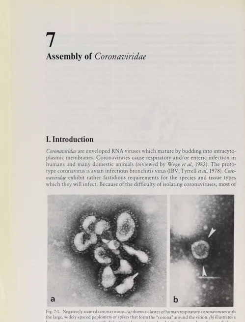
Fig. 7-1. Negativelystainedcoronavirions,(a) shows a Clusterofhuman respiratory coronaviruses with
the large,widely-spaced peplomers orspikes thatform the “corona”around the virion, (b)illustratesa
human entericcoronavirus with delicate peplomers (arrowheads).Suchviruses have frequently been visualized in preparationsof human fecal material,butthey are very difficult to propagate in vitro.
Molecular Organization 101 them were first classified by their virion morphology in negatively stained prepara tions (Tyrrell et al., 1975). The virions are characterized by large, club-shaped peplo-
mers or spikes about 20 nm long and 7 nm wide at their tips (Almeida and Tyrrell, 1967; Tyrrell et al., 1968; Fig. 7-1). Avian coronaviruses vary in diameter from 70 to 120 nm, but murine coronaviruses have more uniform diameters, approximately 90 nm. More recently, common features of virus replication and biochemistry have confirmed the assignment of coronaviruses into a single virus family (Tyrrell et al.,
1978; ter Meulen etal., 1981). Thereare at least 3 subgroups of Coronaviridae, based on their mutual lack of antigenic cross-reactivity (Pedersen et al., 1978; Sturman and Holmes, 1983). Avian coronaviruses, such as infectious bronchitis virus (IBV), do not share antigenic determinants with mammalian coronaviruses. Mammalian coronaviruses fall into two distinct groups: one includes mouse hepatitis virus (MHV), bovine coronavirus (BCV), hemagglutinating encephalomyelitis virus of swine (HEV) and human respiratory coronavirus OC43; and a second includes human respiratory coronavirus 229E, transmissible gastroenteritis virus of swine (TGEV), canine coronavirus (CCV), and feline infectious peritonitis virus (FIP). There are also coronaviruses antigenically unrelated to these three major sub groups, such as porcine enteropathic coronavirus, CV777 (Pensaert et al., 1981).
IL Molecular
Organization
We will summarize the molecular composition of coronavirions and events in coro navirus replication and then consider the morphology of the virions and their budding mechanisms in detail.
Transcription
The coronavirus genome is a single-stranded molecule of RNA of M.W. 5.4 to 6.9 × 10b which is capped at its 5' end and polyadenylated at its 3' end (Lai and Stohl- man, 1978; Stern and Kennedy, 1980 a; Siddell et al., 1982; Fig. 7-2). The isolated genomic RNA is infectious and serves as an mRNA within the infected cell. Thus, coronaviruses are positive-stranded RNA viruses. The genomes of IBV and MHV have been mapped by comparing oligonucleotides of genomic RNA with oligo nucleotides of 3' co-terminal, polyadenylated RNAs of different lengths generated by RNAse digestion of virion RNA. These maps have then been compared with the oligonucleotide patterns of the Subgenomic mRNAs found in infected cells (Stern and Kennedy, 1980 b, Lai et al., 1981).
There are 5 Subgenomic mRNAs for IBV, and 6 for MHV. The mRNAs of coro naviruses are unusual in that they form a nested set of molecules of varying length which share a common, polyadenylated 3' end (Stern and Kennedy, 1980 a and b; Weiss and Leibowitz, 1981; Cheley et al,, 1981; Fig. 7-2). Except for a short sequence of nucleotides at the 5' end of each mRNA, the oligonucleotides of each mRNA are completely identical to the 3' end of the next larger mRNA species (LaieZtf/., 1982 a).
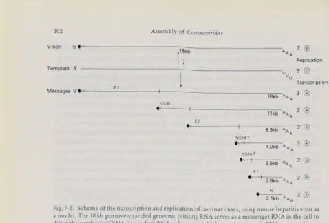
Assemblyof Coronaviridae Virion Template 3' ∣18kb
I
Messages 3∙ © Replication 5- © Transcription 3' © 3' © 3' © 3-© 3-© 3-© 3'©Fig. 7-2. Scheme ot the transcription andreplication of coronaviruses, using mouse hepatitis virus as amodel. The 18 kb positive-strandedgenomic (virion)RNAserves asamessengerRNA in the cellto direct the synthesis ofRNA-dependent RNApolymerasewhich uses thegenomic RNAas a template to
makea full-length negative-stranded RNA. From this template, new 18 kb genomic RNA and6 sub-
genomic mRNAs are made; allspecies form a nested setwith common3' ends. All of these positive-
stranded RNAspecies are capped(«---) and polyadenylated (AAA).Ashort leader sequence, indicated bythe wavy line, isfoundon the5' end of eachmRNA butoccursOnlyonceonthegenomic
strand. For each mRNA, only the gene atthe 5' end (to the shortvertical line)is translated; theprotein
productof eachmRNAis indicated by theletters above the 5' gene. The twosmallestmRNAshave been clonedandsequenced. (AdaptedfromSturman andHolmes,1983)
The 5' end of each mRNA consists of a cap plus a short common nucleo tide sequence referred to as the leader sequence (Lai et al., 1983).
In vitro translation of the 6 Subgenomic mRNAs of MHV has demonstrated that
each species codes for only one polypeptide and that the translated gene is at the 5' end of the mRNA (Siddell etal., 1980,1981 c; Leibowitz and Weiss, 1981; Leibowitzet
al., 1982; RottieretaZ., 1981 a). A tentative map based upon these studies is shown in
Fig. 7-2.
The structural proteins of different coronaviruses appear to follow a common pattern, and the terminology for MHV proteins will be used here. There are gener ally three structural polypeptides: a nucleocapsid protein N, and two envelope gly coproteins, El and E2 (Garwes, 1980; Siddell et al., 1982; Sturman and Holmes, 1983 a).
Replication
Coronavirus replication has recently been the subject of several comprehensive re views (ter Meulen et al., 1980; Siddell et al., 1982 and 1983; Sturman and Holmes, 1983 a; Rottier et al., 1984), and the reader is referred to these for details which are
Molecular Organization 103
beyond the scope of this chapter. The major features of coronavirus replication and transcription will be summarized here briefly.
Because of the positive-stranded nature of its genome, the coronavirion con tains no RNA-dependent RNA polymerase. This enzyme is synthesized in cells soon after infection and first directs the synthesis of full-length negative strands (Brayton et al., 1982; Dennis and Brian, 1981, 1982; Lai et al., 1982 b). Synthesis of
negative strands is complete within 5—6 hours after infection; it is not clear how continued synthesis of negative strands is inhibited. From this template, the RNA- dependent RNA polymerase directs the synthesis of genomic RNA and 5 subgeno- mic mRNAs (IBV; Stern and Kennedy, 1980 a and b) or 6 subgenomic mRNAs (MHV; Spaan et al., 1981, 1982; Wege et al., 1981). A cap and the same short leader nucleotide sequence are added to the 5' end of each mRNA, either by RNA splicing or by allowing the common leader sequence to serve as a primer for synthesis of each mRNA (Baric et al., 1983). The primer model is supported by the observation
that caps and leaders are found on nascent mRNA strands (Lai etal., 1983 b), but no
free leader RNA has yet been isolated from infected cells.
Two distinct peaks of RNA polymerase activity occur during virus replication (Brayton et al., 1982). The first involves primarily synthesis of the full-length nega
tive-stranded template, and the second coincides with synthesis of new genomic RNA and mRNAs. The virus protein species responsible for polymerase activity have not yet been identified.
Throughout the replicative cycle, the ratios of the different subgenomic RNAs remain constant (Stern and Kennedy, 1980 a; Wege etal., 1981; Spaan etal., 1981; Lei bowitz et al., 1981). Each of the mRNAs appears to direct the synthesis of a single
viral polypeptide (Fig. 7-2).
Virus Proteins
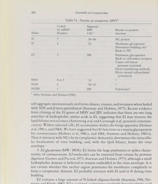
Table 7-1 summarizes the properties of the 3 structural proteins of MHV. The func tions of the non-structural proteins of corona-viruses are not known.
1. N protein (MW: 50 K). The NC protein, N, is phosphorylated (Stohlman and Lai, 1979). With the positive-stranded viral genomic RNA, the N protein forms a helical NC which is RNAse-sensitive. Regulatory or enzymatic functions of N have not been identified.
2. Elglycoprotein (MW: 23 K). El is an unusual virus glycoprotein in several ways. It has 3 domains: external, Intramembranous, and cytoplasmic (Sturman and Holmes, 1977, 1983). The short amino-terminal external domain (about 5 K) con tains all of the El oligosaccharides (Sturman and Holmes, 1977). In MHV, short oligosaccharides are O-glycosidically linked to serine or threonine residues of El (Niemann etal., 1982), unlike the complex oligosaccharides N-glycosidically linked
to asparagine residues which are found on nearly all other virus glycoproteins (Sha ron and Lis, 1981). The El glycoprotein of IBV contains only N-Iinked oligosaccharides (Stern et al., 1982; Stern and Sefton, 1983). The significance of this diversity of glycosylation patterns among coronaviruses is not known.
The El molecule is very hydrophobic, particularly when its external domain has been removed. Like a small number of integral membrane proteins, it tends to
AssemblyofCoronaviridae
1AfterSturman and Holmes (1983).
Table 7-1. Proteins of coronavirusMHf
Name Coded bymRNA Number Apparent MW ×10~3 Knownorputative function N 7 50-60 NC protein El 6 23 Membrane glycoprotein Determines budding site? Binds toNC
E2 3 180 Peplomeric glycoprotein
Binds to cell-surfacereceptors Causes cellfusion
(protease activated)
Elicits neutralizingantibody Elicits naturalcell-mediated
cytotoxicity
NS14 4 or 5 14 >
NS30 2 30-35 >
NS200 1 200 Polymerase?
self-aggregate spontaneously and forms dimers, trimers, and tetramers when boiled with SDS and /Tmercaptoethanol (Sturman and Holmes, 1977). Recent evidence from cloning of the El genes of MHV and IBV indicates that there are two long stretches of hydrophobic amino acids in El, suggesting that El may traverse the lipid bilayer several times (Armstrong et al. and Cavanagh et al., personal communi
cation). Within infected cells, El accumulates within the Golgi apparatus (Holmes
et al., 1981 a, and 1984). We have suggested that El functions as a matrix glycoprotein
for coronaviruses (Holmes et al., 1981 a, and 1984; Sturman and Holmes, 1983 a).
Thus it interacts with NCs by its cytoplasmic domain(s), determines the intracellu lar localization of virus budding, and, with the lipid bilayer, forms the virus envelope.
3. E2glycoprotein (MW: 180 K). E2 forms the large peplomers or spikes charac
teristic of coronavirions. E2-molecules can be removed from virions by protease digestion (Garwes and Pocock, 1975; Sturman and Holmes, 1977), although a small hydrophobic domain is believed to remain embedded in the virus envelope. It is not certain whether this anchor region penetrates the membrane completely to form a cytoplasmic domain. E2 probably interacts with El and/or N during virus budding.
E2 contains a large amount of N-Iinked oligosaccharide (Sturman, 1981; Nie mann and Klenk, 1981). E2 is acylated, containing covalently bound palmitic acid (Niemann et al., 1982; Schmidt, 1982 a), presumably located at or near the lipid bi layer (Schmidt, 1982 a and b). E2 is proteolytically cleaved at a late stage in virus maturation (Sturman and Holmes, 1983, and 1984; Holmes et al., 1984). In MHV- A59, cleavage appears to be dependent upon the host cell and yields two molecules, 90 A and 90B, which comigrate in electrophoresis in SDS-containing polyacryl amide gels with an apparent molecular weight of 90 K. These have different amino
Synthesis, Transportand Processingof Virus Proteins 105
acid compositions and only 90 A contains covalently bound palmitic acid. Cleavage of the large peplomeric glycoprotein of IBV in chicken cells may be very efficient, since only the two cleavage products ofE2, gp 93 and gp84, are found on the virion (Cavanagh, 1981; Wadey and Westaway, 1981; Stern et al., 1982).
E2 is a large, multifunctional molecule. It is responsible for virus-induced cell fusion (Holmes et al., 1981 b; Collins et al., 1982) and its cleavage is required for cell
fusion (Sturman and Holmes, 1983, and 1984). E2 on the plasma membrane renders cells subject to cell-mediated cytotoxicity (Welsh etal., 1983). It induces neutralizing antibody (Garwes et al., 1976,1978/79; Hasony and Macnaughton, 1982; Schmidt
and Kenny, 1981,1982) and binds the virion to receptors on the plasma membranes of susceptible cells (Holmes etal., 1981 b). E2 also plays an important role in the pH- dependent thermolability of coronaviruses (Sturman, 1981). The anchoring region of E2 probably interacts with El in the virus envelope.
III.
Synthesis,
Transport
and
Processing
of Virus
Proteins
The synthesis of N protein is directed by the smallest mRNA, which is derived from the 3' end of the viral genome (Siddell etal., 1980; RottierrZd/,, 1981 a). Synthesis of N occurs on free polysomes, although even in very short pulse-labeling experi ments some N is found on RER and smooth membranes isolated from homog enized cells (Frana and Holmes, unpublished observations). Thus, some mRNAs on membrane-bound polysomes may direct the synthesis of N protein. Immuno fluorescence studies with anti-N antibody reveal delicate flecks of fluorescence in the cytoplasm by 2 to 3 hours after virus inoculation. These foci increase in size and number during infection.
The N protein is phosphorylated at serine and threonine residues (Stohlman and Lai, 1979). Although protein kinase activity has been demonstrated in the virion (Siddell etal., 1981 a), it is not yet Clearwhetherthis activity represents a virus specific or cellular enzyme. The role (if any) of phosphorylation and déphosphory lation in modulation of the biological functions of N is not known. Pulse-chase studies show that although large amounts of N are synthesized in the infected cell, only a very small fraction of N is chased out of the cell into mature virions (Holmes
et al., 1984; Rottier et al., 1981 b; Siddell et al., 1981 b). This suggests that N protein
may have several functions during virus replication in addition to formation of NCs. Only one species of N appears in the virion of each coronavirus strain. However, even between closely related strains of MHV the apparent molecular weight of the virion-associated N varies, ranging from 50 K to 60 K (Siddell et al., 1982). Whether this difference is due to phosphorylation has not been determined. Several species of N which migrate more rapidly than the virion N have been detected within infected cells late in the infectious cycle or following immunopre cipitation (Cheley and Anderson, 1981). Their functions are unknown.
The synthesis of the peplomeric glycoprotein E2 (Rottier et al., 1981a; Siddell et
al., 1981b; Leibowitz et al., 1981), encoded in mRNA3 occurs on RER membranes
plasmic granules at 4 hours after infection. The glycoprotein then disperses rapidly throughout the cytoplasm, presumably upon intracytoplasmic membranes (Holmes et al., 1981 b). After 7 hours, delicate flecks of fluorescent staining are ob
served on the plasma membrane. These gradually spread to cover the cell surface. The first species ofE2 to be detected in pulse-label experiments is a 150 K form (Siddell et al., 1981 b) which is Cotranslationally glycosylated at asparagine residues by transfer of core oligosaccharides from dolichol-phosphate intermediates. This form of E2 is sensitive to endoglycosidase H. It is transported to the Golgi appara tus where further trimming and glycosylation of the N-Iinked oligosaccharides and acylation occur under the direction of cellular enzymes (Niemann et al., 1982). Pulse-chase studies show that in the 17 Cl 1 line of BALB/c mouse fibroblasts, syn thesis ofE2 is well-balanced with its release into virions (Holmes etaL,V)%∖ b); labeled
E2 is quantitatively chased into released virions within 2 hours. At least early in infection, there is no large excess of E2 in the cell. At a late stage in the processing of E2, when or just before it reaches the plasma membrane, proteolytic cleavage by a trypsin-like cellular enzyme occurs (Holmes et al., 1984). In tunicamycin-treated cells, E2 is synthesized as a 120 K non-glycosylated polypeptide which is not incor porated into virions (Niemann and Klenk, 1981).
The matrix glycoprotein, El, of MHV-A 59 is synthesized on membrane bound ribosomes as a non-glycosylated 20 Kpolypeptide (Niemann etal., 1982). Itis transported to the Golgi, where cell fractionation studies show that it is post- translationally glycosylated by the addition of several sugars to serine or threonine residues. This process of O-Iinked glycosylation, which is probably done by cellular enzymes, is not well understood. Each molecule of El receives from 1 to 3 oligo saccharide chains.
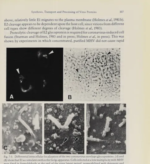
Immunofluorescence studies with monospecific or monoclonal antibodies to El and E2 have shown that the intracellular transport of the two coronavirus glyco proteins is different. E2, like the peplomeric glycoproteins of most other enveloped viruses, is transported from the RER through the Golgi to the plasma membrane. In contrast, the El glycoprotein is transported only as far as the Golgi apparatus, where it accumulates during virus infection (Fig. 7-3A and B). This has been demonstrated by simultaneously staining El with immunofluorescent antibody and marking the
trans cisternae of the Golgi by a histochemical reaction for thiamine pyrophospha
tase (Sturman and Holmes, 1983; DollereZa/., in preparation). It appears likely that the restricted intracellular transport of the El glycoprotein may account for the intracellular budding site of coronaviruses (Holmes et al., 1981 b; Sturman and Holmes, 1983). It is interesting to speculate whether other virus groups which mature by budding from intracellular membranes, such as bunya- and flavi- viruses may show a similarly restricted pattern of intracellular transport of a NC- binding protein.
Coronaviruses cause cell fusion in vitro and in vivo. This cell fusion appears to be
mediated by E2 glycoprotein on the plasma membrane of infected cells, since incu bation in the presence of anti-E2 antibody prevents virus-induced cell fusion (Holmes et al., 1981 b; Collins et al., 1982). Fluorescent antibody staining and immu
noelectron microscopy (Figs. 7-3 C-E) show that the E2 glycoprotein is on the plasma membrane in large amounts late in the infectious cycle, whereas, as noted
Synthesis,Transport and Processing of Virus Proteins 107 above, relatively little El migrates to the plasma membrane (Holmes et al., 1981 b).
E2 cleavage appears to be dependent upon the host cell, since virions from different cell types show different degrees of cleavage (Holmes et al., 1983).
Proteolytic cleavage of E2 glycoprotein is required for coronavirus-induced cell fusion (Sturman and Holmes, 1983 and in press; Holmes et al., in press). This was
shown by experiments in which concentrated, purified MHV did not cause rapid
Fig.7-3.Differential intracellular localizationof thetwo coronavirus envelope glycoproteins.(√4Jand
(B) show that El accumulateswithintheGolgiapparatus.Cells infected at a low multiplicity with MHV were fixed in formaldehyde at the endof the viruslatentperiod, permeabilized with detergentand
labeledwith monospecificanti-El antibody andfluorescent anti-rabbit IgG (A). The same cells were reacted with a cytochemicalmarker for theGolgi apparatus, thiamine pyrophosphatase (B). The El antigenin thecells of(A) (arrowheads) is locatedin the Golgi region shown in(B) (arrowheads). E2, the peplomeric glycoprotein, does not accumulate in the Golgi, but is transported to the plasma membrane,as shownin (C)to (E). MHV-infected cells 24 hours after infection werefixed and labelled
withnormal rabbit serum (C),or monospecific anti-E2 antibody(D)1 or anti-El antibody (E) followed
byStaphylococcus aureusprotein A conjugated to peroxidase.The presence ofantigen is shownby
development of agranular reactionproduct following reaction with hydrogenperoxideand 3,3'
diaminobenzidine. The cells werecoated withmetal and examined by scanning electron microscopy.
E2 is transported to theplasma membrane as shown by the granularlabelingin (D)(arrow heads), butElis nottransported to the plasmamembrane, becausepanel(E),like the normal rabbit serum control,(C), shows no granular reaction product on the plasmamembrane. Magnifications: (A)
Assembly ofCoronaviridae
fusion of susceptible cells in the presence of cycloheximide, whereas trypsin-treat ed, purified and concentrated MHV caused rapid cell fusion. (The only effect of trypsin on the virions was the quantitative cleavage of E2 180 to E2 90). Activation of plaque formation, hemagglutination and/or virus cytopathic effect by trypsin have been shown for several coronaviruses (reviewed in Sturman and Holmes, 1983). Whether cleavage of the E2 glycoprotein is required for virus infectivity is not yet clear, however, since at present there is no method available to obtain homogeneous populations of virions with uncleaved E2. Some coronaviruses which have been difficult to propagate in vitro, such as human enteric coronaviruses (Fig. 7-lb), may undergo a single cycle of infection yielding large numbers of non- infectious virions, as shown in negatively stained preparations (Caul and Eggle- stone, 1977). It may be necessary to identify a cell type in which cleavage of E2 occurs or to identify a different protease which may activate infectivity in order to propagate these fastidious viruses in vitro.
IV.
Assembly
of
Virus
Components
In this section, we will review the assembly of the A59 strain of MHV, since it has been studied most extensively. Where other coronaviruses differ from this model, this will be noted in the text.
NC Assembly
The specific interaction of N with genomic RNA has not yet been studied. It is like ly that only genomic RNA is encapsidated, since encapsidation of mRNAs would interfere with their translation. If this is true, we postulate that an encapsida tion signal is present near the 5' end of the genomic RNA, since all other regions are shared by Subgenomic mRNAs.
Fig. 7-4. Helical NC from the coronavirion. A long strand of helical NC purified from a humancoro
navirusby densitygradient ultracentrifugation is shown in a negatively stained preparation. Such
images have been difficult toobtain withmany coronaviruses.Magnification:× 205,000. (Courtesy of Dr. O. Caul, from Sturman and Holmes,1983, with permission ofAcademic Press)
Assemblyof Virus Components 109
The NC of coronaviruses is helical, but the turn-to-turn bonding of the NC strands appears to be very weak. The tubular appearance of the NC is readily ob served in thin sections of budding virions (David-Ferriera and Manaker, 1965; Becker et al., 1967; Oshiro et al., 1971), but is not often apparent in released virions,
suggesting that it may undergo a conformational change after virus budding and release (Holmes and Behnke, 1981). IntracytopIasmic inclusions of coronavirus NCs were not recognized for many years, perhaps in part due to the great flexibility of the NC strands. Large inclusions of NCs have been observed in coronavirus-in fected cells late in the infectious cycle (Caul and Egglestone, 1977). NCs with a density of 1.25 to 1.30g∕cc have been isolated from virions or from infected cells and shown to consist of genomic RNA and N protein with or without El (Kennedy and Johnson-Lussenberg, 1975/76; Sturman et al., 1980). Coronavirus NCs are apparently not tightly coiled, so that isolated NCs often appear as thin, kinky strands rather than helical coils. Only when virions are gently or spontaneously dis rupted, is it possible to observe or to purify long helices of NC with a diameter of 9 to 11 nm (Macnaughton et al., 1978; Davies et al., 1981; Caul et al., 1979; Fig. 7-4).
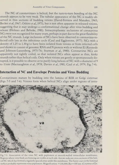
Interaction of NC and Envelope Proteins and Virus Budding
Coronaviruses mature by budding into the Iumina of RER or Golgi cisternae (Figs. 7-5 and 7-6). Virions form when helical NCs align under regions of intra
Fig. 7-5. Association of the virus NCwithpeplomers atsmooth-walled intracellular membranes.
Regions where virus buds are forming are visible at each side. Arrows indicate cross-sections of the heli
cal NC which has formed a regularly spaced array underthemembrane. Thefuzzy coat on theIumenal surfaceprobablyTepresentsviruspeplomers.Magnification: × 130,000; Bar=IOO nm.(CourtesyofDr.
IlO
Fig. 7-6. A binucleate cellinfectedwithMHV inwhich largenumbers of virions have accumulatedin
the lumen of the RER, where a budding virus is seen(arrow). The virionsare also commonly seen inthe
lumenof the nuclearenvelope (arrowheads). In (b), MHV virions bud (arrowheads) froman area of cytoplasm containing numerousstrands of NC(arrow). The centers of the virions are electron-lucent, since the NC is attached to the virus membrane. Magnifications: (a) × 15,000; (b)×80,000. [(a)
Assembly of Virus Components 111
cellular membranes containing virus glycoproteins (David-Ferriera and Manaker, 1965; Oshiro, 1973; Massalski etal., 1981,1982; Ducatelle, 1981; Dubois-Dalcq etal., 1982). NCs may associate with the forming envelope by interacting with the cyto plasmic domain of the El glycoprotein (Holmes et al., 1981 b). Virions form and are released from cells treated with tunicamycin, although these cells contain little E2 and such virions are completely devoid of E2 (Holmes et al., 1981 a). Thus E2 is evidently not required for the formation of coronavirions. Virions synthesized in the presence of tunicamycin contain normal amounts of N and fully glycosylated El, but are not infectious, probably because, lacking E2 peplomers, they cannot attach to virus receptors.
Virions in the RER appear to have large peplomers on their envelopes (Chasey and Alexander, 1976). An important but still unanswered question is whether the glycoproteins El and E2 are processed on intact virions as these migrate through the Golgi apparatus, or whether the glycoproteins are fully processed first and then assembled into virions. For the latter to be true, budding of virions from the RER would require migration of glycolysated E2 from the Golgi back to the RER, but such retrograde transport has never been demonstrated. This question can be re solved by isolation of virions from intracellular compartments and characteriza tion of their structural proteins.
Virions are released from membranes by pinching off into the Iumina of the RER or Golgi apparatus. Large numbers of virions may accumulate within the lumen of the RER (Fig. 7-6a). In the RER, the virions are spherical particles with electron-lucent centers and NC strands beneath the envelope (Fig. 7-6a and b). Re lease of virions into the lumen apparently does not involve cellular actin, since actin has not been detected routinely in coronavirions, with the possible exception of IBV (Lomniczi and Morser, 1981).
Release of Virions from the Cell and Post-Release Maturation
Following their release into the RER and Golgi, coronavirions appear to escape from the cell within smooth-walled vesicles that migrate to the cell membrane and fuse with it (Doughri et al., 1976; Oshiro et al., 1971). Time lapse cinematography of MHV-infected cells shows that during the period of maximum virus release, cells are not lysed, although they may undergo fusion (Holmes, unpublished observa tion). This process of release is similar to secretion of zymogens from pancreatic acinar cells (Holmes etal., 1981 a). Thus, coronaviruses apparently depend on several specialized host cell functions for virus maturation and release, including two mechanisms of protein glycosylation, two pathways of intracellular transport of glycoproteins, and a cellular secretion pathway.
After coronaviruses are released from the cell, many virions remain adsorbed to the plasma membrane (Oshiro et al., 1971; Oshiro, 1973; Doughri and Storz, 1977; Sugiyama and Amano, 1981; Fig. 7-7a-c). It is not clear why such large numbers of virions bind to infected cells. Are they simply virions adherent to the smooth walled vesicles which have not detached from the membrane following fusion of the vesicle with the plasma membrane? Or were these virions first released into the
Assembly of Virus Components 113
medium and then bound to receptors on the plasma membrane? If the latter is true, then why are there very different numbers of virions bound to cells in the same cul ture (Fig. 7-7b and c)? Do the virions bind to cellular receptors which are differently expressed in different cells, possibly due to cell cycle differences, or do they bind to a virus glycoprotein such as E2, which is present on the plasma membrane?
As virions of the A59 strain of MHV are transported through the Golgi-asso ciated vesicles in some cell types, they become flattened, disc-shaped and electron- dense (Holmes et al., 1981a; Fig. 7-7d and e). Possibly, these changes could
be associated with release of the NC from its attachment to the envelope. They might be triggered by changes in pH or ionic concentrations in different intra cellular membrane compartments. Alternatively, they may reflect processing of virus components which takes place as virions migrate through different intra cellular compartments. For instance, since cleavage of the 180 K E2 to 90 K E2 appears to be an event which occurs immediately prior to release of the virions (Holmes et al., 1984), it is possible that the condensation of virions may be triggered
by that event. Since proteolytic cleavage of E2 appears to be dependent upon the host cell, this might explain why post-release condensation of virions is not seen for all coronaviruses or even for MHV-A59 in all cell types.
Many enveloped viruses show polarized budding from epithelial cells (Rodri- guez-Boulan and Sabatini, 1978; Chapter 1). Since coronaviruses do not bud from the plasma membrane, variations in the transport of the E2 glycoprotein to apical or basolateral domains of the plasma membrane would be expected to have no effect on virus budding. However, many polarized cells show marked polarity in secretion of cellular proteins and this polarized secretion might affect release of coronaviruses from post-Golgi vacuoles. Although extensive studies on the release of coronaviruses from cells in epithelial tissues have not been done, release of por cine coronavirus from both apical and basal regions of intestinal cells has been demonstrated (Doughri and Storz, 1977). It is clear that MHV virions may be released from the lower surface of mouse fibroblasts, since membrane fragments of cells which remain attached to the substrate after virus-infected cells peel away show large numbers of adherent, flattened, disc-shaped virions (Fig. 7-7e, Holmes and Turner, unpublished observations).
Fig. 7-7. Large numbers ofcoronavirions adsorbed to the plasma membrane of infected cells, (a)
shows human respiratory coronaviruses attached to cells invitro,(b) and (c) are a Stereopairofscanning electronmicrographsof MHV-infected cells demonstratingthat adjacent cells may show great differ
ences in thenumber of adherent virions. On the uppercell,virions (largearrow, c) are tightlypacked
onthemembrane, whereas onthe lower cell, virions (smallwhite arrow, c)are widely scattered over the
plasmamembrane andmicrovilli, (d) and (e) show that MHVA59 virions on the plasma membrane often appearflattened and disc-shaped. In sections ofinfected cells observed by transmission electron microscopy (d), virions (arrows) at the plasma membrane areflattened and electron-dense.In (e),flat tened virionsare adsorbedtofragments of cell membrane left adheringto the substrate after an
MHV-infected cellhas detached. The preparation was fixed, dehydrated,driedby thecriticalpointmethod, and observedwitha high voltage electron microscope. Large numbers of virions (arrows)adheretothe membranesbeneaththe cells. Magnifications: (a) × 35,000; (b) and (c)×9400; (d) × 52,000;(e)
× 33,000. [(a) Courtesy of Dr.L. Oshiro,from Oshiro et al.,1971, with permission of Cambridge University]
Assembly of Qoronaviridae
Fig.7-8. Inclusionscharacteristic ofcoronavirus-infectedcells.In (a), reticularinclusions formedof specialized infoldings of RER membranes are shown in acell infected with a human coronavirus, (b)
and (c) show cytoplasmicinclusionsof MHVNC seenin differentiated nerve cells in vitro. Inclusions formed by the wild Type JHM strainof MHV contain core-like structurescomposed oftwisted NC strands (arrowheads) in(c). Inclusionsformed by a ts mutant of theJHM strain aremostlymade of gran ules (b). Magnifications: (a) × 50,000; (b)×51,000; (c) × 68,200. [(a)fromOshiro et al., 1971, with permissionof Cambridge University;(b) and(c) from Dubois-Dalcq et al., 1982; withpermission of
Assemblyof Virus Components 115
Defective Assembly
In this section, we will consider circumstances in which the synthesis of virus components is not synchronized with release of mature virions. Under these condi tions, morphological events can be observed which may provide insight into virus assembly. Coronaviruses do not normally bud from the plasma membrane (Oshiro, 1973), although such budding is occasionally seen late in the infectious cycle (Dubois-Dalcq et al., 1982) when some El may have been transported to the plasma membrane. After release of virions has ceased, cytoplasmic inclusions of viral NCs have been observed (Caul and Eggleston, 1977; Watanabe, 1969; Massalski et al., 1982; Dubois-Dalcq et al., 1982; Figs. 7-6b and 7-8b and c). These inclusions vary
considerably in appearance and their protein composition has not yet been deter mined. Reticular cytoplasmic inclusions, which consist of a convoluted network of tightly apposed membranes continuous with the RER, are another characteristic of coronavirus infection (David-Ferriera and Manaker, 1965; Oshiro et al., 1971;
Fig. 7-8a).
Coronavirus-infected cells often contain spherical inclusions about IOO to 400 nm in diameter, which contain fine fibrils and are bounded by double mem branes apparently derived from RER (David-Ferriera and Manaker11965; Sturman and Holmes, 1983; Fig. 7-9). These closely resemble structures called type 1 cytopa- thic vesicles (CPV-I) seen in cells infected with alpha- or Aaviviruses (Murphy, 1980; Grimley et al., 1972; see Chapter 8). They may represent aberrent virion formation
or be associated with virus RNA synthesis, as has been suggested for alphaviruses (Grimley et al., 1972).
Fig. 7-9. Membrane-bound inclusions(arrowheads)containing thin electron-dense filaments.These
inclusions incoronavirus-infected cells resemble the type1 Cytopathic vesicles seen in cells infected with alpha- and flaviviruses (see Chapter 8,Figs.8-5a and 8-13). They may be involved invirus RNA
synthesis. Magnification: × 75,000
Organizationof theVirion 117 Late in infection with MHV, and in tunicamycin-treated MHV-infected cells, long rigid tubules about 50 nm in diameter are seen in the lumen of the RER and in smooth-walled vesicles (Dubois-Dalcq et al., 1982; Holmes et al., 1981 a; Fig. 7-10a).
Similar tubules have been assembled in vitro from the matrix protein of a para myxovirus (Heggeness et al., 1982 a). Since El functions as a M-Iike protein which associates with membranes, it has been suggested that the coronavirus tubules are formed of excess El (Sturman and Holmes, 1983).
Empty virus particles have been isolated from coronavirus-infected cells by density gradient ultracentrifugation (Macnaughton and Davies, 1980), but the mechanism of their formation and release is not understood.
To analyze interactions of NCs with virus-specific membrane proteins, investigators of other virus systems have studied the formation of pseudotypes with different viruses. Attempts to isolate pseudotypes of coronaviruses with other virus types have been limited to pseudotypes of MHV with murine leukemia virus (Yoshikura and Taguchi, 1978). Further studies of coronavirus pseudotypes with other viruses that bud into the RER and Golgi may elucidate the assembly of coronaviruses.
Treatment of MHV-infected cells with monensin arrests virus budding (Niemann etal., 1982; Fig. 7-10b), and removal of monensin results in rapid budding
of virions into the Golgi apparatus. These effects are probably caused by monensin- induced inhibition of intracellular transport of MHV glycoproteins.
Ts mutants have been used to study the morphogenesis of several groups of en veloped viruses. Although several independent collections of ts mutants of corona viruses have been generated (Robb etal., 1979; Haspel etal., 1978; Koolen etal., 1983), most of these mutants are of the RNA negative (RNA—)phenotype. The few RNA+ mutants which make structural proteins at the non-permissive temperature have not been examined for defects in virus maturation.
V.
Organization
of the Virion
A summary of the events in coronavirus assembly is shown in Fig. 7-11. A model of the organization of the structural components of MHV is shown in Fig. 7-12. This general model seems to be valid for most coronaviruses (Sturman and Holmes, 1983), with the possible exception of BCV, which may have an additional glycoprotein (King and Brian, 1982). In negatively stained preparations, corona virions have only one type of peplomer, with the possible exception of BCV and
Fig. 7-10. Late in MHV infection, long tubules, 25 to30 nm in diameter,mayaccumulateinthe lumen of the RER (arrow a). These may represent excessElglycoprotein in association withmembranes,(b)
shows that monensinmay inhibit the maturation of coronaviruses. In monensin-treated cells, partially
budded virionsare seeninlarge numbers in smooth-walled vesicles associatedwiththe Golgi. Magnifi cations: (a) × 53,000; (b)× 60,000.[(a) from Dubois-Dalcq et al., 1982;with permissionof Academic
Assemblyof Coronaviridae
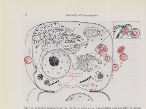
Fig. 7-11. A model summarizing the eventsin replication, transcriptionandassemblyof mouse hepatitis virus.It islikely thatthe virus RNAenters the cell byfusionof the virus envelope withthe plasma membrane.Primary translation yields the RNA polymerase which copies thegenomic RNAto makea fulllength negative-stranded template. Then, synthesis of the6Subgenomic mRNAs occurs
andthe 5'terminal end of eachof these is translated tomake a single protein species.The mRNAs for El
and E2 envelope glycoproteins are translated on membrane-bound ribosomes. E2is glycosylatedwith N-Iinked oligosaccharides and is acylated. In contrast, El is glycosylated with O-Iinked oligo saccharides andis not acylated. E2istransported through the Golgi to theplasmamembrane, but intra cellular transport ofEl is terminated at the Golgi. Meanwhile, the othermRNAs appear to betranslated onfree polysomesto yieldthe non-structuralproteins and the NC protein N.Nassembleswith new
virus genomic RNA to form a helical NC.The NCprobably interactswith thecytoplasmic domainof El in the membranesof the RERor Golgi at sites where E2 is also present. Buddingofvirions into the
RERorGolgi is followed by their releasefrom theintact cell bya process analogous to protein secretion (see text). The window shows virions budding from theRERand Golgi membranes. Cleavage ofthe E2
glycoprotein by a host cellprotease appearstooccur just prior to virusrelease. E2istransported to the plasma membrane where itmay inducefusion with adjacent cells
one strain of MHV (Bridger et al., 1978; Greig et al., 1971; Sugiyama and Amano, 1981). The smaller type of peplomer may contain the additional glycoprotein. Some glycosaminoglycan, a host cell component, has been detected even in highly puri fied coronaviruses (Garwes etal., 1976; Sturman, 1981). No function forthis has been identified.
The molar ratio of structural proteins within the virion is N : El : E2 = 8 :16 :1 (Sturman, 1981). This model indicates that the El glycoprotein interacts with both the NC and the E2 glycoprotein. The organization of the NC within the released virion is uncertain. Although the NC in the budding virus appears to be a well- formed helix attached to the inside of the virus envelope, in the released virion the NC may be less firmly bound to the membrane and may be partially uncoiled.
Organization of the Virion 119
Fig. 7-12.A model of the organization ofthe MHV virion. The Nprotein isassociated with the virus
RNA genomein the helical NC. Duringbudding, the NC probably associates withthe cytoplasmic domain ofthe El glycoproteinwhich acts asa matrix protein. Thepeplomers are formed of the large
glycoprotein E2, which probably interacts with the El in the virus envelope. A small amountofhost glycosaminoglycan (g) is associated with purifiedcoronavirions.(After Sturman andHolmes, 1983)