S179D-Human PRL, a Pseudophosphorylated Human
PRL Analog, Is an Agonist and Not an Antagonist
SOPHIE BERNICHTEIN, SANDRINA KINET, SE´ BASTIEN JEAY, MARTA LLOVERA,
DOMINIQUE MADERN, JOSEPH A. MARTIAL, PAUL A. KELLY, AND
VINCENT GOFFIN
INSERM, U-344, Molecular Endocrinology, Faculte´ de Me´decine Necker (S.B., S.K., S.J., M.L., P.A.K., V.G.), 75730 Paris, France; Laboratory of Molecular Biology and Genetic Engineering, University of Liege (S.K., J.A.M.), 4000 Sart-Tilman, Belgium; Laboratory of Molecular Biophysics, Institut de Biologie Structurale, Commissariat a` l’e´nergie atomique-Centre National de la Recherche Scientifique (D.M.), 38027 Grenoble, France
For many years, our group has been involved in the develop-ment of human PRL antagonists. In two recent publications, S179D-human PRL, a human PRL analog designed to mimic a putative S179-phosphorylated human PRL, was reported to be a highly potent antagonist of human PRL-induced prolifera-tion and signaling in rat Nb2 cells. We prepared this analog with the aim of testing it in various bioassays involving the homologous, human PRL receptor. In our hands, S179D-human PRL was able to stimulate 1) the proliferation of rat Nb2 cells and of human mammary tumor epithelial cells (T-47D), 2) transcriptional activation of the lactogenic hormone response element-luciferase reporter gene, and 3) activation
of the Janus kinase/signal transducer and activator of tran-scription and MAPK pathways. Using the previously charac-terized antagonist G129R-human PRL as a control, we failed to observe any evidence for antagonism of S179D-human PRL toward any of the human PRL-induced effects analyzed, in-cluding cell proliferation, transcriptional activation, and sig-naling. In conclusion, our data argue that S179D-human PRL is an agonist displaying slightly reduced affinity and activity due to local alteration of receptor binding site 1, and that the antagonistic properties previously attributed to S179D-human PRL cannot be confirmed in any of the assays analyzed in this study. (Endocrinology 142: 3950 –3963, 2001)
P
RL IS a pituitary hormone involved in a wide spectrum of biological activities (1), the majority of which are related to lactation and reproduction, as emphasized by the phenotype of mice in which the genes encoding either the hormone (2) or its receptor (3) have been invalidated. The PRL receptor (PRLR) is a member of the cytokine receptor superfamily (4) that closely resembles the receptor of GH (5). As is true for the GH receptor, the PRLR is activated by ligand-induced homodimerization, which involves the se-quential interaction of two regions on the hormone (called binding sites 1 and 2) with two PRLR molecules, leading to the formation of an active trimer (6, 7). Among the numerous signaling pathways triggered by the ligand-bound receptor (1, 8), activation of the Janus kinase 2 (JAK2)/signal trans-ducer and activator of transcription 5 (STAT5) cascade prob-ably represents the most widely distributed hallmark of PRLR signaling (9 –11).PRL exists in several molecular isoforms resulting from various posttranslational modifications, including proteo-lytic cleavages, glycosylation, and phosphorylation (for a review, see Ref. 12). The occurrence in vivo of PRL phos-phorylation has been clearly demonstrated in bovine pitu-itary (13), where serine 90 was identified as the major phos-phorylation site of bovine PRL (14). In other species the in
vivo demonstration of PRL phosphorylation has been less
definitive (15, 16), and the physiological relevance of phos-phorylation sites identified by in vitro incorporation of32P into PRL (17, 18) remains questionable. In humans, although pituitary-purified human PRL (hPRL) preparations obtained from the NIDDK were shown to contain traces of phosphor-ylated forms (19, 20), definite identification of phosphory-lated amino acid(s) awaits further investigation.
Several reports have shown that phosphorylation alters the biological properties of PRL. For example, phosphory-lated bovine PRL displays reduced mitogenic activity in the Nb2 cell proliferation bioassay compared with the nonphos-phorylated isoform (21). Accordingly, dephosphorylation of hPRL slightly increases its bioactivity (19, 20). Still more remarkably, phosphorylation of serine 177 confers antago-nistic properties to rat PRL (rPRL) in the same Nb2 cell proliferation assay (18, 19). This serine residue is highly conserved among PRLs from different species (22) and is homologous to serine 179 in hPRL. Based on the antagonistic properties demonstrated for phosphorylated rPRL, a molec-ular mimic of a putative S179-phosphorylated hPRL has been engineered in Walker’s laboratory by substituting an aspar-tate for the wild-type (WT) serine 179, generating the so-called S179D-hPRL analog (20). In a first report these re-searchers reported that S179D-hPRL strongly antagonizes the mitogenic activity of hPRL on Nb2 cells, thereby extrap-olating to the human hormone the functional consequences of Ser177phosphorylation earlier reported for rat PRL (20). Surprisingly, contrary to all previous reports describing the
Abbreviations: hPRL, Human PRL; IPTG, isopropylthiogalactoside; JAK, Janus kinase; LHRE, lactogenic hormone response element; MM, molecular mass; PRLR, PRL receptor; rPRL, rat PRL; STAT, signal trans-ducer and activator of transcription; TBST, Tris-buffered saline-Tween 20; Vel, elution volume; WT, wild type.
mechanism of action of PRL or GH antagonists (23–27), S179D-hPRL appeared to antagonize hPRL effects through a noncompetitive inhibition of receptor activation (20). This encouraged Walker and colleagues to investigate the molec-ular basis of this unusual, although apparently highly potent, mechanism of antagonism. Using the immunoblot approach to investigate activation of the JAK2/STAT5 cascade, these researchers showed that S179D-hPRL strongly activates STAT5 while minimally activating JAK2 (28). Based on these observations, they suggested the involvement of a kinase other than JAK2 in STAT5 activation, and hence, they hy-pothesized that the antagonistic activity of S179D-hPRL may result from activation of signaling molecules/pathways other than those known to be involved in hPRL signaling (1) and still to be identified.
For many years the development of hPRL antagonists that could be of use in reducing breast cancer progression has been one of our goals (8, 29). In this perspective we have previously engineered several hPRL analogs, one of which (G129R-hPRL) appeared to display competitive antagonistic properties in various PRLR-mediated bioassays (25, 26), in-cluding hPRL-induced signaling and proliferation of breast cancer cells (27). However, due to its lower affinity, the analog had to be used at a significant molar excess vs. WT hPRL (1:10 to 1:50 ratio) to exhibit significant antagonism, which justified further efforts in developing more potent hPRL antagonists. In this respect the conclusions claimed by Walker et al. in their two recent reports (20, 28) appeared of great interest and prompted us to take further advantage of the antagonistic properties of S179D-hPRL. Thus, the present work was initiated with the aim to engineer more potent hPRL antagonist by combining the two point mutations [S179D (20) and G129R (25)] individually conferring antag-onism to hPRL analogs. However, we failed to confirm the conclusions of Walker’s articles regarding S179D-hPRL properties, as this mutant clearly acts as an agonist, and not an antagonist, on the PRLR. The goal of the present report is to better characterize the biological properties of this analog in various cell bioassays and to discuss why antagonistic properties were erroneously attributed to S179D-hPRL.
Materials and Methods Cultures and reagents
Culture media, FCS, horse serum, geneticin (G-418), trypsin, and glutamine were purchased from Life Technologies, Inc. (Cergy Pontoise, France). Luciferin and cell lysis buffer were from Promega Corp. (Mad-ison, WI), and luciferase activity was measured in relative light units (Lumat LB 9501, Berthold, Nashua, NH). Iodogen was purchased from Sigma (St. Louis, MO), and carrier-free Na[125I] and ampholytes were
obtained from Amersham Pharmacia Biotech (Orsay, France).
Hormones
All of the hormones used in this study were produced by recombinant technology: WT hPRL (30), the binding site 2 analog G129R-hPRL (Gly129
replaced with Arg) (31), and the molecular mimic of phosphorylated hPRL, so-called S179D-hPRL (Ser179replaced with Asp). The mutants
were produced in Escherichia coli BL21(DE3) using the pT7L expression vector (32), then purified as described previously (25, 31–33).
Cells
Rat Nb2 lymphoma cells were obtained from P. W. Gout (Vancouver, Canada). As previously reported (26), HL5 cells are a clone of 293
fibroblastic cells stably transfected with the plasmids encoding the hPRLR (34) and a PRL-responsive reporter gene. The latter contains the sequence encoding the luciferase gene under the control of a six-repeat sequence of the lactogenic hormone response element (LHRE; which is the DNA-binding element of STAT5), followed by the minimal thymi-dine kinase promoter. T-47D cells (human mammary tumor epithelial cell line) were provided by M. N. Norman (Bristol, UK).
Site-directed mutagenesis
Construction of the S179D-hPRL mutated cDNA was performed by the oligonucleotide-directed mutagenesis method using the Chameleon double stranded, site-directed mutagenesis kit from Stratagene (La Jolla, CA), following the manufacturer’s instructions. The pT7L-hPRL expres-sion vector (32) was used as a template for mutagenesis. The sequence of the mutated oligonucleotide was the following (5⬘33⬘ noncoding strand, mutated codon underlined): GTC GAT TTT ATG GTC ATC CCT GCG TAG. The selection primer (XmnI restriction site; see manufactur-er’s instructions) was the following: 5⬘-CAT CAT TGG AAA ACG CTC TTC GGG GCG-3⬘. Clones containing the expected mutation were iden-tified by DNA sequencing on the pT7L-hPRL plasmid (32).
Biochemical properties
Production and purification of proteins. Recombinant WT hPRL and hPRL
analogs were overexpressed in a 1-liter culture of E. coli BL21(DE3) and purified as previously described (32). Briefly, when the OD600of
bac-terial cultures reached 0.7– 0.9, overexpression was induced using 1 mm isopropylthiolgalactoside (IPTG) for 2 (20) or 4 h (32) (OD600,⬃2.5 after
4 h). Cell lysis was performed using a cell disintegrator (Basic Z, Cell D, Roquemaure, France). Proteins were overexpressed as insoluble inclu-sion bodies that were solubilized in 8 m urea (5 min at 55 C, then 2 h at room temperature) and refolded by continuous dialysis (72 h, 4 C) against 50 mm NH4HCO3, pH 8. Proteins collected at this step of the
protocol were referred to as nonpurified. Alternatively, refolded pro-teins were concentrated by tangential flow ultrafiltration (500 ml/min flow rate) using a YM10 Miniplate bioconcentrator (Millipore Corp.-Amicon, Bedford, MA). After centrifugation (10 min, 9000⫻ g) of con-centrated protein, supernatants were purified by gel filtration chroma-tography using a high resolution Sephacryl S-200 column (Amersham Pharmacia Biotech) equilibrated in 50 mm NH4HCO3and 150 mm NaCl,
pH 8. Fractions corresponding to monomeric hPRL (WT and analogs) were collected, pooled, quantified by the Bradford method (Bio-Rad Laboratories, Inc., Ivry-sur-Seine, France), aliquoted, and stored at ⫺20 C. This final fraction was referred to as purified.
SDS-PAGE and protein quantification. Protein size and purity were
as-sessed using 15% SDS-PAGE under reducing (-mercaptoethanol) or nonreducing conditions (35). Gels were stained using Coomassie blue. Protein preparations were quantified by Bradford protein assay, using BSA as the reference.
Circular dichroism spectroscopy. A Jobin Yvon CD6 circular dichroism
spectropolarimeter with a thermostated sample holder was used. Data were recorded at 25 C using 0.1-cm cells with an interval of 1 nm and an integration time of 5 sec. The sample concentration was 0.4 mg/ml in 25 mm NH4HCO3and 150 mm NaCl, pH 8. Each spectrum is an
average of three scans, corrected for buffer. Helicity was calculated as described (36).
Apparent molecular mass. The apparent molecular mass (MM) of WT
hPRL and hPRL analogs was estimated using analytical gel filtration chromatography (HR S-200) according to calibration from several MM markers: dextran blue (void volume), BSA (68 kDa), carbonic anhydrase (30 kDa), and myoglobin (17.5 kDa).
Isoelectric focusing. The isoelectric point (pI) of hPRL (WT and mutants)
was determined by isoelectrofocusing. Electrophoresis was performed under continuous cooling; electrode solutions were 20 mm acetic acid and 20 mm NaOH. The gel contained polyacrylamide (5.5%), glycerol (10%), and ampholytes (5.5%) in a range of pH 3–10. A prerun of 15 min (200 V) was applied to the gel before sample loading to allow formation of the pH gradient. Ten micrograms of protein diluted in sample buffer (5.5% ampholytes and 10% glycerol) were loaded on the gel, then the run
was performed at 200 V until the visible band corresponding to methyl red (pI 3.75) was focused. Gels were fixed in 20% trichloroacetic acid, then in 40% ethanol/10% acetic acid. Staining was performed using Coomassie blue and destaining in 40% ethanol/10% acetic acid. The pI of hPRL samples were estimated by comparison with pI markers (Am-ersham Pharmacia Biotech).
Biological properties
Binding studies. Binding affinity of S179D-hPRL was determined using
cell homogenates of the HL5 clone (expressing the human PRLR), fol-lowing the procedure previously described (26). Briefly, hPRL was io-dinated using Iodogen (33), and its specific activity was in the range of 40 –50Ci/g. Binding assays were performed overnight at room tem-perature using 150 –300g cell homogenate protein in the presence of 30,000 cpm [125I]hPRL and increasing concentrations of unlabeled WT
hPRL or S179D-hPRL. Results are representative of three independent experiments performed in duplicate. The relative binding affinity of the mutant was calculated as the ratio of its IC50with respect to that of WT
hPRL.
In vitro cell bioassays.
Nb2 cell proliferation assay. The reference bioassay for lactogenic
hor-mones is the lactogen-induced proliferation of rat Nb2 lymphoma cells (37, 38). The Nb2 cell line was routinely maintained in RPMI 1640 supplemented with 10% horse serum, 10% heat-inactivated FCS, 2 mm glutamine, 50 U/ml penicillin, 50g/ml streptomycin, and 100 m -mercaptoethanol. The proliferation assay was performed as initially described (37) with minor modifications. Briefly, the assay was per-formed in 96-well plates using 2⫻ 104cells/well on a starting day in a
final volume of 200l, including hormones. As it will be explained in
Results, lower cell densities were also tested according to the protocol of
Chen et al. (20). Cell proliferation was estimated after 3 d of hormonal stimulation by adding 10l WST-1 tetrazolium salt (Roche, Meylan, France) as previously described (39). This survival reagent is metabo-lized by mitochondria of living cells, which leads to an increase in the OD measured at 450 nm (OD450) in a manner that is proportional to the
number of cells counted by hemocytometer (data not shown). A mini-mum of 30 min was required to get homogenous colorimetric reaction, then OD450values increased for the various experimental conditions
proportionally to the time of incubation with WST-1. The linearity of OD450/cell density ratios up to absorbance values of 3–3.5 was
con-firmed by performing preliminary cell dilution experiments (not shown). We report data obtained after 2– 4 h of reaction (which fall within this absorbance range) to allow detection of minimal cell pro-liferation under low stimulation conditions. Antagonistic properties of hPRL mutants were assessed in competition assays using 200 pg/ml or 1 ng/ml WT hPRL. The experiments were performed at least three times in triplicate or quadruplicate.
Human PRLR transcriptional bioassay. The HL5 clone (human
PRLR-LHRE-luciferase, clone 5) (26) was routinely cultured in DMEM-Nut F12 medium supplemented with 10% FCS, 2 mm glutamine, 50 U/ml pen-icillin, 50g/ml streptomycin, and 700 g/ml G-418 (clonal selection). The assay was performed in 96-well plates using 5⫻ 104cells/100
l䡠well in medium containing only 0.5% FCS. Cells were allowed to adhere overnight, then 100l hormones diluted in FCS-free medium were added to each well. After 24 h of stimulation, cells were lysed (50 l), then luciferase activity was counted for 10 sec in 15 l cell lysate. The absolute values of luciferase activity were found to vary depending on the number of cell passages; however, this did not significantly affect the fold induction of luciferase activity (calculated as the ratio between the relative light units of stimulated vs. nonstimulated cells). Maximal fold induction (routinely⬃15- to 20-fold) was obtained in the range of 1–10 g/ml WT hPRL. To avoid interassay variations, all analogs to be compared were systematically tested within each experiment, and data obtained in one experiment representative of three experiments per-formed in duplicate are shown.
Cell cycle analyses. T-47D cells were routinely cultured in DMEM/Ham’s
F-12 (1:1) medium containing 10% FCS, 2 mm glutamine, penicillin (50 U/ml), and streptomycin (50g/ml). Cells were aliquoted in six-well plates (0.5–1⫻ 106cells/well) and serum-deprived in 0.5%
charcoal-stripped serum for 24 h before addition of hormones. After 24-h stim-ulation, culture medium was collected to recover floating cells by cen-trifugation, and attached cells were harvested by brief trypsinization. Cells were pooled and permeabilized using 40l DNA-Prep Reagent (Coulter Corp., Miami, FL) before 30-min incubation at 37 C in 0.5 ml DNA intercalculator propidium iodide (DNA-Prep stain propidium iodide). Cell cycle distribution was analyzed by flow cytometry on a FACScan using CellQuest software (Becton Dickinson and Co., Moun-tain View, CA) and manual gating.
Immunoprecipitation and Western blots. Nb2 cells were starved overnight
in FCS-free medium before hormonal stimulation. The next day, cells were pelleted and aliquoted in FCS-free medium at 8⫻ 107cells/ml (1
ml/tube) and allowed to equilibrate/recover for 30 min at 37 C before PRL stimulation. Cells were stimulated (5 min at 37 C) using various concentrations of WT hPRL or hPRL analogs, as indicated in the figures. At the end of the stimulation, cells were washed twice with ice-cold stopping buffer as previously described (27), and cell pellets were kept frozen until used. Cells were solubilized in 1 ml lysis buffer (30-min rotation at 4 C) (27). Lysates were centrifuged for 10 min at 13,000⫻ g, then supernatants were quantified for their protein content by Bradford assay and used for immunoprecipitation. A similar procedure was used for Western blot experiments using T-47D cells, except that cells were stimulated for 30 min by the various hormones.
For immunoprecipitation studies (Nb2 cells), 3 mg total lysate were incubated with polyclonal anti-JAK2 (2.5l/ml; Upstate Biotechnology, Inc., Schiltigheim, France) or polyclonal anti-STAT5 (C-17, Santa Cruz Biotechnology, Inc., Santa Cruz, CA), used at 5l/ml. After overnight rotation at 4 C, immune complexes were captured using 20l protein A-Sepharose slurry (Amersham Pharmacia Biotech) for 1 additional h of rotation at 4 C. Protein A complexes were precipitated by centrifugation, and pellets were washed three times in lysis buffer and boiled in 15l reducing SDS sample buffer for 5 min at 95 C. Finally, immunoprecipi-tated samples were analyzed using 7.5% SDS-PAGE. Analysis of MAPK activation was performed on total lysates of T-47D cells using 50 –100g protein/lane on 10% SDS-PAGE. Electrophoretic transfer onto nitrocel-lulose membranes (Bio-Rad Laboratories, Inc.) was performed as pre-viously described (27). Membranes were blocked with 5% skimmed milk in Tris-buffered saline-Tween 20 (TBST) for 2 h at room temperature. After washing in TBST, they were incubated overnight (4 C) in 3% BSA/TBST containing 4G10 antiphosphotyrosine antibody (Upstate Biotechnology, Inc.; 1:10,000 dilution) or anti-threonine 202/tyrosine 204-phosphorylated MAPK 1 and 2 (also referred to as antiactive ERK1/2; dilution 1:1,000; New England Biolabs, Inc.). Membranes were again washed in TBST and incubated for 1 h at room temperature with a 1:4,000 dilution of horseradish peroxidase-conjugated antimouse an-tibody (Amersham Pharmacia Biotech). After washing, immunoblots were revealed by 1-min ECL reaction (Renaissance chemiluminescence kit, NEN Life Science Products, Boston, MA), followed by autoradiog-raphy (various exposure times). When required, the membranes were dehybridized as previously described (27). After extensive washing and reblocking, membranes were reprobed using anti-JAK2, anti-STAT5, or anti-MAPK polyclonal antibodies (using antirabbit horseradish perox-idase as secondary antibody). Densitometric analysis of autoradiogra-phies was performed using the image analysis software Scion Image (Scion Corp., Frederick, MD).
Results Biochemical properties
Protein purification. After solubilization of inclusion bodies,
proteins were refolded by continuous dialysis and purified by gel filtration. Figure 1A represents the chromatogram obtained for S179D-hPRL. As previously described for wild-type hPRL (32), the refolded protein is eluted in two major peaks that were analyzed using reducing and nonreducing SDS-PAGE (see insets in Fig. 1A). The first peak corresponds to the void volume of the molecular sieve [elution volume (Vel), 118 ml] and contains both noncovalent and disulfide-bonded aggregates of S179D-hPRL and most of the
contam-inants, whereas the second corresponds to the monomeric form of the protein according to column calibration with proteins of known mol wt. Its Vel(198 ml) is identical to that of monomeric WT hPRL (23 kDa). Between these two peaks, the chromatogram remains higher than the baseline, reflect-ing the presence of low mol wt multimers (dimers, trimers,
etc.). This experiment indicates that refolded, nonpurified
S179D-hPRL [as previously reported for WT hPRL (32)] is heterogeneous and contains multiple forms of the protein, which justifies a purification step by gel filtration to isolate monomeric S179D-hPRL.
As Walker and colleagues performed all of their
experi-FIG. 1. Biochemical characterization. A, Purification. Renatured S179D-hPRL was purified by gel filtration using a high resolution Sephacryl 200 column (upper panel), and each fraction was analyzed by SDS-PAGE in reducing (⫹) or nonreducing (⫺) conditions (bottom panels). Nonpurified S179D-hPRL (before column) contains high MM aggregates that remain at the top of nonreducing gels (arrowheads) and disappear in reducing gels, suggesting that they represent disulfide-linked multimers of the mutant. These aggregates are eluted in the first peak of the chromatogram (Vel, 118 ml), which contains the multimeric forms of S179D-hPRL and various contaminants. The second peak (Vel, 198 ml)
elutes between carbonic anhydrase (CA) and myoglobin (Myo) and contains the monomeric form of the analog (⬎95% pure based on Coomassie blue staining). The difference in S179D-hPRL electrophoretic mobility in reducing and nonreducing conditions is due to the change in the hydrodynamic radius, which is sensitive to the presence or absence of internal disulfide bonds (32). B, Isoelectric focusing. Electrophoresis of WT hPRL or hPRL analogs (S179D or G129R) was performed as indicated in Materials and Methods. S179D-hPRL and WT hPRL display the same migration profile, with a major band focusing at pI 6.2 and a minor one at pI 5.9 as indicated. G129R-hPRL focuses in two bands, the major one at pI 6.4 (31). C, Circular dichroism. Protein samples were diluted in 25 mMNH4HCO3and 150 mMNaCl, pH 8, and spectra of WT
hPRL (dotted line) or S179-hPRL (continuous line) were measured in the UV range (190 –260 nm). Molar ellipticity (⑀) is expressed in degree ⫻ cm2⫻ d
M⫺1. Both spectra exhibit the profile typical of all␣-helical proteins, with two minima (208 and 222 nm) and one maximum (195 nm). D, Binding assay. The curves represent competition of [125I]hPRL (30,000 cpm) bound to the hPRLR by serial dilutions of unlabeled WT hPRL
(f) or S179D-hPRL analog (F). Each point is the average of duplicate measurements calculated from three independent experiments (error bars represent theSD). Based on the IC50, S179D-hPRL displays an affinity 20-fold lower than that of WT hPRL.
ments using refolded, nonchromatographically purified S179D-hPRL, we systematically included in all our bioassays both nonpurified and purified (monomeric) fractions of the three proteins used in this study (WT hPRL, S179D-hPRL, and G129R-hPRL). Data obtained with nonpurified hor-mones are only presented for experiments using Nb2 cells to allow direct comparison with Walker’s group results.
Isoelectric focusing. Figure 1B represents a typical
isoelectro-focusing experiment of purified WT and hPRL analogs in the pH range of 3–10. As previously described (32), purified hPRL exhibits two bands, the major one focusing at pI 6.2, and the minor one at pI 5.9. According to the addition of a positive charge, the major band of G129R-hPRL focuses at pH 6.4 as reported previously (31). Despite the addition of a negative charge, S179D-hPRL displays the same profile as WT hPRL, with a major band at pI 6.2 and a minor one at pI 5.9.
Circular dichroism. As point mutations can affect regular
sec-ondary structures and/or global protein folding, we assessed the␣-helical content of S179D-hPRL by circular dichroism as previously reported for other hPRL analogs (31, 33, 40). The profile obtained for S179D-hPRL was almost identical to that obtained with WT hPRL (Fig. 1C), with the two minima at 208 and 222 nm and the maximum at 195 nm typically observed for all ␣-helix proteins, suggesting no strong (detectable) alteration of the overall content in secondary structures. The calculated helicity was 45% for WT hPRL and 42% for S179D-hPRL.
Biological properties
Binding studies. The affinity of S179D-hPRL for the hPRLR
was estimated by its ability to compete with [125I]hPRL for binding to the receptor. Typical competition curves are shown in Fig. 1D. The affinity of WT hPRL, as calculated by Scatchard analysis, indicated a Kdof 3.4⫾ 1.3 ⫻ 10⫺10m. As the competition curves of hPRL and S179D-hPRL are almost parallel in the linear part of the sigmoids (Fig. 1D), the IC50 ratio reflects the relative affinity of the mutant for the hPRLR. Averaged from three independent experiments, the IC50 val-ues of hPRL and S179D-hPRL were 9.5⫾ 1.0 and 181.7 ⫾ 28.9 ng/ml, respectively, indicating a 20-fold reduced affinity of the analog for the homologous receptor. As previously re-ported (26), the affinity of G129R-hPRL for hPRLR is 10-fold lower than that of hPRL, i.e. slightly higher than that of S179D-hPRL.
Bioactivity studies.
Rat Nb2 cells: proliferation assay. We first tested the agonistic
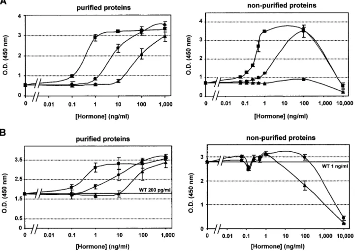
properties of S179D-hPRL in the Nb2 assay using the classical procedure recommended by Gout and colleagues (37, 38), who isolated the Nb2 cell clone and established the lactogen-induced proliferation assay. Figure 2A shows cell prolifer-ation induced by increasing concentrprolifer-ations of purified (left
panel) or nonpurified (right panel) WT hPRL, S179D-hPRL,
and G129R-hPRL. As expected, monomeric hPRL induces cell proliferation with a maximal effect at about 1 ng/ml, whereas the curve obtained for S179D-hPRL is shifted by 1 log unit toward the higher concentrations. As previously reported (26, 31), the agonistic dose-response curve obtained
with G129R-hPRL is shifted by 2 log units compared with that obtained with WT hPRL (Fig. 2A, left). It is noteworthy that the two mutants are able to induce a maximal prolifer-ation response after 3 d of stimulprolifer-ation when added at a sufficient concentration; this was confirmed by cell cycle studies performed using FACS analysis (not shown). Although the agonistic parts of the curve obtained for non-purified WT hPRL and S179D-hPRL (Fig. 2A, right) were similar to their purified counterparts, they were more mark-edly bell-shaped at the higher concentration tested (10g/ ml). The down-slope in dose-response curves for PRL and GH receptors is thought to reflect a phenomenon of self-antagonism (6, 23). However, as no self-self-antagonism was observed at the highest concentration with purified proteins (1000 ng/ml in this study, up to 100g/ml in Refs. 26, 31, and 33), it is possible that bell-shaped curves obtained using nonpurified hormones could result from an inhibitory effect of contaminants present at high doses of nonpurified prep-arations (see Fig. 1A and Discussion). Finally, nonpurified G129R-hPRL failed to induce proliferation.
We next investigated the antagonistic properties of hPRL analogs. Based on its agonistic properties, S179D-hPRL, whether purified or not, expectedly failed to display any antagonism. Indeed, when increasing amounts of S179D-hPRL were added to 0.2 ng/ml WT S179D-hPRL (producing⬃50% of maximal cell growth; see Fig. 2A, left), an increased dose response of proliferation was obtained from approximately 1 ng/ml of the analog (Fig. 2B, left), which appeared to be superimposed on the agonistic curve obtained when testing S179D-hPRL alone (Fig. 2A, left). The same observation was made for G129R-hPRL (compare left panels of Fig. 2, A and B). These experiments confirm the intrinsic agonistic prop-erties of both hPRL mutants in the Nb2 cell proliferation assay. When 1 ng/ml WT hPRL was competed with non-purified S179D-hPRL (Fig. 2B, right), the diminution of cell growth observed in the higher range of analog concentration was also superimposed on the curve obtained when testing nonpurified S179D-hPRL alone (see Fig. 2A, right), indicating that this effect is not a true antagonism, but reflects self-antagonism and/or contaminant effects, as discussed above. On the other hand, the curve obtained with nonpurified G129R-hPRL (Fig. 2B, right) may reflect true antagonism, as this protein fraction is intrinsically inactive (Fig. 2A, right). Finally, we noticed that in all of the antagonism experiments performed using nonpurified proteins (including WT hPRL), a small decrease in cell response occurred around 0.1– 0.2 ng/ml, which may reflect a slight antagonism at this very narrow range of low concentrations, although this was not seen with purified preparations (see Discussion).
Our results are in total contradiction with Chen’s report (20), which claimed an antagonistic activity for S179D-hPRL. These researchers performed their Nb2 proliferation assays using experimental conditions differing from the classical bioassay protocol, using a lower cell density. Whereas the standard protocol recommends a cell density of 105cells/ml on the starting day (37), Chen et al. used 2.5⫻ 104cells/ml for agonistic assays and still less (5⫻ 103cells/ml) for an-tagonistic assays. Therefore, we repeated all of our experi-ments using Chen’s protocol. With respect to agonism, we obtained dose-response curves qualitatively similar to those
presented in Fig. 2A, except that the changes in OD450values of the colorimetric assay (WST-1 reagent) were lower due to lower cell density (not shown). With respect to antagonistic assays, although using 1000 cells/well (200l) led to very small changes in OD450values (⬍0.1 absorbance unit) that were not very reliable in our hands, we never observed any evidence for S179D-hPRL antagonism (not shown).
In summary, our results clearly establish S179D-hPRL as a weak agonist, and not an antagonist, in the Nb2 cell pro-liferation assay regardless of the status of protein purification or cell density.
Activation of the JAK/STAT pathway. Based on the
obser-vation that S179D-hPRL activates STAT5 without signifi-cantly activating the tyrosine kinase JAK2, Walker’s (28) group proposed that this analog may exert its antagonistic properties by triggering alternate intracellular signals. As in our hands S179D-hPRL failed to display any antagonistic properties, but clearly acts as an agonist, it was important to confirm its effect on the JAK2/STAT5 pathway in Nb2 cells. Figure 3 shows dose-dependent activation of JAK2 (A) and
STAT5 (B) in Nb2 cells using purified (left panels) or nonpu-rified (right panels) hormone preparations. Regardless of the purification status, S179D-hPRL is able to activate JAK2 in a manner similar to WT hPRL, which contrasts to the report of Coss et al. (28). Even at 5 ng/ml, a faint band corresponding to phosphorylated JAK2 is already visible, indicating that detection of JAK2 activation does not require stimulation with supraphysiological concentrations of the analog. In con-trast, G129R-hPRL failed to induce strong activation of JAK2, even at a concentration of 500 ng/ml (7-fold less phosphor-ylated JAK2 compared with WT hPRL stimulation; Fig. 3A) or higher (not shown). This clearly indicates that the thresh-old of activation of signaling pathways does not correlate with that of mitogenic activity (compare Figs. 2 and 3). With respect to STAT5, the same observations were globally drawn (Fig. 5B), as both hPRL and S179D-hPRL (purified) achieved a similar level of STAT5 activation. Again, the level of STAT5 activation was markedly lower using G129R-hPRL, as it was when using nonpurified S179D-hPRL.
As reported previously (28), JAK2 and STAT5
coprecipi-FIG. 2. Nb2 cell proliferation assay. Proliferation assays of Nb2 cells in the presence of purified (left panels) or nonpurified (right panels) WT
hPRL (f), S179D-hPRL (F), or G129R-hPRL (Œ) were performed as indicated in Materials and Methods, using 105cells/ml on the starting day.
Cell proliferation was assessed using WST-1 colorimetric reagent. The data presented are representative of at least three independent experiments that were performed in triplicate or quadruplicate. Error bars represent theSD. A, Agonism. Cell proliferation induced by WT hPRL occurs in the range of 0.1–1 ng/ml. Proliferation curves observed in the presence of purified S179D-hPRL or G129R-hPRL are displaced to the high concentration by 1 and 2 log units, respectively, and reach maximal proliferation (same plateau as WT hPRL). Nonpurified WT hPRL and S179D-hPRL lead to bell-shaped curves, whereas nonpurified G129R-hPRL fails to induce cell growth. B, Antagonism. Purified hPRL analogs failed to antagonize cell division induced by 200 pg/ml WT hPRL. The decrease in cell growth in the presence of high concentrations of nonpurified analogs is discussed in the text.
tated when using anti-STAT5B antibodies, even in unstimu-lated cells (bottom panels of Fig. 3B). The only difference between WT hPRL and S179D-hPRL that we repeatedly ob-served in these studies was a lower phosphorylation level of the pool of JAK2 coimmunoprecipitating with STAT5
(ar-rowhead in Fig. 3B). This observation was not analyzed
fur-ther because it falls out of the scope of this study.
Another finding reported by Coss et al. (28) was the ability of S179D-hPRL to competitively inhibit hPRL-induced JAK2 phosphorylation, correlating to some extent with the
antag-onism observed in the Nb2 proliferation assay by these re-searchers (20). Using similar approaches, we investigated the antagonistic activity of our S179D-hPRL preparations (pu-rified or not) on hPRL-induced activation of JAK2 (Fig. 4), using G129R-hPRL as a control. S179D-hPRL failed to inhibit the effect of 500 ng/ml hPRL on JAK2 phosphorylation even when added in a 50-fold molar excess, and this was true whether purified or nonpurified analog was used. In con-trast, a 50-fold excess of G129R-hPRL clearly competed WT hPRL for JAK2 activation, as the phosphorylation of the
FIG. 4. Antagonism of hPRL and hPRL
analogs on JAK2 phosphorylation in Nb2 cells. Cell treatment and immuno-precipitations using JAK2 anti-bodies were performed as described in Fig. 3 (see also Materials and Methods). Antagonism of hPRL analogs was as-sessed by monitoring tyrosine phos-phorylation of JAK2 in the presence of a fixed concentration of WT hPRL (500 ng/ml) vs. increasing concentration of analogs (ratio indicated as WT:analog). When added in a 50-fold molar excess, S179D-hPRL failed to alter hPRL-induced JAK2 activation, whereas 10-and 50-fold excesses of G129R-hPRL decreased the level of JAK2 phosphory-lation by approximately 1.5- and 8-fold, respectively (arrowhead).
FIG. 3. Agonism of hPRL and hPRL an-alogs on the JAK2/STAT5 pathway in Nb2 cells. Nb2 cells (8⫻ 107) were
stim-ulated (5 min) with WT hPRL or hPRL analogs as indicated. Immunoprecipi-tations (IP) were performed on 3 mg cell lysates using JAK2 (A) or anti-STAT5 (B) antibodies. Immunoprecipi-tates were analyzed by Western blot-ting (WB) using antiphosphotyrosine antibody (P-Tyr). Blots were then stripped and reprobed using anti-JAK2 and/or anti-STAT5 antibodies as indi-cated at the bottom of each panel. Whether purified or not, S179D-hPRL is able to induce tyrosine phosphoryla-tion of JAK2 to a similar level as WT hPRL (STAT5 activation was lower us-ing nonpurified S179D-hPRL). In con-trast, using identical time exposure for autoradiography, G129R-hPRL fails to activate JAK2 and STAT5 above a min-imal threshold. The arrowhead indi-cates the lower level of phosphorylated JAK2 coprecipitating with STAT5 in S179D-hPRL-stimulated cells. IgGH, Ig
kinase was strongly impaired (by⬃10-fold) in the presence of the antagonist (Fig. 4, right). The possibility that this in-hibition may result from a toxic effect was ruled out, because no cell death was observed over a 3-d proliferation assay using even higher concentrations of this analog (31). It is noteworthy that the antagonistic effect observed on JAK2 was not directly reflected by the level of STAT5 phosphor-ylation (not shown), which is not fully understood (see
Discussion).
Human PRLR-mediated bioassays. All of the experiments
per-formed using rat Nb2 cells failed to confirm data previously claimed by others and, rather, supported the conclusion that S179D-hPRL acts as an agonist. Therefore, it was important to assess the properties of this analog in other cell systems, especially in cell bioassays using the homologous (human) PRL receptor. For this purpose we used two bioassays that we recently characterized as good models for highlighting antagonistic properties of human lactogen analogs (26, 27, 39).
Transcriptional activity (HL5 clone). We have previously
established a hPRLR-mediated transcriptional bioassay (so-called HL5 clone; see Materials and Methods) that enables us to unambiguously identify lactogens displaying antagonistic properties [e.g. mutants of hPRL (25, 26) or hGH (39)]. In this assay G129R-hPRL fails to act as an agonist even when tested at extremely high concentrations (120g/ml; Fig. 5A). How-ever, as it binds to the receptor it acts as an antagonist (Fig. 5B) (26). In contrast, S179D-hPRL again behaved as a weak agonist, as its curve was slightly displaced toward high con-centrations compared with WT hPRL (Fig. 4A). Averaged from three experiments, the ED50of WT hPRL was 2.3⫾ 0.3 g/ml, whereas a 4-fold higher concentration of the analog (9.7⫾ 0.5g/ml) was required to achieve a similar luciferase response. Surprisingly, the maximal activity obtained at the highest concentration tested (120g/ml) was systematically higher for purified S179D-hPRL than for WT hPRL
(respec-tively, 23.6⫾ 3.0- and 17.8 ⫾ 1.8-fold inductions; n ⫽ 6; Fig. 5A, left). In agreement with its (super) agonistic activity, no evidence of S179D-hPRL antagonism was observed, as high-lighted by the dose-dependent increase in luciferase activity when the analog and WT hPRL were tested together (Fig. 5B). These experiments were repeated using nonpurified mate-rial, and similar observations were made, i.e. that S179D-hPRL activated the S179D-hPRLR, whereas G129R-S179D-hPRL did not (not shown).
Proliferation of human mammary tumor cells (T-47D). We
recently reported that G129R-hPRL was able to competi-tively inhibit hPRL-induced proliferation of T-47D cells, and this was demonstrated by monitoring both cell density (WST-1) and cell cycle status in time-course experiments (27). The same procedure was repeated here for S179D-hPRL. As expected from the data presented above, this analog stimu-lated T-47D cell division, as reflected by the FACS profile monitored after 24 h of treatment. Due to its lower affinity for the hPRLR, however, a similar level of cell division re-quired higher amounts of S179D than WT hPRL (Fig. 6 and data not shown). In agreement with our previous report, G129R-hPRL (even at high concentrations) failed to activate T-47D cells. In competition experiments the latter efficiently competed with hPRL for stimulating entry of T-47D cells into the division cycle, whereas S179D-hPRL failed to exert any antagonistic effect.
Stimulation of the MAPK pathway in human mammary tumor cells (T-47D). One of the hallmarks of the antagonistic effect
of G129R-hPRL in T-47D cell division is its inhibitory effect on the activation of the MAPK pathway induced by WT hPRL (27). This assay was thus performed to definitely assess the effect of S179D-hPRL on the hPRLR at the molecular level. In agreement with proliferation data, S179D-hPRL did activate ERK1 and ERK2, although a little less efficiently than WT hPRL, whereas G129R-hPRL had no stimulatory effect
FIG. 5. LHRE-luciferase transcriptional assay. 293 cells stably expressing the hPRLR and the LHRE-luciferase reporter gene (clone HL5) were stimulated for 18 –24 h with increasing concentrations of purified WT hPRL (f), S179D-hPRL (F), or G129R-hPRL (Œ) as described in Materials
and Methods. One typical experiment performed in duplicate and representative of at least three experiments is shown. A, Agonism. Although
G129R-hPRL is almost inactive, the curve obtained for S179D-hPRL is slightly displaced to the right, but reaches higher final values (125%) compared with WT hPRL. B, Antagonism. Both G129R and S179D hPRL analogs were tested for their ability to compete a fixed concentration of WT hPRL (1g/ml, whose luciferase activity was normalized to 100%). G129R-hPRL inhibits the transcriptional activity induced by WT hPRL, whereas S179D-hPRL has an additive effect, reflecting its agonistic activity in this assay (see A).
(Fig. 7A) (27). In antagonistic experiments, the level of MAPK activation induced by 100 ng/ml hPRL (leading to mid scale activation of MAPK; see Fig. 7A) was competed by G129R-hPRL in a dose-dependent manner. In contrast, S179D-G129R-hPRL had an additive effect, leading to maximal levels of MAPK phosphorylation. These observations clearly indicate that S179D-hPRL exerts agonistic, but absolutely no antagonistic, activity on the human PRLR (Fig. 7B).
Discussion
In two recent studies (20, 28) Walker and colleagues re-ported that S179D-hPRL 1) does not induce the proliferation of Nb2 cells, 2) dramatically antagonizes the growth-promoting activity of WT hPRL on these cells, and 3) acti-vates STAT5 without (or minimally) activating JAK2. In this report we show that the same hPRL analog 1) acts as an agonist of the PRLR in the Nb2 as well as in various human receptor-mediated bioassays, 2) lacks any antagonistic ac-tivity toward WT hPRL in any of these bioassays, and 3) is a potent activator of JAK2, STAT5, and MAPK. The goal of this study was thus to explore why opposing conclusions were drawn for this hPRL analog despite the fact that it was produced using the same recombinant technology and char-acterized using the same bioassay (Nb2 cells). We
hypoth-esized that these differences could result from the intrinsic properties of protein preparations, the experimental proce-dures used for the cell-based assays, and/or different bio-logical tools, e.g. antibodies.
Upon refolding of inclusion bodies, about 50% of hPRL molecules assemble in high MM aggregates containing both covalent and noncovalent multimers. As PRL polymers have been long known to display reduced biological activity (for a review, see Ref. 41), we routinely eliminate these multimers from refolded hPRL through a single gel filtration step (25, 26, 31–33, 42). As illustrated by SDS-PAGE analysis, this step also eliminates most contaminants. In contrast, Walker et al. performed all of their studies using refolded, nonchromato-graphically purified S179D-hPRL. These researchers justified omitting the purification step by claiming that limiting the time of protein overexpression (IPTG) to 2 h, instead of 4 h as recommended in the initial protocol (32), reduces the amount of high MM contaminants pelleting within inclusion bodies (20), and their protein preparations did not contain oligomeric forms based on nonreducing SDS-PAGE analysis (data not shown in Ref. 20). We disagree with the latter argument, as noncovalent multimers cannot be detected by denaturing electrophoresis. Moreover, as we obtained indis-tinguishable chromatograms whether IPTG induction was
FIG. 6. Cell cycle analyses on human mammary tumor epithelial cells (T-47D). T-47D cells were serum-deprived for 24 h, then incubated in 0.5% charcoal-stripped serum in the absence (control) or presence of hormones, added alone for agonism experiments (100 ng/ml WT hPRL, 10g/ml G129R-hPRL, 10 g/ml S179D-hPRL), or in competition with WT hPRL for antagonism experiments (100 ng/ml WT hPRL combined to 10g/ml S179D-hPRL or G129R-hPRL). Cell cycle analyses were performed after 24 h. DNA content (2n and 4n indicated) vs. cell number is presented in each panel; from left to right, numbers indicate the percentage of cells in apoptosis (area below 2n label on x-axis), in G0/G1(peak
centered on 2n label) or in S/M phase (area above 2n label). Note that G129R-hPRL has no effect per se compared with the assay medium with respect to cell division or apoptosis (upper, right), but it completely antagonizes the proliferative effect of hPRL (lower, right). In contrast, S179D-hPRL stimulates cell division (upper, middle) and fails to antagonize WT hPRL in competition experiments (lower, middle). The results were identical using nonpurified hormones (not shown).
performed for 2 h (not shown) or 4 h, we anticipate that the nonpurified hormone preparations used by Chen and col-leagues are heterogeneous and contain a significantly amount of high MM proteins. Unfortunately, due to the absence of a gel filtration profile in their report, the true monomer/multimer ratio of their preparations cannot be assessed. Of more concern is that our nonpurified fraction probably contains bacterial contaminants normally eluted in the void volume of the molecular sieve, which may interfere with bioassays. The pronounced bell-shaped curves ob-served for nonpurified preparations compared with their purified counterparts may actually reflect such a PRL-inde-pendent effect resulting from bacterial contaminants pre-sumably present in significant amount at high doses of hPRL analogs. Our policy is to always use purified proteins for characterizing hPRL analogs. Therefore, we will not go into detailed analysis of data obtained using our nonpurified material, because interpretation of such results could be mis-leading. Their use was solely to assess that the purification parameter alone did not support the contradiction between Walker’s and our observations. Our data conclusively show that nonpurified S179D-hPRL acts as an agonist in the Nb2 proliferation assay. These observations were further con-firmed, first using the luciferase bioassay developed in our laboratory and mediated by the homologous (human) PRLR, and second by monitoring T-47D cell proliferation or
acti-vation of the MAPK cascade known to be triggered by PRL in this human mammary tumor cell line. Depending on bio-assay sensitivity, S179D-hPRL was clearly able to activate the hPRLR with an efficiency decreased by approximately 1 log unit, in perfect agreement with its 20-fold reduced binding affinity for this receptor. In summary, the discrepancy be-tween both reports cannot result from different intrinsic properties of the protein preparations used.
For 20 yr, monitoring Nb2 cell proliferation has been es-tablished as the reference bioassay for quantifying lactogen activity (37, 43). Although the classical experimental proce-dures recommend using 105cells/ml on the starting day of the assay, Walker and colleagues used 4 times less cell den-sity in their bioassays, which we assumed may have influ-enced some characteristics of cell responses to hormone stim-ulation. Supporting this hypothesis, Chen et al. (20) reported unusual dose-response curves, with maximal response peak-ing at 200 – 400 pg/ml, whereas normally maximal cell growth is seen at 1–2 ng/ml hPRL (37). Still more confusing, they observed hPRL self-antagonism (reflected by bell-shaped curves) at concentrations as low as 1.6 ng/ml (20), whereas we (31) and many others (44 – 47) failed to detect any self-antagonism of lactogens on Nb2 cells at much more elevated concentrations (up to 1g/ml). To clarify this issue, we repeated all of our experiments following Chen’s protocol (2.5⫻ 104cells/ml), and our results appeared qualitatively
FIG. 7. Agonism and antagonism of hPRL and hPRL analogs on ERK1/2 in T-47D cells. T47D cells were cultured until semiconfluence in six-well plates, then stimulated (30 min) with various doses of WT hPRL or hPRL analogs as indicated (A, agonism). In antagonistic experiments (B), a fixed amount of WT hPRL (100 ng/ml) was competed with increasing doses of hPRL analogs. Fifty micrograms of total cell lysates were loaded per lane. Blots were then ana-lyzed using anti-active MAP kinase an-tibodies (P-ERK1/2), then stripped and reprobed using anti-MAPK (ERK1/2) antibodies as indicated at the bottom of each panel. S179D-hPRL is able to in-duce tyrosine phosphorylation of MAPK to a level similar to, albeit slightly lower than, WT hPRL (compare 1g/ml con-centration for both). In contrast, using identical time exposure for autoradiog-raphy, G129R-hPRL fails to activate MAPK as previously reported (27). In antagonism experiments, although G129R-hPRL inhibits the activation of MAPK induced by WT hPRL at a 10:1 ratio, S179D-hPRL has an additive ef-fect on this pathway, reflecting its in-trinsic agonistic activity in this assay.
similar (not shown), i.e. that S179D-hPRL (purified or not) acted as a weak agonist on Nb2 cell proliferation.
As a first conclusion regarding agonistic assays, all of our data support that the agonistic properties of S179D-hPRL are real and were missed in Walker’s reports. The most probable reason for this misinterpretation is that their bioassays were performed using an inappropriate concentration range of ligand. In fact, the growth-promoting effect of S179D-hPRL in the Nb2 assay occurs between 1–100 ng/ml, whereas the concentrations tested by Chen and colleagues did not exceed 1.6 ng/ml. This hypothesis was definitely confirmed when a S179D-hPRL preparation provided by Walker was shown in our hands to exhibit agonistic properties very similar to those of our preparation (not shown) in both Nb2 and HL5 assays. As discussed previously (25), Nb2 cell proliferation is an extremely sensitive bioassay and therefore is probably not the most appropriate bioassay for highlighting the antago-nistic properties of hPRL analogs. For example, although G129R-hPRL (25–27) and G120R-hGH (or G120K-hGH) (24, 39, 48) have been characterized as potent antagonists toward the PRLR in various cell-based assays, both are weak agonists and fail to antagonize WT lactogens in the Nb2 cell prolif-eration assay (31, 39, 44). This can be explained by the fact that maximal Nb2 cell proliferation is achieved at very low receptor occupancy (49), as highlighted by immunoblot anal-yses where maximal activation of signal transducers (e.g. JAK2 and STAT5) requires concentrations as high as 500 ng/ml, when 1 ng/ml hPRL is sufficient to promote maximal cell proliferation. Hence, PRL/GH analogs whose ability to dimerize the receptor is strongly, but not totally, abolished maintain the ability to induce receptor dimer formation to a minimal level, sufficient to promote Nb2 cell division (50). In view of this experimental background, the highly potent antagonistic properties of S179D-hPRL in the Nb2 assay re-ported in Chen’s study appeared intriguing (20). They were still more questionable in view of the agonistic properties exhibited by S179D-hPRL in our hands. Expectedly, we failed to observe any evidence for antagonism when this analog was tested in competition experiments in the Nb2 assay. To circumvent the paradoxical response of Nb2 cells to hPRL analogs otherwise acting as potent antagonists, we have re-cently developed the PRLR-LHRE transcriptional assay that previously allowed unambiguous identification of PRL/GH mutants exhibiting antagonistic properties (25, 26, 39). As the transcriptional response, in contrast to cell division, is not (or minimally) affected by intracellular limiting factors, one can consider that the luciferase dose-response curve parallels that of receptor dimer formation (25, 50). In this bioassay, S179D-hPRL clearly lacks any antagonistic activity, whereas G129R-hPRL, the most potent hPRL antagonist currently available, does antagonize WT hPRL. These observations were further strengthened using human mammary T-47D cells. We (27) and others (48) have previously shown that PRL antagonists inhibit lactogen-induced proliferation of mammary tumor cells as well as the major PRLR-triggered signaling cascades, including JAK2; STAT1, -3, and -5; and MAPKs (27). In this study we monitored the inhibition of MAPK by hPRL analogs. Clearly, whereas G129R-hPRL leads to extinction of the phosphorylation signal induced by the intermediate dose of hPRL, S179D-hPRL still increases
MAPK activation. Therefore, we must conclude that S179D-hPRL is completely devoid of any detectable antagonistic activity. To understand why contradictory results were ob-tained, we repeated the experiments involving Nb2 cells using the protocol described in Chen’s study (20). In our hands, using a cell density as low as 5⫻ 103cells/ml ap-peared unreliable, as the OD450values were so low that the variation in absorbance (change in OD450,⬍0.1 U) was not significant. We repeatedly observed a small decrease in Nb2 cell response in a very narrow range of low concentrations using nonpurified hPRLs (including WT hPRL) in competi-tion experiments. Although we cannot propose any molec-ular mechanism to support this observation, such a phenom-enon limited in amplitude and in concentration range may be reminiscent of what Walker and colleagues interpreted as reflecting S179D-hPRL antagonism.
In a second report (28) the same group suggested that S179D-hPRL may activate alternative intracellular signaling cascades, although such pathways were not identified. In a more general fashion their data argued for a dissociation between JAK2 and STAT5 activation, in total contradiction with the conventional model of JAK-induced phosphoryla-tion of STATs (51). Again, we absolutely failed to confirm these observations, as S179D-hPRL, purified or not, appeared able to induce JAK2 phosphorylation in a fashion similar to WT hPRL in Nb2 cells. A tentative explanation for the dis-crepancy between the results of Walker’s group and ours might be that the level of JAK2 phosphorylation was, in general, low in their hands, but detectable (Fig. 3A in Ref. 28), which may have led the researchers to suggest the involve-ment of another kinase in STAT5 activation. Accordingly, we obtained stronger signals using the anti-JAK2 antibody from Upstate Biotechnology, Inc., compared with that from Santa Cruz Biotechnology, Inc., used in their study (data not shown), which, however, does not support the discrepancy, as the antibody from Santa Cruz Biotechnology, Inc., suc-cessfully detected JAK2 phosphorylation induced by WT hPRL (28). Moreover, we observed that G129R-hPRL in-duced only a limited level of JAK2 phosphorylation, dem-onstrating that our experimental procedures were sensitive enough to identify analogs exhibiting lower ability to acti-vate this signaling cascade. According to these observations regarding agonistic effect, we failed to confirm the antago-nistic activity of S179D-hPRL on hPRL-induced JAK2 acti-vation previously reported by these researchers, whereas the antagonism of G129R-hPRL could be detected, in agreement with its inhibitory effect on PRLR signaling pathways re-cently demonstrated in breast cancer cells (this study and Ref. 27). The only difference repeatedly observed between S179D-hPRL and WT hPRL is a decrease in the pool of phos-phorylated JAK2 coprecipitating with STAT5, which may reflect a difference in the stoichiometry and/or conformation of PRLR/JAK2/STAT5 complexes depending on the ligand (see below). Obviously, the importance of this observation will require further investigation, which, however, is out of the scope of the present study.
All of the results presented in this study clearly establish S179D-hPRL as an agonist (although weaker than hPRL), not as an antagonist, of the PRL receptor. However, our report raises many questions, the first of which is how this hPRL
analog should be categorized. Several arguments suggest that S179D-hPRL is a binding site 1 rather than a binding site 2 analog. Based on the three-dimensional model that we established (52), serine 179 is predicted to belong to helix 4, the region of binding site 1 containing the highest number of binding determinants (6). Moreover, the dose-response curves obtained for S179D-hPRL in the various bioassays investigated here are displaced to the right compared with WT hPRL and achieve a maximal response (or supramaxi-mal, see below), two features that fit with alteration of bind-ing site 1 (6, 33, 42, 50). Although S179D-hPRL can be con-sidered as a site 1 analog, this does not necessarily mean that serine 179 is intrinsically involved in an interaction with the PRLR. One of the most powerful methods for identifying binding determinants of PRLs or GHs has proved to be alanine scanning (for review, see Ref. 6). Although Chen et
al. (20) reported that alanine replacement of serine 179
abol-ishes hPRL activity, which would argue that this residue is functionally relevant, we cannot exclude that the putative agonistic activity of the S179A-hPRL analog could have been missed, as it was for S179D-hPRL (see above). In contrast to all binding determinants identified to date (6), serine 179 is predicted to point toward the hydrophobic core of the pro-tein (52), which presumably precludes this residue from di-rectly interacting with the receptor. This structural hypoth-esis is corroborated by isoelectric focusing experiments showing that the pI of S179D-hPRL is identical to that of WT hPRL despite the addition of a negative charge (Asp for Ser). We expect that Asp179(and Ser in WT hPRL) is buried and that its putative charge does not influence electrophoretic mobility. If true, this would suggest that the reduced ago-nism of S179D-hPRL would not result from the mutation of Ser179per se, but, rather, from the effect this mutation has on overall protein structure. The estimation of S179D-hPRL ap-parent MM using analytical gel filtration did not reveal any detectable change with respect to WT hPRL, but this ap-proach is obviously not very sensitive to small structural variations. Circular dichroism previously enabled us to de-tect structural disturbance of hPRL analogs (e.g. C58A or S26W) (31, 33). Using this approach, we failed to detect any significant alteration of secondary structure content of S179D-hPRL, as its spectrum and calculated helicity were not significantly different from those of WT hPRL (this study and Refs. 31 and 33). In agreement with Chen and colleagues, who used RIA recognition to monitor conformational dis-turbances, we thus conclude that the S179D substitution does not dramatically influence the overall hPRL structure.
As Ser179is presumably buried and, in addition, mutation of this residue leads to reduction of affinity and of agonistic activity without any detectable structural alteration, we hy-pothesize that aspartate substitution results in local distur-bance of regions involved in binding site 1, most probably of helix 4. Recent investigation of Walker’s group indicates that Ser179can be phosphorylated in vitro in the human hormone (Lorenson, M. Y., and A. Walker, personal communication), as already demonstrated for the homologous Ser177in rPRL (18), which, however, does not mean that these residues actually are phosphorylated in the pituitary in vivo. Eluci-dating the mechanism by which an amino acid predicted to be buried inside the protein becomes accessible to a kinase
remains open to investigation. Whether S179D-hPRL actu-ally exhibits the same properties as a putative S179-phos-phorylated hPRL, thereby justifying being referred to as a molecular mimic of phosphorylated hPRL remains an open question (20, 53).
The final and probably most important questions relate to the intrinsic properties that S179D-hPRL exerts in vivo when injected to animals. Within the last year, several reports on the effect of S179D-hPRL in various rodent models have been presented. Nonexhaustively, S179D-hPRL was reported to reduce the tumor incidence of cancer prostate cells injected into nude mice (54) and in rats to alter maternal behavior in nulliparous female (55) and to affect T cell survival (56), which is in good agreement with the antagonistic properties claimed by Walker and colleagues from their in vitro studies (20, 28), but obviously does not agree with our conclusions. In contrast, S179D-hPRL was also shown to promote lobu-loalveolar differentiation and casein expression during rat pregnancy (57). Moreover, a very recent publication by the same group described S179D-hPRL as an even more potent agonist than WT hPRL on the inhibition of osteoblastic al-kaline phosphatase activity and bone development in rats (58), which fits with the agonistic activity demonstrated for this analog in the present work. Although one of our con-cerns about the relevance of these preliminary in vivo studies is whether nonpurified proteins obtained from bacteria are a suitable material to be injected into animals, especially for investigations of immunological parameters, the paradoxical properties of S179D-hPRL both in vitro and in vivo obviously argue that this hPRL mutant should be viewed with much caution and certainly not as a full antagonist as suggested previously (20, 55, 56). Unexpectedly, our transcriptional bioassay revealed that although S179D-hPRL behaves as a slightly weaker agonist than hPRL (dose-response curve dis-placed to the right), it induces a luciferase response of higher amplitude (⬃125%) compared with WT hPRL, thereby dis-playing superagonistic properties in vitro. As this phenom-enon was also observed using the hormone preparation pro-vided by Walker (not shown), it may correlate with the superagonistic activity reported for S179D-hPRL on the in-hibition of bone formation (58), although this reported effect would be opposed to the reduced bone development we have reported in PRL receptor knockout mice (59). This sug-gests that once the functional hormone-receptor complex is formed (which requires higher hormone concentration due to reduced affinity), S179D-hPRL might be able to trigger signaling cascades more efficiently than the natural ligand. Whether this property is related to prolonged signal trans-duction, a different kon/koffaffinity constant, conformational changes in the PRLR, or some other molecular feature re-mains to be explored.
In conclusion, the present report clearly establishes S179D-hPRL as a PRLR agonist and suggests that the antagonistic properties claimed for this analog were deduced from inap-propriate bioassays. Although it is assumed that phosphor-ylated PRL plays a role in autoregulation of PRL secretion from the pituitary or is an intracellular component acting as a regulator of trafficking and/or storage of PRL molecules, the physiological relevance of this PRL variant remains poorly understood (12). Based on injection of S179D-hPRL to
pregnant rats, it was recently proposed that the ratio of nonphosphorylated vs. phosphorylated hPRL, rather the lat-ter per se, may be physiologically relevant. We strongly feel that extrapolation of findings with S179D-hPRL to the po-tential physiological relevance of phosphorylated hPRL in
vivo remains to be explored and should await the definite
demonstration that the S179D mutation actually confers properties similar to those specifically exhibited by phos-phorylated PRL.
Acknowledgments
The authors are grateful to Dr. A. M. Walker for extensive discussion of experimental data, supply of their S179D-hPRL analog, and the friendly communication of unpublished observations despite the sci-entific controversy. Dr. P. W. Gout (Vancouver, Canada) is acknowl-edged for providing Nb2 cells, Dr. C. Gerday (Liege, Belgium) for lend-ing the cell disintegrator, and Roland Vaudry for his precious help in running bioassays.
Received December 6, 2000. Accepted May 9, 2001.
Address all correspondence and requests for reprints to: Dr. Vincent Goffin, INSERM, U-344, Molecular Endocrinology, Faculte´ de Me´decine Necker, 156 rue de Vaugirard, 75730 Paris Cedex 15, France. E-mail: [email protected].
This work was supported in part by INSERM, the Association pour la Recherche sur le Cancer, and the Ligue Nationale contre le Cancer.
References
1. Bole-Feysot C, Goffin V, Edery M, Binart N, Kelly PA 1998 Prolactin and its receptor: actions, signal transduction pathways and phenotypes observed in prolactin receptor knockout mice. Endocr Rev 19:225–268
2. Horseman ND, Zhao W, Montecino-Rodriguez E, et al. 1997 Defective mam-mopoiesis, but normal hematopoiesis, in mice with a targeted disruption of the prolactin gene. EMBO J 16:6926 – 6935
3. Ormandy CJ, Camus A, Barra J, et al. 1997 Null mutation of the prolactin receptor gene produces multiple reproductive defects in the mouse. Genes Dev 11:167–178
4. Kelly PA, Djiane J, Postel-Vinay MC, Edery M 1991 The prolactin/growth hormone receptor family. Endocr Rev 12:235–251
5. Goffin V, Kelly PA 1996 Prolactin and growth hormone receptors. Clin En-docrinol (Oxf) 45:247–255
6. Goffin V, Shiverick KT, Kelly PA, Martial JA 1996 Sequence-function rela-tionships within the expanding family of prolactin, growth hormone, placental lactogen and related proteins in mammals. Endocr Rev 17:385– 410 7. Wells JA 1996 Binding in the growth hormone receptor complex. Proc Natl
Acad Sci USA 93:1– 6
8. Llovera M, Touraine P, Kelly PA, Goffin V 2000 Involvement of prolactin in breast cancer: redefining the molecular targets. Exp Gerontol 35:41–51 9. Lebrun JJ, Ali S, Sofer L, Ullrich A, Kelly PA 1994 Prolactin-induced
pro-liferation of Nb2 cells involves tyrosine phosphorylation of the prolactin re-ceptor and its associated tyrosine kinase JAK2. J Biol Chem 269:14021–14026 10. Rui H, Lebrun JJ, Kirken RA, Kelly PA, Farrar WL 1994 Jak2 activation and cell proliferation induced by antibody-mediated prolactin receptor dimeriza-tion. Endocrinology 135:1299 –1306
11. Wakao H, Gouilleux F, Groner B 1994 Mammary gland factor (MGF) is a novel member of the cytokine regulated transcription factor gene family and confers the prolactin response. EMBO J 13:2182–2191
12. Sinha YN 1995 Structural variants of prolactin: occurence and physiological significance. Endocr Rev 16:354 –369
13. Brooks CL, Kim BG, Aphale P, Kleeman BE, Johnson GC 1990 Phosphor-ylated variant of bovine prolactin. Mol Cell Endocrinol 71:117–123 14. Kim BG, Brooks CL 1993 Isolation and characterization of phosphorylated
bovine prolactin. Biochem J 296:41– 47
15. Oetting WS, Tuazon PT, Traugh JA, Walker AM 1986 Phosphorylation of prolactin. J Biol Chem 261:1649 –1652
16. Mastro RM, Dannies PS 1995 Lack of correlation of distribution of prolactin (PRL) charge isoforms with induction of PRL storage. Endocrinology 136: 69 –74
17. Aramburo C, Montiel JL, Proudman JA, Berghman LR, Scanes CG 1992 Phosphorylation of prolactin and growth hormone. J Mol Endocrinol 8:183–191 18. Wang YF, Liu JW, Mamidi M, Walker AM 1996 Identification of the major site
of rat prolactin phosphorylation as serine 177. J Biol Chem 271:2462–2469 19. Wang YF, Walker AM 1993 Dephosphorylation of standard prolactin
pro-duces a more biologically active molecule: evidence for antagonism between
nonphosphorylated and phosphorylated prolactin in the stimulation of Nb2 cell proliferation. Endocrinology 133:2156 –2160
20. Chen TJ, Kuo CB, Tsai KF, Liu JW, Chen DY, Walker AM 1998 Development of recombinant human prolactin receptor antagonists by molecular mimicry of the phosphorylated hormone. Endocrinology 139:609 – 616
21. Wicks JR, Brooks CL 1995 Biological activity of phosphorylated and dephos-phorylated bovine prolactin. Mol Cell Endocrinol 112:223–229
22. Luck DN, Gout PW, Beer CT, Smith M 1989 Bioactive recombinant methionyl bovine prolactin: structure-function studies using site-specific mutagenesis. Mol Endocrinol 3:822– 831
23. Fuh G, Cunningham BC, Fukunaga R, Nagata S, Goeddel DV, Wells JA 1992 Rational design of potent antagonists to the human growth hormone receptor. Science 256:1677–1680
24. Fuh G, Colosi P, Wood WI, Wells JA 1993 Mechanism-based design of pro-lactin receptor antagonists. J Biol Chem 268:5376 –5381
25. Goffin V, Kinet S, Ferrag F, Binart N, Martial JA, Kelly PA 1996 Antagonistic properties of human prolactin analogs that show paradoxical agonistic activity in the Nb2 bioassay. J Biol Chem 271:16573–16579
26. Kinet S, Bernichtein S, Kelly PA, Martial JA, Goffin V 1999 Biological properties of human prolactin analogs depend not only on global hormone affinity, but also on the relative affinities of both receptor binding sites. J Biol Chem 274:26033–26043
27. Llovera M, Pichard C, Bernichtein S, et al. 2000 Human prolactin (hPRL) antagonists inhibit hPRL-activated signaling pathways involved in breast can-cer cell proliferation. Oncogene 19:4695– 4705
28. Coss D, Kuo CB, Yang L, Ingleton P, Luben R, Walker AM 1999 Dissociation of Janus kinase 2 and signal transducer and activator of transcription 5 acti-vation after treatment of Nb2 cells with a molecular mimic of phosphorylated prolactin. Endocrinology 140:5087–5094
29. Goffin V, Touraine P, Pichard C, Bernichtein S, Kelly PA 1999 Should prolactin be reconsidered as a therapeutic target in human breast cancer? Mol Cell Endocrinol 151:79 – 87
30. Cooke NE, Coit D, Shine J, Baxter JD, Martial JA 1981 Human prolactin: cDNA structural analysis and evolutionary comparisons. J Biol Chem 256: 4007– 4016
31. Goffin V, Struman I, Mainfroid V, Kinet S, Martial JA 1994 Evidence for a second receptor binding site on human prolactin. J Biol Chem 269:32598 –32606 32. Paris N, Rentier-Delrue F, Defontaine A, et al. 1990 Bacterial production and purification of recombinant human prolactin. Biotechnol Appl Biochem 12: 436 – 449
33. Goffin V, Norman M, Martial JA 1992 Alanine-scanning mutagenesis of human prolactin: importance of the 58 –74 region for bioactivity. Mol Endo-crinol 6:1381–1392
34. Lochnan HA, Buteau H, Richards S, Edery M, Kelly PA 1995 Functional activity of the human prolactin receptor and its ligands. Mol Cell Endocrinol 114:91–99
35. Laemmli UK 1970 Cleavage of structural proteins during the assembly of the head of bacteriophage T4. Nature 227:680 – 685
36. Andrade MA, Chacon P, Merelo JJ, Moran F 1993 Evaluation of secondary structure of proteins from UV circular dichroism spectra using an unsuper-vised learning neural network. Protein Engin 6:383–390
37. Tanaka T, Shiu RP, Gout PW, Beer CT, Noble RL, Friesen HG 1980 A new sensitive and specific bioassay for lactogenic hormones: measurement of pro-lactin and growth hormone in human serum. J Clin Endocrinol Metab 51: 1058 –1063
38. Gout PW, Beer CT, Noble RL 1980 Prolactin-stimulated growth of cell cultures established from malignant Nb rat lymphomas. Cancer Res 40:2433–2436 39. Goffin V, Bernichtein S, Carrie`re O, Bennett WF, Kopchick JJ, Kelly PA 1999
The human growth hormone antagonist B2036 does not interact with the prolactin receptor. Endocrinology 140:3853–3856
40. Goffin V, Struman I, Goormaghtigh E, Martial JA 1993 The addition of nine residues at the C-terminus of human prolactin drastically alters its biological properties. Eur J Biochem 214:483– 490
41. Sinha YN 1992 Prolactin variants. Trends Endocrinol Metab 3:100 –106 42. Kinet S, Goffin V, Mainfroid V, Martial JA 1996 Characterization of lactogen
receptor binding site 1 of human prolactin. J Biol Chem 271:14353–14360 43. Shiu RP, Elsholtz HP, Tanaka T, et al. 1983 Receptor-mediated mitogenic
action of prolactin in a rat lymphoma cell line. Endocrinology 113:159 –165 44. Dattani MT, Hindmarsh PC, Brook CD, Robinson IF, Kopchick JJ, Marshall
NJ1995 G120R, a human growth hormone antagonist, shows zinc-dependent agonist and antagonist activity on Nb2 cells. J Biol Chem 270:9222–9226 45. Gertler A, Shamay A, Cohen N, et al. 1986 Inhibition of lactogenic activities
of ovine prolactin and human growth hormone (hGH) by a novel form of modified recombinant hGH. Endocrinology 118:720 –726
46. Binder L, Gertler A, Elberg G, Guy R, Vogel T 1990 Site-directed mutations of human growth hormone that selectively modify its lactogenic activity and binding properties. Mol Endocrinol 4:1060 –1068
47. Sakal E, Elberg G, Gertler A 1997 Direct evidence that lactogenic hormones induce homodimerization of membrane-anchored prolactin receptor in intact Nb2–11C rat lymphoma cells. FEBS Lett 410:289 –292
48. Fuh G, Wells JA 1995 Prolactin receptor antagonists that inhibit the growth of breast cancer cell lines. J Biol Chem 270:13133–13137