Contents lists available atScienceDirect
Meat Science
journal homepage:www.elsevier.com/locate/meatsci
Carnobacterium maltaromaticum as bioprotective culture in vitro and in
cooked ham
Gabriela Maia Danielski
a,b, Pedro Henrique Imazaki
b, Caroline Maria de Andrade Cavalari
a,
Georges Daube
c, Antoine Clinquart
b, Renata Ernlund Freitas de Macedo
a,⁎aLaboratory of Agrifood Food Research and Inovation (LAPIAgro), Graduate Program in Animal Science, School of Life Sciences, Pontifícia Universidade Católica do
Paraná, Imaculada Conceição, 1155, 80215-901 Curitiba, Brazil
bLaboratory of Food Technology, Faculty of Veterinary Medicine, Fundamental and Applied Research for Animals & Health (FARAH), University of Liège, Avenue de
Cureghem 10, 4000 Liège, Belgium
cLaboratory of Food Microbiology, Faculty of Veterinary Medicine, Fundamental and Applied Research for Animals & Health (FARAH), University of Liège, Avenue de
Cureghem 10, 4000 Liège, Belgium
A R T I C L E I N F O
Keywords: Antilisterial activity Bioprotective culture Meat product Lactic acid bacteria
A B S T R A C T
The bioprotective effects of Carnobacterium maltaromaticum (CM) strains were assessed in vitro and in sliced cooked ham. CM strains were tested in vitro against Listeria monocytogenes (LM), Escherichia coli O157:H7 (EC) and Salmonella Typhimurium (ST). In vitro effect was evaluated using co-culture (with and without EDTA) and cell-free supernatant (CFS). CFS was tested by agar well diffusion and minimum inhibitory concentration. In cooked ham, the inhibitory effect of CM on L. innocua (LI) and on the physicochemical parameters were eval-uated for 7 days at 4 °C. In co-cultures at−1 °C and 4 °C, all CM isolates inhibited LM. A slight inhibition was observed against the Gram-negative bacteria with the addition of EDTA. CFS did not show inhibitory effect under the studied conditions. In cooked ham, CM inhibited LI growth and did not affect the physicochemical parameters of the product during storage. CM strains show potential to be used as bioprotective cultures in cold-stored cooked ham and improve its safety.
1. Introduction
Foodborne disease outbreaks are caused by the ingestion of con-taminated food with pathogenic microorganisms. The World Health Organization (WHO) considers foodborne outbreaks as critical threats to global health (WHO, 2015). In the Unites States, an estimated 9 million people get sick, 56,000 are hospitalized, and 1300 die of foodborne disease each year (IFSAC, 2019). Salmonella spp. is reported as the cause of 14% of the foodborne diseases in the European Union (EFSA & ECDC, 2018). Other bacteria play an essential role in these outbreaks including Escherichia coli O157:H7 and Listeria mono-cytogenes. These three pathogens are associated to the highest level of frequency, severity of ilnnes, hospitalizations and deaths caused by foodborne illness, linked mainly with chicken, pork and beef (EFSA & ECDC, 2018;IFSAC, 2019).
Meat products characteristics such as high protein content, low acidity, and high-water activity make them susceptible to microbial growth (Sánchez-Ortega et al., 2014). According to the European Food Safety Authority (EFSA) and the European Centre for Disease
Prevention and Control (ECDC) (2017) in 2015, meat products were linked to the most critical cases of foodborne outbreaks in the European Union. Moreover, in Brazil, meat and meat products s were responsible for 12.8% of the outbreaks from 2009 to 2018 (Brazil, 2019). However, the frequency is probably higher than that reported due to the lack of notification of foodborne illness when the symptoms are mild, and patients do not seek medical assistance (de Oliveira, de Paula, Cardoso, & Tondo, 2010).
The food industry has largely investigated the potential use of LAB as biopreservatives. Their inhibitory effect against pathogenic and spoilage bacteria (Alves, Martinis, Destro, Vogel, & Gram, 2005; dos Reis et al., 2011;Hammi et al., 2016;Ho, Lo, Bansal, & Turner, 2018; Huang, Ye, Yu, Wang, & Zhou, 2016;Rivas, Castro, Vallejo, Marguet, & Campos, 2014) is an important indicator of their possible use as pro-tective cultures in food matrices, where they could replace some syn-thetic preservatives (Engelhardt, Albano, Kiskó, Mohácsi-Farkas, & Teixeira, 2015;Huang et al., 2016).
Carnobacteria are ubiquitous lactic acid bacteria (LAB) isolated from cold and temperate environments and can be found as natural
https://doi.org/10.1016/j.meatsci.2019.108035
Received 23 September 2019; Received in revised form 10 December 2019; Accepted 11 December 2019
⁎Corresponding author.
E-mail address:renata.macedo@pucpr.br(R.E.F.d. Macedo).
Available online 16 December 2019
0309-1740/ © 2019 Elsevier Ltd. All rights reserved.
microbiota of chilled meat, fish and dairy products. Among the 11 species of Carnobacterium, two species, Carnobacterium divergens and Carnobacterium maltaromaticum, are frequently isolated from food and show the ability to inhibit pathogenic and spoilage microorganisms in diverse food matrices (Leisner, Laursen, Prévost, Drider, & Dalgaard, 2007). Thus, their use as bioprotective cultures in food has been con-sidered (Iskandar et al., 2017;Orihuel et al., 2018). The antimicrobial properties of Carnobacterium spp. have been studied in vitro (Hammi et al., 2016;Tulini et al., 2014), in cold-smoked salmon (Brillet-Viel, Pilet, Courcoux, Prévost, & Leroi, 2016), ricotta (Spanu et al., 2018), cooked and peeled shrimp, and as a feed additive for rabbits to improve meat microbial quality and safety (Koné et al., 2018). The use of Car-nobacterium spp. has also been investigated as a probiotic culture in broiler chickens (Smialek, Burchardt, & Koncicki, 2018). However, the effect of C. maltaromaticum as a bioprotective culture in meat products, especially ready-to-eat products, has not been assessed.
This study aimed to evaluate the antimicrobial effect of C. mal-taromaticum towards different food pathogenic bacteria in vitro and to assess the potential of its use as a bioprotective culture in cold-stored sliced cooked ham.
2. Material and methods
2.1. Bacterial strains, media, and growth conditions
Three different strains of C. maltaromaticum (CM_B824, CM_B827, and CM_B829), obtained from Australian vacuum packaged chilled beef (longissimus thoracis et lumborum) with a long shelf life (140 days at −1 °C) were used in this study (Imazaki et al., 2015). These strains were selected among 11 CM strains after the sequencing of their genome, which revealed the existence of three main phylogenetic groups. Therefore, one strain of each group was selected to be used in the present study.
Foodborne pathogenic bacteria, L. monocytogenes ATCC®19117™ (LM), E. coli O157:H7 ATCC®35150™ (EC) and Salmonella Typhimurium ATCC®14028™ (ST) were used for the in vitro essay, and a pool of Listeria innocua (LI_33314, LI_33016 and LI_HPB586), isolated from meat products, belonging to the culture collection of LAPIAgro, was used as surrogate for LM in the cooked ham essay.
CM strains were grown in brain heart infusion (BHI) broth (Kasvi, São José dos Pinhais, Brazil) at 25 °C for 48 h, and pathogenic and LI strains at 37 °C for 24 h in the same medium. Growth was verified by optical density at 540 nm for the pathogenic bacteria and LI and 620 nm for CM (Gutiérrez, Martínez-Blanco, Rodríguez-Aparicio, & Ferrero, 2016).
2.2. In vitro assay
2.2.1. Antimicrobial effect of CM in co-culture
The antimicrobial effect of CM in co-culture, where both CM strains and pathogens were grown together (cell-to-cell contact), was de-termined inoculating Falcon® flasks with 30 mL of BHI broth with each strain of CM at 6.0 log CFU/mL in order to account for natural con-tamination levels and allow the enumeration of the inoculum and one of the pathogenic strains (LM, EC, ST) at 3.0 log CFU/mL. Negative controls were considered flasks with BHI inoculated with each pa-thogen at 3.0 log CFU/mL and positive controls were consideredflasks inoculated only with CM strains. Controls and treatments were con-ducted in triplicate.
Flasks were incubated at−1 °C for 28 days, 4 °C for 14 days and 25 °C for 48 h in a shaker (Rotamax 120, Heidolph, Schwabach, Germany) at 150 rpm. Pathogenic bacteria counts were performed using specific chromogenic media: RAPID'L.mono, RAPID'E.coli 2 and, RAPID'Salmonella (BioRad, Marnes, France). CM population was esti-mated as the difference between counts on plate count agar (PCA) (BioRad, Marnes, France) and chromogenic media. The plating was also
conducted in triplicate.
2.2.2. Antimicrobial effect of CM in co-culture with the addition of EDTA The influence of the addition of ethylenediaminetetraacetic acid (EDTA) (VWR, Radnor, USA) on the effect of CM against pathogens was investigated, since this chelating agent showed synergestic interaction with bacteriocins from gram-positive bacteria targeting gram-negatives (Mathur et al., 2017). Co-cultures were carried out inflasks containing BHI broth with EDTA 1.0 mM, incubated at 25 °C for 48 h, in a shaker at 150 rpm. Bacterial counts were performed following the same proce-dure described above. Tofind the concentration of EDTA that would not interfere in the bacterial growth by itself, a previous experiment was conducted. Serial concentrations of EDTA (1, 5, 10, 20 and 40 mM) were added to the broth medium with each pathogen (LM, EC, ST) (Bordignon-Junior et al., 2012). The concentration that showed no difference in growth compared to negative control, inoculated broth without EDTA, was selected.
2.2.3. Antimicrobial effect of CM cell-free supernatant (CFS) using agar well diffusion
To check if the antimicrobial effect of CM was mediated by the production of antimicrobial molecules in the culture supernatant, three tubes containing 10 mL of BHI broth were inoculated with each CM strain. The tubes were incubated at 25 °C for 48 h and centrifuged (Model Eppendorf Centrifuge 5804, Hamburg, Germany) at 16,000 g for 10 min. The supernatant of two tubes was treated with sodium hy-droxide (NaOH) 1 M (VWR, Radnor, USA) until pH 6.5 to neutralize antimicrobial effect related to the undissociated form of organic acids potentially produced by CM. Finally, the supernatant of the last tube wasfiltered through 0.2 μm sterile Minisart syringe filters (Sartorius, Germany), resulting in a cell-free supernatant (CFS). The supernatants were inoculated in wells made in three PCA plates, previously spread with 100μL of each pathogenic bacterium (LM, EC, ST) at 6.0 log CFU/ mL. Four treatments were applied on each plate: (1) sterile BHI broth (blank), (2) centrifuged supernatant, (3) centrifuged supernatant treated with NaOH, and (4) centrifuged supernatant treated with NaOH andfiltered (CFS). The halo of inhibition was measured after 48 h of incubation at 37 °C, and all treatments were performed in triplicate. 2.2.4. Minimum inhibitory concentration (MIC) of CM cell-free supernatant (CFS) towards LM
CM_B824, CM_B827 and CM_B829 at 6.0 log CFU/mL were in-cubated at two different growth conditions (at 25 °C for 48 h and 4 °C for 14 days) in co-culture with and without LM (3.0 log CFU/ mL). The combination of the different parameters resulted in 12 treatments: (1) CM_B824 at 25 °C (2) CM_B824 + LM at 25 °C; (3) CM_B824 at 4 °C, (4) CM_B824 + LM at 4 °C, (5) CM_B827 at 25 °C, (6) CM_B827 + LM at 25 °C; (7) CM_B827 at 4 °C, (8) CM_B827 + LM at 4 °C, (9) CM_B829 at 25 °C, (10) CM_B829 + LM at 25 °C; (11) CM_B829 at 4 °C, (12) CM_B829 + LM at 4 °C.
After co-culture incubation, CFS of different treatments was ob-tained as described previously. CFS obob-tained from co-cultures and LM at 6.0 log CFU/mL (10μL), grown in BHI broth, were inoculated in 24-well plates at different concentrations: 62.5, 50, 37.5 and 18.75%, that were composed by the combination v/v of CFS and sterile BHI broth. Plates were incubated at 37 °C for 24 h at 150 rpm, in triplicate. The growth of LM was verified visually. The minimum inhibitory con-centration (MIC) was considered as the lowest concon-centration of the supernatant where no visible growth was observed (CLSI, 2012). 2.3. Cooked ham essay
The meat matrix essay was performed considering the results ob-tained in the in vitro essay. Therefore, the bioprotective effect of CM strains was assessed in a meat matrix stored at refrigeration against Listeria sp. A pool of L. innocua (LI) was used as surrogate for LM in this
essay.
2.3.1. Effect of CM against LI in sliced cooked ham
Cooked ham was processed by a medium scale producer in Southern Brazil with pork ham muscles (M. semimembranosus, M. semitendinosus and M. biceps femoris) (4% fat). Muscles were injected with a brine solution (25 g brine/100 g ham meat), containing the following in-gredients (g/ Kg): water, 100; salt, 20; sodium tri-polyphosphate, 3.0; sodium pyrophosphate 1.0; NaNO2, 0.130; NaNO3, 0.260; sodium
er-ythorbate, 0.5; monosodium glutamate, 2; natural carmine dye, 0.3. After injection, meat was tumbled under vacuum at 4 °C. After tum-bling, the product was stuffed in multilayer shrinkable plastic casings (Schur, Barueri, São Paulo, Brazil) and placed in stainless steel molds (110 mm width, 120 mm height, 230 mm length). The product was then cooked in water bath at 85 °C until core temperature of 72 °C was reached. After cooking the product was pre-cooled in an ice bath until the core temperature was 50 °C and then cooled in a chilling room at 0 °C until reaching 4 °C. After a minimum of 24 h of cooling, the mold was removed, and the product was sliced (dimensions 3 mm × 105 mm width × 115 mm height) and packaged in expanded polystyrene trays and covered with low density polyvinyl chloridefilm.
Cooked ham slices from the same batch, with a shelf-life of 7 days, were purchased at the producer store. Slices were sterilized at 121 °C for 15 min (Alves, Martinez, Lavrador, & De Martinis, 2006) to avoid the interference of the natural microbiota and chilled overnight at 4 °C. LI cold adaptation was performed as follows: strains were in-oculated in BHI at 37 °C until stationary phase and subsequently cul-tured in BHI broth at 4 °C until stationary phase, which was confirmed by colony count in PCA agar plates. Equal volumes of each LI strain were mixed in a sterileflask to form a pool at the concentration of 3.0 log CFU/ mL. Then, three slices were distributed into eight treatments: (1) NC (negative control); (2) CM_B824; (3) CM_B827; (4) CM_B829; (5) LI; (6) CM_B824 + LI; (7) CM_B827 + LI; and (8) CM_B829 + LI.
Each side of ham slice was inoculated with 50μL of the LI pool at 3.0 log CFU/ mL, which was spread evenly with a sterile Drigalski spatula. NC was inoculated with 50μL 0.1% peptone water. To allow the absorption of the inoculum, the slices were kept in a laminarflow cabinet for 1 h at room temperature. Then, the slices were inoculated with 50μL of each CM strain at 5.0 log CFU/ mL, except treatment 5 (LI). After 1 h, the slices were packed in sterile expanded polystyrene trays and covered with polyvinyl chloride film (permeability of 1400 cm3O
2/ m2/ 24 h/ 22.8 °C). The trays were stored in a low
temperature incubator (Fanem 347 CD, São Paulo, Brazil) at 4 °C for 7 days. Samples were withdrawn at 0, 2, 5 and 7 days of storage for the determination of pH, instrumental color and bacterial counts. 2.3.2. Determination of instrumental color
The instrumental color was evaluated using a portable colorimeter (Model CR 410, Konica Minolta, Tokyo, Japan). The color was mea-sured at three different points on the surface of ham slices. Measurement parameters were color space– CIE L*a*b*, light source – D65, opening diameter– 50 to 53 mm, and angle of observation – 2°. The hue value [h = ARCTAN (b/a)] and chroma (C* =√a2+ b2), which indicate intensity of discoloration and color saturation, respec-tively, were calculated.
2.3.3. Determination of pH
For pH determination, 3 g of each sample were homogenized in 30 mL of deionized water for 1 min in a stomacher blender (Model Masticator Basic 2000, IUL, Barcelona, Spain). The pH value of the suspension was determined in triplicates using a pH-meter (Model HI 99163, Hanna, Póvoa de Varzim, Portugal) calibrated with buffer so-lutions at pH 4.0 and 7.0.
2.3.4. Microbiological analysis
For microbiological analysis, the method adopted agreed with
normative instruction number 62 from Ministry of Agriculture, Livestock and Food Supply (Brazil, 2003). 10 g of each sample were homogenized with 90 mL of sterilized peptone-water (0.1% w/v) (Kasvi, São José dos Pinhais, Brazil) for 2 min using a stomacher blender (Masticator Basic 2000, IUL, Barcelona, Spain). Serial tenfold dilutions were performed, and 100μL of the selected dilution was in-oculated on PCA for CM and PCA overlaid with 10 mL of melted PALCAM agar (Sigma Aldrich, MO, USA) for LI. CM population was estimated as the difference between counts on PCA and PCA overlaid with PALCAM agar. Moreover, the colonies of CM and LI were di ffer-entially counted in PCA agar based on colony size (colonies of CM being smaller). The experiment was carried out in duplicate. The plates were incubated at 37 °C for 48 h (LI) and at 25 °C for 48 h (CM).
2.4. Statistical analysis
The in vitro effect of CM on the growth of EC, LM and ST at different temperatures (−1 °C, 4 °C and 25 °C) and with the adittion of EDTA at 25 °C was analyzed by one-way ANOVA and means were compared by Tukey test (P < .05), using VassarStats online.
Microbiological and physicochemical data obtained from cooked ham essay were analyzed using a random block design, considering a mixed linear model including treatment and storage time asfixed ef-fects and replication as a random effect. Means were compared by Tukey test (P < .05). Cooked ham assay was performed two times independently and differences between replicates were not significant (P < .05). The microbiological analyses were conducted in duplicate (n = 4) and physicochemical analyses in triplicate (n = 6). For sta-tistical analysis, bacterial counts with values below the limit of detec-tion were considered as 0.69 log CFU/g. The analysis was performed using the software Statgraphics® Centurion XVI version 16.1.11 (Statpoint Technologies, Warrenton, Virginia, USA).
3. Results and discussion 3.1. In vitro essay
3.1.1. Antimicrobial effect in co-culture
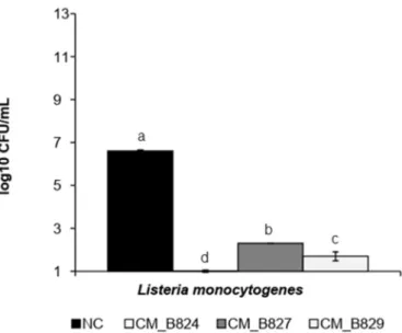
LAB can inhibit spoilage and pathogenic microorganisms by com-petitive growth and synthesis of antagonistic compounds such as or-ganic acids and bacteriocins (Gómez-Sala et al., 2016). When in co-culture at−1 °C, CM strains were able to reduce the population of LM from 6.6 (control) to < 1.0 (CM_B824), 2.3 (CM_B827), and 1.7 (CM_B829) log CFU/ mL (Fig. 1). However, EC and ST were not in-hibited when co-cultured at−1 °C with any of the CM strains (data not shown). Moreover, at 4 °C, all CM isolates inhibited the growth of LM (P < .05), showing a count reduction of a least 5.5 log CFU/ mL as compared to LM alone (negative control) (Fig. 2). Still, EC and ST were not inhibited at the condition of 4 °C. The ability of CM, differently from others LAB, to grow under low temperatures (Leisner et al., 2007) al-lows a competition with other bacteria in this kind of environment.
At 25 °C, CM_B824 and CM_B827 showed a weak but significant inhibition effect towards LM (P < .05) when in co-cultures (Fig. 3E). CM did not show any inhibitory effect when grown in co-culture with EC and ST at 25 °C (Fig. 3A and 3C). However, when EDTA was added in the co-culture broth, all CM isolates reduced the growth of EC (P < .05) (Fig. 3B), and CM_B824 and CM_B827 inhibited the growth of ST and LM (P < .05) (Fig. 3D and 3F).
As Carnobacterium is phylogenetically related to other genera of LAB it is possible to compare these bacteria (Hammes & Hertel, 2006). Other authors also demonstrated the antilisterial activity of LAB.Rivas et al. (2014)showed the antilisterial activity of Lactobacillus curvatus and its purified bacteriocin, sakacin Q on cooked meat.Ho et al. (2018)found antilisterial activity in co-cultures of Lactococcus lactis, Lact. ra ffino-lactis, Leuconostoc mesenteroides, Leuc. pseudomesenteroides, Weissella soli, and W. viridescens. Furthermore,Huang et al. (2016)demonstrated
the inhibition of LM when in co-culture with Enterococcus faecium B1/ B2.
Regarding the genus Carnobacterium,Alves et al. (2005)reported the antilisterial activity of a strain of CM (formerly Carnobacterium piscicola) isolated from Brazilian smokedfish.Dos Reis et al. (2011)also observed the antilisterial activity of CM in fish models. Moreover, Hammi et al. (2016) demonstrated an anti-Listeria activity by a new class IIa bacteriocin, termed maltaricin CPN, produced by a CM strain isolated from mold-ripened cheese.
Other authors observed inhibition of Gram-negative pathogens when bacteriocins from Gram-positive bacteria were analyzed in the presence of EDTA (Camargo, de Paula, Todorov, & Nero, 2016;Field et al., 2017;O'Connor, Ross, Hill, & Cotter, 2015;Prudêncio, Vanetti, & Prieto, 2015). A bacteriocin produced by CM UAL307, termed carno-cyclin A, showed an antimicrobial activity against Gram-negative
bacteria including EC and Pseudomonas aeruginosa, when incubated with EDTA (Martin-Visscher, Yoganathan, Sit, Lohans, & Vederas, 2011). Bacterial metabolites can become more effective biopreserva-tives when used in combination with other hurdles such as chelating agents. This combination is a strategy to increase the activity of the bacteriocins produced by Gram-positive bacteria. As the composition of membranes from Gram-positive bacteria and Gram-negative bacteria are different, multiple approaches for increasing the activity of a bac-teriocin or other bacterial metabolites are necessary (Hwanhlem, Ivanova, Haertlé, Jaffrès, & Dousset, 2017). The chelating capacity of EDTA, which acts by removing Mg2+ and Ca2+, promotes the de-stabilization of the outer membrane from Gram-negative bacteria, al-lowing metabolites to access the cytoplasmic membrane (Field et al., 2017;Mathur et al., 2017).
3.2. Antimicrobial effect of CFS
Regarding the CFS essays, there was no inhibition effect on LM, EC and ST growth in agar well diffusion at the tested conditions. In the MIC determination, there was no inhibition of LM using the different CFS treatments and concentrations. Similarly, Schillinger and Holzapfel (1990)did not observe any antilisterial effect of CFS obtained from 37 isolates of Carnobacterium spp.Arena et al. (2016)also did notfind any inhibitory effect of CFS obtained from 79 Lactobacillus plantarum iso-lates towards LM, EC, Salmonella sp. and Staphylococcus aureus using CFS in a well-diffusion assay. However, when these pathogens were challenged in the presence of the cells of L. plantarum an inhibitory effect was observed for 17 strains.
Based on these results, the isolates of CM used in this study are not likely to produce bacteriocins under the studied conditions. The bac-teriocin production is related to the maximum cell growth and shows primary metabolic kinetics. So, the metabolization of bacteriocins are strictly related to optimal conditions of growth for the bacteriocino-genic strain, which depends on environmental conditions such as pH, temperature, media composition, aeration, salinity, agitation and in-cubation atmosphere (Elayaraja, Annamalai, Mayavu, & Balasubramanian, 2014; Malheiros, Sant'Anna, Todorov, & Franco, 2015;Yang et al., 2018). For C. maltaromaticum, the best temperatures to maximum production of bacteriocin were found to be around 19 °C (Gursky et al., 2006).
Thus, the LM inhibition observed at the co-culture essays may be explained by the production of organic acids or other antibacterial metabolites, the competition of nutrients, the need of a more direct interaction between bacteria to activate the antimicrobial mechanisms or even, by the production of bacteriocins, although this production was not enough to cause a significant inhibition (Arena et al., 2016; Chanos & Mygind, 2016;Yang et al., 2018).
Each isolate of each species appears to have an exclusive optimal condition to produce bacteriocins, and these conditions should be de-termined for each parameter and producer isolate (Masuda, Perez, Zendo, & Sonomoto, 2016;Pérez, González, Agrasar, & Guerra, 2013; Sidooski, Brandelli, Bertoli, Souza, & Carvalho, 2018).
3.3. Cooked ham essay 3.3.1. Microbiological analysis
The use of LAB as protective cultures to improve safety and prolong the shelf life of the meat products, including cooked meat products, is a concept that has been suggested by many authors (Comi, Andyanto, Manzano, & Iacumin, 2016; Metaxopoulos, Mataragas, & Drosinos, 2002;Vermeiren, Devlieghere, & Debevere, 2004).
There was significant interaction between ‘treatment’ and ‘storage time’ for the count of CM in cooked ham (P < .05) (Table 3). For LI count, there was no interaction between thefixed effects, but there was independent effect of ‘treatment’ and ‘storage time’ (P < .05).
During storage, the count of CM varied significantly, however,
Fig. 1. Count of L. monocytogenes at co-culture with C. maltaromaticum isolates at−1 °C for 28 d.
NC = negative control; CM = Carnobacterium maltaromaticum.
No common superscript indicates that there is a significant difference among treatments (P < .05).
Fig. 2. Count of L. monocytogenes at co-culture with C. maltaromaticum isolates at 4 °C for 14 d.
NC = negative control; CM = Carnobacterium maltaromaticum.
No common superscript indicates that there is a significant difference among treatments (P < .05).
count differences were < 0.4 log CFU/g in all CM-treatments from day 1 to day 7 of storage, showing adaptation of CM strains to the cured cooked meat matrix. Conversely, the count of LI in the CM + LI treatments reduced significantly during storage, whereas the count of LI in the LI-treatment did not differ during storage (P > .05). At 2 days of storage, the count of LI in the CM + LI treatments decreased to non-detected level (< 0.69 log CFU/g) and the addition of CM_B829 re-duced the count of LI > 2.0 log CFU/ g after 7 days of storage. CM_B824 and CM_B827 also caused a significant reduction of LI in ham during storage (P < .05). This inhibition of L. innocua may be attrib-uted to the greater ability of Carnobacterium to grow and to adapt to this food matrix and storage conditions in comparison to Listeria (Amézquita; Brashears, 2002). This ability leads to a depletion of nu-trients hindering the growth of the pathogen, as well as the occupation of the food matrix before the pathogen (Nilsson et al., 2005;Vermeiren et al., 2004).
Although the ability of Carnobacterium strains to produce bacter-iocins under the studied conditions has not been proven, the bacteriocin production by LAB does not always lead to increased inhibitory activity towards pathogenic bacteria. Vermeinen et al. (2004) studied the an-tilisterial activity of LAB, able or not to produce bacteriocins, in cooked ham stored at 4 and 7 °C and observed that strains that did not produce bacteriocin had a greater inhibition of the growth of Listeria sp. The occurrence of resistant L. monocytogenes target organisms has led to the
suggestion that bacteriocin-negative LAB may be more suitable for practical use as bioprotective agents against L. monocytogenes in ready-to-eat foods (Nilsson et al., 2005;Vermeiren et al., 2004). Indeed, L. monocytogenes is inhibited by carnobacteria that do not produce bac-teriocins, and this is partly due to glucose depletion (Leisner et al., 2007).
3.3.2. Color and pH
There were significant interactions between ‘treatment’ and ‘storage time’ for pH and instrumental color coordinates of cooked ham (P < .05) (Tables 1 and 2). A slight increase in luminosity (L*) and a slight decrease in pH was observed in ham from all treatments during storage. The addition of CM strains in cooked ham did not cause sig-nificant changes in the pH of the product, which remained similar to NC and LI during storage. After 7 days of storage, CM_824 + LI showed lower pH than other treatments (P < .05), however, the pH values in all treatments were within the normal range for this type of meat product, i.e., 5.6–6.2 (Arnau, Guerrero, Casademont, & Gou, 1995). Gao, Li, and Liu (2015)found that the addition of L. sakei C2 in vacuum packed sliced cooked ham decreased the pH value during the storage at refrigerated temperature. Conversely to other aciduric LAB genera such as Lactobacillus, Leuconostoc and Pediococcus, Carnobacterium is not a strong acid producer. Even if carbohydrate catabolism by carnobacteria appears to result in a diverse number of metabolites, these have
Fig. 3. Count of pathogenic bacteria (E. coli O 157:H7 (A-B), S. Thyphimurium (C-D), L. monocytogenes (E-F) in co-culture with C. maltaromaticum isolates at 25 °C for 48 h, with and without EDTA.
generally a limited effect on the sensory attributes of foods (Leisner et al., 2007).
Regarding color parameters, there were small, but significant dif-ferences in instrumental color values between treatments during sto-rage. However, those differences are likely due to intrinsic character-istics of the product itself than to the influence of the treatments.
The acceptability of cooked ham by the consumers is strongly re-lated to the color of the product (Lloret, Picouet, Trbojevich, & Fernández, 2016). Changes in the color of a meat product caused by Carnobacterium was reported only byPeirson, Guan, and Holley (2003) who observed that a strain of C. viridans induced greening in cured bologna. However, this effect was detected only after the opening of the vacuum packages and took 2 days or less at 9 °C and 3 days at 4 °C. Nonetheless, the authors state that representative strains of most known species of Carnobacterium failed to cause discoloration in this type of cooked meat product.
As Carnobacterium are frequently predominant members of the LAB microbiota of non-spoiled raw meat and processed meat products, in-cluding ham and bacon, irrespective of whether products have been stored aerobically, vacuum packaged, or subjected to modified atmo-spheres, they normally do not cause changes in the physicochemical and sensory characteristics of the product (Li et al., 2018). Additionally, high concentrations of bacteria (> 106- 107CFU/g) in food are typi-cally required before their activity is enough to influence the sensory properties of a product (Leisner et al., 2007).Gao et al. (2015)and Comi et al. (2016)reported that Lactobacillus sakei and Lactococcus lactis affected the growth of spoilage bacteria and L. monocytogenes and did not negatively affect the physicochemical properties of sliced cooked ham and cooked bacon, respectively. On the other hand, the addition of C. maltaromaticum in meat products as a protective culture has not been
assessed. Thus, it could be considered that the addition of the CM strains in the present study did not negatively affect the physico-chemical parameters of the product.
The C. maltaromaticum isolates used in this study present several properties that are desirable for biopreservative cultures: the isolates were able to growth and the count remained stable in the cooked ham during storage, the isolates did not cause significant changes in pH and color of the product and the fast inhibition of L. innocua, at the second day of storage, is another advantage of these isolates.
As the artificial contamination of ham in this study extrapolated the natural contamination observed in the food industry by Listeria sp., the C. maltaromaticum could show more successful antimicrobial results in practical conditions. Despite the promising results observed in the present study, the possible impact of the addition of the Carnobacterium isolates as protective culture on sensory properties of the meat product should be further investigated.
4. Conclusion
The three C. maltaromaticum strains tested showed an antilisterial potential in vitro, which was more important at−1 °C and 4 °C than at 25 °C. When applied in cooked ham, the antilisterial potential was confirmed, since the growth of Listeria spp. was inhibited by the addi-tion of the strains of C. maltaromaticum, without affecting the physi-cochemical quality of the product. Therefore, C. maltaromaticum strains show potential to be used as bioprotective culture in cooked meat product to improve its safety.
Table 1
Effect of the addition of three different strains of C. maltaromaticum and L. innocua on lightness (L*), redness (a*) and yellowness (b*) of cooked ham during storage at 4 °C for 7 days.
Treament Storage days
0 2 5 7 P value
L*
NC 60.10 ± 0.66abB 63.05 ± 0.27abA 63.26 ± 0.57aA 62.91 ± 0.62aA 0.0017
LI 60.28 ± 0.71abB 63.65 ± 0.39aA 64.20 ± 0.24aA 65.35 ± 0.94aA 0.0001
CM_824 59.90 ± 0.66abB 63.56 ± 0.23aA 63.83 ± 0.34aA 64.55 ± 0.36aA 0.0000
CM_827 61.04 ± 0.41abC 62.60 ± 0.48abBC 63.52 ± 0.58aAB 64.70 ± 0.59aA 0.0006
CM_829 59.18 ± 0.85bB 62.07 ± 0.47abA 62.83 ± 0.40aA 64.13 ± 0.28aA 0.0000 CM_824 + LI 61.23 ± 0.70abA 60.92 ± 0.59bcA 63.10 ± 0.48aA 62.97 ± 0.81aA 0.0538 CM_827 + LI 61.99 ± 0.54abA 59.71 ± 0.44cA 61.03 ± 1.92aA 63.17 ± 0.29aA 0.1430 CM_829 + LI 62.32 ± 0.61aA 63.11 ± 0.71aA 61.20 ± 0.96aA 63.98 ± 0.44aA 0.4327 P value 0.0208 0.0000 0.1007 0.0454 a* NC 17.13 ± 1.01abA 15.51 ± 0.32aA 17.33 ± 0.32aA 16.38 ± 0.56aA 0.1822 LI 17.32 ± 0.64abA 16.10 ± 0.54aA 17.40 ± 0.30aA 16.37 ± 0.56aA 0.2248 CM_824 16.84 ± 0.57abA 16.44 ± 0.42aA 17.53 ± 0.42aA 15.73 ± 0.88aA 0.2321 CM_827 17.63 ± 0.57aA 15.34 ± 0.15abB 16.94 ± 0.26abA 14.59 ± 0.27aB 0.0000 CM_829 16.09 ± 0.62abAB 16.73 ± 0.44aAB 17.71 ± 0.43aA 15.88 ± 0.18aB 0.0374 CM_824 + LI 15.17 ± 0.21abA 13.25 ± 0.70bB 16.15 ± 0.50abA 16.43 ± 0.37aA 0.0005 CM_827 + LI 14.81 ± 0.59bBC 13.27 ± 0.62bC 16.79 ± 0.25abA 15.96 ± 0.26aAB 0.0002 CM_829 + LI 15.73 ± 0.63abA 15.58 ± 0.53aA 15.54 ± 0.45bA 14.67 ± 0.31aA 0.3356 P value 0.0159 0.0000 0.0024 0.0349 b* NC 12.31 ± 0.60bcA 11.25 ± 0.40abA 11.51 ± 0.61aA 11.64 ± 0.34aA 0.5059 LI 13.55 ± 0.65bcA 11.50 ± 0.48abA 11.92 ± 0.50aA 12.73 ± 0.58aA 0.0750 CM_824 10.94 ± 0.16cB 11.75 ± 0.07aAB 11.99 ± 0.62aAB 12.71 ± 0.36aA 0.0246 CM_827 14.56 ± 0.29abA 11.33 ± 0.45abB 11.44 ± 0.49aB 12.04 ± 0.35aB 0.0000 CM_829 17.19 ± 0.99aA 11.59 ± 0.66abB 11.13 ± 0.15aB 12.30 ± 0.25aB 0.0000 CM_824 + LI 11.52 ± 0.46cAB 10.50 ± 0.33abB 12.60 ± 0.34aA 12.40 ± 0.44aA 0.0048 CM_827 + LI 11.76 ± 0.75cA 9.89 ± 0.13bB 12.37 ± 0.31aA 11.61 ± 0.36aAB 0.0052 CM_829 + LI 11.75 ± 0.44cA 12.34 ± 0.39aA 11.60 ± 0.49aA 12.30 ± 0.20aA 0.5228 P value 0.0000 0.0071 0.3624 0.2701
NC = negative control; CM = Carnobacterium maltaromaticum; LI– Listeria innocua. Means ± standard error. Different lowercase letters in the same column show significant differences among treatments (P < .05). Different uppercase letters in the same row show significant differences among storage time (P < .05).
Declaration of Competing Interest
The authors declare that there is no conflict of interest. This statement is to certify that all authors have seen and approved the manuscript being submitted.
We warrant that the article is the authors' original work. We warrant that the article is not under consideration for publication elsewhere.
On behalf of all co-authors, the corresponding author shall bear full
responsibility for the submission.
We attest to the fact that all authors listed on the title page have contributed significantly to the work, have read the manuscript, attest to the validity and legitimacy of the data and its interpretation, and agree to its submission toMeat Science.
Table 2
Effect of the addition of three different strains of C. maltaromaticum and L. innocua on chroma (C*), hue (h) and pH of cooked ham during storage at 4 °C for 7 days.
Treatment Storage days
0 2 5 7 P value
C*
NC 21.15 ± 1.10bcdA 19.18 ± 0.20abA 20.85 ± 0.26aA 20.17 ± 0.33abA 0.1266
LI 22.02 ± 0.71abcA 19.80 ± 0.51aB 21.12 ± 0.36aAB 19.80 ± 0.25abB 0.0100
CM_824 20.08 ± 0.52cdA 20.22 ± 0.34aA 21.25 ± 0.65aA 19.70 ± 0.35abA 0.1630 CM_827 23.23 ± 0.50abA 19.09 ± 0.31abB 20.43 ± 0.47aB 18.92 ± 0.29bB 0.0000 CM_829 24.61 ± 0.71aA 20.37 ± 0.71aB 20.93 ± 0.34aB 20.09 ± 0.15abB 0.0000 CM_824 + LI 18.76 ± 0.47dAB 16.92 ± 0.69bcB 20.51 ± 0.36aA 20.44 ± 0.58BaA 0.0003 CM_827 + LI 18.45 ± 0.26dB 16.58 ± 0.45cC 20.87 ± .026aA 19.76 ± 0.18abA 0.0000 CM_829 + LI 19.63 ± 0.56cdA 19.82 ± 0.57aA 20.00 ± .36aA 19.15 ± 0.29abA 0.6064 P value 0.0000 0.0000 0.4157 0.0335 h NC 35.77 ± 1.11bcA 35.96 ± 1.42aA 33.55 ± 1.76abA 35.01 ± 1.60aA 0.6609 LI 38.02 ± 1.45bcA 35.55 ± 1.50aA 34.37 ± 1.25abA 40.13 ± 1.90aA 0.0675 CM_824 33.02 ± 0.85cB 35.60 ± 0.74aAB 33.75 ± 1.14abB 40.11 ± 2.09aA 0.0048
CM_827 40.50 ± 1.59abA 36.39 ± 1.09aAB 33.86 ± 0.88abB 39.60 ± 1.07aA 0.0028
CM_829 46.01 ± 1.33aA 34.58 ± 1.06aBC 32.20 ± 0.81bC 37.77 ± 0.77aB 0.0000 CM_824 + LI 37.05 ± 0.92bcA 38.56 ± 1.16aA 38.02 ± 1.37aA 37.00 ± 0.69aA 0.6754 CM_827 + LI 37.18 ± 2.07bcA 36.88 ± 1.55aA 36.35 ± 0.85abA 36.04 ± 1.11aA 0.9463 CM_829 + LI 36.74 ± 0.74bcA 38.22 ± 0.94aA 37.54 ± 1.64abA 39.99 ± 0.70aA 0.2078 P value 0.0000 0.2915 0,0191 0.0461 pH NC 6.42 ± 0.02aA 5.92 ± 0.02aB 5.70 ± 0.02bC 5.88 ± 0.02aB 0.0000 LI 6.33 ± 0.11aA 5.61 ± 0.01cC 5.72 ± 0.05bBC 5.87 ± 0.02aB 0.0000 CM_824 5.93 ± 0.01bA 5.64 ± 0.02cB 5.61 ± 0.02bB 5.87 ± 0.02aA 0.0000 CM_827 6.46 ± 0.02aA 5.85 ± 0.04abB 5.61 ± 0.01bC 5.89 ± 0.03aB 0.0000 CM_829 6.47 ± 0.02aA 5.94 ± 0.03aB 5.62 ± 0.01bC 5.93 ± 0.03aB 0.0000 CM_824 + LI 5.83 ± 0.01bB 5.93 ± 0.02aA 5.89 ± 0.02aAB 5.66 ± 0.02bC 0.0000 CM_827 + LI 5.82 ± 0.01bAB 5.94 ± 0.01aA 5.90 ± 0.02aAB 5.80 ± 0.06aB 0.0196 CM_829 + LI 5.85 ± 0.03bA 5.76 ± 0.07bcA 5.90 ± 0.02aA 5.91 ± 0.02aA 0.0649 P 0.0000 0.0000 0.0000 0.0000
NC = negative control; CM = Carnobacterium maltaromaticum; LI– Listeria innocua. Means ± standard error. Different lowercase letters in the same column show significant differences among treatments (P < .05). Different uppercase letters in the same row show significant differences among storage time (P < .05).
Table 3
Count of the different strains of C. maltaromaticum and the pool of L. innocua in sliced cooked ham stored at 4 °C for 7 days.
Treatment Storage days
0 2 5 7 P
Count of C. maltaromaticum (log CFU/g)
CM_B824 5.60 ± 0.02aA 5.60 ± 0.02aAB 5.26 ± 0.02abC 5.33 ± 0.03aBC 0.0120
CM_B827 5.42 ± 0.07aA 5.41 ± 0.07aA 5.08 ± 0.03bA 5.40 ± 0.05aA 0.0034 CM_B829 5.49 ± 0.05aA 5.48 ± 0.05aA 5.34 ± 0.08aA 5.35 ± 0.03aA 0.2163 CM_B824 + LI 4.75 ± 0.00bA 4.75 ± 0.00bA 4.64 ± 0.01cB 4.31 ± 0.01cC 0.0000 CM_B827 + LI 4.50 ± 0.14bA 4.49 ± 0.13bA 4.60 ± 0.01cA 4.34 ± 0.02cA 0.2174 CM_B829 + LI 4.46 ± 0.03bB 4.46 ± 0.03bB 4.71 ± 0.05cA 4.54 ± 0.03bAB 0.0222 P 0.0000 0.0002 0.0056 0.0075
Count of L. innocua (log CFU/g)
LI 3.06 ± 0.02aA 2.60 ± 0.11aA 2.59 ± 0.50aA 2.40 ± 0.09aA 0.1190
CM_B824 + LI 2.15 ± 0.15bB NDbB NDbB 1.350 ± 0.50bAB 0.0355
CM_B827 + LI 2.00 ± 0.00bB NDbB NDbB NDbB 0.0298
CM_B829 + LI 2.15 ± 0.15bB NDbB 1.50 ± 0.50bAB NDbB 0.0079
P 0.0067 0.0004 0.0042 0.0133
CM = Carnobacterium maltaromaticum; LI– Listeria innocua. ND = non-detected (limit of detection = 0.69 log CFU/g).
Means ± standard error. Different lowercase letters in the same column show significant differences among treatments (P < .05). Different uppercase letters in the same row show significant differences among storage time (P < .05).
Acknowledgements
This work was financed by the Pontifical Catholic University of Paraná and the University of Liège (“Fonds Spéciaux de la Recherche – FMV – 2017”). The authors are grateful to the funding and all the technical support provided.
References
Alves, V., Martinis, E., Destro, M., Vogel, B., & Gram, L. (2005). Antilisterial activity of a Carnobacterium piscicola isolated from Brazilian smokedfish (surubim
[Pseudoplatystoma sp.]) and its activity against a persistent strain of Listeria mono-cytogenes isolated from surubim. Journal of Food Protection, 68(10), 2068–2077.
Alves, V. F., Martinez, R. C. R., Lavrador, M. A. S., & De Martinis, E. C. P. (2006). Antilisterial activity of lactic acid bacteria inoculated on cooked ham. Meat Science, 74(4), 623–627.
Arena, M. P., Silvain, A., Normanno, G., Grieco, F., Drider, D., Spano, G., & Fiocco, D. (2016). Use of Lactobacillus plantarum strains as a bio-control strategy against food-borne pathogenic microorganisms. Frontiers in Microbiology, 7, 1–10.
Arnau, J., Guerrero, L., Casademont, G., & Gou, P. (1995). Physical and chemical changes in different zones of normal and PSE dry cured ham during processing. Food Chemistry, 52(1), 63–69.
Bordignon-Junior, S. E., Miyaoka, M. F., Costa, J., Benavente, C. A. T., Couto, G. H., & Soccol, C. R. (2012). Inhibiting Gram-negative bacteria growth in microdilution by Nisin and EDTA treatment. Journal of Biotechnology and Biodiversity, 3(4), 127–135.
Brazil (2003). Ministry of Agriculture, livestock and food supply. Normative Instruction No. 62. Brasília, DF: Official journal of the union. Section, 1, 14.
Brazil (2019). Ministry of Health. National Agency of Sanitary Surveillance. Foodbourne outbreaks in Brazil - Report 2018http://portalarquivos2.saude.gov.br/images/pdf/ 2018/julho/02/Apresentacao-Surtos-DTA-Junho-2018.pdf.
Brillet-Viel, A., Pilet, M.-F., Courcoux, P., Prévost, H., & Leroi, F. (2016). Optimization of growth and bacteriocin activity of the food bioprotective Carnobacterium divergens V41 in an animal origin protein free medium. Frontiers in Marine Science, 3, 1–13.
Camargo, A. C., de Paula, O. A. L., Todorov, S. D., & Nero, L. A. (2016). In vitro evaluation of bacteriocins activity against Listeria monocytogenes biofilm formation. Applied Biochemistry and Biotechnology, 178(6), 1239–1251.
Chanos, P., & Mygind, T. (2016). Co-culture-inducible bacteriocin production in lactic acid bacteria. Applied Microbiology and Biotechnology, 100(10), 4297–4308.
CLSI (2012). Methods for dilution antimicrobial susceptibility tests for bacteria that grow aerobically. CLSI document M07-A9. Wayne, PA: Clinical and Laboratory Standards Institute.
Comi, G., Andyanto, D., Manzano, M., & Iacumin, L. (2016). Lactococcus lactis and Lactobacillus sakei as bio-protective culture to eliminate Leuconostoc mesenteroides spoilage and improve the shelf life and sensorial characteristics of commercial cooked bacon. Food Microbiology, 58, 16–22.
dos Reis, F. B., de Souza, V. M., Thomaz, M. R. S., Fernandes, L. P., de Oliveira, W. P., & De Martinis, E. C. P. (2011). Use of Carnobacterium maltaromaticum cultures and hydroalcoholic extract of Lippia sidoides Cham. against Listeria monocytogenes infish model systems. International Journal of Food Microbiology, 146(3), 228–234. EFSA and ECDC (European Food Safety Authority and European Centre for Disease
Prevention and Control) (2018). The European Union summary report on trends and sources of zoonoses, zoonotic agents and food-borne outbreaks in 2017. EFSA Journal, 16(12), 5500. 262 pphttps://doi.org/10.2903/j.efsa.2018.5500.
Elayaraja, S., Annamalai, N., Mayavu, P., & Balasubramanian, T. (2014). Production, purification and characterization of bacteriocin from Lactobacillus murinus AU06 and its broad antibacterial spectrum. Asian Pacific Journal of Tropical Biomedicine, 4, 305–311.
Engelhardt, T., Albano, H., Kiskó, G., Mohácsi-Farkas, C., & Teixeira, P. (2015). Antilisterial activity of bacteriocinogenic Pediococcus acidilactici HA6111-2 and Lactobacillus plantarum ESB 202 grown under pH and osmotic stress conditions. Food Microbiology, 48, 109–115.
Field, D., Baghou, I., Rea, M., Gardiner, G., Ross, R., & Hill, C. (2017). Nisin in combi-nation with cinnamaldehyde and EDTA to control growth of Escherichia coli strains of swine origin. Antibiotics, 6(4), 35.
Gao, Y., Li, D., & Liu, X. (2015). Effects of Lactobacillus sakei C2 and sakacin C2 in-dividually or in combination on the growth of Listeria monocytogenes, chemical and odor changes of vacuum-packed sliced cooked ham. Food Control, 47, 27–31.
Gómez-Sala, B., Herranz, C., Díaz-Freitas, B., Hernández, P. E., Sala, A., & Cintas, L. M. (2016). Strategies to increase the hygienic and economic value of freshfish: Biopreservation using lactic acid bacteria of marine origin. International Journal of Food Microbiology, 223, 41–49.
Gursky, L. J., Martin, N. I., Derksen, D. J., Van Belkum, M. J., Kaur, K., Vederas, J. C., & McMullen, L. M. (2006). Production of piscicolin 126 by Carnobacterium mal-taromaticum UAL26 is controlled by temperature and induction peptide concentra-tion. Archives of Microbiology, 186(4), 317–325.
Gutiérrez, S., Martínez-Blanco, H., Rodríguez-Aparicio, L. B., & Ferrero, M. A. (2016). Effect of fermented broth from lactic acid bacteria on pathogenic bacteria pro-liferation. Journal of Dairy Science, 99(4), 2654–2665.
Hammes, W. P., & Hertel, C. (2006). The genera Lactobacillus and Carnobacterium. In M. Dworkin, S. Falkow, E. Rosenberg, K. H. Schleifer, & E. Stackebrandt (Eds.). The prokaryotes (pp. 320–403). New York: Springer.
Hammi, I., Delalande, F., Belkhou, R., Marchioni, E., Cianferani, S., & Ennahar, S. (2016). Maltaricin CPN, a new class IIa bacteriocin produced by Carnobacterium
maltaromaticum CPN isolated from mould-ripened cheese. Journal of Applied Microbiology, 121(5), 1268–1274.
Ho, V. T. T., Lo, R., Bansal, N., & Turner, M. S. (2018). Characterisation of Lactococcus lactis isolates from herbs, fruits and vegetables for use as biopreservatives against Listeria monocytogenes in cheese. Food Control, 85, 472–483.
Huang, Y., Ye, K., Yu, K., Wang, K., & Zhou, G. (2016). The potential influence of two Enterococcus faecium on the growth of Listeria monocytogenes. Food Control, 67, 18–24.
Hwanhlem, N., Ivanova, T., Haertlé, T., Jaffrès, E., & Dousset, X. (2017). Inhibition of food-spoilage and foodborne pathogenic bacteria by a nisin Z-producing Lactococcus lactis subsp. lactis KT2W2L. LWT - Food Science and Technology, 82, 170–175. Interagency Food Safety Analytics Collaboration (IFSAC). (2019). Foodborne illness
source attribution estimates for 2017 for Salmonella. Escherichia coli O157, Listeria monocytogenes, and Campylobacter using multi-year outbreak surveillance data, United States (pp. 2019). GA and D.C.: U.S. Department of Health and Human Services, CDC, FDA, USDA-FSIS. https://www.cdc.gov/foodsafety/ifsac/pdf/P19-2017-report-TriAgency-508.pdf.
Iskandar, C. F., Borges, F., Taminiau, B., Daube, G., Zagorec, M., Remenant, B., & Revol-Junelles, A. M. (2017). Comparative genomic analysis reveals ecological differ-entiation in the genus Carnobacterium. Frontiers in Microbiology, 8, 1–14.
Koné, A. P., Zea, J. M. V., Gagné, D., Cinq-Mars, D., Guay, F., & Saucier, L. (2018). Application of Carnobacterium maltaromaticum as a feed additive for weaned rabbits to improve meat microbial quality and safety. Meat Science, 135, 174–188.
Leisner, J. J., Laursen, B. G., Prévost, H., Drider, D., & Dalgaard, P. (2007).
Carnobacterium: Positive and negative effects in the environment and in foods. FEMS Microbiology Reviews, 31(5), 592–613.
Li, X., Li, C., Ye, H., Wang, Z., Wu, X., Han, Y., & Xu, B. (2018). Changes in the microbial communities in vacuum-packaged smoked bacon during storage. Food Microbiology, 77, 26–37.
Lloret, E., Picouet, P. A., Trbojevich, R., & Fernández, A. (2016). Colour stability of cooked ham packed under modified atmospheres in polyamide nanocomposite blends. LWT - Food Science and Technology, 66, 582–589.
Malheiros, P. S., Sant’Anna, V., Todorov, S. D., & Franco, B. D. G. M. (2015). Optimization of growth and bacteriocin production by Lactobacillus sakei subsp Sakei 2a. Brazilian Journal of Microbiology, 46(3), 825–834.
Martin-Visscher, L. A., Yoganathan, S., Sit, C. S., Lohans, C. T., & Vederas, J. C. (2011). The activity of bacteriocins from Carnobacterium maltaromaticum UAL307 against Gram-negative bacteria in combination with EDTA treatment. FEMS Microbiology Letters, 317(2), 152–159.
Masuda, Y., Perez, R. H., Zendo, T., & Sonomoto, K. (2016). Nutrition-adaptive control of multiple-bacteriocin production by Weissella hellenica QU 13. Journal of Applied Microbiology, 120(1), 70–79.
Mathur, H., Field, D., Rea, M. C., Cotter, P. D., Hill, C., & Ross, R. P. (2017). Bacteriocin-antimicrobial synergy: A medical and food perspective. Frontiers in Microbiology, 8, 1–18.
Metaxopoulos, J., Mataragas, M., & Drosinos, E. H. (2002). Microbial interaction in cooked cured meat products under vacuum or modified atmosphere at 4°C. Journal of Applied Microbiology, 93(3), 363–373.
Nilsson, L., Hansen, T. B., Garrido, P., Buchrieser, C., Glaser, P., Knøchel, S., & Gravesen, A. (2005). Growth inhibition of Listeria monocytogenes by a nonbacteriocinogenic Carnobacterium piscicola. Journal of Applied Microbiology, 98(1), 172–183.
O’Connor, P. M., Ross, R. P., Hill, C., & Cotter, P. D. (2015). Antimicrobial antagonists against food pathogens: A bacteriocin perspective. Current Opinion in Food Science, 2, 51–57.
de Oliveira, A. B. A., de Paula, C. M. D., Cardoso, M. R. I., & Tondo, E. C. M. (2010). Foodborne diseases, main etiologic agents and general aspects: a review. Revista HCPA, 30(3), 279–285.
Orihuel, A., Bonacina, J., Vildoza, M. J., Bru, E., Vignolo, G., Saavedra, L., & Fadda, S. (2018). Biocontrol of Listeria monocytogenes in a meat model using a combination of a bacteriocinogenic strain with curing additives. Food Research International, 107, 289–296 2017.
Peirson, M. D., Guan, T. Y., & Holley, R. A. (2003). Aerococci and carnobacteria cause discoloration in cooked cured bologna. Food Microbiology, 20(2), 149–158.
Pérez, R. A., González, E. A., Agrasar, A. T., & Guerra, N. P. (2013). A review on some important factors affecting bacteriocin production by Lactococci, Lactobacilli and Pediococci. Current Biochemical Engineering, 1(1), 9–24.
Prudêncio, C. V., Vanetti, M. C. D., & Prieto, M. (2015). Tolerance of Salmonella enterica serovar Typhimurium to nisin combined with EDTA is accompanied by changes in cellular composition. Food Research International, 69, 281–288.
Rivas, F. P., Castro, M. P., Vallejo, M., Marguet, E., & Campos, C. A. (2014). Sakacin Q produced by Lactobacillus curvatus ACU-1: Functionality characterization and antil-isterial activity on cooked meat surface. Meat Science, 97(4), 475–479.
Sánchez-Ortega, I., García-Almendárez, B. E., Santos-López, E. M., Amaro-Reyes, A., Barboza-Corona, J. E., & Regalado, C. (2014). Antimicrobial ediblefilms and coatings for meat and meat products preservation. The Scientific World Journal, 2014, 1–18.
Schillinger, U., & Holzapfel, W. H. (1990). Antibacterial activity of carnobacteria. Food Microbiology, 7, 305–310.
Sidooski, T., Brandelli, A., Bertoli, S. L., de Souza, C. K., & de Carvalho, L. F. (2018). Physical and nutritional conditions for optimized production of bacteriocins by lactic acid bacteria– A review. Critical Reviews in Food Science and Nutrition, 0(0), 1–11.
Smialek, M., Burchardt, S., & Koncicki, A. (2018). The influence of probiotic supple-mentation in broiler chickens on population and carcass contamination with Campylobacter spp. - Field study. Research in Veterinary Science, 118, 312–316.
Spanu, C., Piras, F., Mocci, A. M., Nieddu, G., De Santis, E. P. L., & Scarano, C. (2018). Use of Carnobacterium spp protective culture in MAP packed ricotta fresca cheese to control Pseudomonas spp. Food Microbiology, 74, 50–56.
Martinis, E. C. P. (2014). Purification and characterization of antimicrobial peptides fromfish isolate Carnobacterium maltaromaticum C2: Carnobacteriocin X and carno-lysins A1 and A2. International Journal of Food Microbiology, 173, 81–88.
Vermeiren, L., Devlieghere, F., & Debevere, J. (2004). Evaluation of meat born lactic acid bacteria as protective cultures for the biopreservation of cooked meat products. International Journal of Food Microbiology, 44(1–2), 41–45.
World Health Organization (2015). WHO estimates of the global burden of foodborne dis-eases: Foodborne disease burden epidemiology reference group 2007–2015. WHO Institucional repository.
Yang, E., Fan, L., Yan, J., Jiang, Y., Doucette, C., Fillmore, S., & Walker, B. (2018). Influence of culture media, pH and temperature on growth and bacteriocin produc-tion of bacteriocinogenic lactic acid bacteria. AMB Express, 8(1).