Regulation of T ceil function by interaction between a TNF receptor family member DcR
31TR6and a TNF family member LIGHT
Xiaochun Wan
Département de Médecine Faculté de Médecine Université de Montréal
Thèse présentée à la Faculté des études supérieures en vue de l’obtention du grade de
Philosophiae Doctor (Ph.D.) en Sciences Biomédicales
September, 2003
j1,,2DJ3 LC Dz
)
q
/
k fUniversité
dl1
de Montréal
Direction des bibliothèques
AVIS
L’auteur a autorisé l’Université de Montréal à reproduire et diffuser, en totalité ou en partie, par quelque moyen que ce soit et sut quelque support que ce soit, et exclusivement à des fins non lucratives d’enseignement et de recherche, des copies de ce mémoire ou de celle thèse.
L’auteur et les coauteurs le cas échéant conservent la propriété du droit d’auteur et des droits moraux qui protègent ce document. Ni la thèse ou le mémoire, ni des extraits substantiels de ce document, ne doivent être imprimés ou autrement reproduits sans l’autorisation de l’auteur.
Afin de se conformer à la Loi canadienne sur la protection des
renseignements personnels, quelques formulaires secondaires, coordonnées ou signatures intégrées au texte ont pu être enlevés de ce document. Bien que cela ait pu affecter la pagination, il n’y a aucun contenu manquant.
NOTICE
The author of this thesis or dissertation has granted a nonexclusive license allowing Université de Montréal to reproduce and publish the document, in part or in whole, and in any format, solely for noncommercial educational and research purposes.
The author and co-authors if applicable retain copyright ownership and moral
rights in this document. Neither the whole thesis or dissertation, nor
substantial extracts from it, may be printed or otherwise reproduced without the author’s permission.
In compliance with the Canadian Privacy Act some supporting forms, contact
information or signatures may have been removed from the document. While this may affect the document page count, it does not represent any loss of content from the document.
Université de Montréal Faculté des études supérieures
Cette thèse intitulée:
«REGULATION 0F T CELL FUNCTION BY INTERACTION BETWEEN A TNF RECEPTOR FAMILY MEMBER DCR3/TR6 AND A TNF FAMILY MEMBER LIGHT)>
Présentée par:
Xiaochun Wan
A été évaluée par un jury composé des personnes suivantes:
Yves Raymond Président rapporteur
Jiangping Wu Directeur de recherche
Claude Perreault Membre du jury
David Hoskin Examinateur externe
Richard Bertrand Représentant du doyen de la FES
Thèse acceptée le:
SUMMARY
TR6, also named DcR3, M68, is a recently identified soluble receptor belonging to the TNFR superfamily. FasL, LIGHT and TL1 are 3 so-far identified ligands for TR6, and
they all belong to the TNF ligand superfamily. TR6 can interfere with fasL and Fas
interaction. It can also interfere with the interaction between LIGHT and HveA, and
between TL1 and DR3. Some tumors have high TR6 expression. It has been
hypothesized that TR6-secreting tumors use this molecule to evade immune surveillance and gain survival advantage.
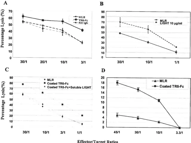
In this project, the role of TR6 in immune regulation was invcstigated. We demonstrated that hurnan TR6 could cross-react with mouse LIGHT. As the mouse counterpart of TR6 unlikely exists according to Genebank search, this finding allowed us to use human TR6 in the mouse system. In the mouse system, soluble human TR6 could suppress IL-2, IL-5 and GM-CSF secretion by mitogen-activated T ceils, and downregulate cytotoxic T-cell development in vitro. In vivo, soluble TR6 could suppress graft versus host disease and allograft rejection. These effects of TR6 are probably achieved by its interference with the interaction between LIGHT and HveA on T cells.
In human system, we found that solid phase TR6, in the presence of suboptimal solid phase anti-CD3, could significantly costimulate T cells in terms of proliferation. Blocking studies using soluble LIGHT and Fas indicated that LIGHT likely mediated the costimulation. This bas revealed a novel mechanism of TR6 triggered reverse signaling
through a ligand, LIGHT. Base on this finding, it is likely that the suppressive effect of soluble TR6 in the mouse system is due to its interference of the two-way costimulation between HveA and LIGHT. Intriguingly, soluble TR6 could augment T-cell proliferation, lymphokine production and cytotoxic T-cell activity in the human system. The opposite effect of soluble TR6 in the human and mouse systems is probably due to different affinity of TR6 to human and mouse LIGHT. The dimeric TR6-Fc might have higher afflnity to human LIGHT, hence capable of triggering strong reverse costimulation through LIGHT. Although it rnight block the two-way costimulation between HveA and LIGHT and abate the immune response, the overall effect is dominated by reverse costimulation through LIGHT. In the mouse system, affinity of human TR6 to mouse LIGHT might be lower, and cannot effectively trigger LIGHT reverse signaling; its overali effect is thus biased to the blocking of the two-way interaction between HveA and LIGHT, hence repressed immune responses.
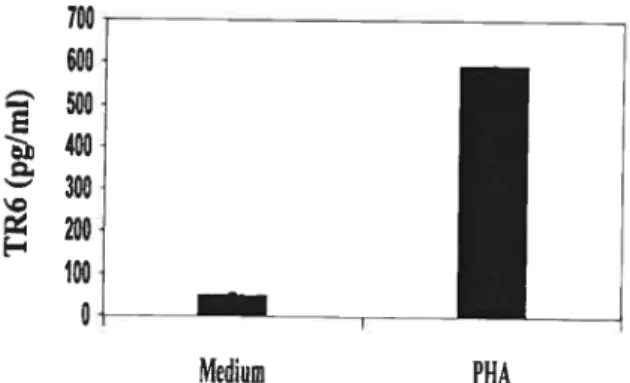
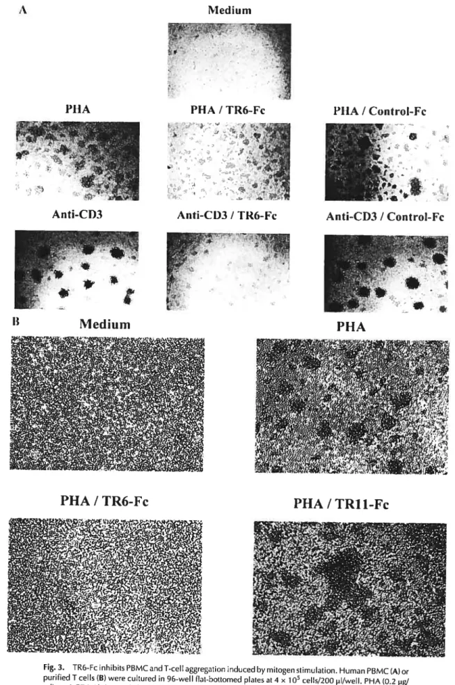
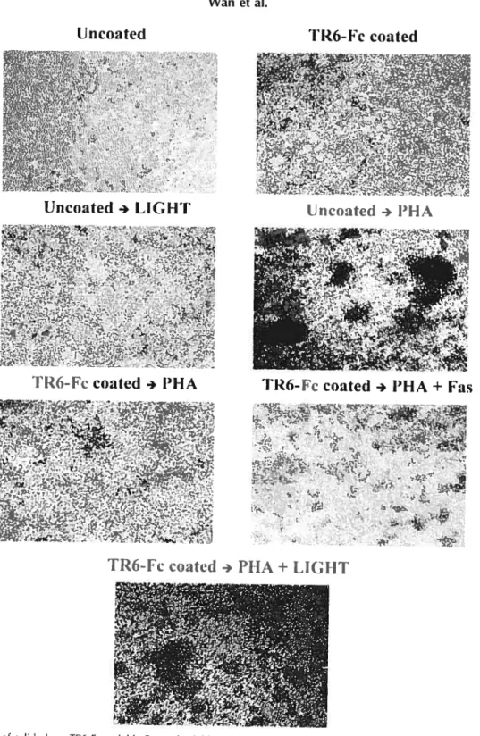
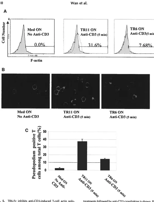
We also found that human peripheral blood mononuclear cells could secrete TR6 and the secretion was enhanced by T-cell activation. Interestingly, both soluble and solid phase TR6 was able to suppress mitogen-induced T-cell aggregation. T cells pretreated with TR6 had reduced actin polymerization and pseudopodium formation, which are both important for the celi-celi interaction. These results suggest that TR6 might regulate the duration of T-cell interaction with other cells, and allow T cells to disengage from antigen presenting cells or fellow T ceils once the interaction becornes unnecessary.
This study has discovered three important functions of TR6 in immune regulation. Some of the findings, such as the suppressive effect of soluble TR6 in immune response and enhancing effect of solid phase TR6 could be exploited for clinical applications. Our findings have also broadened our knowÏedge on TR6 in immune regulation.
Key words: TR6/DcR3; LIGHT; reverse signaling; costimulation
Résumé
Récemment, TR6 aussi connu sous le nom de DcR3 ou M68, a été identifié comme un
récepteur soluble appartenant à la superfamille du récepteur TNf. Les ligands FASL, LIGHT et TL1, qui font partie de la superfamille du ligand TNF, ont la capacité de lier le récepteur TR6. TR6 peut interférer au niveau de l’interaction de Fas et FasL, de LIGHT et HveA ou encore de TL1 et DR3. Puisque certaines tumeurs expriment fortement le récepteur TR6, il a été proposé que les tumeurs sécrétant TR6 pourraient échapper à la surveillance du système immunitaire, par un avantage de croissance.
Ce projet explore le rôle du récepteur TR6 dans la régulation immunitaire. Nous avons démontré que le récepteur TR6 humain peut interagir avec le ligand LIGHT de souris. Cette observation nous a permis d’utiliser le récepteur TR6 humain dans un modèle de souris, puisque l’équivalent murin de TR6 n’existe pas selon les recherches effectuées à partir de Genebank. Chez le modèle murin, le récepteur humain TR6 soluble peut inhiber la sécrétion de IL-2, IL-5 et GM-CSF produite par les cellules T activées et régule négativement le développement des cellules T cytotoxiques in vitro. In vivo, le récepteur TR6 soluble peut supprimer la présence de greffes en favorisant le développement de maladies auto-immunes et le rejet des allogreffes. Ces effets biologiques associés à TR6 sont probablement initiés par l’interférence de TR6 dans l’interaction de LIGHT et HveA dans les cellules T.
Chez un modèle humain, nous avons trouvé qu’en présence d’une concentration suboptimale de la phase solide d’un anti-CD3, la phase solide de TR6 costimule
significativement la prolifération des cellules T. Des études d’inhibition impliquant le ligand LIGHT soluble et Fas indiquent que LIGHT est responsable de la costimulation. Ceci suggère un nouveau mécanisme impliquant une inversion de la signalisation liée à l’activation de TR6 par le ligand LIGHT. Basé sur cette observation, l’effet suppresseur du récepteur TR6 soluble dans le modèle de souris est dû à l’interférence de la costimulation à deux sens entre HveA et LIGHT. Étonnamment, le récepteur TR6 soluble peut augmenter la prolifération des cellules T, la production de lymphokine et l’activité des cellules T cytotoxiques dans le modèle humain. L’effet opposé du récepteur soluble TR6 dans le modèle de souris et humain est probablement le résultat d’une différence d’affinité de TR6 pour le LIGHT humain et le LIGHT murin. Le dimère TR6-Fc pourrait avoir une plus grande affinité pour le LIGHT humain, d’où sa capacité à déclencher une forte costimulation inversée induite par LIGHT. Dans le modèle de souris, l’affinité du TR6 humain pour le LIGHT murin pourrait être plus faible et, par conséquent, incapable de déclencher efficacement la signalisation inversée. Cet effet est alors biaisé par l’inhibition de l’interaction à deux sens de HveA et LIGHT, d’où la répression de la réponse immunitaire.
Nous avons également trouvé que les cellules mononucléaires du sang périphérique peuvent sécréter le récepteur TR6, sécrétion qui serait augmentée après une activation des cellules T. De façon intéressante, les phases soluble et solide de TR6 sont capables d’inhiber l’agrégation des cellules T activées. Les cellules T pré-traitées avec TR6 ont une réduction de la polymérisation de l’actine ainsi qu’une diminution de la formation des pseudopodes, toutes deux importantes pour les interactions cellules-cellules. Ces résultats
suggèrent que TR6 régule la durée des interactions des cellules T avec les autres cellules, conduisant à un désengagement des cellules présentatrices d’antigènes ou des autres cellules T, une fois les interactions devenues non essentielles. Cette étude a permis la découverte de trois fonctions importantes du récepteur TR6 dans la régulation immunitaire. Certaines de ces découvertes, telles que l’effet inhibiteur du récepteur TR6 soluble dans la réponse immunitaire ainsi qu’une augmentation de l’effet du TR6 de la phase solide, pourraient être exploitées pour des applications cliniques. Nos observations ont également permis d’élargir nos connaissances sur le rôle du récepteur TR6 dans la régulation immunitaire.
TABLE 0F CONTENTS
Summary .111
Résumé.VI
List of Figures .Xll
List of Tables XIV
List ofAbbreviations XV
I. INTRODUCTION 1
I.1. TNFand TNF receptor superfamilies 2
1.2. TNFRfarnily 3
1.2.1. The structural features ofthe TNFR superfarnily 6
1.2.2. The biological features ofthe TNFR superfarnily $
1.2.3. Subsets of the TNFR superfarnily 11
1.2.3.1. TRAF associated subgroup 12
1.2.3.2. Death receptor subgroup 13
1.2.3.3. Decoy receptor subgroup 14
1.2.3.4. General characteristics ofTR6 15
1.2.3.5. The TR6 Ligands 17
1.2.3.6. The identified biological function ofTR6 1$
1.2.4. The other TNF receptors related with TR6 19
1.2.4.1. HVEM/TR 2 19
1.2.4.2. Fas 21
1.3. TNF ligand superfamily 22
1.3.1. The structural features ofTNF ligands .22
1.3.2. The Soluble Form ofTNf Ligands 23
1.3.3. The biological features ofTNF ligands 24
1.3.4. Reverse signaling 25
1.3.5. fas ligand 26
1.3.6. LIGHT 27
1.3.6.1. LIGHT expression and distribution 29
1.3.6.2. The receptors of LIGHT 30
1.3.6.3. The biological functions of LIGHT 30
1.3.6.4. LIGHT transgenic and knockout mouse models 32
1.4. Interaction ofTNF SFP ligands with their receptors 34
1.5. TNFR SFP signaling 34
1.6. Hypotheses 3$
1.6. (1) TR6 modulates immune responses such as T cell costimulation by
interrupting several pairs ofTNFSF and TNFRSF interactions 39
1.6. (2) TR6 on solid phase might trigger reverse signaling through the ligand(s) 39
II. ARTICLES 41
Article 1: Modulation ofT-cell responses to alloantigens by TR6/DcR3....42 Article 2: A TNFFamily Member LIGHT Transduces Costirnulatory Signais
into Human T Ceils 53
Article 3: DcR3/TR6 Modulates Immune Celi Interactions 63
III. Discussion 74
111.1. Monomer TR6 without aggregation is required to inhibit hurnan T-cell
111.2. The biological significance ofTR6’s inhibition on T-cell agegato.77
111.3. The in vivo foie ofTR6 in immune responses 77
111.4. TR6 in tumorigenesis $0
111.5. The significance of our study $2
111.6. future perspectives 82
IV. REFERENCES 84
V. Appendix 11$
VI. ACKNOWLEDGEMENTS 121
LIST 0F FIGURES
Figure 1. Structural features ofTNf receptors 7
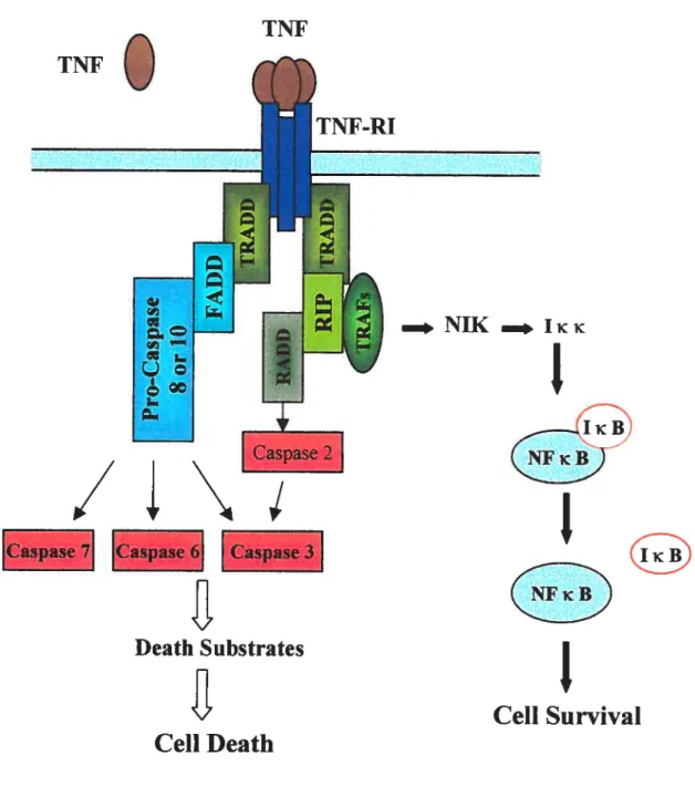
Figure 2. Signaling pathways ofTNF andTNF-RI 37
Figure 3. TR6 and related ligands and receptors 40
Article 1:
Fig 1. TR-6 binds to ceil surface LIGHT and competes withTR2 for its binding to
LIGHT 47
Fig 2. Hurnan TR6 cross-reacts with mouse LIGHT 47
Fig 3. Inhibition of in vivo and ex vivo spienic alloactivation in mice by TR6-Fc 4$
Fig 4. TR6-Fc and TR6 represses the development ofCTL in mice 49
Fig 5. TR6-Fc modulates in vitro lyrnphokine production in H2d alloantigen-stirnulated
2C spleen ceils 49
Fig 6. TR6-Fc prolongs heart aÏlograft survival ofthe mice 50
Article 2:
Fig 1.TR6-Fc strongly prornoted proliferation ofPHA- or anti-CD3-stimulated PBMC
and T celis 56
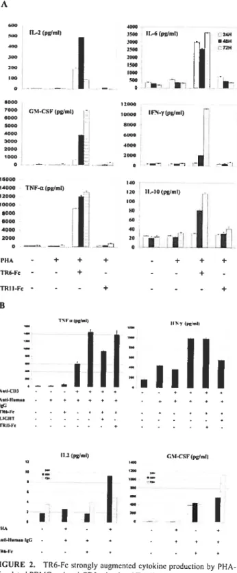
Fig 2. TR6-fc strongly augmented cytokine production by PHA-stimuÏated PBMC and
anti-CD3-stirnulated T celis 5$
Fig 3. LIGHT was the major TR6-Fc ligand on activated Thi and Th2 celis 5$
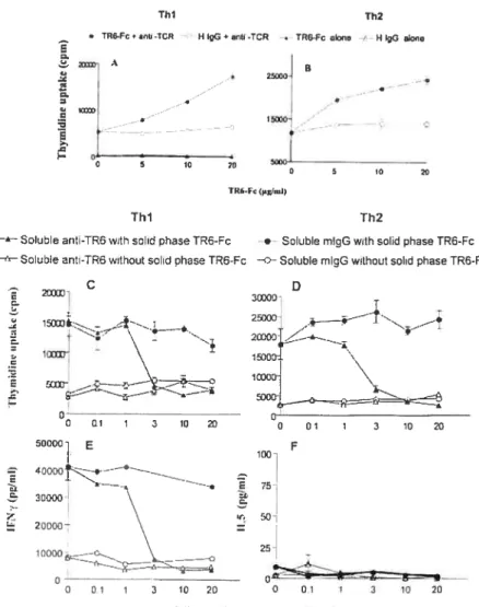
Fig 4. Effects ofTR6 costirnulation on proliferation and Ïymphokine production ofThl
Fig 5. Effect of LIGHT reverse signaling on CTL development 60
Article 3:
Fig 1. TR6 is secreted by activated T celis 61
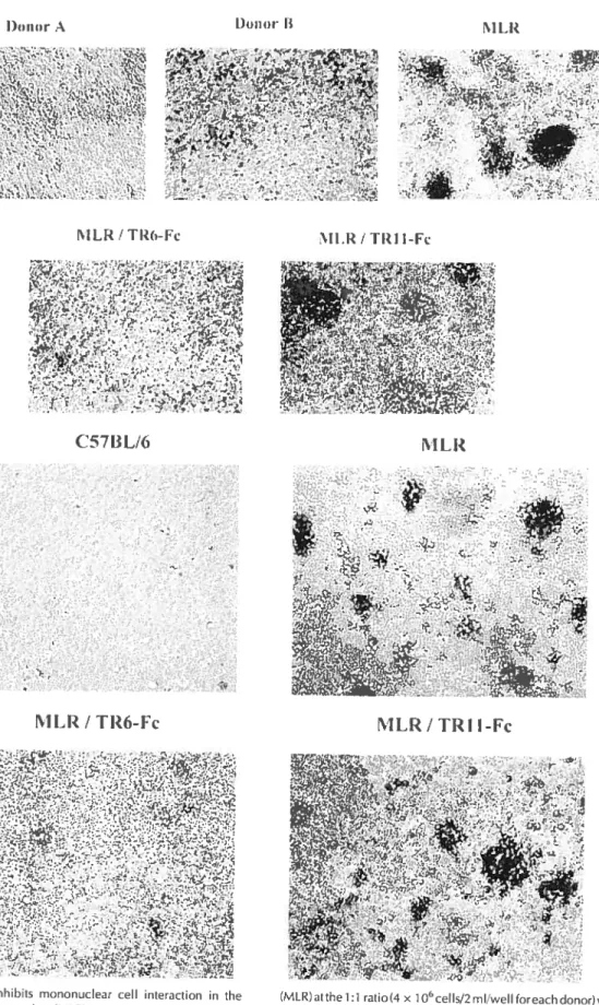
Fig 2. TR6-Fc inhibits mononuclear cell interaction in the MLR 67
Fig3. TR6-Fc inhibits PBMC and T-cell aggregation induced by mitogen stirnulation..6$ Fig 4. Effect of solid phase TR6-Fc, soluble Fas and soluble LIGHT on T-cell
aggregation 69
Fig 5. TR6-Fc inhibits anti-CD3-induced T-cell actin polymerization and pseudopodium
formation 71
Appendix:
Figure 1. Mutated Fc does flot bind to human PBMC 119
Figure 2. Gastric cancer lymphocyte infiltration is reversely correlated to serum TR6
level 120
LISI 0F TABLES
Introduction:
Table 1. TNFR Superfamily Members 4
Table 2. TNf Ligand Superfamily Members 5
Article 1:
Table 1. JÇq values for LIGHT binding to TR6-Fc, TR6, and TR2-Fc as determined by
BlAcore analysis 46
Table 2. Effect ofTR6-Fc on alloactivation-induced splenomegaly in BALB/c x
LIST 0F ABBREVIATIONS
AA Arnino acid residues
ACBP Acyl-CoA binding protein
MF Apoptosis-inducing factor
Ag Antigen
AP-1 Transcription factor activator protein 1
Apaf- 1 apoptosis protease activating factor- I
APC Antigen-presenting celis
BSAP B ceil lineage-specific activator protein
Caspase Cytosolic aspartate-specific protease
CRD Cysteine rich domain
CIL Cytotoxic T lymphocyte
DC Dendntic celi
DcR3 Decoy receptor 3
BD Death domain
DR Death receptor
EST database Expressed sequence tagged database
FADD Fas-associated death dornain
FasL Fas ligand
GPI Glycosylphosphatidylinositol
GvHD Graft versus host disease
HtrA2 High temperature required A2
IAP liihibitor of apoptosis protein
1g immunoglobulin IL-2 Interleukin 2 IFN-y Interferon-y KO Knockout LC Langerhans ceils LN Lymph nodes LPS Lipopolysaccharide LTa Lymphotoxin a LTf3 Lymphotoxin
f3
LTf3R Lymphotoxin beta receptor
HVEM/TR2 Herpesvirus entry mediator
JNK c-j un N-terminal kinase
MAPK Mitogen activated protein kinase
MLR Mixed lymphocyte reaction
NF-icB Transcription factor nuclear factor-icB
NIK NF-icB-inducing kinase
NK Natural killer
OPG Osteoprotegrin
PARI? Poly (ADP-ribose) polyrnerase
PUA Phytohemaggiutinin
PMA Phorbol 1,2-myristate 1,3-acctate
PP Peyer’s patch
SFP Superfamily protein
Smac Second mitochond-derived activator of caspase
SODD Silencer of death domain
Tg Transgenic
Thu/2 Thelper ceil and 2
TNF Tumor necrosis factor
INFR TNF receptor
TRAF TNF receptor associated factor
TRADD TNFR-associated death domain protein TRANCE TNF related activation induced cytokine
I. INTRODUCTION
I. INTRODUCTION
Cytokines are important glycoprotein messenger molecules capable of transmitting signais from one ce!! to another. Most cytokines exist in secreted forrn but some are either expressed at the celi surface or stored in the extra-cellu!ar space. To date more than 200 cytokines have been identified including interieukins, growth factors, chemokines, interferons, and a host of others (Caliard et al., 1999; Locksley et aÏ, 2001). Cytokines need to interact with their receptors expressed on the surface of the target ce!!s, thereby triggering compiex interceliuiar signaiing cascades, which u!timateÏy control gene expression required for the cellular response. Under normal circumstances, the production of cytokines and the expression of their receptors are under tight and complex biologicai contro!, including negative and positive feedback by the cytokines themselves. Cytokines can be divided into severai groups such as the hematopoietins, the interferons, the tumor necrosis factor (TNF)-related molecules, the immtmoglobulin (1g) superfamily members, and the chemokines. Among these groups, the TNF superfamily is unique since members of this superfarnily are mainly concentrated in the immune system and display cruciai functional roles in regulating immune responses (Gruss and Dower, 1995; Gruss et aÏ., 1996).
1.1. TNFandTNfreceptor supeifaindies
TNF ligand superfamiiy was originated from two proteins, TNf-aipha (TNF Œ) and
lymphotoxin a (LT a). These two stmcturaiiy and functionaliy reiated but distinct proteins, identified by the property of tumor celi lysis, were the prototypic members of
the TNF superfamily (Carswell et al., 1975; Gray et al., 1984, Pennica et al., 1985). The
receptors ofTNf Πare TNFR-I (p55) and TNFR-ll (p75) whereas the receptors for LTa
are TNFR-I, TNFR-II and HVEM/TR2 (Locksley et al., 2001). These receptors then constituted a new TNF receptor (TNFR) related gene famity (Gntss and Dower, 1995; Locksley et al., 2001). Both TNFR and TNF ligand superfamilies have experienced rapid
expansion over the past decade ami many molecules were identified as TNf or TNFR
superfamily proteins ($FPs) (Srnith et aï., 1994; Idriss and Naismith, 2000; Locksley et al., 2001).
for the past decades there was no well-coordinated, systernatic naming system and the
nomenclatures for SFPs in these two superfamilies were complicated and sornewhat redundant. It was common that some members had multiple names given by different groups (as presented in Table 1 and Table 2). Based on this situation, a standard, official designation system was foniially introduced for both TNF ligand and TNFR SFPs while sorne popular, well-accepted narnes are still being used in parallel (Locksley et al., 2001). (Details refer to Table 1 for related receptors and Table 2 for related Iigands).
1.2. TNfRJainily
Cunently, this stiil growing family has incorporated more than 20 different membrane proteins and several open viral reading frames encoding related molecules (Locksley et aÏ., 2001; Adams et al., 2002). As summarized in Table 1, the mammalian TNTR superfamily now includes: TNFR-I, TNFR-II, Fas, 0X40, CD4O, CD27, CD3O, 4-133, DcR1, DcR2, TR6 (DcR3), OPG, DR3, DR4, DR5, DR6, HVEM (TR2), RANK, TACI, BAFFR, EDAR, 3CM, RELT, SOBa, Tnfthl, TAI (Armitage, 1994; Smith, 1994;
r) tri (ID tri. H r) r (ID (ID tri. r r) r (ID (ID H r D z H r r > D r) X r) z
Table
1.
TNFR
Superfamïly
Members
Officiai Other Common Names References Symbol TNFRSF1A P5 5-R, CDI2Oa, TNF-R-I p55, TNF-R, TNFAR, TNF-R55, Van Arsdale and Ware, 1994 p55TNFR,TNFR6O TNFRSF1B CD12Ob, p75, TNF-R, TNF-R-II, TNFR8O, TNFR2, TNF-R75, TNFBR, Santee and Owen-Schaub, 1996; Smith et aI., 1990 p75TNfR LTBR TNFRSf3, TNFR2-RP, CD 1$, TNFR-RP, TNfCR, TNf-R-III Gruss and Dower, 1995 TNFRSF4 OX-40, ACT35, TXGP1L Godfrey et aI., 1994 TNfRSF5 P50, 3p50, CD4O Banchereau et aI., 1994 TNFRSf6 FAS, CD95, APO-1, APT-1 Itoh and Nagata, 1991 TNFRSf6B DcR3, TR6 Pitti et al., 1998; Bai et aI., 2000 TNFRSF7 Tp55, S152, CD27 Camerini et al., 1991; Loenen et aI., 1992 TNFRSf8 Ki-1, DISI66E, CD3O Smith et aI., 1993; Gruss et aI., 1995 TNfRSF9 4-133, CD 137, ILA Kwon et aI., 1994 TNFRSFIOA DR4, Apo-2, TRAILR-1 Pan et aI., 1997 TNfRSF1O3 DR5, KILLER, TRICK2A, TRAIL-R2, TRICKB Pan et aI., 1997; Sheridan et aI., 1997 TNFRSfIOC DcR1, TRAILR3, LIT, TRID Sheridan et al., 1997; TNfRSF1OD DcR2, TRUNDD, TRAILR4 Pan et al., 1998; Marsters et al., 1997 TNfRSf11A RANK Anderson et al., 1997 TNFRSF1 13 OPG, OCIF, TRi Emery et al., 1998; Simonet et al., 1997 TNFRSFI2 DR3, TRAMP, WSL-1, LARD, WSL-LR, DDR3, TR3, APO-3 Chinnaiyan et aI., 1996; Masters et al., 1996 TNFRSF12L DR3L Grenet et al., 1998 TNFRSF 133 TACI Xia et al., 2000; Gross et al., 2000 TNFRSF13C BAfFR Thompson et al., 2001 TNFRSf 14 HVEM, ATAR, TR2, LIGHTR, HVEA Mauri et al., 1998; Kwon et al., 1997 NGFR TNFRSFI6 Gruss and Dower, 1995 EDAR EDAR Srivastava et al., 1997 TNfRSf 17 3CM, 3CMA Gross et aI., 2000 TNFRSF18 AITR, GITR Kwon et al., 1999; TNFRSF19 TAJ, TROY Hu et al., 1999 TNFRSFÏ9L RELT Sica et al., 2001 TNFRSf2I DR6 Pan et al., 1998 TNFRSF22 SOBa, Tnfrh2 Clark et aI., 2002 TNFRSf23 MSOB, Tnfrhl Clark et al., 2002E
z z
z
e
Anderson et al., 1997; Ashkenazi and Dixit, 1998; Gruss and Dower, 1995; Locksley et aï., 2001). Some rnost recent identified members such as 3CM, RELT, SOBa, Tnfrhl and TAJ are currently stili flot well defined (Locksley et al., 2001). The viral open reading frames encoding soluble TNFRs such as crmB (Hu et al., 1994), Va53 (Smith et al., 1990) G4RG (Howard et ctl., 1991), and SfV-T2 (Upton et al., 1987) were also identified.
1.2.1. The structzcraÏfeatures ofthe TNFR supe,famiÏy
The mammalian TNFR family members are type I membrane proteins, whose extracellular N terminal part contains ligand-binding domain. A remarkable feature for TNFR superfarnily is the low degree of sequence homology in their extracellular ligand binding domain (20-25%) (Gniss and Dower, 1995). These SFPs are mostÏy trirneric
(Gruss and Dower, 1995; Armitage et al., 1994; Aggarwal et al., 1996; Bazzoni and Beutler, 1996).
The definition of the TNFR superfarnily is mainly based on the conserved motif of “cysteine-rich repeats” in the extracellular N-terminal region. These common conserved cysteine-rich domains are also terrned as cysteine—rich motif or cysteine-rich domain (CRD), which consists of multiple cysteine-rich repeats of approxirnately 30-40 amino acids (Smith, 1994). In general, each member of this family contains varying numbers
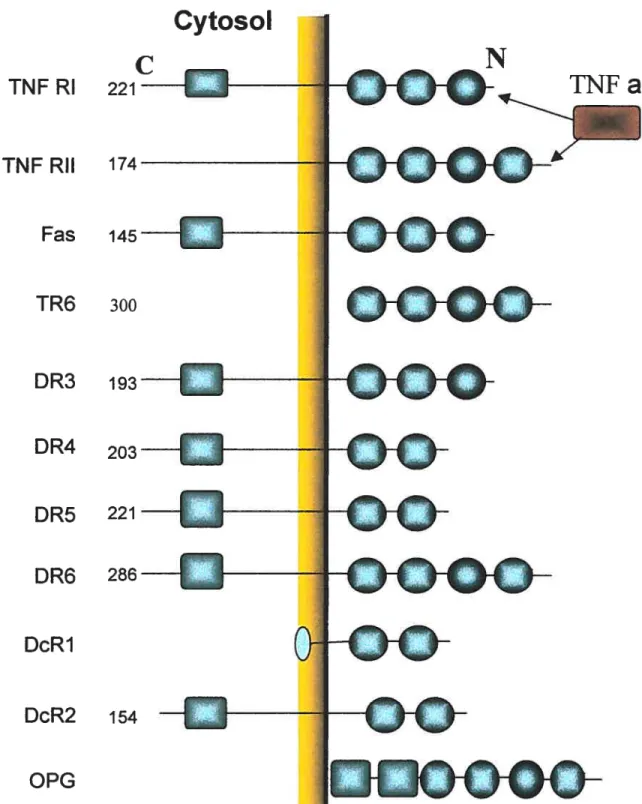
(2-6) of CRDs as shown in Figure 1. Each CRD is featured by the presence of
approximately 6 cysteine residues that are interspersed within CRD domain. The structure of CRD is supported by 3 intrachain disulfide bonds fonried by these 6 highly conserved cysteines (Smith et al., 1994). These multiple cysteine-rich domains in the
Cytosol
TNFRI
221INFRII
174Fas
145—c
1-TR6
300DR3
1930
DR4
203DR5
221DR6
286—-DcR1
DcR2
154OPG
o
TNFa
o-oøG
oo
œ
1YF
œ
GcF
OOGŒGO
figure
1.
Structural features of TNF receptors. There are two structural domains forcommon TNF receptors. The cysteine nch domains (CRD)
O)
are located in the extracellular N-terminal portion. Several receptors aso have a death domain (DD)
(
O)
extracellular part have been shown to be involved in ligand binding (Locksley et al.,
2001).
The extracellular structure of TNFR SfPs can be best illustrated by TNFR and DR5. These two receptors have been well studied for their crystal structure, which represents common structural features of ail TNFR SFPs (Naismith et al., 1996; Hyrnoowitz et al., 1999). for example, DR5 forms an extended rod-like shape consisting of 3 CRDs which form the interface to bind its cognate ligand(s). DR5 had a total of 7 disulfide bridges: 6 are in CRD 2 and CRD3 (three for each) and I in C-terminal part. These disulfide bonds form a structural scaffold and two patches formed are located there for ligand binding. The combination of structural conservation and variable arnino acid sequences in the ligand contacting region confer the ligand-binding specificity. Notably, the structure of TNFR is rather flexible and can be optimized for its interaction with ligand through a series ofhinging movements (Idriss and Naisrnith, 2000).
In addition to the membrane-bound form, many TNF receptors also exist in a soluble form. These soluble molecules are, in many cases, generated by proteolytic cleavage of ceil surface receptors. Soluble TNTR-I, TNFR-II, CD27, CD3O, CD4O, and fas are ail generated in this fashion (Gruss and Dower, 1995). The only exception is 4-1BB, whose soluble form is generated by alternative splicing (Grnss and Dower, 1995). The necessity of the soluble forms of these receptors is not fully understood.
1.2.2. The biologicalfeatures of TNfR superfamity
The TNFR SFPs have emerged as prominent regulators of the immune system (Tracey and Cerami, 1994). In the immune system, TNFR SfPs are well known for their critical
roles in regulating immune responses such as celi activation, proliferation, differentiation, apoptosis, immunoglobulin (1g) class switching, immune evasion, and immune suppression (Smith et al., 1994; Aggarwai et al., 1996; Tewari and Dixit, 1996; Baker et al., 1996; Locksley et al., 2001). Sorne SFPs are also involved in the generation and developrnent of iymphoid organs (Matsumoto et aï., 1997).
The primary feature of TNFR superfamily members is ce!! death induction including tumor killing and this effect was observed severa! decades ago (Carswell et al., 1975). Many, if not ail, TNFR members are related to ce!! death induction. Members such as TNF, Lia, CD3OL, CD95L, 4-1BBL are a!! capable of inducing cytotoxic ce!! death (Gruss et al., 1996; Gruss and Dower, 1995). Probably this ability to induce ceil death is one of the unique features with remarkab!e adaptive va!ue that TNT/TNFR SFPs have developed (Gniss and Dower, 1995; Locksley et al., 2001). As will be detailed in the next section, severa! TNF receptors contain a structure called “death domain” which is responsible for ceil death induction and these receptors are termed “death receptors” (DR). There are 8 receptors containing “death domain” in TNFR superfarni!y and at least 6 of them can induce apoptosis through activation of caspases (Screaton and Xu, 2000; Raff, 1998). Meanwhile, other TNF/TNFR SFPs lacking death domains can potentially modulate the response to DRs or directly influence ce!! deathlsurvival. for instance, TNFR-II markedly enhances TNFR-I induced T celi death and CD4O can augment Fas induced B celi death (Garrone et al., 1995; Chan et al., 2000b).
In contrast to the death induction feature of TNFR SFPs, it is interesting that they are also closely related to !ymphocyte survival including T/B celi proliferation and differentiation (Locks!ey et al., 2001). Indeed, the major docurnented function of TNFR SfPs is
associated with lymphocyte survival. For instance, Blys (officiai symbol: TNFSFI3B, or THANK, BAFF, see Table 2) expressed on activated dendritic celis can interact with the TACI and BCMA receptors (both are TNFR members, see Table 1) on B ceils and promote B ceil survival (Laabi and Stresser, 2000, Moore et al., 1999). Similarly, T ceil activation is also regulated by TNF/TNFR SFPs. For instance, LT3R enhances T celi activation and promotes T celi clone expansion by engaging with its ligand LIGHT (Tamada et aÏ., 2002). Some other members such as CD4O and Fas may also stirnulate T
cell survival and proliferation by engaging with their receptors (Cayabyab et aÏ., 1994; Suzuki et al., 199$).
In addition to modulating lymphocyte deathlsurvival, SFPs of this superfamily are also associated with antigen presenting celis (APC) survival and maturation. For instance, dendritic celis (DCs) are potent APCs for antigen presentation but fully differentiated and mature DCs wilÏ undergo rapid apoptosis. The life span of mature DCs can be prolonged substantially by TNF SFPs such as CD4OL, TNF, TRANCE (TNF related activation induced cytokine) and recently identified member LIGHT (Wong et al., 1997; Tamada et al., 2002).
Some, if not most, TNFR SfPs are involved in T celi costimulation. For example, both TNFRJ and Fas can co-stimulate T celi activation under diffei-ent settings (Siegel et al., 2000; Suzuki et aï., 2000A, 2000B). In addition, the SFPs such as 0X40, CD27, and
4-1BB regulate the activation and expansion of CD4+ and CD$+ T celis responding to
dendritic ceils beanng their respective ligands. Costimulation effects were also observed for LTR/LIGHT pair. It was found most likely that this pair provides costimulation for T ceil activation and plays a critical role in T celi activation independent of B7/CD2$
costimulation pathway (Tamada et al., 2000; Wang et al., 2001). Thus, it is suggested that TNFR SFPs might be critically important in negative selection and determine the activation and/or apoptosis of T celis (Sebzdaetal., 1999; Wang et al., 2001).
Another welI-docurnented function of TNFR superfamily is to orchestrate permanent lymphoid organ structure. A remarkable example is LTf3R that belongs to TNFR superfamily and plays critical roles in the immune system. Several studies showed that
LTJ3W’ mice lacked lymph nodes (LN) and Peyer’s patch (PP) and displayed severe
disorganization of spienic architecture, featured by the absence of T/B celi segregation, marginal zones, follicular dendritic ccli (FDC) networks, and germinal centers (GC) (De Togni et al., 1994; Banks et al., 1995; Wu et al., 1999). Depletion ofRANK or RANKL lead to the disappearance of ail peripheral and mesenteric lymph nodes while Peyer’s patches remain intact and the spienic architecture is unaffected (Kim et al., 2000; Kong et al., 1999; Dougall et al., 1999). Studies dernonstrated that the requirements for RANX/RANKL and LTalj32 or LTR do flot compensate for each other. Ail these data suggest that SFPs of TNFR superfamily are necessary for the formation and development of lymphoid immune organs, sorne of them being indispensable.
In addition to critical functions in the immune system, other functions were also identified for TNfR SFPs. For instance, Edar is a death domain containing protein and recent studies indicate that this protein is important in the development of hair, teeth and other ectodermal derivatives (Headon et al., 2001).
1.2.3. Subsets oJTNFR superfainiÏy
Based on their structural and biological features, the TNFR sttperfarnily SFPs can be further divided into different groups according to the presence of featured domains in the
intracellular portion of the receptor. The SFPs in the first group featured by a TNf R
associated factor (TRAF) binding domain that enabies coupling to TRAFs, that in tum activate a signaling cascade that resuits in the activation of Nf-KB and initiation of transcription (Rothe et ctÏ., 1995; Wallach et aÏ., 1999). The second group ofreceptors is featured by a 60- amino acid globular structure named “death dornain” (DD) and these death dornain-containing receptors are termed as death receptors (DRs) (Tartaglia et aï., 1993; Itoh and Nagata 1993). The third group bas drawn more attention recentiy and members of this group are designated as decoy receptors (DcRs) that include DcR1, DcR2, DcR3/TR6 and OPG (Ashkenazi and Dixit, 1999). Ail members in this group except DcR2 do not have cytoplasmic domain and thus they may act as inhibitors and compete with other signal-transducing receptors for ligand binding (Ashkenazi and Dixit,
1999; Ashkenazi, 2002).
1.2.3.1. TRAf associated subgrottp
The TRAF s are adaptor ptoteins belonging to ring and zinc-linger proteins. The members
ofthis group are TNFRII, CD4O, CD3O, CD27, LTF3R, 4-YBB, 0X40, NGFR, HVEM, GITR and RANK (as presented in Table 1). They bind directly to the receptor’s cytoplasmic tau. They also bind to one another in homotypic and/or heterotypic interactions. They are believed to convey certain signais in proliferative and pro inflammatory responses. This is achieved, in large part, by activation of NF-KB, a transcription factor involved in nurnerous proliferative and pro-inflarnmatory events
(Jabara et al., 2002; Wallach et al., 1999; Harrop et al., 199$). So far at least 6 TRAFs
have been found to associate with non-DD TNF receptors (Locksley et aï., 2001). Most
TNFR SFPs have no death domains but contain a consensus motif which is capable of binding to the TRAF proteins (Ye etal., 1999).
1.2.3.2. Death receptor subgroup
The above mentioned “death dornain” was first coined from studies of deletion mutagenesis involving TNFR-I rnediated apoptotic celi death (Tartaglia et al., 1993).
The proteins harboring “death domains” were thus defined as death receptors (DRs) and they form DR group within the TNFR superfamily (Ashkenazi and Dixit, 1999; Locksley
et aï., 2001). This death-domain-containing receptor subgroup now includes up to $
members: TNF-R1, Fas, and recently discovered DR3 (Chiimaiyan et al., 1996; Marsters et al., 1996; Kitson etal., 1996; Bodmer et al., 1997; Screaton et al., 1997), DR4 (Pan et
aï., 1997), DR5 (Pan et al., 1997) and DR6 (Pan et al., 1998), EDAR (lucher et al., 2000) and NGFR (Gruss, 1996). The typical conserved death dornain (DD) is a 6$ amino acid segment located within the cytoplasmic region of the receptor. Upon ligation with either cognate ligands or specific agonistic antibodies, death receptors can activate an apoptotic signaling pathway (Nagata, 1997).
These death receptors have common structural features such as 2-6 CRDs in their extracellular domains and an intracellular death domain. The death domain most likely functions as a protein interaction dornain and provides docking sites for signaling molecules, therefore enabling each receptor to couple to the caspase cascades which are critical for the induction ofapoptosis.
1.2.3.3. Decoy receptor subgroup
Another subgroup of the TNFR superfamily was termed “decoy molecule”. This decoy receptor subgroup now includes DcR1 (decoy receptor 1, also known as TRID or TRAIL R3) (Pan et al., 1997; Degi-Esposti et al., 1997; Sheridan et al., 1997; Pitti et al., 1998), DcR2 (decoy receptor 2, also known as TRUNDD or TRAIL-R4) (Pan et al., 1998; Degli-Esposti et al., 1997), DcR3 (TR6) (Pitti et al., 1999) and osteoprotegerin (OPG)
(Simonet et aÏ., 1997; Ashkenazi and Dixit, 1999). DcR1 and DcR2 are ceil surface
proteins whereas DcR3 and OPG are soluble molecules. Both DcR1 and DcR2 bind to TRAIL with a similar affinity (Pan et al., 1997A; Pan et al., 1997B; Sheridan et al.,
1997). DcR1 is a glycosylphosphatidylinositol (GPI)-linked protein without an
intracellular death domain whereas DcR2 is a transmembrane receptor but with a partially deleted death domain and accordingly, both are incapable of transmitting apoptotic signais (Ashkenazi and dixit 1999; Deli-Esposti et al., 1999). The product of OPG is a secreted protein that also binds to TRAIL but with much weaker affinity compared with other receptors. DcR3 (TR6), as will be described in details below, is doser to OPG and exists as a secreted protein (Pitti et al., 1998).
Both DcR1 and DcR2 are believed mainly to function as anti-apoptotic decoy receptors that compete with DR4 and DR5 for TRAIL binding and consequently protect those receptor-bearing ceils from TRAIL-induced apoptosis (Ashkenazi and dixit, 1998; Deli
Esposti et aÏ., 1999). DcR1 is a GPI-linked protein and its role in active signal
transduction has flot been fully eÏucidated. Ernerging evidence suggests that DcR2 may be involved in activating signal transduction since it lias been shown to activate NF-icB in
some systems (Degli-Esposti et al., 1997). In contrast, there are reports of failure of DcR2 to activate Nf-KB in certain systems (Meng, 2000). Thus more studies are necessary to explore the role of DcR1 and DcR2 in anti-apoptosis signal transduction and in apoptosis.
1.2.3.4. General characteristics of TR 6
TR6 is a nove! TNF decoy receptor with the officiai designation of TNFR$F6B (see Table 1). By screening expressed sequence tagged (EST) database, Pitti (1998) identified
a previously unknown fu!I-!ength cDNA from a human fetal lung library. This cDNA
showed homology to other TNFR superfamily members. The protein encoded by this gene was then named decoy receptor 3 (DcR 3) (Pitti et ctl., 199$). This gene was also independently identified by other groups and named TR6 (Yu et al., 1999) or M6$ (Bai et al., 2000), respectively. The hurnan TR6 gene is mapped at position 20q13.3 (Pitti et al., 199$), whi!st its mouse counterpart might not exist since a BLAST search of the mouse genome did not reveal sequences with any significant homology. Since the position of 20q 13 is also named “cancer amplicon” where genes responsible for rnany types of tumors are !ocated, TR6 may be a candidate gene associated with certain types of malignancy (Kom et aÏ., 1999; Medeiros et aï., 1999; Stubbs et al., 1999; Savelieva et al., 1997;Sonoda et aÏ., 1997; Sakakura et aï., 1999).
The fuIl-Iength open reading frame ofTR6 encodes 300 amino acid residues with the first 29 amino acid residues as signal sequence. The mature fonri ofTR6 has 271 amino acid
residues with no transmembrane region. There is only one potential N-!inked
glycosylation site at Asn-173. Like OPG, TR6 vas regarded as a soluble secreted protein.
TR6 shares remarkable amino acid sequence homology with OPG (31%) and TNFR-II (29%) but less liomology with Fas (17%) (Pitti et al., 1998). TR6 was reported to have
two complete and two incomplete cysteine-rich motifs, which is the hallmark of TNF
receptors.
The TR6 mRNA lias prorninent expression in lung and colon cancers, whereas it is
weakly detectable in several hematopoietic celi unes, and is hardly detectabÏe in many tissue samples (Pitti et al., 199$). In the immune system, TR6 is highly expressed in both lymph nodes and spleen, but the expression in thymus is weak (Pitti et al., 199$; Bai et al., 2000). In addition, TR6 is expressed in an endothelial celi une and its expression is
inducible by phorbol 12-myristate 13-acetate (PMA) /ionomycin in Jurkat T leukernia
celis (Yu et al., 1999). Moreover, TR6 mRNA over-expression was reported in
gastrointestinal cancers but without gene amplification (Bai et al., 2000). Also, the
expression of TR6 can be detected in malignant glioma ceils as well as in human
glioblastomas. In addition to human tumors, the TR6 gene is also over-expressed in silicosis or systemic lupus patients (Otsuki et aÏ., 2000).
Mild et al. investigated a large number of colorectal cancers and found nearly 63% (185 out of 294) of patients with DcR3 gene amplification (Mild et al., 2002). In another study, it was found that TR6 was amplified and over expressed in EB virus- and human T celi leukemia virus-I— associated lymphomas (Ohshima et aÏ., 2000). This expression trend is also true at the protein level of TR6. For instance, TR6 protein was found over expressed in human adenocarcinomas of the esophagus, stomach, colon, and rectum while no gene amplification was detected (Bai et al., 2000). It was reported recently that up to 73% (163 out of 223 patients) of colorectal patients showed up-regulated TR6
protein level (Mild et aÏ., 2002). These data suggested that certain types of turnor as welÏ
as EBV and human T ce!! leukemia virus-1 may use TR6 to escape from immune
surveillance during lymphomagencsis, or that virus infected Ïymphoma ceils with TR6 expression might be selected during multi-step tumorigenesis (Ohshima et al., 2000). Recent studies in our laboratory as well as in others suggested that TR6 over-expression correlates with the grade of malignancy and could be used as diagnostic and prognostic parameter (Wu et al., 2003; Roth et al., 2001).
1.2.3.5. The TR6 Ligands
The FasL (Fas ligand) was the first ligand identified for TR6 (Pitti et aï., 199$). FasL is one ofthe major effectors ofcytotoxic T lymphocytes (CIL) and natural killer (NK) cells and a major inducer of apoptosis. It is also involved in the establishment of peripheral tolerance in the activation-induced celi death of lymphocytes (Zhang et al., 2000; Ju et al., 1999). Moreover, the expression ofFasL in non-lymphoid and tumor cells contributes to their maintenance (Nagata, 1997). LIGHT (also termed HVEM-L) is another cognate ligand for TR6 (Yu et al., 1999). Very recently, another novel member of TNF ligand superfamily TLÏA was found to be a third ligand for TR6 (Migone et aï., 2002). TL1A is considered as the longer variant ofTLl and this molecule was designated as VEGI when it was identified in endothelial ce!! DNA libraries (Zhai et al., 199$; Yue et al., 1999). Studies at the mRNA level show that TLÏA is highly expressed in endothelial cells and is inducible by TNF and IL-1. TL1A is the ligand for both DR3 and TR6 (Migone et al., 2002). DR3 is a TNFR member and has the capability of inducing NF-icB activation and apoptosis upon over-expression. DR3 shares another ligand TWEAK (or Apo3L) with a
novel receptor TWEAK (Masters et aï., 1998; Wiley et ctl., 2001). But the relationship between these proteins and the significance of their interactions have yet to be fully studied
1.2.3.6. The identfled biotogicalfimctioizs oJTR6
It has been demonstrated that TR6 could neutralize the biologicai effects of FasL by
inhibiting fas-FasL interaction (Pitti et al., 1998). This decoy receptor substantialiy biocks the FasL-induced apoptosis in Jurkat ceils as well as FasL-dependant NK celi activity (Comol1y et al., 2001). These resuits suggest that TR6 could interact with FasL under certain conditions and modulate immune responses, especially apoptosis mediated by the Fas/FasL pathway.
TR6 can suppress LIGHT binding to both LTR and TR2 (Yu et al., 1999; Mauri et al., 1998), and it is conceivable that such interference can inhibit LTf3R- and TR2-mediated responses in immune ceils. It was demonstrated that TR6 can interact with LIGHT and thus inhibit LIGHT —mediated cytotoxicity of H29 ceils. These resuits suggest that TR6 acts as a natural inhibitor of LIGHT mediated tumor celi killing.
In addition to modulation of immune responses, it was found in a recent study that TR6 profoundly modulates dendritic ceils LDCs) differentiation and maturation from CD14 monocytes (Hsu et al., 2002). Interestingly, it was reported that TR6 enhances CD86/37.2 expression, whereas CD8O/B7.1, CD4O, CD54/ICAM-1, CDY.a and HLA DR were ail repressed. In addition, DcR3-treated DCs dramatically enhanced 1L4 secretion by naïve T celis (CD4CD45RA), thus favoring Th2 dcvelopment.
The ligand TL1A can also function as a T ceil costimulator. The T ccli stimulation induced by TL1A resuÏts in an increased responsiveness to IL-2 and other
pro-inflammatory cytokines (Migone et al., 2002). TR6 competes with DR3 for TL1A binding with similar affinity. Therefore, it is possible that TR6 may modulate the duration and magnitude of immune responses mediated by DR3. Moreover, since DR3 can induce apoptosis, blocking the interaction between TL1A and DR3 by TR6 might provide protection from apoptosis to certain types oftumors (Migone et al., 2002).
1.2.4. The other TNF receptors related with TR6
Obviously, TR6 may have important functions stili to be elucidated in cancer development and in the immune system. In the latter, TR6 might compete with other TNf receptors for core common ligands such as LIGHT and FasL and thus interfere with their
biological functions. Among all the possible TNFR SFPs that share LIGHT and FasL
with TR6, TR2 and Fas are predominantly expressed in the immune system and are
critically important in regulating immune responses. As such, to decipher the nature of TR6, identify with which ligand TR6 interacts and the related receptor(s) such as TR2 and Fas will certainly help to better understand the underlying mechanisrns of the regulatory roles of TR6 both in vitro and in vivo.
1.2.4.1. HVEM/TR2
The receptor herpes virus entry mediator (HVEM or TR2) is a recently discovered TNFR superfamily member with broad tissue and cell type expression especially in the immune system (Montgomery et cd., 1996; Kwon et al., 1997; Tan et aÏ., 1997). This molecule was also identified by screening expressed sequence tag (EST) cDNA database for sequence hornology with cysteine-rich motifs of the TNFR superfamily (Harrop et al.,
1998). This molecule was discovered through its ability to mediate HSV infection. The TR2 gene locates at chromosome lp36, a position close to other TNFR members such as CD3O, 4-1BB, OX-40, and TNfR-II (Montgomery et al., 1996). Three ligands so far have been defined for TR2: the HSV surface envelope glycoprotein gD, lymphotoxin a (LT a) and the newly described TNF ligand family member LIGHT (Montgomery et al., 1996; Mauri et aÏ., 1998).
The TR2 mRNA can be readily detected in lung, spleen, thymus, monocytes, dendritic ceils, B and T lymphocytes, but not in liver, brain, and skeletal muscle (Morel et al., 2000). Moreover, RNA analysis unveiled that most solid tumor unes do flot express TR2. On the other hand, some hematopoietic celi lines do express TR2 (Kwon et al., 1997). The full-length TR2 encodes a 283 amino acid protein. It is a type I transmembrane protein containing a 50-amino acid cytoplasmic region without a death domain (Harrop et al., 199$). At the protein level, expression studies indicated that TR2 protein has wide distribution and can be readily detected by flow cytometric analysis in peripheral blood T and B lymphocytes, NK ceils, and monocytes (Mord et al., 2000). Interestingly, it was reported in a recent study that TR2 and one of its ligands LIGHT display reciprocal expression at T ceil surfaces as detected by flow cytometric analysis (Mord et al., 2000). LIGHT is hardly detectable in resting T celis, but its expression was enhanced tipon activation (Morel et al., 2000). On the other hand, HVEM showed down-regulation upon T ceil activation. Data from confocal microscopy and intracellular staining by flow cytometry showed the existence of intracellular LIGHT in unprimed T ceils. After activation, there is a pronounced induction of LIGHT both intracellularly and at the cell surface. This suggests that de novo synthesis and redistribution of LIGHT both contribute
to the enhancement of LIGHT expression at ceil surface. The detailed mechanism of down-regulation ofTR2 expression by LIGHT is stiil flot well understood.
The biological function ofTR2 was an active area of investigation in the past few years. Current evidence suggests that TR2 is closely associated with T ceil activation and a number of T celi responses such as proliferation, cytokine production, and expression of celi surface activation molecules (Hanop et al., 1998). LIGHT, as its ligand, stimulates the proliferation of activated T ceils expressing TR2 (Harrop et aÏ., 1998), stimulates NF iB activation, and induces apoptosis in celis expression both TR2 and LTR (Zhai et al., 1998; Harrop et al., 1998). Recently, studies show that LIGHT can costimulate T celi responses by interacting withTR2 (Kwon et al., 1997; Tamada et al., 2000A; Tamada et al., 20003) and TR2 is critically important in T celi costimulation. T celi derived LIGHT can readily deliver stimulation to TR2 on dendritic celis, that in tum up-regulate T ceil
activity (Shaikh et al., 2001). Moreover, TR2 on T cells can also receive LIGHT
stimulation directly from LIGHT expressing T ceils (Wang et al., 2001).
1.2.4.2. fas
Fas (also named APO-1 or CD95) is the primary receptor for fasL and this molecule is a type I membrane protein belonging to the TNFR family (Suda et al., 1993). Fas is abundantly expressed in various tissues and celi types (Suda et aÏ., 1993), especially thymocytes, activated T cells and virus transformed T cells. Resting B cells do flot express fas, but its expression can be induced by CD4O ligand or endotoxins (Briones et al., 2002; Hahne et al., 1996). Fas has broad tissue distribution, but is most abundantly expressed in the thymus, liver, heart and kidney (Itoh and Nagata, 1993). Cross-linking of
Fas by an agonistic antibody or ligation by FasL will cause Fas clustering which is necessary for receptor activation and death signal initiation (Kischkel, 1995).
1.3. TNf Ïigand superfamiÏy
As listed in Table 2, the mammalian TNF ligand superfamily also exhibited rapid expansion in recent years and it currentÏy includes TNF Œ, LTA (lymphotoxin-Œ) or LTB (lymphotoxin—t3), FasL, CD27L, CD3OL, CD4OL, 4-1BBL (Gruss et al., 1995), TRAIL
(Wiley et al., 1995), TRANCE/RANKL/OPGL (Lacey et al., 1998), TWEAK (Chicheportiche et al., 1997); APRIL/TALL-2 (Hahue et aï., 199$; Shu et al., 1999), AITRL (Kwon et al., 1999), VEG1 (Zhai et al., 1999), BAFF/TALL1 (Shu et al., 1999; Schneider et cii., 1999), LIGHT (Mauri et al.,1998), TRANCE (Wong et al., 1997), TLI/VEGI (Zhai et ai., 1999), TL1A(Migone et al., 2002), TL6/hGITRL (Kwon et al., 1999). AIl these ligands share, to some extent, structural features, which might be important for their functions. Meanwhile, these ligands, in a mechanism similar to other cytokines and growth factors, exert their effects through receptor-ligand interactions that induce downstream signal transduction events (Gntss and Dower, 1995).
1.3.1. The structural features of TNf ligands
The members of TNF ligand superfamily are highly diverse in sequence and have an average of 20% (range from 12% to 36%) sequence homology in their extracellular domain. These ligands exist mainly in membrane-bound forms and their biologically active forms are trimeric/multirneric complexes (Gruss and Dower, 1995). Their monomers are composed oftwo stranded beta pleated sheets (Armitage et aÏ., 1994; Lotz
et al., 1996). TNF ligands are synthesized as type II transmembrane proteins characterized by their extracellular C—terminus. The typical TNF ligand structure consists of a short cytoplasmic segment (10-80 amino acid residues) and a relatively long extracellular region (140-215 amino acid residues). Several members of this superfamily have rnoderate-sized cytoplasmic regions. The cytoplasmic regions of TNF tigands are not conserved among farnity members, but highly conserved across species. This cross species conservation implicates sorne important biological functions such as signal transduction for the cytoplasmic region (Smith, 1993), as also demonstrated for OX4OL and CD4OL (Stuber et al., 1995; van Essen et ctl., 1995).
1.3.2. The Soluble form ofTNFLigands
Although members of the TNF ligand superfamily norrnally exist as trimeric or multimeric membrane bound proteins, many ofthem are also expressed and functional in a soluble forrn. For example, Fas ligand (Suda et al., 1993; Tanaka et al., 1995), TNF a (Kriegler et al., 1988), CD4OL (Graf et al., 1995), OX 40L (Stuber and Strober, 1996), CD27L (Lens et al., 1998), 4-1BBL (Salih et al., 2001), LIGHT (Harrop et al., 1998), TRAIL (Wajant et aÏ., 1995) were ail found to exist in soluble forrns that are biologically active. The release of soluble ligands from the celi surface is mediated by proteolytic cleavage and likely regulates receptor/ligand interactions between ceils. Cleavage of membrane-bound ligand to an active soluble forrn would alter both proxirnal and distal cellular responses, including celi survival and costimulatoiy or inflarnrnatory responses. Currently the mechanism of coexistence of both forms is not fully understood. Interestingly, studies on TNFŒ discovered that while membrane-bound and soluble forms
are both biologically active, soluble TNFŒ is more potent than the membrane-bound form (Decoster et aÏ., 1995).
Lymphotoxin—beta (LT-f3) is an exception to this feature. It onÏy exists as a membrane bound form (Browning et al., 1996). Human LT-3 (also narned as p33) is a 33 kDa glycoprotein cloned from T celi hybridorna celi une. On the celi surface, LT forms a trimeric complex with TNF 3 in either LT Œ2f3 1 (major form) or LT al J32 (minor form) ratio (Hochman et al., 1995).
1.3.3. The biologicalfeatures oJTNf Ïigands
The TNF ligand SFPs play multiple roles in both innate and adoptive immune responses (Smith et al., 1994), obviously through their interaction with TNFR SFPs. One fundamental feature of TNF ligand superfamily is the biological activity related to T-cell mediated immunity (Suda et al., 1993; Armitage et aÏ., 1993)
The ability to induce celi death (either necrosis or apoptosis) is the most thoroughly investigated feattire of TNT ligands as established for TNF, LTa, CD3OL, 4-1BBL and FasL (Gray et aÏ., 1984; Smith et aÏ., 1993; Alderson et al., 1994; Suda et al., 1993; Liu et al., 1989; Gruss et aÏ., 1994). TNF ligands are also directly involved in lymphoid organ generation and development. For instance, mice genetically deficient for LTalf32 do flot develop secondary organs such as lymph nodes, or Peyer’s patches, and have defective spleen structure and humoral immunity (Fu and Chaplin, 1999). Among these TNF ligands, LIGHT and FasL will be discussed in details for their biological functions. Interestingly, these two ligands also have the capability of reverse signaling and receptor function.
1.3.4. Reverse signaling
The reverse signaling phenomenon had been observed several ycars ago and recent studies have provided more evidence for TNFR famiiy members’ reverse signaling capability (Hsu et al., 2002; Suzuki et al., 1998). Members such as CD3OL, CD4OL,
TRANCE, TRAIL and DR4 are reported to transmit reverse signais into ligand bearing celis. The reverse signaling of these ligands has important biological functions. For instance, reverse signaling through CD4OL is associated with different immune responses such as T celi costimulation and germinal center formation (Rooney et al., 2000).
Moreover, CD4OL is able to trigger brief CD4 T celi activation as weli as regulatory cytokine product secretion and apoptosis (Blair et al., 2000). Cross-linking OX-40L on
CD4OL-stimulated B ceils resuits in enhanced B celi proliferation and down-reguiation of the transcription factor B ceil iineage-specific activator protein (BSAP) ÇStuber et al.,
1995). More studies showed that CD4OL reverse signaling ieads to T celi costimulation (van Essen et al., 1995). In addition, CD4OL reverse signaling was associated witli
protein tyrosine phosphorylation, Ca 2+ influx, and activation ofLck, protein kinase C, c jun N terminal kinase, and p38 mitogen-activated protein kinase activation in EL-4 thyrnoma ceils (Brenrier et al., 1997a, Brenner et ai., 1997b). In a recent report, it was further demonstrated that maximal proliferation of CTL requires reverse signaling via Fas-L (Suzuki and Fink, 1998). In addition, reverse signaling through CD27/CD7O has been demonstrated to induce pronounced proliferation of a subset of leukemic B celis, an effect that is synergisticaliy enhanced by ligation of CD4O (Lens et aÏ., 1999). In a more recent report, Chen demonstrated that TRANCE enhanced IfN y production in activated
1h ceils (Chen et al., 2001). CD3OL cross-linking activates neutrophils (Wily et al., 1996) and inhibits 1g class switching in B ceils (Cerutti et al., 2000). Crosslinking of TRANCE enhances IFN ‘ secretion by Thl celis (Chen et al., 2001). Crosslinking of
TRAIL induces p38 mitogen activated protein kinase activation (Chou et al., 2001). Studies on CD40L reveal that reverse signaling associated with TRAIL cross-Iinking also induces p38 mitogen activated protein kinase activation (Brenner et al., 1997A). Taken together, these findings illustrate the importance of reverse signaling in immune system activation. It would be interesting to know whether bi-directional signaling also cxists with other members of the TNF superfarnily. For example, we investigated the reverse signaling properties ofTR6 via its ligands.
1.3.5. fas ligand
The Fas ligand (FasL or CD95L) belongs to TNF ligand superfamily (Suda et aÏ., 1993). fasL is expressed as a trirneric molecule either in membrane-bound or soluble fomi. FasL
is predominantly expressed in activated T celis and natural killer (NK) ceils and also
expressed constitutively at immune privileged sites (Nagata, 1997; Oshimi et al., 1996; Suda et al., 1993). It is a type II membrane protein and its extracellular region consists of
a 150 amino acid stretch displaying remarkably low homologies (20-25%) with other
members ofthis family. The cytoplasmic region ofFasL has 77 amino acid residues. FasL-triggered apoptosis is the fundarnental regulatory factor for cell survival and maintenance of normal inm-iune functions, and dysregulation of this system has been shown to resuit in many hurnan pathological conditions such as SLE (Wu et al., 1996). Recently it was reported that DcR3 cornpetitively bind to FasL, and therefore block the
fas/FasL engagement which ultimately resulted in the blockage of apoptosis (Pitti et al., 1998).
The interaction of Fas-L with the extracellular ligand-binding dornain of the Fas receptor can induce Fas trimerization and activate of the apoptotic celi death pathway. In addition, the Fas/f as-L system plays a critical role in CIL activity and regulation of immune response amplitude (Nagata, 1997). This Fas-L induced celi death utilized by CTL system could be involved in the immune response against tumor celis and other cytotoxic activities. The expression of FasL in the plasma membrane of numerous turnor ceils allows them to kili Fas bearing immune celis in vitro (Nagata, 1997). These observations have suggested that tumor celis may use FasL to induce a specific immune tolerance. However, in the in vivo setting, FasL expression rather induces tumor celi rejection. The quantity and the environment of FasL expressed on turnor celis could determine whether tumor ceils are toÏerated or rejected (Suzuki et al., 2000)
1.3.6. LIGHT
LIGHT is a recently identified and characterized core member of TNF ligand superfamily. The term “LIGHT” stands for “homologous to lymphotoxins, showing inducible expression, and competing with HSV glycoprotein D for herpes virus entiy mediator, a receptor expressed by T lymphocytes” (Mauri et al., 1998; Harrop et al., 1998). The standardized symbol of LIGHT is TNFSF14 (refer to Table 2 and Lockslay et aÏ., 2001). Since LIGHT can competitively block the engagement ofHSV glycoprotein D
to HVEM, it was also named HVEM ligand (HVEM-L) (Hanop et aï., 199$).
The LIGHT gene maps on human chromosome 19, adj acent to other TNF ligands such as
CD27L, 4-1BBL (Granger et aÏ., 2001). The primary structure of LIGHT as predicted
from the cDNA sequence contains 240 amino acid residues, no predicted signal cleavage site, and a stretch of 22 hydrophobic residues as a transmembrane region, characteristic of a type 2 transmembrane protein. The extracellular dornain of LIGHT consists of a short membrane extension of 39 arnino acid residues close to the receptor-binding domain. There is only one N-linked glycosylation site identified for LIGHT that lies within the major receptor-binding loop (A-A’Ç3 strand) (Harrop et al., 1998).
LIGHT is closely related to LTΠand LTf3 according to amino acid sequence homologies and shares receptor specificity but is genetically different. The similanty of LIGHT to lymphotoxins outside the scaffold regions is reflected in the conservation of tyrosine 173 located in the D-E loop, a contacting region ofLTa for TNFR (Banner, 1993). Structural study indicates that LIGHT has a secondary structure of anti-parallel 3 sandwich conformation that favors the formation of a homotrimer structure.
Ibis is also suggested by sequence homology studies in the TNF ligand family (Rooney et al., 2000B). Species conservation between human and mouse LIGHT is 76% (Mauri et aÏ., 1998). LIGHT exhibits significant sequence homology with the C-terminal receptor binding domains of LTf3 (34% identity), Fas ligand (30%), 4-1BB ligand (29%), TRAIL
(28%), LTa (27%), TNF (27%), and CD4OL (26%). No sequence homology is found
with HSV-1 envelope glycoprotein D. Sequence homology is mainly limited to residues forming the
J3
sandwich structure and assembling as a trimer.1.3.6.1. LIGHTexpression cmd distribution
It was found that LIGHT mRNA was abundantly expressed in spleen and lymph nodes,
but weakly in peripheral blood, thymus, appendix, and in bone marrow as well. Visceral organs, heart, colon, small intestine, lung, and liver also exhibit weak expression. Reports about LIGHT expression in the brain are conflicting (Harrop et al., 1998; Mauri et al.,
1998). LIGHT expression in T ceils requires activation stimuli similar to those for LTΠand LTD. Monocytes and granulocytes may also express LIGHT (Zhai et al., 1998). But
these resuits have not been well confirmed at protein level (Harrop et al., 1998).
Mitogen-activated CD4 and CD8 T cell subpopulations from peripheral blood have readily detectable LIGHT mRNA. LIGHT can be detected on the surface of activated T cells (Mauri et ciL, 1998), macrophages and immature dendritic cells (Tamada et al.,
2000). Also, activated T celis do not appear to produce soluble LIGHT. However, while LIGHT was expressed by HEK 293 cells, its observed molecular mass was 28-29 kDa while the molecular mass of LIGHT from activated II-23 T celI une was 30 kDa. The analysis of a truncated fomi of LIGHT (at position G85) suggests that a soluble form of LIGHT protein retains receptor-binding activity (Harrop et al., 1998). lii contrast to LTΠor LTD, LIGHT is also expressed by the monocytic cell line THP-1 following activation by phorbol ester (Zhai et al., 1998).
for the II-23 T ceIl hybridoma (CD4 T celis), both phorbol ester (PMA) and ionomycin are required for the enhanced expression of LIGHT, while PMA is sufficient for LTŒI3 complex (Yu et al., 1999; Kuprash et al., 1996; Zhai et al., 1998). Other reagents that
activate T ceils, such as PHA or anti-CD3, or specific antigens, induce the expression of LIGHT.
These resuits suggested that LIGHT had a broader tissue expression pattem than LTΠand LTJ3 which are limited to activated T and B lymphocytes and NK celis. Although it is wel! accepted that activated T ceil and dendntic ce!! have ce!! surface LIGHT expression, recent findings in our laboratory suggested that LIGHT indeed can 5e detected at resting
T ceil surface without activation (Shi et al., 2002).
1.3.6.2. The receptors ofLIGHT
Three receptors from TNFR superfarni!y have been identified for LIGHT: LT R on stromal ceils, HVEM (herpes virus entry mediator, also know as HveA) on T ceils (Mauri et aï., 1998), and the soluble receptor TR6/DcR3 (Pitti et al., 1998;Yu et aÏ., 1999).
1.3.6.3. The biologicalfiinctions ofLIGHT
LIGHT has been intensive!y studied for its functions. Accumu!ating evidence suggests that LIGHT p!ays critical roles in regulating immune responses by interacting witli HVEM, LTj3R and possibly with TR6 as well.
f irst, LIGHT is demonstrated as a novel costimu!atory rno!ecu!e for T ce!! activation,
which directly resu!ts in increased T ceil pro!iferation, enhanced Thi type cytokine production, and NF-kB trans!ocation. Moreover, the LIGHT triggered T ce!l activation was found independent of the 37/CD28 pathway (Tamada et al., 2000A; Tamada et al., 2000B).
In addition to induction of T celI activation, LIGHT a!so regulates antigen presenting celi (APC) development. For instance, DCs are APCs most potent in initiating primary T ce!! responses. It was found that blockade of LIGHT can inhibit the optimal induction of
primary T ceil responses by ailogeneic DCs, suggesting that LIGHT is a pivotai costimulator for DC triggered stimulation of primary T ccli responses and the costimulation is rnost likely through HVEM (TR2) (Tamada et al., 2000) on DCs. In one report, it was found that LIGHT signaling via HVEM was in cooperation with CD4O signaling for DC maturation. The DCs then achieves activation and thus are able to elicit an enhanced anti-tumoral CTL response (Mord et al., 2001).
LTÇR is one of the receptor of LIGHT that piays a major role in the formation of secondary lyrnphoid tissue during embryogenesis (Futterer, 199$). Both LTŒ1 and
LTI3W’ mice showed disappearance of lymph nodes (LN) and Peyer’s patches (PP) and a
severe disorganization of spienic structure (Fu et al., 1999; De Togni et al., 1994). To study whether T ccii derived LIGHT and its interaction with LTf3R are sufficient to support the fonriation of lymphoid tissues, LIGHT transgenic (LIGHT Tg) mice were deveioped and backcrossed with either LTŒ or LTW’ mice (Wang et al., 2002). The resuits showed that LIGHT—complemented LTŒ mice (LIGHT Tg!LTŒj dispiay recovery of secondary lymphoid tissues and restoration of spienic architecture. In addition, blockade of endogenous LIGHT activity in LTt3R mice give risc to more severe disturbed spienic structure, suggesting the importance of LIGHT in the developrnent and maintenance ofthe lyrnphoid organs and tissues (Wang et al., 2002). LIGHT couid trigger apoptosis in some types of tumors both in vitro and in vivo. One
important supporting experiment is that LIGHT gene transfected into a human breast carcinorna une resuited in the compiete inhibition of tumor development in T ceil deficient athymic nude mice (Zhai, et aÏ., 1998; Harrop et aï., 199$). It was found that gene transfection of LIGHT mediates turnor rejection through the generation of tumor
specific CTL. This was supported by data showing that blockade of LIGHT ameliorates acute graft-versus host disease (GVHD) by anergizing host-specific CTL (Tarnada et ctl., 2000). However, the in vivo effects of LIGHT, particularly on T celis, can only be well
understood by using LIGHT transgenic (LIGHT Tg) and/or kiiockout (KO) animais.
1.3.6.4. LIGHT transgenic and knockout mouse models
Shaikh et aï., generated T ce!! LIGHT transgenic (Tg) mice and found that LIGHT Tg mice displayed abnormal !yrnphoid organ structure, chaotic lymphocyte distribution in addition to organ inflammation and destruction (Shaikh et al., 2001). T ceils from LIGHT Tg mice exhibit an abnormally activated phenotype and elevated Thl cytokine activity (Wang et al., 2001). Wang et aï., also found that LIGHT Tg mice exhibited an unusually enlarged T celi compartrnent and a hyper-activated peripheral T ce!l population. In addition, LIGHT Tg mice spontaneous!y develop severe autoimmune disease characterized by !ymphadenopathy, glomemlonephritis, splenomega!y, enhanced !eve!s of autoantibodies and severe lymphocytes infiltration of different peripheral tissues. Blockade of LIGHT activity by HVEM-Ig decreases the severity of T ce!!-mediated disease. Using the same model, it was found that LIGHT might be one of the important costimulatory molecules functioning in the T-T celis interaction and activation required for the complete expansion of peripheral T celis. The dysregu!ation or over-expression of LIGHT may play an important ro!e in the pathogenesis of T celi mediated chronic inflammation and autoimmunity. Moreover, the in vivo data showed that LIGHT is
sufficient to cause the activation and expansion of peripheral T ce!!s that subsequently lead to the breakdown ofperipheral tolerance (Wang et al., 2001).
These findings together suggested a profound role for endogenous LIGHT in regulating
T celi activation, presumably through costimulation to TR2 on T ceils. Moreover, these
findings aiso indicated a critical roic for LIGHT as an important T ceil costimulatory TNF ligand in T celi activation and expansion, the dysregulation of LIGHT leading to aitered T celi homeostasis, breakdown ofperipherai tolerance and autoimrnune diseases. A logical question: what are the consequences of LIGHT knockout in animais? One study from LIGHT deficient (LIGHTj mice showed that lymphoid organs are largely
intact and function norrnally as T ceiis and APCs do. But CTL (CD8) induction and cytokines related to CTL deveiopment were reduced from LIGHT’ mice (Tamada et al., 2002). By using LIGHT’ mice in an allograft rejection study, it was found that the mean allograft survival tirne of LIGHT mice is only slightly prolonged whist in combination with CsA, the survival tirne is significantly enhanced compared with normal LIGHT’ mice (Ye et aÏ., 2002). Scheu et al., found that LIGHT’ mice actuaiiy deveiop intact lymphoid organs whereas LIGHT’ LT’ double deficient mice have low percentage of mesenteric iymph nodes. Interestingiy, in the LIGHT, CD28’ allogeneic skin graft rejection mice model, it was found that LIGHTCD28’ showed a skin survival of 19 d,
i.e. 6 days (d) longer than single deficient or WT mice. This suggested that LIGHT
together with CD28 plays important role in aÏÏo-graft rejection. The reason couid be the reduced development of cytotoxic T lymphocytes as shown in the followup MLR study on LIGHT’ mice (Schew et al., 2002).
These resuits suggested that LIGHT is necessary for activation of CD8 but flot CD4 T lymphocytes (Tarnada et aÏ., 2002). This may be further implicated in the decrease of allogeneic CTL activities and the delay of ailogeneic graft rejection. Together, these
resuits suggested that LIGHT and its receptor TR2 may contribute to the organogenesis of secondary lymphoid tissues and the important involvement of T celi costimulation in T ceil mediated immune responses.
1.4. Interaction of TNF SfF Ïigands with their receptors
The biological function of TNFR!TNF ligand SFPs as well as their associated diseases are dependent on an essential signaling stoichiometry. Signaling is assumed to be achieved by ligand-induced trimerization of the monomeric receptor chains. This was demonstrated by crystallography of the extracellular ligand binding domain bound to LT
CL (TNF
f3)
which showed a three to three syrnmetry of the ligand-receptor complex(Banner et aÏ., 1993). The X-ray structure for both TNF a and TNF [3 unveiled that both proteins exist as a “triangular colle” trimer tEck et al., 1992) and the ligand trimer binds three receptor molecules, one at each of three TNF monomer-monomer interfaces (Banner et al., 1993). The trimeric ligand makes contacts within the CRD 2-CRD 3 region and thus forms a hexameric complex unit containing three receptors and a ligand trimer. Substantial data indicates that other TNF cytokine-receptor interact similarly to this typical, obligatory 3:3 symmetry and lead to receptor activation (Idriss and Naismith, 2000).
1.5. TNfR SfF signaling
The last few years have witnessed a proliferation in the knowledge of the proteins participating in the signaling of the TNF system. TNF receptors are activated by interacting with their specific ligands followed by receptor trimerization or