Contents lists available atScienceDirect
Mechanisms of Development
journal homepage:www.elsevier.com/locate/modCochlear connexin 30 homomeric and heteromeric channels exhibit distinct
assembly mechanisms
Jean Defourny
⁎, Nicolas Thelen, Marc Thiry
GIGA-Neurosciences, Unit of Cell and Tissue Biology, University of Liège, C.H.U. B36, B-4000 Liège, Belgium
A R T I C L E I N F O Keywords: Hearing loss Cochlea Connexin 26 Connexin 30 Gap junctions Actin A B S T R A C T
Many of the mutations in GJB2 and GJB6, which encode connexins 26 and 30 (Cx26 and Cx30), impair the formation of membrane channels and cause autosomal syndromic and non-syndromic hearing loss. In cochlear non-sensory supporting cells, Cx26 and Cx30 form two types of homomeric and heteromeric gap junctions. The biogenesis processes of these channels occurring in situ remain largely unknown. Here we show that Cx30 homomeric and Cx26/Cx30 heteromeric gap junctions exhibit distinct assembly mechanisms in the cochlea. When expressed as homomeric channels, Cx30 preferentially interacts with β-actin in the peripheral non-junctional membrane region, called perinexus, and strongly relies on the actin network for gap junction plaque assembly. In contrast, we found that Cx26/Cx30 heteromeric gap junction plaques are devoid of perinexus and associated actin network, and resist to actin-depolymerizating drug. This supports that Cx26/Cx30 oligomers could be directly delivered from the interior of the cell to the junctional plaque. Altogether, our data provide a novel insight in homomeric and heteromeric gap junction plaque assembly in the cochlea.
1. Introduction
Hearing loss is the most common congenital sensory deficit. About 1–3 in 1000 children are affected at birth or during early childhood by severe hearing loss, which is defined as prelingual deafness, with at least half of all cases attributable to genetic causes (Korver et al., 2017). Mutations in GJB2 and GJB6, which encode connexins 26 and 30 (Cx26 and Cx30) involved in inner ear homeostasis, are found in patients with autosomal dominant or recessive non-syndromic hearing loss (del Castillo et al., 2002;Grifa et al., 1999;Kelsell et al., 1997). Beside these non-syndromic forms of deafness, GJB2 and GJB6 mutations also cause several types of skin disorders which are associated or not with hearing deficits (Xu and Nicholson, 2013). In mammals, sounds are perceived through mechanosensory hair cells located within the sensory epithe-lium of the cochlea (i.e. the organ of Corti). Within the organ of Corti, sensory inner and outer hair cells, and non-sensory supporting cells are organized in a regular mosaic pattern that extends along the basal-to-apical axis of the cochlear duct. Cx26 and Cx30 gap junction proteins allow the rapid removal of K+away from the base of sensory hair cells, resulting in the recycling of this ion back to the endolymph to maintain cochlear homeostasis (Kikuchi et al., 2000). However, gap junctions may serve additional roles in the cochlea, such as providing networks for nutrient transfer (Chang et al., 2008;Jagger and Forge, 2015). In the
cochlea, Cx26 and Cx30 assemble in two types of gap junctions, which form a syncytium extending from the spiral limbus to the cochlear spiral ligament. On the one hand, Cx30 is expressed as homomeric channels in Deiters' cells, i.e. the supporting cells which surround the outer hair cells (Jagger and Forge, 2015;Sun et al., 2005). On the other hand, Cx30 co-assembles with Cx26 as heteromeric channels in other supporting cell types (Ahmad et al., 2003;Sun et al., 2005) (Fig. 1). Although these two channel components are well characterized, the gap junction plaque assembly mechanisms occurring in situ remain largely unknown. Beside mutations that affect the channel function itself, many of the disease-causing mutations in GJB2 or GJB6 impair the trafficking and assembly of Cx26 and Cx30, what prevents the formation of gap junctions (Berger et al., 2014; Hoang Dinh et al., 2009; Xu and Nicholson, 2013). Thus, deciphering the processes of gap junction biogenesis occurring in situ when Cx30 is expressed as homomeric or heteromeric channels with Cx26 should represent an advance in un-derstanding the pathogenic significance of these mutations.
Gap junction assembly usually occurs in a kind of“two-step me-chanism”. First, hexameric connexons assembled in the trans-Golgi network are trafficked to the non-junctional plasma membrane (Falk et al., 2016;Thévenin et al., 2013). Secondly, hemichannels associate with the cortical actin through actin-binding proteins zonula occludens (ZOs) which regulate delivery of connexins from the periphery to the
https://doi.org/10.1016/j.mod.2018.10.001
Received 6 September 2018; Accepted 4 October 2018
⁎Corresponding author.
E-mail address:[email protected](J. Defourny).
Available online 05 October 2018
0925-4773/ © 2018 Published by Elsevier B.V.
gap junction plaque (GJP) (Hervé et al., 2014;Thévenin et al., 2013). This peripheral membrane region containing non-junctional hemi-channels and surrounding the gap junction plaque is called“perinexus” (Rhett et al., 2011). Because of the relatively short half-life of connexins (usually 1–5 h), the junctional plaque is in a dynamic state, constantly remodeled through both recruitment of newly synthesized connexons to the periphery and endocytosis of older components from the center of the plaque (Gaietta et al., 2002).
Here we show that cochlear Cx30 exhibits distinct gap junction assembly mechanisms when expressed as homomeric or heteromeric channels with Cx26. In Deiters' cells, homomeric Cx30 preferentially interacts with the β-actin isoform in the perinexus of the GJP. Moreover, the assembly of Cx30 into GJPs is strongly disturbed in the presence of actin-depolymerizating drug. In inner sulcus cells, in con-trast, Cx26/Cx30 heteromeric GJPs are devoid of adjacent perinexus and associated actin network, and resist to treatment with actin-depo-lymerizating drug. Altogether, our data provide a novel insight in homomeric and heteromeric GJP assembly in the cochlea.
2. Materials and methods 2.1. Animals
Mice of the BALB/c strain were group-housed in the animal facility of the University of Liège under standard conditions with food and water ad libitum and were maintained on a 12-h light/dark cycle. All animals were taken care in accordance with the Declaration of Helsinki and following the guidelines of the Belgian ministry of agriculture in agreement with EC laboratory animal care and use regulation (2010/ 63/UE, 22 September 2010).
2.2. Tissue processing and immunostainings
Cochleae of newborn mice werefixed for 2 h in 4% formaldehyde. Whole-mount cochleae or organotypic explants were incubated over-night at 4 °C with primary antibodies directed against connexin 30 (mouse monoclonal antibody; 1:100; Santa Cruz Biotechnology; RRID: AB_2532309; rabbit monoclonal antibody; 1:500; Thermo Fisher Scientific), connexin 26 (RRID: AB_2533903; rabbit polyclonal anti-body; 1:500; Invitrogen), ZO-1 (RRID:AB_628459; rat monoclonal an-tibody, 1:50; Santa Cruz Biotechnology) and ZO-2 (RRID:AB_2203577; mouse monoclonal antibody, 1:50; Santa Cruz Biotechnology). TRITC-conjugated phalloidin (1:500; Sigma-Aldrich) was used as an F-actin marker. Tissues were then incubated for 1 h with either Rhodamine Red X- or FITC-conjugated goat anti-mouse, anti-rabbit or anti-rat IgGs secondary antibodies (Jackson Immunoresearch Laboratories).
2.3. In situ proximity ligation assay
In order to characterize endogenous protein interactions, we used the Duolink in situ proximity ligation assay reagent (Olink Biosciences, Uppsala, Sweden). Whole-mount cochleae were treated and handled as for immunolabelling (see above). Oligo-labelled anti-mouse plus and anti-rabbit minus probes were then used as recommended by the manufacturer. Two combinations of primary antibodies were used for incubation overnight at 4 °C: anti-connexin 30 (RRID: AB_2532309; rabbit monoclonal antibody, 1:500; Thermo Fisher Scientific) and ei-ther anti-β-actin (RRID: AB_476692; mouse monoclonal antibody, 1:100; Sigma-Aldrich) or anti-γ-actin (RRID: AB_2289264; mouse monoclonal antibody, 1:100; Sigma-Aldrich). Negative controls were obtained by omitting one of the two primary antibodies. Cochleae were then labelled using FITC-conjugated anti-connexin 30 (mouse mono-clonal antibody; 1:100; Santa Cruz Biotechnology) and FluoProbes 647H – Phalloidin (1:100, Cheshire Sciences, Chester, UK) then mounted using Duolink In Situ Mounting Medium. Proximity ligation assay images combined with connexin 30 and F-actin staining were acquired as for immunolabelling above.
2.4. In vitro organotypic assay
Organs of Corti were isolated from two-day-old mice and cultured onto Millicell Culture Insert (Millipore) as previously described (Defourny et al., 2015). Organotypic cultures were incubated for 5 h with dimethyl sulfoxide (DMSO, vehicle) or cytochalasin D (10μM; Sigma-Aldrich).
2.5. Confocal microscopy, image analysis and quantification
Confocal fluorescence images were acquired using the Olympus Fluoview FV1000 confocal system (Olympus Europa GmbH). For comparison between different culture conditions, all preparations were analysed at the same time, using the same acquisition parameters. For each culture condition, the GJPs of 90 Deiters' cells and 90 inner sulcus cells from three independent experiments were measured and summed, and data were plotted. Deiters' and inner sulcus cells were randomly chosen and gap junction plaques were measured using ImageJ software. Mean pixel intensities in GJPs and in adjacent perinexus were measured using ImageJ software.
2.6. Statistics
All data are presented as mean ± SEM. Data were statistically analysed using two-tailed Student's t-test. P-values < 0.05 were con-sidered significant (⁎⁎P < 0.01,⁎⁎⁎P < 0.001).
2.7. Data availability
The data that support thefindings of this study are available from the corresponding author upon reasonable request.
3. Results
3.1. Cx30 homomeric and Cx26/Cx30 heteromeric channels exhibit distinct co-localization patterns with the cortical actin network
Cx26 and Cx30 are strongly expressed in cochlear supporting cells from two-day-old in mice (Sun et al., 2005). Once they have reached the plasma membrane, most connexins are linked to the submembrane actin network through ZOs. ZOs contain three PDZ domains and con-nexins interact with them via a PDZ-binding motif (Hervé et al., 2014; Thévenin et al., 2013). Interestingly, the genomic duplication and overexpression of TJP2/ZO-2 cause a progressive non-syndromic hearing loss in humans (Walsh et al., 2010). We thus examined whether
Fig. 1. Schematic distribution of Cx30 and Cx26/Cx30 GJPs in cochlear sup-porting cells.
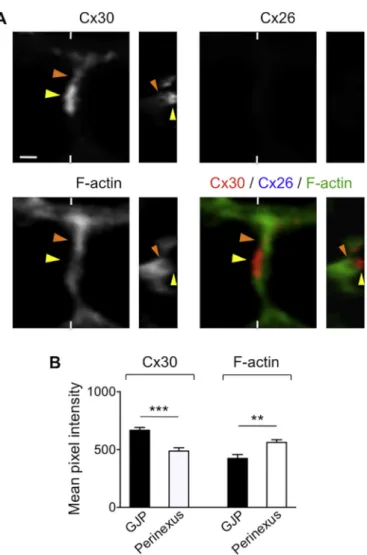
Cx30 associates with cortical actin and ZO proteins when expressed as homomeric channels in Deiters' cells or as heteromeric channels with Cx26 in inner sulcus cells at two-day-old. These latter non-sensory cells, located close to the inner hair cell layer, were previously considered to address the role of Cx26 in the assembly of the gap junction macro-molecular complex in the cochlea (Kamiya et al., 2014). The capacity of Cx30 to bind to ZO proteins is experimentally controversial, although Cx30 does not contain any predicted PDZ-binding motif at its C-term-inal extremity (Thévenin et al., 2013). In Deiters' cells, we observed that Cx30 gap junction plaques exhibit unclear, diffuse boundaries. The dense Cx30 immunolabelling, likely corresponding to the junctional plaque, quite faintly co-localizes with F-actin (yellow arrowhead in Fig. 2A). In contrast, the weaker Cx30 immunolabelling present ad-jacent to the plaque, which could correspond to non-junctional hemi-channels accruing to the plaque, overlaps with a dense actin network (orange arrowhead in Fig. 2A). The mean pixel intensity of Cx30
immunolabelling is significantly stronger in the GJP and weaker in the adjacent perinexus. In contrast, mean pixel intensity measurement re-vealed that the cortical actin network is weaker at the level of the GJP itself, and enriched in the perinexus (Fig. 2B). Although Cx30 appar-ently co-localizes with cortical actin in Deiters' cells, it does not interact with ZO proteins (Fig. S1). In inner sulcus cells, where Cx30 is co-ex-pressed with Cx26, the GJPs are devoid of cortical actin and exhibit sharp boundaries. Moreover, a gap is visible between each of the lateral boundaries of the GJP and the closest submembrane actin network, suggesting the absence of any adjacent perinexus (yellow arrowheads in Fig. 3).
3.2. Homomeric Cx30 interacts withβ-actin in the perinexus of the gap junction plaque
Several types of membrane channel proteins are believed to interact directly with the actin network, which regulate channel trafficking and function (Sasaki et al., 2014). Since Cx30 does not associate with ZO proteins in Deiters' cells, we tested whether homomeric Cx30 directly interacts with ubiquitously expressed actin isoformsβ-actin and γ-actin. Both of these isoforms are strongly expressed in Deiters' cells (Furness et al., 2005). Of note, mutations in ATCB and ATCG1 genes encoding humanβ-actin and γ-actin, respectively, cause a Baraitser-Winter cra-niofrontofacial syndrome frequently including sensorineural hearing loss (Rivière et al., 2012;Verloes et al., 2015). Althoughβ-actin and γ-actin are nearly identical proteins that differ by only four biochemically similar amino acids, they might have specific protein binding affinities and perform distinct cellular functions (Perrin and Ervasti, 2010). Using a proximity ligation assay (PLA) aimed to detect in situ protein-protein interactions (Söderberg et al., 2006), we observed a strong Cx30/β-actin PLA signal in the region directly adjacent to the Cx30 GJP (orange arrowhead inFig. 4A) and a weaker signal at the level of the dense junctional plaque itself (yellow arrowhead inFig. 4A). A PLA negative control performed by omitting one of the primary antibodies did not show any positive signal (Fig. 4B). In contrast no Cx30/γ-actin PLA signals were observed neither adjacent nor within the GJP (Fig. S2). Thesefindings suggest that β-actin rather than γ-actin especially pro-motes the recruitment of peripheral Cx30 from the perinexus to the GJP. Although it was shown that Cx30-GFP GJPs are replenished at the outer edges in vitro (Kelly et al., 2015), the asymmetry of the Cx30/β-actin PLA signal we observed in Deiters' cells suggests that peripheral Cx30 is preferentially recruited to only one side of the GJP. In inner sulcus cells, where Cx30 co-assembles with Cx26, no Cx30/β-actin PLA signals were observed (Fig. 4C), suggesting the absence of any peri-nexus and distinct gap junction assembly mechanisms.
3.3. Cx30 homomeric but not Cx26/Cx30 heteromeric gap junction assembly requires the actin network
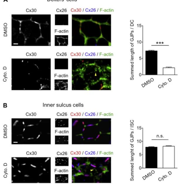
To examine the requirement of cortical actin in homomeric and heteromeric gap junction assembly in the cochlea, we performed an in vitro organotypic assay aimed to disrupt the actin network. To this end, explants of organ of Corti isolated from two-day-old mice were treated with DMSO and cytochalasin D as vehicle and actin-depolymerizating drug, respectively. In Deiters' cells, where Cx30 is expressed as homo-meric channels, cytochalasin D strongly disturbed gap junction as-sembly (Fig. 5A). In contrast, cytochalasin D treatment had no effect on Cx26/Cx30 GJPs in inner sulcus cells (Fig. 5B). The effect of cytocha-lasin D on actin networks was ascertained by the presence of actin aggregates throughout the cytoplasm (Schliwa, 1982) (yellow arrow-heads inFig. 5A, B). These data suggest a distinct requirement of actin networks in homomeric Cx30 and heteromeric Cx26/Cx30 gap junction assembly.
Fig. 2. Cochlear Cx30 exhibits dual co-localization patterns with the actin network.
(A) Cx30 immunolabelling and F-actin staining of whole-mount cochlea of a two-day-old mouse. In Deiters' cells, in which Cx30 is expressed as homomeric channels, the dense Cx30 labelling likely corresponding to the GJP is quite faintly co-localized with the cortical actin network (yellow arrowhead). In contrast, the weaker Cx30 labelling present in the membrane region adjacent to the GJP (perinexus) overlaps with a dense actin network (orange arrowhead). (B) Mean pixel intensity measurement showing that Cx30 immunolabelling is stronger in the GJP and weaker in the adjacent perinexus (n = 10). In contrast, the cortical actin network is weaker in the GJP itself, and enriched in the perinexus (n = 10). Statistical significance was determined using Student's t-test. Data are presented as mean ± SEM. n.s. = not significant,
4. Discussion
Although Cx26 and Cx30 belong to the sameβ-group of connexins and share 77% protein sequence identity in mouse (Dahl et al., 1996), our data show that Cx30 homomeric and Cx26/Cx30 heteromeric GJPs exhibit different assembly mechanisms in the cochlea. Just as Cx26 has been shown to be the key organizer of the heteromeric gap junction macromolecular complex in the cochlea (Kamiya et al., 2014), our data suggest that intrinsic Cx26 gap junction assembly features dominates the ones of Cx30 in situ. We observed that cochlear Cx26/Cx30 GJPs exhibit sharp boundaries and are apparently devoid of adjacent peri-nexus. In addition, the submembrane actin network is not required for GJP assembly and/or stability. This supports that Cx26/Cx30 oligomers could be directly delivered from the interior of the cell to the junctional plaque without trafficking through any surrounding perijunctional
space. Although this model does notfit with the classical connexin oligomerization and trafficking pathway (Laird, 2006), it has been shown that Cx43 hemichannels can be directly delivered to junctional plaques from the interior of the cell (Shaw et al., 2007). This hypothesis of an unusual trafficking and delivery pathway of Cx26/Cx30 hexamers is supported by previous data. Whereas connexins usually oligomerize in the trans-Golgi network, Cx26 oligomers are already found in the endoplasmic reticulum (Diez et al., 1999) and can reach the cell surface via a route bypassing the Golgi complex (George et al., 1999;Martin et al., 2001). In this context, an in vitro cell-free transcription/transla-tion system has shown that Cx26 exhibits a singular membrane-in-tegration behaviour and integrates directly in a post-translational manner into plasma membranes. Protecleavage studies of Cx26 in-tegrated into plasma membranes indicated a similar native transmem-brane topography to that of Cx26 integrated co-translationally into
Fig. 3. Cx26/Cx30 heteromeric channels do not co-localize with the cortical actin network.
Cx26/Cx30 immunolabelling and F-actin staining of whole-mount cochlea of a two-day-old mouse. In inner sulcus cells, where Cx30 co-assembles with Cx26, the GJP is devoid of cortical actin and exhibit sharp boundaries. A space is visible between each of the lateral edges of the GJP and the closest submembrane actin network, suggesting the absence of any adjacent perinexus (yellow arrowheads). Scale bar in (A) represents 1μm.
microsomes (Ahmad and Evans, 2002;Zhang et al., 1996). Cx26 oli-gomerization and assembly into hemichannels thus occurs in-dependently of the conventional biogenesis of gap junctions involving connexin trafficking and oligomerization via membrane components of the secretory pathway (Ahmad and Evans, 2002).
When expressed as homomeric channels in Deiters' cells, Cx30 likely assembles into junctional plaques in a more classical way. Although Cx30 was previously found to associate with the actin cytoskeleton in vitro (Qu et al., 2009), we observed that cochlear Cx30 preferentially interacts with theβ-actin isoform in the perinexus adjacent to the GJP. This suggests that F-actin especially promotes the recruitment of per-ipheral Cx30 from the perinexus to the GJP and, to a lesser extent, contributes to the stability of the junctional plaque. Of note, the sen-sitivity of Cx30 GJPs to actin-depolymerizating drugs varies among cell
types. Cx30 GJPs are severely disrupted by cytochalasin B in transfected HeLa cells, whereas they resist to the same treatment in REK cells (Kelly et al., 2015;Qu et al., 2009). Given that Cx30-mediated intercellular communication promotes epithelial repair (Forge et al., 2013), our findings should be of interest beyond the hearing system, especially regarding the role of Cx30 in skin physiology and disease.
4.1. Conclusion
Altogether, our data provide a novel insight in homomeric and heteromeric gap junction assembly mechanisms in the cochlea. Since many of the disease-causing mutations in GJB2 and GJB6 impair the formation of GJPs (Berger et al., 2014;Hoang Dinh et al., 2009;Xu and Nicholson, 2013), ourfindings should help further research aimed to
Fig. 4. Homomeric Cx30 interacts withβ-actin in the perinexus of the GJP.
In situ proximity ligation assay (PLA) using anti-Cx30 and anti-β-actin antibodies, combined with FITC-conjugated Cx30 immunolabelling and F-actin staining on whole-mount cochlea of a two-day-old mouse. (A) In Deiters' cells, a strong Cx30/β-actin PLA signal is observed in the perinexus adjacent to the GJP (orange arrowhead), whereas a weaker signal overlaps with the Cx30 GJP itself (yellow arrowhead). (B) The corresponding PLA negative control was performed by omitting the anti-Cx30 primary antibody. (C) In inner sulcus cells, in which Cx26 co-assembles with Cx30, no Cx30/β-actin PLA signals are observed within or adjacent to the GJP. Scale bars in (A–C) represent 1 μm.
decipher the pathogenic significance of these mutations.
Acknowledgements
We thank the GIGA-Cell Imaging platform. This work was supported by the Belgian Fonds de la Recherche Scientifique - FNRS.
Competing interests
The authors declare no competingfinancial interests.
Appendix A. Supplementary data
Supplementary data to this article can be found online athttps:// doi.org/10.1016/j.mod.2018.10.001.
References
Ahmad, S., Evans, W.H., 2002. Post-translational integration and oligomerization of connexin 26 in plasma membranes and evidence of formation of membrane pores: implications for the assembly of gap junctions. Biochem. J. 365, 693–699.https:// doi.org/10.1042/bj20011572.
Ahmad, S., Chen, S., Sun, J., Lin, X., 2003. Connexins 26 and 30 are co-assembled to form gap junctions in the cochlea of mice. Biochem. Biophys. Res. Commun. 307, 362–368. https://doi.org/10.1016/S0006-291X(03)01166-5.
Berger, A.C., Kelly, J.J., Lajoie, P., Shao, Q., Laird, D.W., 2014. Mutations in Cx30 that are linked to skin disease and non-syndromic hearing loss exhibit several distinct cellular pathologies. J. Cell Sci. 127, 1751–1764.https://doi.org/10.1242/jcs.138230. Chang, Q., Tang, W., Ahmad, S., Zhou, B., Lin, X., 2008. Gap junction mediated
inter-cellular metabolite transfer in the cochlea is compromised in connexin30 null mice. PLoS One 3, e4088.https://doi.org/10.1371/journal.pone.0004088.
Dahl, E., Manthey, D., Chen, Y., Schwarz, H.J., Chang, Y.S., Lalley, P.A., Nicholson, B.J., Willecke, K., 1996. Molecular cloning and functional expression of mouse connexin-30,a gap junction gene highly expressed in adult brain and skin. J. Biol. Chem. 271, 17903–17910.https://doi.org/10.1074/jbc.271.30.17903.
Defourny, J., Mateo Sánchez, S., Schoonaert, L., Robberecht, W., Davy, A., Nguyen, L., Malgrange, B., 2015. Cochlear supporting cell transdifferentiation and integration into hair cell layers by inhibition of ephrin-B2 signalling. Nat. Commun. 6, 7017. https://doi.org/10.1038/ncomms8017.
Fig. 5. Actin depolymerization differently affects Cx30 homomeric and Cx26/Cx30 heteromeric GJPs.
Organotypic cultures of two-day-old organs of Corti were treated with DMSO (vehicle) or cytochalasin D and labelled for Cx26, Cx30 and F-actin. (A) In Deiters' cells, cytochalasin D treatment significantly disrupts the formation of Cx30 homomeric GJPs (n = 90). (B) In inner sulcus cells, cytochalasin D treatment does not affect the Cx26/Cx30 heteromeric GJPs (n = 90). The effect of cytochalasin D on actin networks was ascertained by the presence of actin aggregates throughout the cytoplasm (yellow arrowheads in A and B). Statistical significance was determined using Student's t-test. Data are presented as mean ± SEM. n.s. = not significant,
del Castillo, I., Villamar, M., Moreno-Pelayo, M.A., del Castillo, F.J., Alvarez, A., Tellería, D., Menéndez, I., Moreno, F., 2002. A deletion involving the connexin 30 gene in nonsyndromic hearing impairment. N. Engl. J. Med. 346, 243–249.https://doi.org/ 10.1056/NEJMoa012052.
Diez, J.A., Ahmad, S., Evans, W.H., 1999. Assembly of heteromeric connexons in guinea-pig liver en route to the Golgi apparatus, plasma membrane and gap junctions. Eur. J. Biochem. 262, 142–148.https://doi.org/10.1046/j.1432-1327.1999.00343.x. Falk, M.M., Bell, C.L., Kells Andrews, R.M., Murray, S.A., 2016. Molecular mechanisms
regulating formation, trafficking and processing of annular gap junctions. BMC Cell Biol. 17 (Suppl. 1).https://doi.org/10.1186/s12860-016-0087-7.22.
Forge, A., Jagger, D.J., Kelly, J.J., Taylor, R.R., 2013. Connexin30-mediated intercellular communication plays an essential role in epithelial repair in the cochlea. J. Cell Sci. 126, 1703–1712.https://doi.org/10.1242/jcs.125476.
Furness, D.N., Katori, Y., Mahendrasingam, S., Hackney, C.M., 2005. Differential dis-tribution of beta- and gamma-actin in Guinea-pig cochlear sensory and supporting cells. Hear. Res. 207, 22–34.https://doi.org/10.1016/j.heares.2005.05.006. Gaietta, G., Deerinck, T.J., Adams, S.R., Bouwer, J., Tour, O., Laird, D.W., Sosinsky, G.E.,
Tsien, R.Y., Ellisman, M.H., 2002. Multicolor and electron microscopic imaging of connexin trafficking. Science 296, 503–507.https://doi.org/10.1126/science. 1068793.
George, C.H., Kendall, J.M., Evans, W.H., 1999. Intracellular trafficking pathways in the assembly of connexins into gap junctions. J. Biol. Chem. 274, 8678–8685.https:// doi.org/10.1074/jbc.274.13.8678.
Grifa, A., Wagner, C.A., D'Ambrosio, L., Melchionda, S., Bernardi, F., Lopez-Bigas, N., Rabionet, R., Arbones, M., Monica, M.D., Estivill, X., et al., 1999. Mutations in GJB6 cause nonsyndromic autosomal dominant deafness at DFNA3 locus. Nat. Genet. 23, 16–18.https://doi.org/10.1038/12612.
Hervé, J.C., Derangeon, M., Sarrouilhe, D., Bourmeyster, N., 2014. Influence of the scaffolding protein Zonula Occludens (ZOs) on membrane channels. Biochim. Biophys. Acta 1838, 595–604.https://doi.org/10.1016/j.bbamem.2013.07.006. Hoang Dinh, E., Ahmad, S., Chang, Q., Tang, W., Stong, B., Lin, X., 2009. Diverse deafness
mechanisms of connexin mutations revealed by studies using in vitro approaches and mouse models. Brain Res. 1277, 52–69.https://doi.org/10.1016/j.brainres.2009.02. 008.
Jagger, D.J., Forge, A., 2015. Connexins and gap junctions in the inner ear-it's not just about K+ recycling. Cell Tissue Res. 360, 633–644.https://doi.org/10.1007/ s00441-014-2029-z.
Kamiya, K., Yum, S.W., Kurebayashi, N., Muraki, M., Ogawa, K., Karasawa, K., Miwa, A., Guo, X., Gotoh, S., Sugitani, Y., et al., 2014. Assembly of the cochlear gap junction macromolecular complex requires connexin 26. J. Clin. Invest. 124, 1598–1607. https://doi.org/10.1172/JCI67621.
Kelly, J.J., Shao, Q., Jagger, D.J., Laird, D.W., 2015. Cx30 exhibits unique characteristics including a long half-life when assembled into gap junctions. J. Cell Sci. 128, 3947–3960.https://doi.org/10.1242/jcs.174698.
Kelsell, D.P., Dunlop, J., Stevens, H.P., Lench, N.J., Liang, J.N., Parry, G., Mueller, R.F., Leigh, I.M., 1997. Connexin 26 mutations in hereditary non-syndromic sensorineural deafness. Nature 387, 80–83.https://doi.org/10.1038/387080a0.
Kikuchi, T., Kimura, R.S., Paul, D.L., Takasaka, T., Adams, J.C., 2000. Gap junction sys-tems in the mammalian cochlea. Brain Res. Brain Res. Rev. 32, 163–166.https://doi. org/10.1016/S0165-0173(99)00076-4.
Korver, A.M., Smith, R.J., Van Camp, G., Schleiss, M.R., Bitner-Glindzicz, M.A., Lustig, L.R., Usami, S.I., Boudewyns, A.N., 2017. Congenital hearing loss. Nat. Rev. Dis. Primers 3, 16094.https://doi.org/10.1038/nrdp.2016.94.
Laird, D.W., 2006. Life cycle of connexins in health and disease. Biochem. J. 394, 527–543.https://doi.org/10.1042/BJ20051922.
Martin, P.E., Blundell, G., Ahmad, S., Errington, R.J., Evans, W.H., 2001. Multiple pathways in the trafficking and assembly of connexin 26, 32 and 43 into gap junction intercellular communication channels. J. Cell Sci. 114, 3845–3855.
Perrin, B.J., Ervasti, J.M., 2010. The actin gene family: function follows isoform. Cytoskeleton (Hoboken) 67, 630–634.https://doi.org/10.1002/cm.20475. Qu, C., Gardner, P., Schrijver, I., 2009. The role of the cytoskeleton in the formation of
gap junctions by Connexin 30. Exp. Cell Res. 315, 1683–1692.https://doi.org/10. 1016/j.yexcr.2009.03.001.
Rhett, J.M., Jourdan, J., Gourdie, R.G., 2011. Connexin 43 connexon to gap junction transition is regulated by zonula occludens-1. Mol. Biol. Cell 22, 1516–1528.https:// doi.org/10.1091/mbc.E10-06-0548.
Rivière, J.B., van Bon, B.W., Hoischen, A., Kholmanskikh, S.S., O'Roak, B.J., Gilissen, C., Gijsen, S., Sullivan, C.T., Christian, S.L., Abdul-Rahman, O.A., et al., 2012. De novo mutations in the actin genes ACTB and ACTG1 cause Baraitser-Winter syndrome. Nat. Genet. 44, 440–444.https://doi.org/10.1038/ng.1091.
Sasaki, S., Yui, N., Noda, Y., 2014. Actin directly interacts with different membrane channel proteins and influences channel activities: AQP2 as a model. Biochim. Biophys. Acta 1838, 514–520.https://doi.org/10.1016/j.bbamem.2013.06.004. Schliwa, M., 1982. Action of cytochalasin D on cytoskeletal networks. J. Cell Biol. 92,
79–91.https://doi.org/10.1083/jcb.92.1.79.
Shaw, R.M., Fay, A.J., Puthenveedu, M.A., von Zastrow, M., Jan, Y.N., Jan, L.Y., 2007. Microtubule plus-end-tracking proteins target gap junctions directly from the cell interior to adherens junctions. Cell 128, 547–560.https://doi.org/10.1016/j.cell. 2006.12.037.
Söderberg, O., Gullberg, M., Jarvius, M., Ridderstråle, K., Leuchowius, K.J., Jarvius, J., Wester, K., Hydbring, P., Bahram, F., Larsson, L.G., et al., 2006. Direct observation of individual endogenous protein complexes in situ by proximity ligation. Nat. Methods 3, 995–1000.https://doi.org/10.1038/nmeth947.
Sun, J., Ahmad, S., Chen, S., Tang, W., Zhang, Y., Chen, P., Lin, X., 2005. Cochlear gap junctions coassembled from Cx26 and 30 show faster intercellular Ca2+ signaling than homomeric counterparts. Am. J. Phys. Cell Phys. 288, C613–C623.https://doi. org/10.1152/ajpcell.00341.2004.
Thévenin, A.F., Kowal, T.J., Fong, J.T., Kells, R.M., Fisher, C.G., Falk, M.M., 2013. Proteins and mechanisms regulating gap-junction assembly, internalization, and de-gradation. Physiology (Bethesda) 28, 93–116.https://doi.org/10.1152/physiol. 00038.2012.
Verloes, A., Di Donato, N., Masliah-Planchon, J., Jongmans, M., Abdul-Raman, O.A., Albrecht, B., Allanson, J., Brunner, H., Bertola, D., Chassaing, N., et al., 2015. Baraitser-Winter cerebrofrontofacial syndrome: delineation of the spectrum in 42 cases. Eur. J. Hum. Genet. 23, 292–301.https://doi.org/10.1038/ejhg.2014.95. Walsh, T., Pierce, S.B., Lenz, D.R., Brownstein, Z., Dagan-Rosenfeld, O., Shahin, H., Roeb,
W., McCarthy, S., Nord, A.S., Gordon, C.R., et al., 2010. Genomic duplication and overexpression of TJP2/ZO-2 leads to altered expression of apoptosis genes in pro-gressive nonsyndromic hearing loss DFNA51. Am. J. Hum. Genet. 87, 101–109. https://doi.org/10.1016/j.ajhg.2010.05.011.
Xu, J., Nicholson, B.J., 2013. The role of connexins in ear and skin physiology– functional insights from disease-associated mutations. Biochim. Biophys. Acta 1828, 167–178. https://doi.org/10.1016/j.bbamem.2012.06.024.
Zhang, J.T., Chen, M., Foote, C.I., Nicholson, B.J., 1996. Membrane integration of in vitro-translated gap junctional proteins: co- and post-translational machanisms. Mol. Biol. Cell 7, 471–482.