The Plasminogen Activator Inhibitor PAI-1 Controls In Vivo Tumor
Vascularization by Interaction with Proteases, Not Vitronectin:
Implications for Antiangiogenic Strategies
Khalid Bajou,* Véronique Masson,* Robert D. Gerard,
‡Petra M. Schmitt,
§Valérie Albert,* Michael Praus,
‡Leif R. Lund,
储Thomas L. Frandsen,
储Nils Brunner,
储Keld Dano,
储Norbert E. Fusenig,
¶Ulrich Weidle,**
Geert Carmeliet,
‡‡David Loskutoff,
§Desiré Collen,
‡Peter Carmeliet,
‡Jean Michel Foidart,* and Agnès Noël*
*Laboratory of Tumor and Developmental Biology, University of Liège, Tour de Pathologie (B23), B-4000 Liège, Belgium;
‡Center for Transgene Technology and Gene Therapy, Flanders Interuniversity Institute for Biotechnology, Katholieke Universiteit
Leuven, B-3000 Leuven, Belgium; §Department of Vascular Biology, The Scripps Research Institute, La Jolla, California 92037; 储Finsen Laboratory, Rigshospitalet, DK-2100 Copenhagen, Denmark; ¶Division of Carcinogenesis and Differentiation, German
Cancer Research Center (DKFZ), 69120 Heidelberg, Germany; **Roche Diagnostics, D-82372 Penzberg, Germany; ‡‡Laboratory
of Experimental Medicine and Endocrinology (LEGENDO), Katholieke Universiteit Leuven, B-3000 Leuven, Belgium
Abstract.
The plasminogen (Plg)/plasminogen activa-tor (PA) system plays a key role in cancer progression, presumably via mediating extracellular matrix degrada-tion and tumor cell migradegrada-tion. Consequently, uroki-nase-type PA (uPA)/plasmin antagonists are currently being developed for suppression of tumor growth and angiogenesis. Paradoxically, however, high levels of PA inhibitor 1 (PAI-1) are predictive of a poor prognosis for survival of patients with cancer. We demonstrated previously that PAI-1 promoted tumor angiogenesis, but by an unresolved mechanism. We anticipated that PAI-1 facilitated endothelial cell migration via its known interaction with vitronectin (VN) and integrins. However, using adenoviral gene transfer of PAI-1 mu-tants, we observed that PAI-1 promoted tumor angio-genesis, not by interacting with VN, but rather byin-hibiting proteolytic activity, suggesting that excessive plasmin proteolysis prevents assembly of tumor vessels. Single deficiency of uPA, tissue-type PA (tPA), uPA re-ceptor, or VN, as well as combined deficiencies of uPA and tPA did not impair tumor angiogenesis, whereas lack of Plg reduced it. Overall, these data indicate that plasmin proteolysis, even though essential, must be tightly controlled during tumor angiogenesis, probably to allow vessel stabilization and maturation. These data provide insights into the clinical paradox whereby PAI-1 promotes tumor progression and warrant against the uncontrolled use of uPA/plasmin antagonists as tumor angiogenesis inhibitors.
Key words: angiogenesis • tumor invasion • proteolysis • migration • serine protease
Introduction
Tumor progression involves the disruption of anatomical barriers and penetration of tumor cells into normal adjacent host tissues, as well as the infiltration of normal host cells into the tumor. Such migratory and tissue remodeling events are, among others, regulated by different proteolytic systems. Among the proteases that play an active role in these processes are the serine proteases of the plasminogen (Plg)1/plasminogen activator (PA) system (Andreasen et
al., 1997). Urokinase-type (uPAs) and tissue-type plasmino-gen activators (tPAs) both activate the zymoplasmino-gen Plg into its active form, plasmin. uPA is secreted as an inactive pre-cursor (pro-uPA) that binds with high affinity to a specific cell surface glycosylphosphatidylinositol-anchored receptor (the uPA receptor [uPAR]). Concomitant binding of pro-uPA to pro-uPAR and Plg to nonspecific binding sites at the cell surface strongly enhances plasmin generation.
It is generally believed that uPA at the cell surface ini-tiates a proteinase cascade, which in turn leads to break-down of the extracellular matrix and thereby promotes cellular migration. This conclusion is supported by the fact that uPA and uPAR are highly expressed by tumor cells or by surrounding stromal cells, and that they are both in-dependent prognostic indicators in human cancer (Dano et al., 1994; Reuning et al., 1998; Stephens et al., 1999). In Address correspondence to Agnès Noël, Laboratory of Tumor and
Devel-opmental Biology, University of Liège, Tour de Pathologie (B23), Sart-Tilman, B-4000 Liège, Belgium. Tel.: 32-4-366-24-53. Fax: 32-4-366-29-36. E-mail: agnes.noel@ulg.ac.be
1Abbreviations used in this paper: Ab, antibody; AdPAI-1, adenovirus-expressing hPAI-1; hPAI-1, human PAI-1; PA, Plg activator; PAI-1, PA inhibitor 1; Plg, plasminogen; tPA, tissue-type PA; uPA, urokinase-type PA; uPAR, uPA receptor; VN, vitronectin; WT, wild-type.
on September 19, 2006
www.jcb.org
addition, the use of antisense mRNA for uPA and uPAR, of natural or synthetic serine protease inhibitors, or of uPAR antagonists, all reduced tumor invasion (Min et al., 1996; Carmeliet and Collen, 1998). Consequently, uPA/ uPAR/plasmin antagonists are currently being developed as therapeutic strategies to inhibit tumor angiogenesis and progression.
PA inhibitor 1 (PAI-1) is the primary physiological in-hibitor of uPA and tPA. It not only regulates the pro-teolyticactivity of uPA, but also determines the level of uPA bound to uPAR by promoting the rapid endocytosis of the trimolecular uPA–PAI-1–uPAR complex (Conese and Blasi, 1995; Blasi, 1997). However, it has also been implicated in modulating cell migration via alternative mechanisms (Deng et al., 1996; Stefansson and Lawrence, 1996; Blasi, 1997; Loskutoff et al., 1999), even though con-clusive in vivo evidence is lacking. By blocking the inter-action between vitronectin (VN), uPAR, and integrins, PAI-1 may induce cell detachment from the extracellular matrix and thereby promote cellular migration and tumor invasion. However, the relevance of the latter mechanism in overall tumor growth and angiogenesis has not been confirmed in vivo.
Surprisingly high, rather than low, levels of PAI-1 are predictive of poor survival prognosis for patients suffer-ing from a variety of different cancers (Pedersen et al., 1994a,b). To date, the molecular mechanisms of this appar-ent paradox remain largely unexplained, raising concerns about whether therapeutic strategies to suppress tumor growth and angiogenesis should be aimed at inhibiting or increasing uPA/plasmin proteolysis. These questions have been difficult to address, largely because uPA promotes, and PAI-1 suppresses, tumor growth in most available ex-perimental tumor models. However, recently PAI-1 was
found to promote tumor growth and angiogenesis (Bajou et al., 1998) in a highly reproducible mouse tumor model. The availability of this model now allows us to examine the molecular mechanism by which PAI-1 promotes tumor an-giogenesis (i.e., either by inhibiting the proteolytic activity or by interrupting the interaction between VN and uPAR or integrins). To address this question and to further deter-mine the respective role of the different members of the Plg/plasmin system, we have transplanted malignant kerati-nocytes into wild-type (WT) mice and mice lacking uPA, tPA, uPA and tPA, uPAR, Plg, PAI-1, or VN. We also per-formed adenoviral gene transfer experiments using PAI-1 mutants that selectively inhibit PA activity or no longer bind to VN. We provide direct evidence that plasmin is in-volved in the formation of new vessels by host endothelial cells, but that PAI-1 control of proteolytic breakdown is re-quired. These data not only help to explain the apparent paradox that high levels of a proteinase inhibitor promote tumor angiogenesis and are positively correlated with tu-mor progression in patients, but also warrant against the uncontrolled use of uPA/plasmin inhibitors for suppression of tumor angiogenesis. Furthermore, they indicate that neutralization of PAI-1 may be an attractive new target for antiangiogenic therapy
Materials and Methods
MiceHomozygous mice with single (uPA⫺Ⲑ⫺, tPA⫺Ⲑ⫺, uPAR⫺Ⲑ⫺, and VN⫺Ⲑ⫺ mice) or combined deficiencies (tPA⫺Ⲑ⫺/uPA⫺Ⲑ⫺ mice) and their corre-sponding WT with a mixed genetic background of 75% C57BL/6 and 25% 129 SV/SL strain were generated as described previously (Carmeliet et al., 1994; Zheng et al., 1995; Carmeliet and Collen, 1998). Tumor angiogenesis and invasion in PAI-1⫺Ⲑ⫺ (Carmeliet et al., 1993a,b) and WT mice were evaluated in three different sets of experiments using mice with different genetic background obtained by two, three, or four backcrosses with C57BL/6 strain. In each set of experiments, similar results were obtained and therefore the data presented in Table I were pooled. The Plg⫺Ⲑ⫺ mice were derived from those generated previously (Bugge et al., 1995; Ploplis et al., 1998). They have been backcrossed 1 or 11 times with C57BL/6 strain, yielding similar results in two different sets of experiments pre-sented in Table I. Mice of either sex used for experiments were littermate mice, produced by mating of heterozygous brothers and sisters between 8 and 12 wk old. Genotypes of mice were established using tail biopsy DNA preparation by either Southern blot analysis or PCR assays (Carmeliet et al., 1993b, 1994; Bugge et al., 1995).
Cell Culture
Malignant murine keratinocytes (PDVA cells; Fusenig et al., 1978) were routinely grown in DME containing a fourfold concentration of amino ac-ids and vitamins (GIBCO BRL), 10% FCS (GIBCO BRL), and antibiot-ics in a humidified incubator at 37⬚C, 5% CO2. Cells (2 ⫻ 105) were plated on collagen gel (4 mg/ml of type I collagen isolated from rat tail tendons) inserted in teflon rings (Renner GmbH) and maintained in culture for 1 d before transplantation into mice as described previously (Fusenig et al., 1983; Bajou et al., 1998). For transplantation assays in VN⫺Ⲑ⫺ mice, kerati-nocytes were precultured on the collagen gels in the presence of 10% se-rum derived from VN⫺Ⲑ⫺ mice.
Transplantation Assay in Mice
The cell-coated collagen gels were covered with a silicone transplantation chamber (Renner GmbH) and implanted in toto onto the dorsal muscle fascia of mice as described previously (Skobe et al., 1997; Bajou et al., 1998). 2 wk later, transplants were resected, embedded in Tissue Tek (Miles Laboratories, Inc.), and either frozen in liquid nitrogen for cryostat sectioning or embedded in paraffin after zinc formalin fixation.
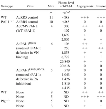
Table I. Tumor Angiogenesis and Invasion in WT, PAI-1⫺Ⲑ⫺,
and Plg⫺Ⲑ⫺ Mice Injected or Not with WT or Mutated
hPAI-1 Adenovirus
Genotype Virus Mice
Plasma level
of hPAI-1 Angiogenesis Invasion
n ng/ml
WT AdRR5 control 11 ⬍0.8 ⫹⫹⫹ ⫹⫹⫹
PAI-1⫺Ⲑ⫺ AdRR5 control 10 ⬍0.8 0 0
AdCMVPAI-1 (WT hPAI-1) 4 300 ⫹⫹ ⫹⫹ 102 0 0 1,699 ⫹ ⫹ 2,805 ⫹ ⫹ AdPAI-1Q123K (mutated hPAI-1: defective in VN binding) 6 106 ⫹⫹ ⫹ 250 ⫹⫹ ⫹⫹ 1,853 ⫹ ⫹ 6,722 ⫹ ⫹ 26,840 ⫹ ⫹ 20,618 0 0 AdPAI-1R346M,M347S (mutated hPAI-1: defective in PA inhibition) 5 579 0 0 1,043 0 0 1,426 0 0 1,816 0 0 4,435 0 0 WT None 9 ND ⫹⫹ ⫹⫹ None 5 ND ⫹⫹⫹ ⫹⫹⫹ Plg⫺Ⲑ⫺ None 5 ND ⫹ 0 None 3 ND ⫹ ⫹
Tumor invasion and angiogenesis were scored as described in Materials and Methods.
on September 19, 2006
www.jcb.org
In all assays, the take rate and growth of transplants were verified by classical histology and some samples were not taken into account. The exclusion criteria were the loss of transplantation chamber, the absence of cells on top of the collagen gel, and the failure of collagen gel adher-ence onto the host tissue. Based on these parameters, we excluded ⵑ20 and 50% of samples resected from mice untreated or injected with ade-novirus (see above), respectively. There were no differences in exclusion rates related to the genotypes of the mice or the type of virus injected.
Adenovirus-mediated PAI-1 cDNA Transfer
A recombinant adenovirus vector bearing WT human PAI-1 (hPAI-1) (AdCMVPAI-1 and control adenovirus [AdRR5]) were propagated as described previously (Gerard and Meidell, 1995; Carmeliet et al., 1997). Recombinant viruses expressing mutant hPAI-1, AdCMVPAI-1Q123K, and AdCMVPAI-1R346M,M347S were generated after substitution of a restriction fragment containing the desired mutation into the pACCMVPAI-1(WT) shuttle plasmid (Gerard and Meidell, 1995). PAI-1 with mutation of Gln 123 to Lys had a specific 100-fold decrease in affinity for VN, but retained full inhibitory activity (Stefansson and Lawrence, 1996). The double point mutant, Arg 346 to Met and Met 347 to Ser (Shubeita et al., 1990), bound to VN with the same affinity as WT PAI-1 but did not inhibit PA activity. We verified that hPAI-1 (WT or mutant forms) interacts efficiently with murine PA and murine VN according to the procedure described previ-ously (Vleugels et al., 2000).
1 d after cell transplantation, mice were intravenously injected with 200 l of control or recombinant adenovirus (7 ⫻ 108 pfu). After 5 d, blood was sampled from the retroorbital sinus and PAI-1 antigen was measured as described (Bajou et al., 1998). On day 14, mice were killed and trans-plants were excised and processed as described above. The hPAI-1 cDNA was used in this study to allow ELISA measurement of circulating PAI-1 levels in all animals, including WT mice.
Immunofluorescence and Morphometry
Cryostat sections (5 m thick) were fixed in acetone at ⫺20⬚C and in 80% methanol at 4⬚C and then incubated with the primary antibodies (Abs). For double immunofluorescence labeling studies, sections were first incu-bated for 1 h at room temperature with the two primary Abs: antitype IV collagen Ab (rabbit polyclonal Ab, diluted 1:100) and antikeratin Ab (guinea pig polyclonal Ab, diluted 1:20; Sigma-Aldrich). The sections were washed three times in PBS for 10 min each and subsequently FITC-or Texas red–conjugated appropriate secondary Abs were applied fFITC-or 30 min: swine anti–rabbit (diluted 1:40; Dakopat) and/or mouse anti–guinea pig (diluted 1:40; Sigma-Aldrich). After three washes in PBS for 10 min each and a final rinse in 10 mM Tris-HCl buffer, pH 8.8, coverslips were mounted and specific labeling was observed using an inverted microscope equipped with epifluorescence optics.
Morphometric measurements of cell invasion (average distance of pen-etration) were performed as described previously (Bajou et al., 1998): mi-gration ⬍50 m was scored as 0; migration from 50 to 150 m, ⫹; mi-gration from 150 to 300 m, ⫹⫹; and migration 300 m, ⫹⫹⫹. Morphometric assessment of angiogenesis was scored as follows: vessels undetected in the collagen gel, 0; vessels infiltrating the collagen without reaching the malignant epithelial layer, ⫹; blood vessels in close apposi-tion to the epithelial layer, ⫹⫹; and blood vessels intermingling with inva-sive epithelial tumor sprouts, ⫹⫹⫹.
In Situ Zymography
Cryostat sections were coated with a mixture containing 2% skim milk, 0.9% agar, and 600 g/ml of Plg (Sigma-Aldrich) (Bajou et al., 1998). An 8% milk stock solution was prepared in PBS, heated at 95⬚C for 30 min, and centrifuged briefly at 3,000 rpm to remove insoluble material. Slides were incubated at 37⬚C in a humidified chamber for 2 h for assessment of total PA activity and for 24 h in the presence of uPA-specific inhibitor
Figure 1. Invasive behavior
of malignant mouse kerati-nocytes (PDVA cells), 2 wk after implantation. In WT (a), uPA⫺Ⲑ⫺ (b), tPA⫺Ⲑ⫺ (c), tPA⫺Ⲑ⫺/uPA⫺Ⲑ⫺ (d), and uPAR⫺Ⲑ⫺ (e) mice, the col-lagen gel has been remodelled and malignant cells formed tu-mor sprouts intermingled with host tissue. The dotted line delineates the bottom of the collagen gel in Plg⫺Ⲑ⫺ (f) and PAI-1⫺Ⲑ⫺ mice (g). Histological sections were stained with hema-toxylin and eosin. C, carcinoma cells; G, collagen gel; H, host connective tissue. Bar, 100 m. on September 19, 2006
www.jcb.org
amiloride (2 mM; Sigma-Aldrich) for assessment of tPA activity. Caseinol-ysis was monitored by examination under a dark field microscope.
Results
Plasmin Proteolysis in Tumor Angiogenesis and Invasion
Malignant murine keratinocytes (PDVA cells; Fusenig et al., 1978) cultured on a collagen gel were implanted onto the dorsal muscle fascia of WT and transgenic mice. In re-sponse to angiogenic stimuli (produced by tumor cells; Skobe et al., 1997), new blood vessels formed in the under-lying stroma, invaded the collagen gel, and reached the malignant epithelial layer. Thereafter, the malignant kera-tinocytes formed tumor sprouts that invaded downwards into the granulation tissue (Fig. 1 a). Within 2 wk after transplantation, these tumor islets were intermingled with closely apposed new vessels. The degree of tumor cell in-vasion was scored by calculating the average distance over which the tumor cells infiltrated in the host mesen-chyme, whereas tumor angiogenesis was semiquantitatively scored after staining for collagen type IV, a component of the capillary basement membrane (Fig. 2 a). Compared with WT mice, tumor angiogenesis (score ⫹⫹⫹) devel-oped to a similar degree and invasion of tumor cells
oc-curred over a similar distance (⬎150 m, ⫹⫹ or ⫹⫹⫹) in uPA⫺Ⲑ⫺, tPA⫺Ⲑ⫺, uPAR⫺Ⲑ⫺ mice, and double tPA⫺Ⲑ⫺/uPA⫺Ⲑ⫺ mice (Fig. 1, b–e, and Fig. 2, b–e). Both tumor invasion and stromal angiogenesis were reduced in Plg⫺Ⲑ⫺ mice (Ta-ble I, Figs. 1 f and 2 f).
In situ zymography in WT mice revealed that uPA activ-ity (Fig. 3 a) was mainly localized to the cellular front, con-taining both invading tumor cells (these malignant kerati-nocytes are in all experiments WT, and hence produce uPA and tPA; Bajou et al., 1998) and newly formed blood vessels, whereas weak tPA activity was largely restricted to the upper part of the transplant, predominantly consist-ing of tumor cells (Fig. 3 b). In uPA⫺Ⲑ⫺ mice, tPA activity was not only restricted to the tumor-rich upper region, but also expressed at increased levels over the invasion zone, containing both infiltrating tumor cells and newly formed vessels (Fig. 3, c and d). These results may suggest a com-pensatory upregulation of tPA activity in the absence of uPA. In tPA⫺Ⲑ⫺ mice, caseinolytic activity appeared simi-lar to that visualized in WT mice and was exclusively me-diated by uPA (Fig. 3, e and f). The lack of host uPAR in uPAR⫺Ⲑ⫺ mice did not influence the intensity or the local-ization of uPA-mediated lysis (Fig. 3, g and h). These data indicate that formation of host-derived stromal vessels re-quires plasmin, normally generated by uPA. However, the two PAs may be somehow redundant.
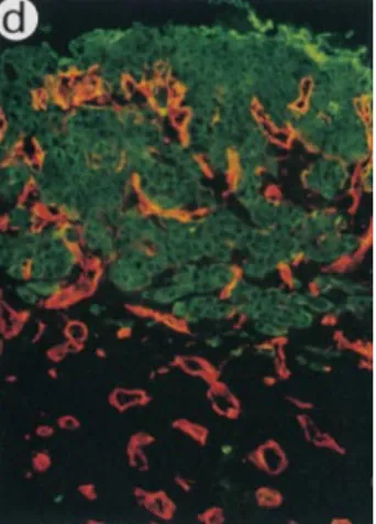
Figure 2. Immunofluorescence
labeling of malignant kerati-nocytes and vessels 2 wk after implantation. PDVA cells were transplanted into WT (a), uPA⫺Ⲑ⫺ (b), tPA⫺Ⲑ⫺ (c), tPA/ uPA⫺Ⲑ⫺ (d), uPAR⫺Ⲑ⫺ (e), Plg⫺Ⲑ⫺ (f), and PAI-1⫺Ⲑ⫺ (g) mice. Malignant cells were de-tected on cryostat sections by using antikeratin Ab (green) and vessels were detected us-ing an anticollagen type IV Ab (red). At all times after graft-ing, collagen type IV labelings were codistributed with endo-thelial cells recognized by the anti–mouse platelet endothe-lial cell adhesion molecule immunostaining (data not shown). The dotted line delineates the bottom of the collagen gel, revealing not only a reduced upward migration of vessels in Plg⫺Ⲑ⫺ mice (e), but also that vessels remained confined beneath the collagen gel in PAI-1⫺Ⲑ⫺ mice (f). C, carcinoma cells; G, collagen gel; H, host connective tissue. Bar, 100 m.
on September 19, 2006
www.jcb.org
Normal Tumor Angiogenesis and Invasion in VN⫺Ⲑ⫺ Mice
In agreement with our previous findings, host-derived ves-sels in PAI-1⫺Ⲑ⫺ mice were unable to migrate towards the tumor cells and remained confined beneath the collagen gel (Table I, Fig. 2 g). In addition, malignant cells failed to invade the host tissue in PAI-1⫺Ⲑ⫺ mice (the average depth of invasion was ⬍50 m, scored 0) and remained as an ir-regular stratified epithelium on top of the collagen gel (Fig. 1 f). Since PAI-1 binds strongly to VN and alters the adhe-sion and migration of cells on this matrix substrate, we an-ticipated that lack of VN should mimic the impaired tumor angiogenesis and invasion phenotype of PAI-1⫺Ⲑ⫺ mice. However, transplantation of malignant keratinocytes into VN⫺Ⲑ⫺ mice was associated with normal, and perhaps even accelerated angiogenesis and tumor infiltration (Fig. 4).
Mechanism of the Tumor-promoting Role of PAI-1
We had demonstrated previously that tumor vasculariza-tion and invasion in PAI-1⫺Ⲑ⫺ mice can be restored by in-travenous injection of a recombinant adenovirus express-ing human PAI-1 (AdPAI-1; Bajou et al., 1998). To further investigate whether the role of PAI-1 in promoting tumor invasion and angiogenesis depended on its ability to block proteolytic activity, or instead on its ability to bind to VN, two additional adenovirus constructs were pro-duced that expressed a mutant form of hPAI-1 that: (a) exhibited normal binding to VN but was inactive in inhib-iting the proteolytic activity of tPA and uPA (AdPAI-1R346M,M347S), or (b) inhibited the PA activity normally but
had a dramatically reduced affinity for VN (AdPAI-1Q123K). Intravenous injection of these adenoviruses
re-sulted in 100–1,000-fold increased plasma levels of hPAI-1
Figure 3. In situ zymography of PDVA cells 2 wk after transplantation into WT (a and b), uPA⫺Ⲑ⫺ (c and d), tPA⫺Ⲑ⫺ (e and f), and uPAR⫺Ⲑ⫺ (g and h) mice. Total PA activity was visual-ized as a dark zone of lysis (dark field images) after incubation for 6 h (a, c, e, and g). In the presence of 2 mM amiloride for 36 h, only tPA activity was detected (b, d, f, and h). C, carci-noma cells; H, host connective tissue. Bar, 1 mm.
on September 19, 2006
www.jcb.org
above normal murine PAI-1 plasma levels of WT mice (2 ng/ml; Table I). Injection of the AdPAI-1Q123K virus into
PAI-1⫺Ⲑ⫺ hosts restored tumor vascularization and inva-sion in five of six mice (Table I). In sharp contrast, injec-tion of the AdPAI-1R346M,M347S virus into PAI-1⫺Ⲑ⫺ hosts
was unable to restore tumor vascularization and invasion in any of the six mice (Table I). Thus, the requirement for PAI-1 in tumor angiogenesis and invasion, in this model, appears not to be due to inhibition of cellular adhesion through its interaction with VN, but rather due to preven-tion of excessive plasmin formapreven-tion.
Discussion
Proteolytic breakdown of extracellular matrices by uPA/ plasmin has been associated with tumor invasion and an-giogenesis (Andreasen et al., 1997; Stephens et al., 1999). However, prognostic studies have indicated that the pro-tease inhibitor PAI-1 is a clinical marker of poor prognosis in a variety of human cancers (Pedersen et al., 1994a,b; Brunner et al., 2000). The molecular mechanisms of action that underlie this apparent paradox remained to date un-explained. Nonetheless, a fundamental understanding of these processes is mandatory because of the growing inter-est to develop uPA antagonists as angiogenesis inhibitors. This study demonstrates that plasmin proteolysis is in-volved in tumor angiogenesis but, at the same time, indi-cates that an excessive plasmin formation, as a result of PAI-1 deficiency, prevents normal assembly and out-growth of newly formed stromal vessels. This explanation of the observed clinical paradox is supported by the exper-iment using mutated PAI-1 constructs and showing that PAI-1 mediates its proangiogenic effect, not by its interac-tion with VN but rather by its antiprotease funcinterac-tion.
The Plg/plasmin system has been implicated in extracel-lular proteolysis during angiogenesis (Montesano et al., 1990). However, studies in gene-inactivated mice have re-vealed that angiogenesis during embryonic development occurs normally in the absence of both PAs and Plg and
that angiogenesis during pathological conditions can occur to a large degree in the absence of either Plg, tPA, or uPA, possibly by redundancy or compensation by other protein-ases (Bugge et al., 1997, 1998; Carmeliet and Collen, 1999; Lund et al., 1999). In accordance with these observations, the present findings show that tumor vascularization was not affected in mice deficient in tPA, uPA, or uPAR. No-tably, stromal/host lack of uPA may in the present tumor model have been compensated by upregulation of tPA ac-tivity. However, in the absence of PA in combined defi-cient tPA⫺Ⲑ⫺/uPA⫺Ⲑ⫺ mice, the invasive and angiogenic phenotype of malignant keratinocytes was similar to that observed in WT mice or mice with single deficiencies (uPA⫺Ⲑ⫺ or tPA⫺Ⲑ⫺ mice). Interestingly, both Plg and ac-tive plasmin were detected by Western blotting in the in-vasive region of tumors transplanted onto WT and double tPA⫺Ⲑ⫺/uPA⫺Ⲑ⫺ mice (data not shown). These observa-tions suggest that in vivo neither tPA nor uPA, produced by host cells individually or in combination, is essential for Plg activation; and other enzyme(s) can compensate the lack of uPA and tPA. Alternatively, the presence of active plasmin might result from activation by uPA or tPA pro-duced by cancer cells (Bajou et al., 1998). The Plg activa-tion pathway(s) occurring in the double-deficient mice remain(s) to be determined, but might involve blood co-agulation factor XII, kininogen, or kalikrein (Colman, 1969; Miles et al., 1983; Carmeliet et al., 1994). The re-duced tumor vascularization and invasion in Plg⫺Ⲑ⫺ mice indicate that plasmin is required for optimal tumor pro-gression. The fact that some tumor angiogenesis was ob-served in these mice suggests that other enzymes may, at least in part, contribute to the angiogenic phenotype.
There are several mechanisms whereby PAI-1 could ex-ert its proangiogenic activity. First, PAI-1 is known to in-teract with VN and, consequently, has been considered as a molecular switch governing uPAR- and/or integrin-mediated cell adhesion and motility (Deng et al., 1996; Chapman, 1997). Binding of PAI-1 to VN blocks the inter-action between integrins and the uPAR–uPA complex with VN, thereby inhibiting cell adhesion and migration (Loskutoff et al., 1999). However, in our transplantation model, the facilitating effects of PAI-1 on angiogenesis and invasion are not dependent on its interaction with VN. Indeed, neither tumor vascularization nor invasion were restored by injection of an adenovirus expressing a mutant PAI-1 that binds normally to VN but is inactive as a PA in-hibitor. Similarly, deficiency of VN in mice did not sup-press the typical malignant keratinocyte invasion and tu-mor vascularization. Second, PAI-1 may affect endothelial cell migration via its competition with uPAR for VN bind-ing. PAI-1 may potentiate tumor cell migration by stimu-lating internalization of uPA–uPAR complexes (Conese and Blasi, 1995). When uPAR is recycled at the cell sur-face, it can facilitate successive rounds of adhesion as the endothelial cells move along their substrates. However, this hypothesis is unlikely in the presented model, since tu-mor vascularization and invasion were not affected in uPAR⫺Ⲑ⫺ mice. Third, PAI-1 may prevent production of the angiogenesis inhibitor angiostatin that can be cleaved from Plg by plasmin (O’Reilly, 1997). However, lack of Plg was associated with a decreased, rather than an increased, formation of blood vessels, suggesting that in this model,
Figure 4. Invasive behavior of malignant mouse keratinocytes
(PDVA cells) 2 wk after implantation into WT mice (a) or Vn⫺Ⲑ⫺ mice (b). Histological sections stained with hematoxylin and eosin revealed tumor cells (C) intermingled with host cells (H) in both WT mice (a) and Vn⫺Ⲑ⫺ mice (b). Bar, 100 m.
on September 19, 2006
www.jcb.org
angiostatin generation is not a critical event. This is in ac-cordance with findings on other tumor models in Plg-defi-cient mice (Bugge et al., 1997, 1998). Fourth, PAI-1 may be implicated in inhibiting the proteolytic activity of both PAs, thereby reducing overall plasmin formation. This possibility is supported by the restoration of tumor angio-genesis in PAI-1–deficient mice after adenovirus-medi-ated transfer of a mutant PAI-1, defective in binding to VN but able to inhibit PA-activity. Presumably, increased plasmin proteolysis in PAI-1–deficient mice may prevent accumulation of fibrin, fibronectin, laminin, and, indirectly via activation of other matrix degrading proteinases, of ad-ditional extracellular matrix components that are known to stimulate endothelial proliferation and outgrowth. By mediating deposition of such a scaffold, PAI-1 also would allow newly formed vessels to acquire the necessary stabil-ity and maturstabil-ity. Excessive degradation of extracellular matrix is incompatible with efficient cellular migration (Montesano et al., 1990; Pepper and Montesano, 1990). The maintenance of a certain degree of extracellular ma-trix integrity is indeed an essential requirement for capil-lary morphogenesis. PAI-1 may thus balance PA-medi-ated pericellular proteolysis, protecting the stroma from excessive proteolysis during endothelial cell invasion.
In conclusion, our data indicate that in vivo, like previ-ously shown in vitro (Pepper and Montesano, 1990; Liu et al., 1995), a critical balance between proteases and PAI-1 is necessary for optimal invasion. However, in contrast to in vitro data, lack of VN, uPAR, uPA, tPA, or both uPA and tPA did not impair tumor formation. This discrepancy between in vitro and in vivo data probably indicates that compensatory mechanisms are active in vivo but not in vitro. Our observations demonstrate that PAI-1 is essen-tial for tumor angiogenesis, not via its interaction with VN but rather via counterbalancing excessive plasmin genera-tion. Our findings also warrant against the uncontrolled use of inhibitors of proteinases (such as of uPA and per-haps also of metalloproteinases) for suppression of tumor angiogenesis. They also indicate that neutralization of PAI-1 activity may be an effective new therapeutic ap-proach for suppression of tumor angiogenesis.
We thank A. Belayew and V. Attenburrow for their collaboration; and P. Gavitelli, V. Laureysens, F. Olivier, and K. Wautrickx for their technical assistance.
This work was supported by grants from the Communauté Française de Belgique (Actions de Recherches Concertées), the Commission of Euro-pean Communities, the Fonds de la Recherche Scientifique Médicale, the Fonds National de la Recherche Scientifique, the Fédération Belge Con-tre le Cancer, the CenCon-tre Anticancéreux près l’Université de Liège, the CGER - Assurances, the Fondation Léon Frédéricq (University of Liège), the Fonds d’Investissements de la Recherche Scientifique (Centre Hospi-talier Universitaire, Liège, Belgium), General RE-Luxembourg; the Deutsche Forschungsgemeinschaft (DFG; Fu 91/5-1) to N.E. Fusenig, the Danish Cancer Society to K. Dano, and the National Institutes of Health (HL31950) to D.J. Loskutoff. A. Noël is a senior research associate from the National Fund for Scientific Research (FNRS, Brussels, Belgium). K. Bajou, V. Masson, and V. Albert are recipients of a grant from FNRS-Télévie.
Submitted: 12 September 2000 Revised: 11 December 2000 Accepted: 2 January 2001
References
Andreasen, P.A., L. Kjoller, L. Christensen, and M.J. Duffy. 1997. The uroki-nase-type plasminogen activator system in cancer metastasis: a review. Int. J. Cancer. 72:1–22.
Bajou, K., A. Noël, R.D. Gerard, V. Masson, N. Brunner, C. Holst-Hansen, M. Skobe, N.E. Fusenig, P. Carmeliet, D. Collen, and J.M. Foidart. 1998. Ab-sence of host plasminogen activator inhibitor 1 prevents cancer invasion and vascularization. Nat. Med. 4:923–928.
Blasi, F. 1997. uPA, uPAR, PAI-1: key intersection of proteolytic, adhesive and chemotactic highways? Immunol. Today. 18:415–417.
Brunner, N., R.W. Stephens, and K. Dano. 2000. Control of invasion and me-tastasis. In Disease of the Breast. J.R. Harris, editor. Lippincott, Williams & Wilkins, Philadelphia, PA. 367–375.
Bugge, T.H., M.J. Flick, C.C. Daugherty, and J.L. Degen. 1995. Plasminogen deficiency causes severe thrombosis but is compatible with development and reproduction. Genes Dev. 9:794–807.
Bugge, T.H., K.W. Kombrinck, Q. Xiao, K. Holmback, C.C. Daugherty, D.P. Witte, and J.L. Degen. 1997. Growth and dissemination of Lewis lung carci-noma in plasminogen-deficient mice. Blood. 90:4522–4531.
Bugge, T.H., L.R. Lund, K.K. Kombrinck, B.S. Nielsen, K. Holmback, A.F. Drew, M.J. Flick, D.P. Witte, K. Dano, and J.L. Degen. 1998. Reduced me-tastasis of Polyoma virus middle T antigen-induced mammary cancer in plas-minogen-deficient mice. Oncogene. 16:3097–3104.
Carmeliet, P., and D. Collen. 1998. Development and disease in proteinase-deficient mice: role of the plasminogen, matrix metalloproteinase and coag-ulation system. Thromb. Res. 91:255–285.
Carmeliet, P., and D. Collen. 1999. Role of vascular endothelial growth factor and vascular endothelial growth factor receptors in vascular development. Curr. Top. Microbiol. Immunol. 237:133–158.
Carmeliet, P., L. Kieckens, L. Schoonjans, B. Ream, A. van Nuffelen, G. Pren-dergast, M. Cole, R. Bronson, D. Collen, and R.C. Mulligan. 1993a. Plasmi-nogen activator inhibitor-1 gene-deficient mice. I. Generation by homolo-gous recombination and characterization. J. Clin. Invest. 92:2746–2755. Carmeliet, P., J.M. Stassen, L. Schoonjans, B. Ream, J.J. van den Oord, M. De
Mol, R.C. Mulligan, and D. Collen. 1993b. Plasminogen activator inhibitor-1 gene-deficient mice. II. Effects on hemostasis, thrombosis, and thrombolysis. J. Clin. Invest. 92:2756–2760.
Carmeliet, P., L. Schoonjans, L. Kieckens, B. Ream, J. Degen, R. Bronson, R. De Vos, J.J. van den Oord, D. Collen, and R.C. Mulligan. 1994. Physiologi-cal consequences of loss of plasminogen activator gene function in mice. Na-ture. 368:419–424.
Carmeliet, P., L. Moons, R. Lijnen, S. Janssens, F. Lupu, D. Collen, and R.D. Gerard. 1997. Inhibitory role of plasminogen activator inhibitor-1 in arterial wound healing and neointima formation: a gene targeting and gene transfer study in mice. Circulation. 96:3180–3191.
Chapman, H.A. 1997. Plasminogen activators, integrins, and the coordinated regulation of cell adhesion and migration. Curr. Opin. Cell Biol. 9:714–724. Colman, R.W. 1969. Activation of plasminogen by plasma kallikrein. Biochem.
Biophys. Res. Commun. 35:273–279.
Conese, M., and F. Blasi. 1995. Urokinase/urokinase receptor system: internal-ization/degradation of urokinase-serpin complexes: mechanism and regula-tion. Biol. Chem. Hoppe Seyler. 376:143–155.
Dano, K., N. Behrendt, N. Brunner, B.V. Ellis, M. Ploug, and C. Pyke. 1994. The urokinase receptor. Protein structure and role of plasminogen activa-tion and cancer invasion. Fibrinolysis. 8:189–203.
Deng, G., S.A. Curriden, S. Wang, S. Rosenberg, and D.J. Loskutoff. 1996. Is plasminogen activator inhibitor-1 the molecular switch that governs uroki-nase receptor–mediated cell adhesion and release? J. Cell Biol. 134:1563– 1571.
Fusenig, N.E., S.M. Amer, P. Boukamp, and P.K. Worst. 1978. Characteristics of chemically transformed mouse epidermal cells in vitro and in vivo. Bull. Cancer. 65:271–279.
Fusenig, N.E., D. Breitkreutz, R.T. Dzarlieva, P. Boukamp, A. Bohnert, and W. Tilgen. 1983. Growth and differentiation characteristics of transformed keratinocytes from mouse and human skin in vitro and in vivo. J. Invest. Dermatol. 81:168s–175s.
Gerard, R.D, and R.S. Meidell. 1995. Adenovirus vectors. In DNA Cloning: A Practical Approach: Mammalian Systems. B.D. Hames, and D. Glovers, edi-tors. Oxford University Press, Oxford. 285–307.
Liu, G., M.A. Shuman, and R.L. Cohen. 1995. Coexpression of urokinase, urokinase receptor and PAI-1 is necessary for optimum invasiveness of cul-tured lung cancer cells. Int. J. Cancer. 60:501–506.
Loskutoff, D.J., S.A. Curriden, G. Hu, and G. Deng. 1999. Regulation of cell adhesion by PAI-1. APMIS. 107:54–61.
Lund, L.R., J. Romer, T.H. Bugge, B.S. Nielsen, T.L. Frandsen, J.L. Degen, R.W. Stephens, and K. Dano. 1999. Functional overlap between two classes of matrix-degrading proteases in wound healing. EMBO (Eur. Mol. Biol. Organ.) J. 18:4645–4656.
Miles, L.A., J.S. Greengard, and J.H. Griffin. 1983. A comparison of the abili-ties of plasma kallikrein, beta-factor XIIa, factor Xia and urokinase to acti-vate plasminogen. Thromb. Res. 29:407–417.
Min, H.Y., L.V. Doyle, C.R. Vitt, C.L. Zandonella, J.R. Stratton-Thomas, M.A. Shuman, and S. Rosenberg. 1996. Urokinase receptor antagonists inhibit an-giogenesis and primary tumor growth in syngeneic mice. Cancer Res. 56:
on September 19, 2006
www.jcb.org
2428–2433.
Montesano, R., M.S. Pepper, U. Mohle-Steinlein, W. Risau, E.F. Wagner, and L. Orci. 1990. Increased proteolytic activity is responsible for the aberrant morphogenetic behavior of endothelial cells expressing the middle T onco-gene. Cell. 62:435–445.
O’Reilly, M.S. 1997. Angiostatin: an endogenous inhibitor of angiogenesis and of tumor growth. EXS. 79:273–294.
Pedersen, H., N. Brunner, D. Francis, K. Osterlind, E. Ronne, H.H. Hansen, K. Dano, and J. Grondahl-Hansen. 1994a. Prognostic impact of urokinase, urokinase receptor, and type 1 plasminogen activator inhibitor in squamous and large cell lung cancer tissue. Cancer Res. 54:4671–4675.
Pedersen, H., J. Grondahl-Hansen, D. Francis, K. Osterlind, H.H. Hansen, K. Dano, and N. Brunner. 1994b. Urokinase and plasminogen activator inhibi-tor type 1 in pulmonary adenocarcinoma. Cancer Res. 54:120–123. Pepper, M.S., and R. Montesano. 1990. Proteolytic balance and capillary
mor-phogenesis. Cell Differ. Dev. 32:319–327.
Ploplis, V.A., E.L. French, P. Carmeliet, D. Collen, and E.F. Plow. 1998. Plas-minogen deficiency differentially affects recruitment of inflammatory cell populations in mice. Blood. 91:2005–2009.
Reuning, U., V. Magdolen, O. Wilhelm, K. Fischer, V. Lutz, H. Graeff, and M. Schmitt. 1998. Multifunctional potential of the plasminogen activation
sys-tem in tumor invasion and metastasis. Int. J. Oncol. 13:893–906.
Shubeita, H.E., T.L. Cottey, A.E. Franke, and R.D. Gerard. 1990. Mutational and immunochemical analysis of plasminogen activator inhibitor 1. J. Biol. Chem. 265:18379–18385.
Skobe, M., P. Rockwell, N. Goldstein, S. Vosseler, and N.E. Fusenig. 1997. Halting angiogenesis suppresses carcinoma cell invasion. Nat. Med. 3:1222– 1227.
Stefansson, S., and D.A. Lawrence. 1996. The serpin PAI-1 inhibits cell migra-tion by blocking integrin alpha V beta 3 binding to vitronectin. Nature. 383: 441–443.
Stephens, R.W., H.J. Nielsen, I.J. Christensen, O. Thorlacius-Ussing, S. So-rensen, K. Dano, and N. Brunner. 1999. Plasma urokinase receptor levels in patients with colorectal cancer: relationship to prognosis. J. Natl. Cancer Inst. 91:869–874.
Vleugels, N., A. Gils, A.P. Bijnens, I. Knockaert, and P.J. Declerck. 2000. The importance of helix F in plasminogen activator-1. Biochim. Biophys. Acta. 1476:20–26.
Zheng, X., T.L. Saunders, S.A. Camper, L.C. Samuelson, and D. Ginsburg. 1995. Vitronectin is not essential for normal mammalian development and fertility. Proc. Natl. Acad. Sci. USA. 92:12426–12430.
on September 19, 2006
www.jcb.org