Fidélité et validité de la mesure clinique du déjettement
du tronc auprès d’enfants et d’adolescents présentant
une scoliose idiopathique.
par Erin Grunstein
École de Réadaptation Faculté de Médecine
Mémoire présenté à la Faculté de Médecine en vue de l’obtention du grade de maitrise
en Sciences Biomédicales option réadaptation
Avril, 2011
Faculté des études supérieures et postdoctorales
Ce mémoire intitulé :
Fidélité et validité de la mesure clinique du déjettement du tronc auprès d’enfants et d’adolescents présentant une scoliose idiopathique.
Présentée par : Erin Grunstein
a été évaluée par un jury composé des personnes suivantes :
Dorothy Barthelemy, président-rapporteur Debbie Feldman, directeur de recherche
Stefan Parent, co-directeur Nathaly Gaudreault, membre du jury
Résumé
La mesure du déjettement du tronc est un élément important pour évaluer la posture en ce qui a trait à la scoliose idiopathique. Cependant, il y a peu d'informations quant à sa mesure et les associations entre le déjettement du tronc et d'autres indicateurs ou facteurs pertinents. Les objectifs de cette étude sont : 1) d’évaluer la validité et la fiabilité de test-retest du fil à plomb pour mesurer le déjettement du tronc de C7 à S1 chez les personnes atteintes de scoliose idiopathique; et 2) d'étudier l'association entre le déjettement du tronc et les facteurs suivants : la douleur, l’angle de Cobb, le type de scoliose, la santé mentale et l’image de soi chez les personnes atteintes de scoliose idiopathique.
Nous avons recruté 55 sujets âgés de 10 à 21 ans atteints de scoliose idiopathique (angle de Cobb : 15º à 60º) de la clinique de scoliose dans un hôpital pédiatrique de soins tertiaires. Le déjettement a été mesuré directement sur les sujets à deux reprises par la même physiothérapeute de même que sur les radiographies prises ce jour-là. Deux mesures ont été prises à chaque fois : une les pieds joints (FT) et l’autre en écartant les pieds (FA). Les sujets ont répondu au questionnaire adressé au patient de la Scoliosis Research Society-22, qui traite de la douleur, de l'image de soi et de la santé mentale. Le type de scoliose et l'angle de Cobb ont été mesurés sur les radiographies prises ce jour-là. Nous avons utilisé la théorie de la généralisabilité pour calculer la fidélité de test-retest pour les mesures FT et FA, en rapportant les coefficients de fiabilité (f) et les erreurs types de mesure (SEM). La validité de la mesure du fil à plomb a été calculée en comparant les mesures prises directement sur les radiographies en utilisant le coefficient de corrélation
de Pearson. Les corrélations de Pearson ont été calculées entre le déjettement du tronc et la douleur, l'angle de Cobb, l'image de soi et la santé mentale. Les corrélations de Spearman ont été calculées entre le déjettement du tronc et le type de scoliose. Nous avons ensuite utilisé des modèles de régression linéaire multiple pour déterminer les associations entre ces variables.
Nos résultats indiquent que les mesures de déjettement du tronc en utilisant un fil à plomb ont une forte corrélation (r = 0,87) avec la mesure obtenue par radiographie. La mesure du déjettement du tronc en utilisant un fil à plomb a démontré une excellente fidélité de test-retest (f: 0,98 pour la mesure FT et 0,99 pour la mesure FA) et les SEM étaient de 2,0 mm pour la mesure FT et 1,8 mm pour la mesure FA. Le déjettement du tronc est corrélé positivement avec l'angle de Cobb (r = 0,32, p = 0,02), mais il n'est pas corrélé à la douleur, la santé mentale, l'image de soi ou le type de scoliose.
Les conclusions de notre étude ont montré que la mesure clinique du déjettement du tronc en utilisant un fil à plomb est une méthode fiable et valide et que le déjettement du tronc est associé à l'angle de Cobb. Une étude longitudinale est nécessaire pour déterminer si le déjettement du tronc est un indicateur pronostique de la progression de la scoliose.
Mots-clés: la scoliose idiopathique, le déjettement du tronc, fil à plomb, posture, déplacement latéral du tronc
Abstract
Measurement of trunk list is an important component of an evaluation of posture in idiopathic scoliosis. However, there is little information regarding its measurement and the associations between trunk list and other relevant indicators or factors. The objectives of this study were to: 1) evaluate the validity and test-retest reliability of the plumbline to measure trunk list from C7 - S1 in persons with idiopathic scoliosis and 2) to explore the association between trunk list and the following factors: pain, Cobb angle, type of scoliosis, mental health and self-image, in persons with idiopathic scoliosis.
We recruited 55 participants aged from 10 to 21 years old with idiopathic scoliosis (Cobb angle: 15º to 60º) from the scoliosis clinic at a tertiary care pediatric hospital. Trunk list was measured directly on participants on two occasions by the same physiotherapist and also on radiographs taken that day. Two measurements were taken each time: with feet together (FT) and feet apart (FA). The participants answered the Scoliosis Research Society-22 patient questionnaire, which addresses pain, self-image and mental health. Type of scoliosis and Cobb angle were measured on radiographs taken that day. We used generalizability theory to calculate test-retest reliability for FT and FA, reporting Dependability Coefficients (f) and Standard Errors of Measurement (SEM). Validity of the plumbline measurement was calculated by comparing to measurements taken directly on radiographs using the Pearson correlation coefficient. Pearson correlations were calculated between trunk list and pain, Cobb angle, self-image and mental health. Spearman correlations were calculated between trunk list and type of scoliosis. We then used multiple linear regression models to determine associations
between these variables.
Our results indicated that the plumbline measurements of trunk list correlated highly (r=0.87) with the measure obtained via radiograph. Plumbline measurements of trunk list demonstrated excellent test-retest reliability (f: 0.98 for FT and 0.99 for FA) and SEMs were 2.0 mm for FT and 1.8mm for FA. Trunk list was positively correlated with Cobb angle (r=0.32, p=0.02) but it was not correlated with pain, mental health, self-image, or type of scoliosis.
The conclusions of our study were that the clinical measurement of trunk list using a plumbline is both reliable and valid and that trunk list was associated with Cobb angle. A longitudinal study is needed to determine whether trunk list is a prognostic indicator of scoliosis progression.
Table of contents
List of Tables………..viii List of Figures………...ix List of Abbreviations……….x Dedication……….xi Acknowledgements………...………...xii Chapter 1: Introduction………..1Chapter 2: Literature Review……….3
2.1 Trunk List……….3
2.1.1 Trunk List Defined………....3
2.1.2 Trunk List Measurement………...4
2.1.3 Trunk List and its Associations……….7
2.2 Adolescent Idiopathic Scoliosis………...9
2.2.1 Introduction ………..9
2.2.2 Classification and types of scoliosis………11
2.2.3 Scoliosis Symptoms……….12
2.2.3.1 Pain………...………12
2.2.3.2 Mental Health, Self-Image and Health Related Quality of Life………..…..13
2.2.3.3 Pulmonary Function………..14
2.2.4 Prognostic Factors………14
2.2.5 Scoliotic Measurements and Evaluation………..16
2.2.5.1 Cobb Angle………...………16
2.2.5.2 Risser-Fergusson Angle………17
2.2.5.3 Postural Evaluation………...……18
2.2.5.4 Scoliosis Research Society Outcomes Instrument…....…20
2.2.5.4.1 Overview…………...……….20
2.2.6 Treatment………..……….………....21
2.2.6.1 Indications for Treatment……….………...22
2.2.6.2 Physiotherapy…………...……….……..…22
2.2.6.3 Bracing………...……...25
2.2.6.4 Surgical Intervention………...26
Chapter 3: Objectives and Hypothesis... 28
3.1 Objectives………….………..……….……….……….28
3.2 Hypothesis………..……….……...……...28
Chapter 4: Methods... ………...29
4.1 Ethics Approval……….……….………...…....29
4.2 Participant Recruitment……….….………....…...29
4.2.1 Inclusion and Exclusion Criteria…………...……….………...30
4.3 Materials………..……...….…..30
4.3.1 Plumbline, ruler and stickers……….………....….30
4.3.2 Digital Camera………...………..……...31
4.3.3 Radiographs………...…...……….……….31
4.3.4 Two position bases for the feet………....……...31
4.3.5 Questionnaires………....32
4.4 Procedure………...…32
4.4.1 Measurement of Trunk List on Participants………...32
4.4.2 Measurement of Trunk List on Radiographs………...……..….33
4.4.3 Measurement of Trunk List on Photographs...………...…33
4.5 Statistical Analysis...……….……34
4.5.1 Objective 1………...………...……34
4.5.2 Objective 2………..37
4.5.2.1 Pearson Correlation Coefficient………...…38
4.5.2.2 Bland and Altman………..…..38
4.5.3 Objective 3………..39
Chapter 5: Article 1: Reliability and Validity of the Clinical Measurement of Trunk List in
Children and Adolescents with Idiopathic Scoliosis………...…………....41
Chapter 6: Article 2: Is Trunk List Associated with Pain, Cobb Angle, Type of Scoliosis, Mental Health and Self-Image in Adolescents with Idiopathic Scoliosis?...57
Chapter 7: Additional Results... .72
7.1 Bland and Altman………...……….…..72
7.2 Additional Correlations………...……….….73 Chapter 8: Discussion………...……...…75 8.1 Summary of Results………...75 8.1.1 Reliability………....75 8.1.2 Validity………...……75 8.1.3 Agreement………...76
8.1.4 Correlations and Associations………....76
8.2 Discussion of Additional Results………..77
8.2.1 Agreement between the Clinical and Radiographic Measurements...77
8.2.2 Additional Correlations………...…78
8.3 Trunk List Normative Value………..79
8.4 Clinical Implications………..80
8.5 Future Research Directions………...83
Chapter 9: Conclusion……….85
Bibliography………..………i
Appendix A: Ethics Approval……….xii
Appendix B: Consent Form……….xv
Appendix C: Health Status Questionnaire………xxii
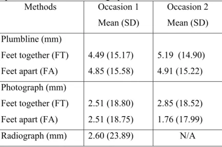
List of Tables
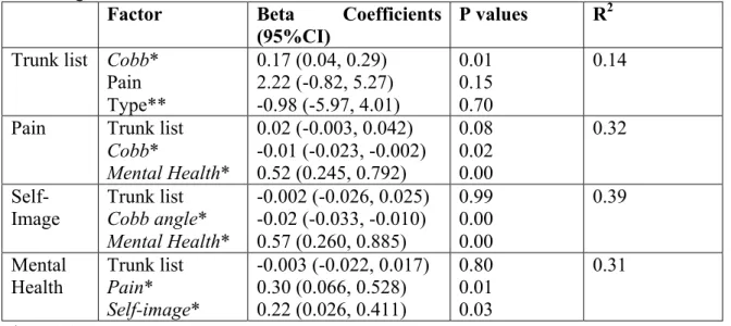
Table 1. Means and standard deviations of trunk list measurement and values of radiographic measurement…….……….56 Table 2. Reliability and validity of the plumbline and photographic methods…………...56 Table 3. Descriptive results of cohort of adolescents with Idiopathic Scoliosis………….70 Table 4. Pearson correlations between trunk list and Cobb angle, pain, mental health and self-image………70 Table 5. Linear regression models describing factors that contribute to trunk list, pain, self-image and mental health………...71 Table 6. Factors associated with trunk list in logistic regression models of trunk list dichotomized at 1cm, 1.5cm and 2 cm………...……….71 Table 7. Bland and Altman limits of agreement for trunk list measurement between the plumbline and radiographic methods………...……...72 Table 8. Correlation matrix between the following variables: trunk list, Cobb angle, pain, mental health and self-image………...………74
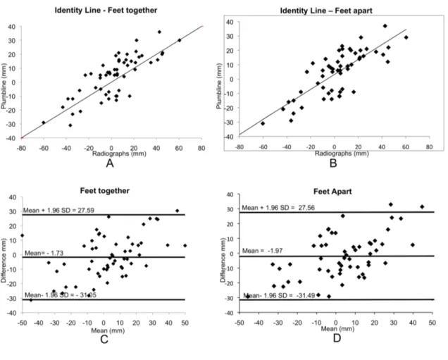
List of Figures
Figure 1. Trunk list measured using a plumbline………...…………...4 Figure 2. Scoliotic Measurements………...………17 Figure 3. Position 2 – Feet Apart on Position Base……….31 Figure 4. Validity analysis comparing plumbline versus radiographs using Bland and Altman analysis………..……….73
List of Abbreviations
AIS Adolescent Idiopathic Scoliosis ANOVA Analysis of Variance
CHU Centre Hospitalier Universitaire D Study Decision Study
FA Feet Apart
FT Feet Together
G study Generalizability Study
ICC Intra-class Correlation Coefficient IS Idiopathic Scoliosis
S Session
P Person
PH Photography
PL Plumbline
r Pearson Correlation Coefficient SD Standard Deviation
SEAS Scientific Exercises Approach to Scoliosis SEM Standard Error of Measurement
SRS Scoliosis Research Society Outcomes Instrument
SRS-22vf Scoliosis Research Society Outcomes Instrument 22 – French version
T Trial
χ2 Chi-squared
ρ2 Generalizability Coefficient
Dedicated to my husband, Alex, and my parents for their constant love and support
Acknowledgements
First and foremost, I would like to thank my research director, Dr. Debbie Feldman, for her constant guidance and support during my time as her student. Her dedication and encouragement were invaluable throughout this process.
Dr. Carole Fortin also deserves my great appreciation for her never-ending support and expertise. She was there whenever I needed her help and direction and this master’s thesis would not have been possible without her.
As well, I would like to sincerely thank my co-director, Dr. Stefan Parent, for all of his expertise and assistance in performing my research and writing my thesis.
For help in performing the statistical analysis, I would like to acknowledge Michelle Houde, as well, thank Julie Joncas for her assistance in recruiting participants and José-Felix Sosa for measuring trunk list on radiographs. My research was based on the cooperation of participants, so they deserve a special thank you for their help in contributing to the further study of scoliosis. Université de Montréal and CHU Ste. Justine have been wonderful institutions that have facilitated my learning and growth over the course of my research and studies. I am very proud of my association with both institutions.
Finally, I wish to thank the Canadian Arthritis Network, OPPQ-REPAR partnership program, Ordre professionnel de la physiothérapie du Québec, and Université de Montréal Faculté de Médecine (Fonds de dépannage) for their financial support in the form of grants and bursaries. .
Chapter 1: Introduction
Trunk list is a lateral deviation of the spine, frequently seen in individuals with scoliosis, disc herniation and low back pain1. It is associated with muscle asymmetry and imbalance2 and may lead to development of pelvic obliquity, curve progression following skeletal maturity and back pain in individuals with idiopathic scoliosis (IS)3, 4. It is an important element of the scoliosis evaluation since it may be predictive of outcome2, 5, 6 and is routinely measured in a clinical setting using a plumbline and determining the horizontal displacement from midline7. When trunk list is mentioned in this thesis, this author is referring to trunk list measured from the C7 to the S1 vertebrae using a plumbline.
Only one study examines reliability of the plumbline used to measure trunk list in persons who do not have IS8 and none investigate its validity. Therefore, despite its widespread use in the clinical evaluation of IS, little is known about the reliability and validity of the plumbline measure in IS. It may also be important in the context of repeated measures of trunk list over time, which has been suggested as a possible marker of scoliosis progression2, 5, 6.
Scientific evidence is needed to discern whether trunk list is associated with other aspects of scoliosis, including pain, Cobb angle, type of scoliosis, mental health and self-image. If trunk list is highly associated with Cobb angle and changes over time, then it may serve as a future marker of progression and may have prognostic value when assessing persons with IS. Exploring its relation with pain, self-image and mental health may lead to improved methods of treatment: if trunk list is
strongly associated with these factors, then treating trunk list may help improve outcomes. There is very little information regarding the relationship between trunk list and its associations with any of these factors in IS.
The objective of our study was to examine trunk list in individuals with Adolescent IS; namely to investigate the reliability and validity of its clinical measurement using the plumbline and from photographs, and to explore its associations with Cobb angle, type of scoliosis, pain, mental health and self-image.
In this thesis, I will first present the available literature on the subject of trunk list and on adolescent idiopathic scoliosis. In chapter 3, I will define the objectives and hypotheses of my study. In chapter 4, I will outline the methods that were used in our study, including a description of the analysis using generalizability theory. In chapters 5 and 6, I will present the two manuscripts, each of which addresses one of the two objectives described above. Chapter 7 includes some additional results and Chapter 8 comprises a general discussion of all the results presented and clinical implications. The final chapter is a conclusion.
Chapter 2: Literature Review
A review of the available literature regarding trunk list will be presented in this chapter. The definition of trunk list, how it is measured, and its importance in terms of associations with scoliosis, pain and function are discussed. Following this, adolescent IS is examined; first it is described, then classification and types, prognosis, symptoms, scoliosis measurements and evaluation and finally treatment are discussed.
2.1 Trunk List
2.1.1 Trunk List Defined
Trunk list is defined as “the lateral displacement of the human thoracic cage relative to the pelvis” 9. It is a common trait seen in individuals with postural disorders, including scoliosis, disc herniation and low back pain9. Trunk list is also known as coronal balance, lateral translation or deviation of the spine, lumbosacral list and lateral trunk shift.
The percentage of individuals presenting with trunk list in the general population is unknown. In 1941, a study of 395 individuals with scoliosis showed that 55% had a trunk list; 39.5% to the right and 15.5% to the left and that the trunk shift was generally to the side of the convexity. The majority (71%) of the trunk lists were less than or equal to ½ inch or 12.7mm10. There is very little information on prevalence of trunk list since that 1941 publication.
2.1.2 Trunk List Measurement
There are various methods to measure trunk list in a clinical setting. These include the plumbline, measurement on radiographs, projecting a shadow from a vertical line, 3D posture analysis systems and digital photography. I will describe these methods in this section.
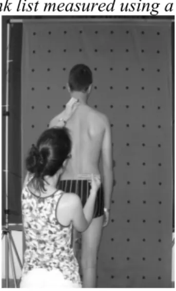
Trunk list is commonly measured in a clinical setting using a plumbline7, where the plumbline is placed at a specific spinous process of the cervical or thoracic regions of the spine and the horizontal distance between S1 and the plumbline is measured (see Figure 1). We chose to evaluate trunk list from the C7 vertebrae because it considers all the thoracic and lumbar vertebrae affected by a scoliosis curve. This simple method has not been studied in depth and there is a dearth of knowledge with regards to its psychometric properties and trunk list in individuals with IS.
Figure 1: Trunk list measured using a plumbline
There is no reported gold standard for measuring trunk list. Studies use radiographs to evaluate trunk list11-13, however on radiographs the central of the vertebral body is the landmark used, not the spinous process. It is not practical to take radiographs of all patients in a clinical setting, nor is it safe to expose them to unnecessary radiation. There are various other methods to measure trunk list, however, many of them require expensive equipment that is not widely available and are therefore not feasible in a clinical setting. These methods include:
1.Projected shadow from a vertical wire onto the skin of the back8. This method is relatively simple but requires a wire and special lighting to attain a shadow. It is also not portable like a plumbline.
2.3D posture analysis systems, including the 3SPACE Isotrak, measure the three-dimensional shape of the back14. However, due to the system requirements (e.g.: special software and equipment), this is difficult to use in a clinical setting.
3.Using digital photography, Fortin et al. developed a computer software program to evaluate posture. Photos are taken of individuals and they are analyzed using this software. Reliability was excellent with a dependability coefficient of 0.9515. They found good concurrent validity for trunk list measurement from digital photographs with radiographs among individuals with IS (r = 0.76)16. Digital photography is a promising method to evaluate posture17. However, the tool is not yet widely available and it is more
McLean and colleagues compared three methods of assessing trunk list: plumbline, projected shadow and 3SPACE Isotrak, a 3D posture analysis system. They demonstrated that the plumbline and projected shadow yielded similar results, yet they selected the plumbline as the preferred method due to its simplicity8. This study had
several important limitations. First, the number of participants was quite limited, varying between 7 and 27 for the different methods. Second, the trunk list measurement was performed between the T12 and S1 vertebrae, instead of between the C7 or T1 and S1 vertebrae, not accounting for the thoracic spine posture and the increased sway that may have been present at the upper back due to a larger distance from the base of support.
In order to measure trunk list, the spinous processes used as reference points should be identified7, 8. In this study, the C7 and S1 vertebrae were used as reference points. As mentioned above, our team previously conducted a study assessing reliability of postural indices in scoliosis, including trunk list, using digital photographs. We found high test-retest reliability (dependability coefficient of 0.98, and standard error of measurement (SEM) of 2.9 mm16. Engsberg and colleagues13 put skin surface markers on the spinous processes of individuals with spinal deformity and then took radiographs. He measured trunk list on the radiographs from the skin surface markers that had been placed at the spinous processes and then directly from the spinous processes. He reported a strong correlation (r = 0.80) between these two measurements13. However, Lenke et al.’s results contradict those of Engsberg. He assessed trunk list in thirty individuals with IS during
upright posture and during gait using videographic gait analysis and via radiographs and he found only weak correlations between all three measurements11. These differences may be attributed to landmark placement (spinous process vs. central of vertebral body) and position during evaluation.
2.1.3 Trunk List and its Associations
Trunk list is an important measurement since it may be a manifestation of scoliosis and pain and also have an effect on self-image and mental health. It has even been suggested to lead to scoliotic curvatures of the spine. In the next sections these potential associations are discussed.
Trunk list is an important measurement, performed by physicians and physical therapists. It has been associated with back pain and intervertebral disc lesions18. Reducing trunk list using reverse trunk list exercises and reverse trunk list traction (i.e. the Harrison treatment method), also decreases pain in persons with low back pain1. A
retrospective study of 2442 children and adolescents with IS examined various aspects potentially related to pain experienced. Trunk list greater than 1 cm was seen in 220 children; in 11% of those presenting with pain (62/560) and in 8% of those not presenting with pain (158/1882), p=0.052. The authors do not mention if there was a difference in pain intensity, duration or frequency in those with trunk list versus those with no trunk list. In addition, in their analysis of pain and trunk list, the authors do not
include 210 individuals who presented with pain after their initial diagnosis of IS19. In a study of 34 patients presenting with lumbar disc herniation, the investigators found that those with trunk list (n=10) had significantly increased nerve root pressure than those without trunk list (82.1 mmHg versus 41.2 mmHg, p<0.05). They do not explain the phenomenon, rather express that further research is warranted20. Souchard believes that trunk list develops as an antalgic posture secondary to pain experienced by individuals6. Therefore, addressing trunk list may also address an individual’s pain and the reverse can be presumed as well, which has ramifications on health related quality of life.
Trunk list may be correlated with type of scoliosis. Gauchard et al.21 found that
trunk list, measured using the plumbline was most common in those with lumbar scoliosis, followed by thoracolumbar scoliosis, then by thoracic scoliosis and finally by double major curves. Gram and Hasan22 reported trunk list, measured using infrared-emitting markers, to be more common in those with thoracic scoliosis than lumbar scoliosis in both standing and sitting positions.
Another study demonstrated that in 298 adults with scoliosis (with a Cobb angle > 30°), persons who had a trunk list > 4cm as measured on radiographs and who had not been operated on had poorer functional level (Oswestry Disability Index; SF-12) and increased pain (SRS-29) compared with the those who had trunk list of < 4cm23. Based on clinical experience, Floman expressed his opinion that trunk list may also be associated with development of pelvic obliquity, scoliotic curve progression following skeletal maturity and back pain4, but this has never been proven. Trunk list has also been
suggested to lead to scoliotic curve formation secondary to increased loads, vertebral growth alteration and postural changes24. Absence of trunk list is also thought to be associated with improved scoliotic prognosis25. Authors have demonstrated that when healthy individuals assume a trunk list, a correlation exists between the degree of the trunk list and Cobb angle, as well as the Risser-Ferguson and lumbosacral angles26. However, these results have not been duplicated in persons with IS. If these correlations do exist in individuals with scoliosis, then perhaps trunk list could serve as an indicator of both progression and severity of scoliosis. Further, treatment of trunk list could then potentially help improve an individual’s scoliotic curve.
2.2 Adolescent Idiopathic Scoliosis
In the following section, Adolescent Idiopathic Scoliosis is dicussed; namely its classification, symptoms, prognosis, evaluation and treatment. As this study deals with scoliosis of an idiopathic nature, the discussion focuses on IS.
2.2.1 Introduction
Adolescent Idiopathic Scoliosis (AIS) is a three-dimensional deformity of the spine, characterized by a lateral deviation and axial rotation of the spine. Studies estimate that 1-3% of children aged 10-16 years are at risk of developing a scoliotic curve with a Cobb angle greater than 10°27-29. Idiopathic scoliosis is more common in
females; with a ratio of 5.4:1 for curves greater than 20 degrees and a ratio of 7:1 for curves under treatment30.
The etiology of IS is unknown, yet several hypotheses and associations do exist. Individuals with scoliosis have a family history of the condition approximately 30% of the time, however this does not appear to be predictive of curve progression or severity25. Other potential hypotheses involve hormonal imbalance, muscle and tissue imbalance (including trunk list), neurological abnormalities and associations to puberty24, 27.
Clinical appearance of scoliosis may include one scapula that protrudes more than the other, inequality in shoulder and pelvis levels, asymmetric appearance of breasts and prominence on one side of the back31. The waist angles are uneven; being larger on the side of the concavity and smaller on the side of the convexity7. The arm on the side of the concavity may hang closer to the body than the other arm7. Asymmetry of the paraspinal muscles leads to bulging and weakening on the convex side and a flattening and shortening of the muscles on the concave side7, 32. Individuals may lack normal flexibility and may have asymmetric side bending. On the convex side of the curve, the rotation can lead to a rib hump (or gibbosity), which is always apparent in a forward bent position and may be seen in standing32. A functional leg length discrepancy (i.e. true leg lengths are equal but they appear not to be) may also be present7.
2.2.2 Classification and types of scoliosis
Scoliosis is classified first by cause – namely idiopathic (of unknown cause), osteopathic (due to spinal disease or bony anomaly), myopathic (due to muscle weakness) or associated with neurological conditions. Idiopathic scoliosis accounts for 80% of scoliotic curves. It is further classified by age of appearance; namely infantile (under 3 years of age), juvenile (4 to puberty), adolescent (puberty years) or adult (mature skeleton)32.
Scoliosis can also be categorized as structural or functional. Functional scoliosis is caused by factors such as pain, poor posture, leg length discrepancy, spondylolisthesis or herniated disc causing muscle spasm; i.e. factors in which the vertebrae are not involved. A functional scoliosis will disappear if the causative factor is addressed. When a fixed curvature of the spine exists, the scoliosis is designated as structural; generally including vertebral rotation and translation, as well as asymmetry of the surrounding soft tissue structures32.
Types of scoliosis are commonly classified by curve pattern designated according to the level of the apex of the curve and the direction. For example, a right thoracic curve has a thoracic apex with a right convexity. A cervical scoliosis’ apex is between the vertebrae C1-C6. A cervico-thoracic curve’s apex is at the C7 or T1 vertebrae. A thoracic scoliosis apex ranges between the T2 and T11 vertebrae. A thoraco-lumbar curve’s apex is either at the T12 or L1 vertebrae. A lumbar scoliosis apex is between the L2 and L4 vertebrae. A lumbo-sacral scoliosis’ apex is at the L5 or S1 vertebrae7.
A scoliotic curve can also be classified as single or double major according to the number of curvatures in the spine. One author states that curve types include: double major curves (37%), thoracic curves (22.1%), lumbar curves (23.6%), thoracolumbar curves (16%) or cervicothoracic curves (1.3%) 33. Another author found that curve types
include: main thoracic (51%), double thoracic (20%), double major (11%), triple major (3%), thoracolumbar/lumbar (12%) and thoracolumbar/lumbar-main thoracic (where the thoracolumbar/lumbar curve is the major curve) (3%)34.
2.2.3 Scoliosis Symptoms
Individuals with scoliosis can have symptoms such as pain, poor self-image and mental health, and impaired pulmonary function, which can hinder quality of life. These are discussed below.
2.2.3.1 Pain
Between 23-73 % of individuals with IS present with back pain19, 31, 35-37. This large range in prevalence of pain in those with IS could be due to confounding factors such as age, weight, other comorbidities and activity level37. In addition, while some looked at present pain, others examined pain within the past year. Ramirez et al.19 performed a retrospective study of 2442 patients with IS, where they investigated the prevalence of back pain. They found an association between back pain and age > 15
years, skeletal maturity, post-menarchal status and a history of injury. 9% of those with pain had an underlying pathological condition, including spondylosis and Scheurmann kyphosis19. Another study by Ramirez et al.38 investigated back pain in 303 individuals with IS wearing back braces. He found that of those with back pain, 26% eventually required spinal fusion surgery because of curve progression, while only 2.6% of the group without pain required fusion. Thus, back pain appears to be associated with curve progression38. Individuals with IS have increased pain when compared to controls, however pain does not appear to limit function39. Some authors show that there is no correlation between curve magnitude and pain19, 39, 40, whereas others show that
correlation ranging from r=0.32-0.37 exists41-43.
2.2.3.2 Mental Health, Self-Image and Health Related Quality of Life in Persons with IS
Adolescents with IS are more likely to exhibit poor mental health35, 42, 44 and
self-image42, 45, 46, which both correlate with Cobb angle (r = -0.27 and -0.50 respectively)42. Payne et al.44 studied 34706 adolescents, 685 of whom had IS, and found that scoliosis is an independent risk factor for suicidal thought, worry and concern over body development. Ascani et al.35 studied individuals with untreated IS after skeletal maturity and found that 19% of his cohort had real psychological disturbances; the majority were female, had thoracic curves and Cobb angles>40°35. In a long term follow-up study of 2092 adults with AIS, individuals with scoliosis perceived themselves to be less healthy
and had poorer body image, however they had improved perception of self as compared to controls46. A review investigating psychosocial issues and quality of life in scoliosis concluded that compared to healthy controls, adolescents with IS have poorer health-related quality of life, body image and psychosocial functioning45.
2.2.3.3 Pulmonary function
Pulmonary function is highly associated with curve size. It is affected as well by number of vertebrae involved, degree of rotation, location of uppermost vertebrae and patient’s age47. Reduced vital capacity, frequent shortness of breath and less commonly
cardiopulmonary compromise, have been associated with curves greater than 50°27. Early onset idiopathic scoliosis patients can present with substantial loss of both vital capacity and forced expiratory volume, which can cause pulmonary hypertension, right heart failure and even death. However, this is quite rarely seen in AIS27.
2.2.4 Prognostic Factors
Scoliosis prognosis varies according to many factors; including age, physical maturity level, sex, curve size and curve pattern. Clinicians use these factors to decide on treatment plans for their patients. In this section, these factors are discussed.
Various factors are predictive of scoliosis progression in individuals with AIS, such as: time of diagnosis compared with puberty, sex, curve severity, curve pattern,
degree of vertebral rotation, as well as other factors, whose associations have not been definitively proven25, 27.
Curve progression often occurs during periods of growth or puberty. If an adolescent or child diagnosed with AIS is at the beginning of the puberty process, his/her curve must be monitored very closely as this is the time of greatest risk for curve progression. The occurrence of menarche, the Risser sign (iliac crest progressive ossification) and the individual’s age are all evaluated when determining progression risk25. Following menarche and passing a Risser Stage II (iliac apophyses 50% ossified) are both factors that reduce risk of scoliosis progression to less than 20%16 25, 48. A
modification to the Risser sign has been proposed that signals the start of rapid growth49. In patients who are physiologically immature, Cobb angles of 30° or more have an increased risk of progression when compared to smaller curves. If a Cobb angle is greater than 50° at skeletal maturity, the curve will likely increase at a rate of 1° per year throughout life50. Therefore curve severity is also an important factor in the prediction of
prognosis25.
Curve pattern may also predict progression. Bunnell25 states that lumbar curves have low risk of progression, thoracolumbar curves have intermediate risk and thoracic curves and double major curves have highest risk of progression. However Bunnell25 does not report what constitutes high, moderate and low risk. Picault et al.51 report
incidence of progression as 67% for thoracolumbar curves, 62% for double major curves, 58% for thoracic curves and 44% for lumbar curves.
Other factors that are possibly associated with curve progression include the presence of chest deformity, vertebral rotation, trunk list2, 5, 6 and increase in height of a subject in one year25, 52. However, Peterson et al.52 showed that in individuals with IS an absence of trunk list was significantly associated with curve progression. They hypothesized that individuals with trunk list may sense their imbalance and try to actively correct it.
2.2.5 Scoliotic Measurements and Evaluation
The following sections address measurement of scoliosis and evaluation of patients with IS.
2.2.5.1 Cobb Angle
Scoliosis is typically measured and quantified by Cobb Angle. The Cobb angle is the gold standard with regards to monitoring scoliosis progression53. It is defined as “the angle between the two straight lines that are tangent to the superior and inferior endplate of the superior and inferior end vertebrae respectively” 54 and it is calculated on radiographs. It is used to quantify a scoliotic curve and the value guides diagnosis, treatment and follow up of these patients. (See figure 2A)
2.2.5.2 Risser-Ferguson angle
A less commonly used measurement is the Risser-Ferguson angle, however certain authors find that this method is a better indicator of scoliosis severity55, 56. It is the angle between the two straight lines that connect the centres of the end vertebrae with the centre of the apical vertebrae54. (See figure 2B)
Figure 2: Scoliotic Measurements
2A: The Cobb angle 2B: The Risser-Fergusson angle
2.2.5.3 Postural evaluation
Risk of progression of IS increases with postural asymmetries, therefore it is very important to evaluate the posture of individuals with IS2, 57, 58. Posture is generally evaluated qualitatively by physicians and physical therapists13, 59, 60. Clinicians evaluate
symmetry of muscles and bony landmarks, presence of kyphosis or lordosis, trunk list, lower extremity position and symmetry in standing, sitting and lying positions7.
An evaluation of an individual with scoliosis begins as soon as he/she walks into the office of the clinician. This way, gait can be evaluated without the patient’s knowledge and therefore without him/her correcting his/her posture61.
The individual should be properly undressed and is then observed in the standing position from the front, side and back. From the front, the clinician observes the position of the head relative to the trunk and the position of the trunk relative to the hips. The clinician observes rib cage, shoulder, waist and hip asymmetry, as well as the position of the arms with respect to the trunk (i.e. if they are equidistant)7. From the side, the natural
curvatures of the spine (thoracic kyphosis and lumbar lordosis) and the presence of anteversion or retroversion of the pelvis are observed7. Finally, from the back, the clinician checks alignment of the spine, symmetry of shoulders, scapulae, waist and hips and position of the head. During all this time, clinicians must look for any deformities61.
The individual’s sitting and lying positions should also be observed and the alignment of the spine and symmetry should be rechecked7. The clinician should note
whether the amount of asymmetry is different in the sitting versus standing positions. Regardless of whether there exists a discrepancy in the symmetry between the lying, sitting and standing positions, leg length discrepancy should be checked61. This is typically done using a tape measure to measure the distance from the anterior superior iliac spine to the medial or lateral malleolus7.
A clinician will ask his/her patient to stand and bend forward in order to see if there is any asymmetry of the ribs, otherwise known as a rib hump. If a hump is present, the clinician uses a tape measure to evaluate the distance between the hump and the hollow7. He/she also evaluates the angle of trunk rotation or inclination, which is the
angle between the horizontal and the plane across the back at the elevation of a rib/lumbar prominence in forward flexion62. The clinician will also evaluate trunk list and sagittal balance, the alignment of the C7 and S1 vertebrae in the sagittal plane, using the plumbline61.
In standing, the position of an individual’s feet can alter his/her posture. For example, standing with one foot forward or with one knee bent can give the impression of a leg length discrepancy. Lenke et al.11 have mentioned that foot position may influence the correlation between surface marker and radiographic measurement. As such, it is very important to standardize foot position for the evaluation of posture. In addition, when evaluating posture, it is important to avoid postural sway, as it will influence the results of certain measurements. When an individual’s feet are close together, he/she has a decreased base of support, therefore decreased balance, leading to
increased postural sway63. Therefore, foot position is an important component of postural evaluation.
Arm position may also affect posture measurements. Vedantam et al.64 studied posture evaluation from a lateral view and found that arm position must be standardized. They stated that the ideal arm position for lateral radiographs is arms at the side.
2.2.5.4 Scoliosis Research Society Outcomes Instrument
Besides objective measures of scoliosis and posture, it is important to assess the patient’s perspective as well. Patient reported outcomes in scoliosis are measured primarily by the Scoliosis Research Society Outcomes Instrument, which is described below.
2.2.5.4.1 Overview
The Scoliosis Research Society Outcomes Instrument (SRS) was developed as a scoliosis-specific, simple questionnaire for patients with idiopathic scoliosis65. It is a patient-based questionnaire, taking into account not only objective measures of an individual’s medical condition, but also his/her self-perception of his/her condition. The questionnaire has evolved and now includes 22 questions (SRS-22) covering the following five domains: pain, function, self-perceived image, mental health and satisfaction with management. Each domain (except satisfaction with management) has 5
corresponding questions that address various aspects of the domain. For example, pain is addressed with five questions regarding intensity, frequency, medication etc. For each domain, a score out of five is obtained; 5 being highest and 0 being lowest health related quality of life. The SRS-22 has been modified into many languages, including a cross-cultural French Canadian adaptation66. The SRS-22 French Canadian version (SRS-22vf) will be discussed in more depth, as it is what was used for our study.
2.2.5.4.2 Psychometric Properties
Beausejour et al.66 performed a cross-cultural French Canadian translation of the
SRS-22. They demonstrated that the SRS-22vf had very good overall reliability (Cronbach α = 0.86). In addition, they compared the SRS-22vf to the SF-12, showing a concurrent validity of 0.79 for the total scores. Moderate ceiling effects were observed in the pain and satisfaction with management domains. The psychometric properties observed in the SRS-22vf are consistent with those in the original version.
2.2.6 Treatment
As mentioned in Section 2.2.4, postural asymmetry can lead to progression of scoliosis. This implies that the treatment of postural asymmetries is important in scoliosis. In the next sections, possible treatments of scoliosis, including medical, surgical and physiotherapy treatments will be explored.
2.2.6.1 Indications for treatment
Scoliosis treatment varies according to the size of the scoliotic curve, as well as the amount of growth remaining27. However, other factors, including pain and
neurological changes will also guide the treatment approach32.
Generally, if a Cobb angle is less than 25°, physicians will monitor the scoliosis every 6 to months looking for a progression of the curve or of symptoms32. If the curve begins to progress, physicians may opt for the following treatment options: physiotherapy, bracing or surgery.
With a Cobb angle ranging between 20° and 45°, physicians will generally recommend physiotherapy. For a Cobb angle that is greater than 25°, bracing is recommended in order to prevent curve progression until reaching skeletal maturity (defined as Risser 3 or 4 – iliac crests ossification of 75% and 100% respectively67), at which point the risk of curve progression diminishes greatly. For a Cobb angle that is greater than 45˚ or in the presence of chronic pain or neurological changes secondary to the curvature, physicians usually opt for corrective surgery32.
2.2.6.2 Physiotherapy
Physiotherapy focuses on preventing the progression of IS, enhancing the effect of a brace if the individual has one, improving neuromuscular control and stability of the
spine, decreasing biomechanical postural collapse and improving breathing function27, 68. Physiotherapy goals include correcting muscle imbalance, coordination, spinal proprioception and decreasing pain27. Different physiotherapeutic methods to treat scoliosis exist, which include side shift exercises69, the Schroth method70, the Scientific
Exercises Approach to Scoliosis (SEAS) method71, the Dobosiewicz method72 and Global Postural Reeducation6. Side shift exercises are an active form of auto-correction where the patient is taught to shift the trunk sideways over the pelvis in the direction opposite to the curve convexity69. The Schroth method corrects scoliotic postures and breathing patterns with the help of propioceptive and exteroceptive stimulation and mirror control70. SEAS is a program of individually adapted exercises based on active self-corrective movements performed to achieve maximum correction71. The Dobosiewicz method is a method that utilizes active three-dimensional auto-correcting concerning the primary curvature and mobilizations towards correction of the curvature72. Finally, global postural reeducation is a combination of overall stretching
positions, which gradually evolve from an initial position of minimal tension to a final position with an overall stretch, with the intention of stretching tissues and decreasing tensions6.
There exists much debate with regards to physical exercise in the treatment of AIS. Two studies concluded that a regime of physical exercise does not improve or halt the progression of scoliosis10, 73. However, the first study did not specify the type of exercise regime10. In the second study, physiotherapists administered home exercise
programs to individuals with IS with Cobb angles ranging between 4°-22° and compliance was only 50%73. A third study indicated that adding a physical exercise regime to bracing had no additional benefits with regards to reducing or maintaining Cobb angles, however these were aerobic exercises whose goal was not to decrease spinal curvatures but rather to improve exercise capacity74. On the other hand, more recent studies have shown positive results. Den Boer69 compared a group receiving side shift physiotherapeutic exercises to another who were given corrective braces and found similar results in both groups: reduced curve progression and even some improvement in the scoliosis. Weiss et al.75 performed a prospective follow-up study, comparing a group
that had not received treatment with a group that had received the Schroth method as therapy and found that progression was 1.5 to 2.9 times higher in the non-treatment group (p<0.01), implying that the Schroth method may decrease progression of IS. A prospective cohort study regarding the efficacy of SEAS versus traditional physiotherapy demonstrated that among those receiving SEAS 11.5% required bracing secondary to progression versus 30.8% in the traditional treatment group71. Negrini et al.68 reviewed the available literature on the subject of therapeutic exercise in IS and concluded that those who received physiotherapy (versus those who did not) tended to either maintain or reduce the degree of spinal curvature irrespective of the amount of baseline curvature. Physiotherapy improves breathing function74, 76, strength77 and postural balance78 in
Although there is still controversy regarding the efficacy of therapeutic exercise in AIS, the recent studies described above support its use. Well-designed randomized controlled trials are needed to establish whether exercise improves or halts progression of scoliosis.
2.2.6.3 Bracing
Braces are typically used for Cobb angles greater than 25°32. According to Willers et al.79, the prevention of curve progression is achieved in 85% to 88% of cases. Many different orthoses have been developed to treat scoliosis. The effectiveness of the bracing seems to be affected by the brace used. Boston braces are effective in preventing progression but do not decrease Cobb angles80, 81. A retrospective study showed that the use of Milwaukee brace did not decrease the need for surgical intervention82. However Carr and associates83, who examined the results of treatment with a Milwaukee brace (without a control group) are of the opinion that the Milwaulkee brace is the most successful in halting progression of scoliosis. Some suggest that the Cheneau brace, used frequently in Europe, may correct a Cobb angle more than 40% from the initial Cobb angle84, 85. Different brace wearing protocols exist – some requiring subjects to wear the brace for 23 hours/day, others only in the evening, however some studies found that exclusive night-time wear is not effective86, 87. An important aspect of bracing is
compliance. Positive associations between compliance in wearing the brace and scoliosis outcome have been demonstrated80, 88, 89. In the most recent study, 34 individuals with IS
brace-wearing habits were monitored using a temperature sensor and logger that was embedded in the brace. The 15 patients whose curves progressed more than 5° had compliance rates of 62% versus a rate of 85% in the curves that did not progress (p = 0.004)88.
2.2.6.4 Surgical Intervention
The objectives of surgery are to stop the progression, achieve correction of the scoliosis in 3 planes, to balance the trunk, to minimize complications27 and to improve quality of life90. The most common surgical procedure is fusion with posterior segmental
instrumentation32. Danielsonn90 reviewed evidence of whether correcting spinal deformity impacts quality of life of individuals with AIS. He found 3 articles91-93 stating that there was a significant improvement in quality of life after spinal fusion as compared to before surgery. Another review study found that in several long-term follow-up studies, a decreased spinal curvature was maintained for 10-20 years94.
Summary
Trunk list is a trait commonly seen in individuals with AIS. It may be associated with scoliotic curve development and progression, back pain and development of pelvic
obliquity4, 24, 25, although there are no studies to date that support these theories. However, before examining associations and correlations between trunk list and other factors, the methods used to evaluate trunk list must be shown to be reliable and valid. In this study, we try to fill this gap in knowledge: i.e. assess the psychometric properties of the clinical trunk list measure with the plumbline.
Chapter 3: Objectives and Hypothesis
3.1 ObjectivesThe general objective of this study is to evaluate trunk list from C7 – S1 in persons with idiopathic scoliosis, using a plumbline.
The specific objectives are:
1) To determine the inter-trial, test-retest reliability and the standard error of measurement of the plumbline and the photographic methods to measure trunk list with feet together and feet apart
2) to verify the validity of this measure compared to radiographs (gold standard) 3) to explore the association between trunk list and other factors including
Cobb angle, type of scoliosis, pain, mental health and self-image.
3.2 Hypothesis
The hypotheses of this research study were :
1) The degree of inter-trial and test-retest reliability will be good for the measure of trunk list using a plumbline (≥ 0.75). The measure of trunk list taken with the plumbline will be comparable to the measurement taken using a photograph (≥ 0.75).
2) The validity of this measurement will be good when compared with x-rays (≥ 0.75).
3) Trunk list will be correlated with Cobb angle, pain and self-image. Trunk list will be correlated with single curvatures more than double major curves.
Chapter 4: Methods
The following section describes the methods used for this study. First, the process of obtaining approval from the ethics committee is described. Then, the process of participant recruitment, inclusion and exclusion criteria, materials required and the procedure used to measure trunk list using the plumbine, via photographs and on radiographs are described. Finally, the methods of acquisition of clinical information, statistical analysis and the justification of the sample size are defined.
4.1 Ethics Approval
Ethics approval was obtained from the Centre de Recherche du CHU Sainte-Justine (Appendix A) and all subjects and the parents of those under the age of 18 years signed an informed consent form prior to participating in the study (Appendix B).
4.2 Participant Recruitment
Potential participants were recruited during scoliosis clinics at Centre Hospitalier Universitaire Sainte-Justine in Montreal.
4.2.1 Inclusion and Exclusion Criteria
The following lists the inclusion and exclusion criteria used while recruiting individuals for this study.
Inclusion Criteria:
• Youth aged between 10 and 21 years inclusively • Diagnosis of Idiopathic Scoliosis
• Participants with X-rays taken on the same day Exclusion criteria:
• Participants who have had back surgery
• Participants who present with a leg length discrepancy > 1.5cm
4.3 Materials
4.3.1 Plumbline, ruler and stickers
A plumbline (string attached to a weight) was required, in addition to a ruler to measure the horizontal distance between the C7 and S1 vertebrae. Two 5 mm round stickers were used as markers on each participant to mark the bony landmarks of C7 and S1 vertebrae.
4.3.2 Digital Camera
A digital camera was required for comparative purposes between the measurement of trunk list by photographs and the manual measure. A Lumix Panasonic camera, model number FX01, was used in this study.
4.3.3 Radiographs
Radiographs taken as part of the orthopaedic evaluation were retrieved and served as the gold standard of comparison. No new x-rays were ordered for this study.
4.3.4 Two position bases for the feet
To assure reproducibility of the findings, position bases were drawn on the floor to indicate where the subject should stand. Two position bases were used. Position 1 was with heels together and feet opened in a «V» with an angle of 30º. According to some authors6, 95, this is the foot position of reference for the evaluation of posture. Position 2 (see figure 3) was with feet apart on the same position base that is used for radiographs.
4.3.5 Questionnaires
Participants responded to a health status questionnaire developed by our team (see Appendix C) as well as a validated questionnaire for adolescents with scoliosis, the french version of the SRS-22 (see Appendix D). The health status questionnaire addresses past medical history, associated conditions, pain, self-perception of posture and treatment.
To evaluate quality of life, including pain, self-image and mental health, we used the French version of the Scoliosis Research Society Questionnaire-22. It is described in depth in the literature review (chapter 2, section 2.2.5.4).
4.4 Procedure
The following section describes how the data was collected using the plumbline method, via digital photography and from radiographs.
4.4.1 Measurement of Trunk list on Participants
One physiotherapist was responsible for conducting all the measurements in a private room, adjacent to the clinic area. Each of the study participants was measured as follows. The physiotherapist marked the bony landmarks of C7 and S1 with the stickers. The participant was asked to stand on a base, positioned feet in a « V » with an angle of 30º between the feet (position 1). During the acquisition, the participant was asked to look straight ahead, to not move and to breathe normally. The trunk list measurement was completed as follows. A plumbline was suspended from C7 and I
measured the horizontal distance between the plumbline and S1. The trunk list was measured in centimetres with a ruler and then converted to millimetres. Subsequent to the plumbline measurement, photos were taken from a posterior view. The same procedure was done in position 2 (feet apart: same position plate used for radiographs). The participant was asked to move from the position and then asked again to stand in position 1 and subsequently in position 2, at which times the trunk list measurement and photographs were taken again. The stickers were then removed from the subject. After an hour delay, during which the participant filled out the SRS-22vf questionnaire (see Chapter 2, Section 2.2.5.4), the participant was repositioned and the measurement was taken again in the two different positions and techniques. The stickers were then removed.
4.4.2 Measurement of Trunk list on Radiographs
A technician performed the trunk list measurements on the radiographs. He marked the C7 and S1 vertebrae and projected a vertical line down from C7. The horizontal distance between C7 and S1 is directly displayed on the digital radiographs.
4.4.3 Measurement of Trunk list on photographs
The trunk list measurements on the photographs were performed by a physiotherapist using a software program. This program was developed in a previous
study at CHU Ste-Justine16. The software uses interactive click-on markers with the computer mouse. The physiotherapist selected the C7 and S1 vertebrae from the graphic interface and placed them directly on the corresponding marked anatomical landmarks. The software automatically calculated and displayed the distance. The physiotherapist was blinded with respect to the photographs: i.e. she did not know which participant’s photograph she was measuring.
4.5 Statistical Analysis
In this section, the statistical analyses for each of the three objectives of this study are described.
4.5.1 Objective 1
For objective 1, which was to determine the inter-trial, test-retest reliability and the standard error of measurement of the plumbline and the photographic methods to measure trunk list in both foot positions, the generalizability theory was used
The generalizability theory is an extension of the intraclass correlation coefficient used in classical theory96. The generalizability analysis quantifies different sources of error or variance (such as person, occasion, trial etc) rather than
designating all variance simply as error. This theory enables researchers to quantify various sources of error and then to determine ways to eliminate them, ultimately allowing for better measurement techniques97, 98.
There are two components when using generalizability theory; first the generalizability study (G study) and then the decision study (D study). The G study allows for the determination of variances attributed to different factors or sources of error96, taking into account systematic and unsystematic error sources. Random or
unsystematic variance stems from error associated with interactions between factors, while systematic variance stems from the factors themselves. The expected source of variance is that attributed to person, since each person evaluated is expected to be different. Other sources of variance include factors related to the particular study, their interactions and the residual random error, being the unexplained error or error caused by interaction between all the factors. In our study, the factors used were person (P), trial (T) and session (S). The difference between occasion and trial is that the stickers are removed between occasions, whereas they are not between trials. Thus, in this study, seven sources of error variance, P, S, T, PT, TS, PS and PTS, can be identified. The sources of error considered in this study are as follows:
σp2 = inter person variance
σt2 = inter trial variance (systematic variance)
σps2 = variance associated with interactions between person and
session (random variance)
σts2 = variance associated with interactions between trial and session
(random variance)
σpt2 = variance associated with interactions between person and trial
(random variance)
σpts2 = variance associated with interactions between person, trial and
session (residual random variance)
The D study determines the reliability of a particular protocol using the results obtained in the G study 96. From this, we are able to design a measurement procedure that minimizes error.
The G- and D-studies allow dependability coefficients (φ) and generalizability coefficients (ρ2) to be calculated. The dependability coefficient, a measure of reliability in the generalizability study, is the ratio between the inter-person variance (σ2p) and the sum of the inter-person variance and all possible
sources of error, i.e. the sum of both systematic and random error variances which is called absolute error variances (σ2abs)97. Like the intra-class coefficient (ICC), the
dependability coefficient ranges between 0 and 1: 0 is null reliability and 1, perfect reliability. Portney and Watkins99 have suggested that values above 0.75 can be
considered as good reliability, those between 0.50 and 0.75 as moderate and those under 0.5 as poor.
The generalizability coefficient (ρ2) is the ratio of the inter-subject variance versus the sum of the inter-subject variance and the relative error variance (only including sources of error where there is interaction with subjects). The generalizability coefficient still takes into account the number of trials and sessions; therefore it can be tailored to any particular protocol. Since the generalizability coefficient only takes into account error where there is interaction with the subject, ρ2 is greater than ϕ. However the generalizability coefficient still ranges from zero to one; one being perfect reliability.
To appreciate the errors in terms of the unit of measurement, the standard error of measurement (SEM), which is the root square of the absolute error variance, was computed 97. We used the GENOVA program for the generalizability analysis100.
4.5.2 Objective 2
For objective 2, which was to verify the validity of this measure compared to radiographs (gold standard), the Pearson correlation coefficient was used to compare distances measured by the plumbline method and those measures on radiographs. The Bland and Altman method also served to document the agreement between the two clinical measurements of trunk list and the radiograph measurement. For this analysis, we used the first trial of each session.
4.5.2.1 Pearson Correlation Coefficient
The Pearson Correlation Coefficient (r) is a measurement of the relationship between two variables. It ranges from -1 to +1; +1 being a perfect positive relationship, -1 being a perfect negative relationship and 0 being no relationship. The closer the value is to -/+1, the stronger the relationship is. Our study will use the following to define the strength of correlations found: <0.25 as little or no relationship; 0.25 to 0.50 as fair; 0.50 to 0.75 as moderate to good; and >0.75 as good to excellent (for absolute values of the aforementioned numbers)101.
4.5.2.2 Bland and Altman
In the process of validating clinical tools, one compares with the gold standard. Although Pearson correlation is the most widely used and accepted method to evaluate validity, it does not show the agreement. For example, a perfect correlation can exist even when the two compared items are not in the same units or in the case where one method is consistently under/overestimating the true value. The Bland and Altman method alleviates this problem by directly comparing the values obtained using the two methods, however the two methods must be measured using the same units of measurement. It plots the mean of the two measurements versus the difference. The mean difference is calculated, as are the maximum and minimum difference values (called limits of agreement); the closer all these values
are to zero, the closer the sample comes to perfect agreement. Studies rarely show perfect agreement between two measures and therefore an accepted difference, based on the construct being evaluated, should be set prior to performing the analysis102.
4.5.3 Objective 3
For objective 3, which was to explore the association between trunk list and each of the following: Cobb angle, type of scoliosis, pain, mental health and self-image; the Pearson correlation coefficient was calculated between trunk list (plumbline measure) and Cobb angle, pain, mental health and self-image. Chi-square and analysis of variance (ANOVA) were calculated between trunk list and type of scoliosis (comparing single versus double curvatures). Means of double versus single curvatures were also compared using the unpaired samples t-test.
Multiple linear regression models were constructed to explore various associations. These were: 1) trunk list as a function of Cobb angle, pain and scoliosis type; 2) pain as a function of mental health, Cobb angle and trunk list; 3) self-image as a function of trunk list, Cobb angle and mental health; and 4) mental health as a function of pain, trunk list and self-image. Type of scoliosis was designated as single versus double for the purposes of the regression models.
4.6 Sample Size Justification
Sample size was calculated based on objective 2: validation of the trunk list measure with measurement on radiographs as the gold standard. If the correlation between these two measures is assumed to be high, i.e. in the area of 0.9 and we are willing to accept an absolute error of 0.3 and an alpha level of 0.05, then 55 subjects would be required.