HAL Id: hal-02676249
https://hal.inrae.fr/hal-02676249
Submitted on 31 May 2020
HAL is a multi-disciplinary open access
archive for the deposit and dissemination of
sci-entific research documents, whether they are
pub-lished or not. The documents may come from
teaching and research institutions in France or
abroad, or from public or private research centers.
L’archive ouverte pluridisciplinaire HAL, est
destinée au dépôt et à la diffusion de documents
scientifiques de niveau recherche, publiés ou non,
émanant des établissements d’enseignement et de
recherche français ou étrangers, des laboratoires
publics ou privés.
Distributed under a Creative Commons Attribution - ShareAlike| 4.0 International
License
Proteinase-activated receptor-2-induced colonic
inflammation in mice : Possible involvement of afferent
neurons, nitric oxide, and paracellular permeability
Nicolas Cenac, Rafael Garcia Villar, Laurent Ferrier, M. Larauche, Nathalie
Vergnolle, N.W. Bunnett, A.M. Coelho, J. Fioramonti, Lionel Bueno
To cite this version:
Nicolas Cenac, Rafael Garcia Villar, Laurent Ferrier, M. Larauche, Nathalie Vergnolle, et al..
Proteinase-activated receptor-2-induced colonic inflammation in mice : Possible involvement of
af-ferent neurons, nitric oxide, and paracellular permeability. Journal of Immunology, Publisher :
Bal-timore : Williams & Wilkins, c1950-. Latest Publisher : Bethesda, MD : American Association of
Immunologists, 2003, 170, pp.4296-4300. �hal-02676249�
of August 19, 2015.
This information is current as
Oxide, and Paracellular Permeability
Involvement of Afferent Neurons, Nitric
Colonic Inflammation in Mice: Possible
Proteinase-Activated Receptor-2-Induced
Anne-Marie Coelho, Jean Fioramonti and Lionel Bueno
Larauche, Nathalie Vergnolle, Nigel W. Bunnett,
Nicolas Cenac, Rafael Garcia-Villar, Laurent Ferrier, Muriel
http://www.jimmunol.org/content/170/8/4296
doi: 10.4049/jimmunol.170.8.4296
2003; 170:4296-4300; ;
J Immunol
References
http://www.jimmunol.org/content/170/8/4296.full#ref-list-1
, 10 of which you can access for free at:
cites 32 articles
This article
Subscriptions
http://jimmunol.org/subscriptions
is online at:
The Journal of Immunology
Information about subscribing to
Permissions
http://www.aai.org/ji/copyright.html
Submit copyright permission requests at:
Email Alerts
http://jimmunol.org/cgi/alerts/etoc
Receive free email-alerts when new articles cite this article. Sign up at:
Print ISSN: 0022-1767 Online ISSN: 1550-6606.
Immunologists All rights reserved.
Copyright © 2003 by The American Association of
9650 Rockville Pike, Bethesda, MD 20814-3994.
The American Association of Immunologists, Inc.,
is published twice each month by
The Journal of Immunology
by guest on August 19, 2015 http://www.jimmunol.org/ Downloaded from by guest on August 19, 2015 http://www.jimmunol.org/ Downloaded from
Proteinase-Activated Receptor-2-Induced Colonic Inflammation
in Mice: Possible Involvement of Afferent Neurons, Nitric
Oxide, and Paracellular Permeability
1
Nicolas Cenac,* Rafael Garcia-Villar,* Laurent Ferrier,* Muriel Larauche,*
Nathalie Vergnolle,
†Nigel W. Bunnett,
‡Anne-Marie Coelho,
‡Jean Fioramonti,* and
Lionel Bueno
2*
Activation of colonic proteinase-activated receptor-2 (PAR-2) provokes colonic inflammation and increases mucosal permeability in mice. The mechanism of inflammation is under debate and could be neurogenic and/or the consequence of tight-junction opening with passage of exogenous pathogens into the lamina propria. The present study aimed to further characterize the inflammatory effect of PAR-2 activation by investigating: 1) the role of NO, 2) the role of afferent neurons, and 3) a possible cause and effect relationship between colonic paracellular permeability changes and mucosal inflammation. Thus, intracolonic infusion to mice of the PAR-2-activating peptide, SLIGRL, increased both myeloperoxidase (MPO) activity and damage scores indicating colonic inflammation, and enhanced colonic permeability to51
Cr-EDTA from 2 to 4 h after its infusion. NO synthase inhibitors, L-NAME and aminoguanidine, as well as the neurotoxin capsaicin and NK1, calcitonin gene-related peptide (CGRP) receptor antagonists, SR140333 and CGRP8 –37, prevented SLIGRL-induced MPO and damage score increases and permeability. In
con-trast, although the tight-junction blocker, 2,4,6-triaminopyrimidine, and the myosin L chain kinase inhibitor, ML-7, prevented SLIGRL-induced increase in permeability, they did not prevent MPO and damage score increases. Taken together our data show that both NO and capsaicin-sensitive afferent neurons are involved in PAR-2-mediated colonic inflammation and paracellular permeability increase. Nevertheless, the inflammation process is not a consequence of increased permeability which results at least in part from the activation of myosin L chain kinase. The Journal of Immunology, 2003, 170: 4296 – 4300.
P
roteinase-activated receptors (PARs)3belong to a family of seven transmembrane domain G-protein-coupled recep-tors that are activated by cleavage of their N-terminal do-main by a proteolytic enzyme (1). The unmasked new N-terminal sequence acts as a tethered ligand that binds and activates the receptor itself. PAR-2 is activated by trypsin and mast cell tryptase, but synthetic peptides, so-called PAR-activating peptides, (NH2-SLIGRL for PAR-2, hereafter referred to as SLIGRL)
cor-responding to the tethered ligand sequence, are also able to selec-tively activate PARs (2). PAR-2 is expressed throughout the gas-trointestinal tract on several cell types, including enterocytes, mast cells, smooth muscle cells, myenteric neurons, and endothelial cells (3). Indeed, at concentrations physiologically present in the
intestinal lumen, trypsin and trypsin-like molecules activate PAR-2 at the apical membrane of enterocytes and stimulate the generation of inositol-1,4,5-triphosphate, arachidonic acid release, and secretion of eicosanoids, which may in turn influence the in-testinal function (3). Moreover, PAR-2 has been shown involved in pathophysiological processes. Thus, in vivo PAR-2-AP injec-tion into the paw of rats produced an inflammatory reacinjec-tion by a neurogenic mechanism involving afferent capsaicin-sensitive sen-sory neurons and both calcitonin gene-related peptide (CGRP) and substance P (SP) release (4). SLIGRL infused directly into the colonic lumen of rats activates nociceptive neurons and causes delayed rectal hyperalgesia which involves the peripheral activa-tion of tachykinin NK1 receptors (5). We have previously shown that intracolonic infusion of SLIGRL, trypsin, or tryptase to mice led to colonic inflammation characterized by granulocyte infiltra-tion revealed by elevated levels of MPO activity, tissue damage, and increased expression of proinflammatory cytokines (6). More-over, the cytokine profile observed after PAR-2-AP-induced in-flammation revealed a Th1-like type of inin-flammation, in which TNF-␣, IL-1, and IFN-␥ mRNA levels are elevated, while IL-4 and IL-10 levels remain stable. This intestinal inflammation was accompanied by changes in paracellular permeability marked by an increased passage of labeled51
Cr-EDTA and the presence of bacteria translocated to peritoneal organs (6). The mechanism(s) triggered by PAR-2 activation are not clearly understood. Either, this PAR-2-mediated increase of colonic paracellular permeability might constitute an initial event, which then allows penetration of pathogens to cause further inflammation or intestinal inflammation can enhance epithelial barrier breakdown like in ulcerative colitis and Crohn’s disease (7, 8). Inducible NO synthase (iNOS) activity was dramatically increased in colonic biopsies from inflammatory *Neuro-Gastroenterology and Nutrition Unit, Institut National de la Recherche
Agronomique, Toulouse, France;†Department of Pharmacology and Therapeutics,
Faculty of Medicine, University of Calgary, Calgary, Alberta, Canada; and‡
Depart-ment of Surgery and Physiology, University of California, San Francisco, CA 94143 Received for publication November 8, 2002. Accepted for publication February 11, 2003.
The costs of publication of this article were defrayed in part by the payment of page charges. This article must therefore be hereby marked advertisement in accordance with 18 U.S.C. Section 1734 solely to indicate this fact.
1This work was supported by a grant from the UPSA Foundation (to L.B.) and the
institutional support of Institut National de la Recherche Agronomique (France).
2Address correspondence and reprint requests to Dr. Lionel Bueno,
Neuro-Gastro-enterology and Nutrition Unit, Institut National de la Recherche Agronomique, 180 Chemin de Tournefeuille, BP.3, 31931 Toulouse cedex 9, France. E-mail address: [email protected]
3Abbreviations used in this paper: PAR, proteinase-activated receptor; MPO,
my-eloperoxidase; MLCK, myosin L chain kinase; IBD, inflammatory bowel disease; CGRP, calcitonin gene-related peptide; SP, substance P; TAP, 2,4,6-triaminopyrimi-dine; NOS, NO synthase; EGTA, ethylene-glycol-bis( ␣-aminoethyl)-N,N,N⬘,N⬘-tet-raacetic acid; iNOS, inducible NOS; cNOS, constitutive NOS.
The Journal of Immunology
Copyright © 2003 by The American Association of Immunologists, Inc. 0022-1767/03/$02.00
by guest on August 19, 2015
http://www.jimmunol.org/
bowel disease (IBD) patients. A correlation between this activity and the severity of bowel inflammation was found for ulcerative colitis (9). In the intestine, NO can be produced by epithelial cells (10) or nonadrenergic noncholinergic nerves (11) and the effects of NO on intestinal epithelial integrity (12) and permeability (13) have been reported. In fact, inhibition of NO production leads to a reversible circulating leukocyte-independent increase in epithelial permeability. Moreover, down-regulation of junctional proteins and their respective mRNA is observed in actively inflamed colon of patients with IBD, while in tissues from inactive IBD patients, only few junctional molecules are affected, and expression of tight junction-associated proteins appears almost unchanged (5).
Consequently, the aims of this work were 3-fold: first, to further characterize the mechanisms of PAR-2-induced colonic inflamma-tion by investigating the involvement of capsaicin sensitive affer-ent neurons and NO; second, to address the issue of a possible cause and effect relationship between the enhancement of paracel-lular permeability and mucosal inflammation mediated by PAR-2 activation by using a chemical blocker of tight-junctions; and third, to investigate the role of epithelial cell cytoskeleton protein con-tractility in the PAR-2-mediated increase of paracellular perme-ability, using a myosin L chain kinase (MLCK) inhibitor.
Materials and Methods
Animals
Male Swiss 3T3 mice were obtained respectively from Harlan (Gannat, France) and Iffa-Credo (L’Arbresle, France). Mice were housed in poly-carbonate cages in a light (12 h/12 h cycle)- and temperature-controlled room (20 –22°C) and were fed standard pellets usine d’alimentation ration-nelle (UAR) A03 (Epinay-sur-orge, France). Water was provided ad libi-tum. Four groups of eight mice received the neurotoxin capsaicin (25 mg/kg in 100l twice daily for 2 days) or its vehicle injected s.c. The efficacy of the capsaicin treatment was verified by the eye-wiping test with 0.1% NH3(14). The experimental protocols described in this study were
approved by the local Institutional Animal Care and Use Committee. Intracolonic injections
Mice were fasted for 12 h before the beginning of experiments. Under mild xylazine/ketamine (5:1 ratio) anesthesia, a small polyethylene catheter (0.3/0.07 mm) was inserted intrarectally at 4 cm from the anus. The PAR-2 agonist peptide, SLIGRL (100g/mouse; 100 l), or its vehicle (saline) was injected into the distal colon through the catheter.
Experimental protocols
Six groups of eight mice received either the NK1 receptor antagonist, SR140333, (60g/mouse in 100 l; i.p.), or the CGRP receptor antagonist, CGRP8 –37, (50g/mouse in 50 l; i.v.) or their vehicle and were treated
30 min later by SLIGRL (100g/mouse) or its vehicle. Four groups of eight mice received the neurotoxin capsaicin or its vehicle 1 wk before SLIGRL (100g/mouse in 50 l vehicle) or saline. Two groups of 10 mice were treated by SLIGRL or its vehicle for NO synthase (NOS) activity assay. Six groups of eight mice received i.p. either a nonspecific NOS inhibitor, N--nitro-L-arginine methyl ester (L-NAME; 20 mg/kg, 200l), the iNOS inhibitor, aminoguanidine (50 mg/kg, 500l), or their vehicle (saline) and were treated 30 min later by SLIGRL (100g/mouse) or its vehicle. Four groups of eight mice were given a chemical blocker of tight junctions, 2,4,6-triaminopyrimidine (TAP; 30mol/mouse) (15), or its ve-hicle, intracolonically through a polyethylene catheter (0.3/0.07 mm) in-serted intrarectally at 4 cm from the anus with a total volume of 150l during 4 h after SLIGRL (100g/mouse) or its vehicle. Four groups of eight mice were treated with a MLCK inhibitor, 1-(5-iodonaphtalene-1-sulfonyl)-1H-hexahydro-1,4-diazepine (ML-7; 2 mg/kg) (16) or its vehicle injected i.p. three times in a volume of 100l 24, 12, and 0.5 h before SLIGRL (100g/mouse) or its vehicle.
Assessment of inflammation
At 4 h after intracolonic injection of PAR-2 agonist, mice were sacrificed and distal colonic tissues were excised. The severity of intestinal inflam-mation was evaluated by using previously described criteria (17) with some modifications. The disease score (0 –10) was estimated by a combination of both gross and histological findings. The gross score was rated 0, presence
of normal beaded appearance; 1, absence of beaded appearance of colon; 2, focal thickened colon; and 3, marked thickness of the entire colon. The histological score was based upon the extent of intestinal wall thickening (0 –3), lamina propria infiltration (0 –3), and presence (0 –1) of ulceration. Colonic samples were assayed for MPO activity as an index of tissue infiltration by granulocytes (18).
Permeability measurements
Mice were anesthetized with a s.c. injection of xylazine/ketamine (5:1 ra-tio). To measure colonic paracellular permeability, 0.7Ci of51Cr-EDTA
(PerkinElmer Life Sciences, Courtaboeuf, France) in 0.5 ml NaCl 0.9% were slowly infused into the colon (0.25 ml/h). After 2 h, mice were sac-rificed by cervical dislocation and the colon was removed. Then the colon and the rest of the body were placed in separate counting tubes in a gamma-counter (Packard Cobra II; Packard Bioscience, Rungis, France). The per-meability was expressed as the ratio between body and total (body plus colon) radioactivities.
NOS activity assay
Tissue NOS activities were determined by measuring the rate of conversion ofL-arginine toL-citrulline using the method of Bush (19) modified by Anton et al. (20). Briefly, tissue samples were homogenized on ice in buffer (pH 7.4) containing Tris-HCl (50 mM), DTT (1 mM), phenylmethylsulfo-nylfluoride (1 mM), EDTA (0.1 mM), and two protease inhibitors: leupep-tin (23.4M) and pepstatin (14.6 M). After centrifugation (13,500 ⫻ g, 30 min, 4°C), 100l of supernatant was added to the reaction mixture containing 50 mM Tris-HCl (pH 7.4), 1.58ML-arginine, DTT (1 mM), valine (50 mM), 200M NADPH as a cosubstrate, 10 M flavine mono-nucleotide, and 10M flavine adenine dinucleotide as prosthetic groups of NOS. Determination of total NOS activity (i.e., NOS-specific activity and nonspecific activity) was performed by adding 2 mM CaCl2to the buffer.
iNOS activity was determined in the presence of a calcium chelator, eth-ylene-glycol-bis(␣-aminoethyl)-N,N,N⬘,N⬘-tetraacetic acid (EGTA, 1 mM). Nonspecific activity was determined in the presence of 1 mM EGTA and 20 mML-NAME, a nonisoform specific NOS inhibitor. After 30 min of incubation at 37°C, the enzymatic reaction was stopped by adding cold HEPES buffer (pH 5.5) containing 1 mM EGTA and 1 mM EDTA.L -citrulline formed in the medium was separated by applying the samples to columns containing pre-equilibrated Dowex AG50W-X8 (Sigma-Aldrich, St. Quentin Fallavier, France), eluting them with water, and measuring the amount of radioactivity with a liquid scintillation beta counter (Kontron Instruments, St Quentin en Yvelines, France). Total NOS specific (i.e., iNOS ⫹ constitutive NOS (cNOS)) activity was determined by the difference between theL-citrulline generated in samples containing 2 mM CaCl2and samples containing 1 mM EGTA and 20 mML-NAME;
the cNOS activity was determined from the difference between total specific NOS activity and iNOS activity in samples containing 1 mM EGTA. Enzyme activity was expressed as picomoles of citrulline formed per milligram of protein per hour.
Chemicals
Peptides (SLIGRL-NH2and LRGILS-NH2) prepared by solid phase
syn-thesis were obtained from Neosystem (Strasbourg, France). The composi-tion and purity of peptides were confirmed by HPLC analysis. SLIGRL was dissolved in 10% ethanol, 10% Tween 80, and 80% saline. SR140333 was graciously given by Dr. X. Edmond-Alt (Sanofi-Synthelabo, Montpel-lier, France) and was dissolved in 50% saline and 50% DMSO. CGRP8 –37,
capsaicin, aminoguanidine,L-NAME, ML-7, and TAP were obtained from Sigma-Aldrich (St. Quentin Fallavier, France). Aminoguanidine and L-NAME were dissolved in saline. Capsaicin was dissolved in 20% etha-nol, 10% Tween 80, and 70% saline. ML-7 was dissolved in 2% ethanol and 98% saline. TAP was dissolved in Tris-HCl (0.1 M) buffer solution. Statistical analysis
Data are presented as means⫾ SEM, and statistical significance was as-sessed by ANOVA followed by Tukey’s multiple comparison test or by Student’s t test where appropriate. Values of p⬍ 0.05 were considered significant.
Results
Values obtained with the different treatments were normalized to SLIGRL values considered as 100% unless otherwise indicated.
4297 The Journal of Immunology
by guest on August 19, 2015
http://www.jimmunol.org/
Effects of NK1 and CGRP receptor antagonists, and sensory denervation on PAR-2-AP-mediated changes in colonic paracellular permeability and inflammation
Antagonism of NK1 receptor by SR140333 or antagonism of CGRP receptor by CGRP8 –37or depletion of afferent fibers by s.c.
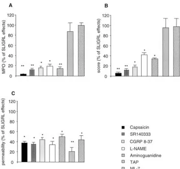
administration of capsaicin (25 mg/kg) twice daily for 2 days, 1 wk before peptide administration, prevented SLIGRL-induced in-crease of MPO activity (respectively, 12.1⫾ 3.8, 15.2 ⫾ 2.5, and 3.3⫾ 0.6%; p ⬍ 0.01) (Fig. 1A) and damage score (11.8 ⫾ 2.5, 14.2⫾ 4.9, and 6.1 ⫾ 1.6%; p ⬍ 0.01) (Fig. 1B), indicating in-hibition of the inflammatory process. Moreover, PAR-2-AP-in-duced increase in paracellular permeability was significantly re-duced in SR140333, CGRP8 –37, and capsaicin-treated mice
(35.4⫾ 3.1, 42.6 ⫾ 2.9, and 37.5 ⫾ 3.1%; p ⬍ 0.05) (Fig. 1C). SR140333, CGRP8 –37, or capsaicin have no per-se effect on MPO,
damage scores, and colonic permeability.
Effects of PAR-2-AP on colonic NOS activity
Intracolonic administration of SLIGRL (100 g/mouse) but not the reverse peptide LRGILS (100g/mouse) caused a significant increase of iNOS activity (60.1⫾ 10.9 vs 12.2 ⫾ 0.8 fmol/mg/h) and a significant decrease of cNOS activity (9.2⫾ 0.5 vs 20.1 ⫾ 9.1 fmol/mg/h) (Table I).
Effects of NOS inhibitors on PAR-2-AP-mediated changes in colonic paracellular permeability and inflammation
Intraperitoneal injection of the iNOS activity inhibitor, aminogua-nidine (50 mg/kg), or the nonisoform-specific NOS inhibitor,L
-NAME (20 mg/kg), 30 min before SLIGRL administration re-duced the PAR-2-AP-inre-duced inflammation as it prevented the increase in MPO activity (14.26⫾ 3.4 and 18.9 ⫾ 4.2%, respec-tively; p⬍ 0.05) (Fig. 1A) and reduced scores (34.3 ⫾ 1.8 and
42.1⫾ 4.1%; p ⬍ 0.05) (Fig. 1B). Moreover, PAR-2-AP-induced increase of paracellular permeability was significantly reduced by aminoguanidine andL-NAME (50.9⫾ 4.3 and 34.1 ⫾ 6.4%; p ⬍ 0.05) (Fig. 1C).L-NAME or aminoguanidine did not affect basal
MPO, scores, and colonic permeability by themselves.
Effects of tight junction blockade on PAR-2-AP-mediated changes in colonic paracellular permeability and inflammation
The tight junction blocker, TAP, infused intracolonically between 0 and 4 h after peptide administration abolished SLIGRL-induced increase in paracellular permeability to51
Cr-EDTA (20.6⫾ 7.4%;
p⬍ 0.01) (Fig. 1C). However, TAP did not prevent the
inflam-mation process as indicated by similar values of both MPO (87.6 ⫾ 17.1%) (Fig. 1A) and damage scores (96.3 ⫾ 18.4%) compared with SLIGRL alone (Fig. 1B). TAP alone did not affect basal MPO, scores, and colonic permeability.
Effects of MLCK inhibitor on PAR-2-AP-mediated changes in colonic paracellular permeability and inflammation parameters
ML-7, a MLCK inhibitor injected i.p. three times 24, 12, and 0.5 h before SLIGRL administration reduced SLIGRL-induced increase of51
Cr-EDTA permeability (44.1⫾ 8.2%; p ⬍ 0.05) (Fig. 1C) but did not prevent MPO increase (99.6⫾ 5.2%) (Fig. 1A) or micro-scopic damage score (98.2⫾ 16.3%) (Fig. 1B) measured 4 h after SLIGRL administration. ML-7 alone did not affect basal MPO, scores, and colonic permeability.
Discussion
The present study confirms our previous observation that intraco-lonic administration of the PAR-2-activating peptide, SLIGRL, re-sults in colonic inflammation and increase in paracellular perme-ability (6). Our present results bring novel data on the mechanisms involved in both effects.
First, colonic inflammation mediated by PAR-2 activation in-volves capsaicin-sensitive afferent neurons and neuromediateurs such as SP and CGRP. Indeed, appropriate pretreatment with the neurotoxin capsaicin inhibits the different hallmarks of inflamma-tion such as the increase of tissue MPO activity, a marker of neu-trophil infiltration, and the enhancement of microscopic damage scores induced by intracolonic SLIGRL. In addition, PAR-2-AP-induced loosening of the mucosal epithelial barrier is also reduced by capsaicin treatment, confirming that both effects of PAR-2 ac-tivation on colonic mucosa are neurally mediated through afferent neurons.
Second, a marked reduction of SLIGRL-induced colonic inflam-mation and permeability was observed after aminoguanidine treat-ment, suggesting a major role of iNOS, in agreement with the increase in iNOS activity and a decrease in cNOS activity ob-served in our study after intracolonic administration of SLIGRL. Third, we also provided evidence in this study that SLIGRL affects epithelial cell permeability by provoking a contraction of
FIGURE 1. Prevention by different inhibitors of SLIGRL-induced co-lonic inflammation and paracellular permeability increase. Values obtained with the different inhibitors were normalized to SLIGRL values considered as 100%. Mice were pretreated by capsaicin (black bars), SR14033 (dark gray bars), CGRP8 –37(light gray bars),L-NAME (open bars),
aminogua-nidine (right hashed bars) TAP (dotted bars), or ML-7 (left hashed bars). Inflammatory parameters were MPO activity (A) and damage score (B). C, Intestinal permeability was assessed by measuring the gut lumen to body passage of labeled51Cr-EDTA. Values are means⫾ SEM; n ⫽ 8. ⴱ, p ⬍
0.05 andⴱⴱ, p ⬍ 0.01 compared with SLIGRL values.
Table I. Effect of intracolonic administration of SLIGRL on NOS activitya
Control SLIGRL
cNOS (fmol/mg/h) 20.1⫾ 9.1 9.2⫾ 0.5ⴱ
iNOS (fmol/mg/h) 12.2⫾ 0.8 60.1⫾ 10.9ⴱ
aExpressed in femtomoles per milligrams per hour of
L-arginine converted into NO. Values are means⫾ SEM (n ⫽ 10).
ⴱ, p ⬍ 0.05 compared to corresponding control.
4298 PAR-2, NEUROGENIC INFLAMMATION, AND PERMEABILITY
by guest on August 19, 2015
http://www.jimmunol.org/
the cytoskeleton. To investigate a possible cause and effect rela-tionship between the enhancement of paracellular permeability and mucosal inflammation mediated by PAR-2 activation, we used a chemical tight-junction blocker (TAP). While SLIGRL-induced increase in paracellular permeability is prevented by TAP, colonic inflammation is unaffected by this compound, leading to the con-clusion that the inflammatory reaction of the lamina propria did not result from an increased paracellular permeability and subsequent penetration of potential pathogens from the lumen. We have fur-ther characterized PAR-2-mediated modulation of permeability by using a specific MLCK inhibitor, ML-7. Similarly, PAR-2-AP-induced increase of paracellular permeability is blocked by ML-7 but the heralding signs of inflammation are still present. Taken together, these observations suggest that SLIGRL-induced inflam-mation does not result from altered tight-junction permeability. These data also indicated that activation of MLCK is probably involved in PAR-2-induced colonic permeability increase.
Under physiological conditions, NO is essentially produced by cNOS and plays an anti-inflammatory role, maintaining mucosal integrity by tonic secretion of low quantities of NO (21). In con-trast, lack of cNOS activity or excess of iNOS activity may lead to gut inflammation (22). In a rat model of acute colitis (23) and in biopsies from patients with active ulcerative colitis or Crohn’s dis-ease, colonic iNOS activity was found to be significantly in-creased, whereas cNOS activity was decreased (24). Our data show that colonic PAR-2 activation increased iNOS activity and decrease cNOS activity, and the fact that a similar protection from PAR-2-induced inflammation and permeability changes was achieved by treatment of mice with aminoguanidine, supports a major involvement of increased iNOS in the proinflammatory influ-ence of local activation of PAR-2. Our findings are consistent with several studies showing attenuated colonic injury in different models of intestinal inflammation treated with iNOS inhibitors (20).
Whereas both sympathetic and parasympathetic efferent nerve activity has a modulatory function in peripheral inflammatory pro-cesses, the afferent neurons have herein a major role in SLIGRL-induced inflammation. In a basal state, the stimulation of afferent neurons produces symptoms similar to those observed during in-flammation: vasodilatation, plasma extravasation, platelet accumu-lation and aggregation, and mast cell activation. Capsaicin-sensi-tive fibers contain notably tachykinins, like SP and neurokinin A, and CGRP. CGRP seems to be mainly responsible for the increase in blood flow, whereas SP and neurokinin A account for vascular leakage. A high proportion of visceral afferent neurons contain SP and its NK-1 receptor, which are involved in the mechanism of visceral pain. A study demonstrated that peripheral NK-1 receptors are implicated in PAR-2-AP-induced delayed rectal hyperalgesia and hypothesized that SLIGRL activates directly a local release of SP from primary spinal afferent neurons through an axonal reflex (5). SP can trigger many events associated with neurogenic in-flammation and the release of algogenic mediators from recruited immune cells. Our present study confirms these hypotheses for visceral inflammation, previous observation made on rat paw edema model in which a diminution of PAR-2-mediated edema also occurs after destroying capsaicin-sensitive afferent fibers by chronic treatment with high doses of capsaicin. Nevertheless, in this model, the mediators released by sensory nerves also protect against the inflammatory effects of the PAR-2 agonist, because ablation of sensory nerves enhanced PAR-2-induced infiltration of neutrophils into the paw (4)
In contrast to basal state in which activation of capsaicin afferent fiber promotes a neurogenic inflammation, we have previously shown in an experimental model of colitis in rats that capsaicin deafferentation and more specifically vagal deafferentation
in-creased the inflammatory response, indicating that vagal afferences exert a protective role in this model (25). PAR-2 activation as acute stimulation of afferences by capsaicin, reduces the inflam-matory response in a chronic colitis model in rat (26). It was also demonstrated that administration of CGRP8 –37or ablation of
sen-sory nerves with capsaicin reverses the protection exerted by PAR-2 in this model confirming that PAR-2 protects from inflam-mation through activation of sensory nerves (26). Finally, our data do not contrast the literature supporting that in basal state, stimu-lation of efferent nerve occurs a neurogenic inflammation and dur-ing the course of inflammation has protective effects (26).
Treatment by capsaicin also reduces the increase in paracellular permeability in response to the PAR-2 agonist, suggesting that capsaicin-sensitive afferent neurons are involved in such effect. However, capsaicin does not completely suppress the increase in paracellular permeability triggered by PAR-2 activation suggest-ing a possible nonneurogenic component in this effect of PAR-2 on permeability.
Interestingly, blockade of SLIGRL-increased paracellular per-meability by TAP did not affect the inflammatory response indi-cating that SLIGRL induced inflammation is not a consequence of epithelial barrier breakdown. To further characterize the pathway by which SLIGRL affects tight-junctions, we used ML-7, a MLCK inhibitor. ML-7 prevented the effect of SLIGRL on paracellular permeability suggesting that PAR-2 activates MLCK to phosphor-ylate myosin L chain, leading to the contraction of epithelial cell cytoskeleton responsible of tight-junction opening (27). PAR-2 may activate directly MLCK in enterocytes by increasing intracel-lular Ca2⫹ (3). However SLIGRL-induced MLCK activation
and/or myosin L chain phosphorylation may be modulated by other intracellular pathways involving mitogen-activated protein kinase (28) or Rho kinase (29). Indeed, by a mechanism that re-quires receptor association into a multiprotein complex, SLIGRL-NH2favors the internalization of PAR-2 by-arrestin and
activa-tion of extracellular signal-regulated kinase 1/2 in hBRIE cells transfected with human PAR-2 which can enhance MLCK activity (30). This hypothesis is supported by the internalization of the receptor in enterocytes after exposure to SLIGRL-NH2 (6).
Fi-nally, our data clearly show that distinct pathways exist for SLI-GRL-induced alterations in inflammation and permeability. How-ever, we cannot exclude that increased permeability is the consequence of a primary neurogenic inflammation. In a previous study performed in rats, we have shown that PAR-2 activation initiates a delayed rectal hyperalgesia associated with increased colonic tight junction permeability to51
Cr-EDTA but without de-tectable inflammation (5). This result suggests that increased para-cellular permeability is of paramount importance to trigger visceral pain independently of any inflammatory reaction. We have also previously reported that activation of colonic PAR-2 increased proinflammatory cytokines and induced a Th-1 profile character-ized by elevated levels of IFN-␥ mRNA (6). In vitro, it has been shown that IFN-␥ disrupts the epithelial barrier function of T84 cells by decreasing the levels of zonula ocludens-1, perturbing the actin cytoskeleton in the tight junction area, and causing the mis-localization of zonula ocludens-2 and occludin (31). In addition, TNF-␣ has been reported to promote the disturbing effects of IFN-␥ on the epithelial barrier (32).
In summary, both NO and capsaicin-sensitive afferent neurons are involved in PAR-2-mediated colonic inflammation and in-crease in paracellular permeability. Nevertheless, the inflammation process is independent of the increase of paracellular permeability which involves MLCK modulation.
4299 The Journal of Immunology
by guest on August 19, 2015
http://www.jimmunol.org/
References
1. Nystedt, S., A. K. Larsson, H. Aberg, and J. Sundelin. 1995. The mouse pro-teinase-activated receptor-2 cDNA and gene. Molecular cloning and functional expression. J. Biol. Chem. 270:5950.
2. Corvera, C. U., O. Dery, K. McConalogue, S. K. Bohm, L. M. Khitin, G. H. Caughey, D. G. Payan, and N. W. Bunnett. 1997. Mast cell tryptase reg-ulates rat colonic myocytes through proteinase-activated receptor 2. J. Clin.
In-vest. 100:1383.
3. Kong, W., K. McConalogue, L. M. Khitin, M. D. Hollenberg, D. G. Payan, S. K. Bohm, and N. W. Bunnett. 1997. Luminal trypsin may regulate enterocytes through proteinase-activated receptor 2. Proc. Natl. Acad. Sci. USA 94:8884. 4. Steinhoff, M., N. Vergnolle, S. H. Young, M. Tognetto, S. Amadesi, H. S. Ennes,
M. Trevisani, M. D. Hollenberg, J. L. Wallace, G. H. Caughey, et al. 2000. Agonists of proteinase-activated receptor 2 induce inflammation by a neurogenic mechanism. Nat. Med. 6:151.
5. Coelho, A. M., N. Vergnolle, B. Guiard, J. Fioramonti, and L. Bueno. 2002. Proteinases and proteinase-activated receptor 2: a possible role to promote vis-ceral hyperalgesia in rats. Gastroenterology 122:1035.
6. Cenac, N., A. M. Coelho, C. Nguyen, S. Compton, P. Andrade-Gordon, W. K. MacNaughton, J. L. Wallace, M. D. Hollenberg, N. W. Bunnett, R. Garcia-Villar, et al. 2002. Induction of intestinal inflammation in mouse by activation of proteinase-activated receptor-2. Am. J. Pathol. 161:1903. 7. Gassler, N., C. Rohr, A. Schneider, J. Kartenbeck, A. Bach, N. Obermuller,
H. F. Otto, and F. Autschbach. 2001. Inflammatory bowel disease is associated with changes of enterocytic junctions. Am. J. Physiol. 281:G216.
8. Arslan, G., T. Atasever, M. Cindoruk, and I. S. Yildirim. 2001.51CrEDTA
co-lonic permeability and therapy response in patients with ulcerative colitis. Nucl.
Med. Commun. 22:997.
9. Guihot, G., R. Guimbaud, V. Bertrand, B. Narcy-Lambare, D. Couturier, P. H. Duee, S. Chaussade, and F. Blachier. 2000. Inducible nitric oxide synthase activity in colon biopsies from inflammatory areas: correlation with inflammation intensity in patients with ulcerative colitis but not with Crohn’s disease. Amino
Acids 18:229.
10. M’Rabet-Touil, H., F. Blachier, M. T. Morel, B. Darcy-Vrillon, and P. H. Duee. 1993. Characterization and ontogenesis of nitric oxide synthase activity in pig enterocytes. FEBS Lett. 331:243.
11. Bult, H., G. E. Boeckxstaens, P. A. Pelckmans, F. H. Jordaens, Y. M. Van Maercke, and A. G. Herman. 1990. Nitric oxide as an inhibitory non-adrenergic non-cholinergic neurotransmitter. Nature 345:346.
12. MacKendrick, W., M. Caplan, and W. Hsueh. 1993. Endogenous nitric oxide protects against platelet-activating factor-induced bowel injury in the rat. Pediatr.
Res. 34:222.
13. Kubes, P. 1992. Nitric oxide modulates epithelial permeability in the feline small intestine. Am. J. Physiol. 262:G1138.
14. Maggi, C., P. Santicioli, F. Borsini, S. Giuliani, and A. Meli. 1986. The role of the capsaicin-sensitive innervation of the rat urinary bladder in the activation of micturition reflex. Naunyn-Schmiedeberg’s Arch. Pharmacol. 332:276. 15. Krejs, G. J., L. L. Seelig, and J. S. Fordtran. 1977. Effect of protonated
2,4,6-triaminopyrimidine, a tight junction blocker, on intestinal transport in dog ileum in vivo. Gastroenterology 72:685.
16. Ma, T. Y., D. Tran, N. Hoa, D. Nguyen, M. Merryfield, and A. Tarnawski. 2000. Mechanism of extracellular calcium regulation of intestinal epithelial tight junc-tion permeability: role of cytoskeletal involvement. Microsc. Res. Tech. 51:156. 17. Mombaerts, P., E. Mizoguchi, M. J. Grusby, L. H. Glimcher, A. K. Bhan, and S. Tonegawa. 1993. Spontaneous development of inflammatory bowel disease in T cell receptor mutant mice. Cell 75:274.
18. Mizoguchi, A., E. Mizoguchi, H. Takedatsu, R. S. Blumberg, and A. K. Bhan. 2002. Chronic intestinal inflammatory condition generates IL-10-producing reg-ulatory B cell subset characterized by CD1d upregulation. Immunity 16:219. 19. Bush, M. A., and G. M. Pollack. 2000. Pharmacokinetics and protein binding of
the selective neuronal nitric oxide synthase inhibitor 7-nitroindazole. Biopharm.
Drug Dispos. 21:221.
20. Anton, P. M., V. Theodorou, V. Bertrand, H. Eutamene, T. Aussenac, N. Feyt, J. Fioramonti, and L. Bueno. 2000. Chronic ingestion of a potential food con-taminant induces gastrointestinal inflammation in rats: role of nitric oxide and mast cells. Dig. Dis. Sci. 45:1842.
21. Miller, M. J., S. Chotinaruemol, H. Sadowska-Krowicka, J. L. Kakkis, U. K. Munshi, X. J. Zhang, and D. A. Clark. 1993. Nitric oxide: the Jekyll and Hyde of gut inflammation. Agents Actions 39:C180.
22. Nishida, K., Y. Ohta, and I. Ishiguro. 1999. Preventive effect of teprenone on stress-induced gastric mucosal lesions and its relation to gastric mucosal consti-tutive nitric oxide synthase activity. Pharmacol Res 39:325.
23. Ferretti, M., P. Gionchetti, F. Rizzello, A. Venturi, P. Stella, F. Corti, J. Mizrahi, M. Miglioli, and M. Campieri. 1997. Intracolonic release of nitric oxide during trinitrobenzene sulfonic acid rat colitis. Dig. Dis. Sci. 42:2606.
24. Rachmilewitz, D., J. S. Stamler, D. Bachwich, F. Karmeli, Z. Ackerman, and D. K. Podolsky. 1995. Enhanced colonic nitric oxide generation and nitric oxide synthase activity in ulcerative colitis and Crohn’s disease. Gut 36:718. 25. Mazelin, L., V. Theodorou, J. More, J. Fioramonti, and L. Bueno. 1998.
Protec-tive role of vagal afferents in experimentally-induced colitis in rats. J. Auton.
Nerv. Syst. 73:38.
26. Fiorucci, S., A. Mencarelli, B. Palazzetti, E. Distrutti, N. Vergnolle, M. D. Hollenberg, J. L. Wallace, A. Morelli, and G. Cirino. 2001. Proteinase-activated receptor 2 is an anti-inflammatory signal for colonic lamina propria lymphocytes in a mouse model of colitis. Proc. Natl. Acad. Sci. USA 98:13936. 27. Ma, T. Y., D. Nguyen, V. Bui, H. Nguyen, and N. Hoa. 1999. Ethanol modulation
of intestinal epithelial tight junction barrier. Am. J. Physiol. 276:G965. 28. Kevil, C. G., T. Oshima, and J. S. Alexander. 2001. The role of p38 MAP kinase
in hydrogen peroxide mediated endothelial solute permeability. Endothelium
8:107.
29. Walsh, S. V., A. M. Hopkins, J. Chen, S. Narumiya, C. A. Parkos, and A. Nusrat. 2001. Rho kinase regulates tight junction function and is necessary for tight junction assembly in polarized intestinal epithelia. Gastroenterology 121:566. 30. DeFea, K. A., J. Zalevsky, M. S. Thoma, O. Dery, R. D. Mullins, and
N. W. Bunnett. 2000.-arrestin-dependent endocytosis of proteinase-activated receptor 2 is required for intracellular targeting of activated ERK1/2. J. Cell Biol.
148:1267.
31. Youakim, A., and M. Ahdieh. 1999. Interferon-␥ decreases barrier function in T84 cells by reducing ZO-1 levels and disrupting apical actin. Am. J. Physiol.
276:G1279.
32. Mahraoui, L., M. Heyman, O. Plique, M. T. Droy-Lefaix, and J. F. Desjeux. 1997. Apical effect of diosmectite on damage to the intestinal barrier induced by basal tumour necrosis factor-␣. Gut 40:339.
4300 PAR-2, NEUROGENIC INFLAMMATION, AND PERMEABILITY
by guest on August 19, 2015
http://www.jimmunol.org/