HAL Id: tel-00814265
https://tel.archives-ouvertes.fr/tel-00814265
Submitted on 16 Apr 2013
HAL is a multi-disciplinary open access archive for the deposit and dissemination of sci-entific research documents, whether they are pub-lished or not. The documents may come from teaching and research institutions in France or abroad, or from public or private research centers.
L’archive ouverte pluridisciplinaire HAL, est destinée au dépôt et à la diffusion de documents scientifiques de niveau recherche, publiés ou non, émanant des établissements d’enseignement et de recherche français ou étrangers, des laboratoires publics ou privés.
Chromatin assembly factors and heterochromatin
organization during cell prolifertaion, tumorigenesis and
in quiescence
Leanne de Koning
To cite this version:
Leanne de Koning. Chromatin assembly factors and heterochromatin organization during cell prolif-ertaion, tumorigenesis and in quiescence. Cellular Biology. Université Pierre et Marie Curie - Paris VI, 2009. English. �NNT : 2009PA066467�. �tel-00814265�
THESE DE DOCTORAT DE L’UNIVERSITE PIERRE ET MARIE CURIE
Spécialité: Biologie Cellulaire et Moléculaire Ecole Doctorale Complexité du Vivant
Présentée par
Leanne DE KONING
Pour obtenir le grade de
DOCTEUR de l’UNIVERSITÉ PIERRE ET MARIE CURIE
Sujet de la thèse :
Facteurs d’assemblage de la chromatine et organisation de
l’hétérochromatine du normal au pathologique
soutenance le 18/09/2009
Devant le jury composé de :
Dr. Geneviève ALMOUZNI Directrice de thèse
Prof. Michelle DEBATISSE Présidente
Dr. Annick HAREL-BELLAN Rapporteur
Dr. Janet HALL Rapporteur
Prof. François SIGAUX Examinateur
Index
Abstract 8 Résumé 9 Preface 10 I. INTRODUCTION 141. Chromatin: a vector of information 15
1.1 Organization of DNA into chromatin 16
1.1.1 The nucleosome: basic unit of chromatin 17
1.1.2 Higher order chromatin structure 18
1.1.3 Nuclear chromatin organization 20
1.2 Chromatin dynamics: histone chaperones and histone variants 22
1.3 Chromatin organization as a source of information 25
1.3.1 DNA methylation 26
1.3.2 Histone variants 27
1.3.3 Histone modifications 29
1.3.4 Chromatin binding proteins and higher order organization 31
1.4 Heterochromatin: the “other type of chromatin” 33
1.4.1 Facultative and constitutive heterochromatin 34
1.4.2 Heterochromatic components 36
1.5 Chromatin organization and tumorigenesis 39
1.5.1 Current prognostic tools 39
1.5.2 Chromatin organization in breast cancer 42
2. Heterochromatin Protein 1 43
2.1 HP1 proteins in different species 44
2.2 Structural organization of HP1 proteins 48
2.3 HP1 partners and functions 50
2.3.1 Heterochromatin formation 50
2.3.2 Heterochromatin maintenance during DNA replication 54
2.3.3 Cell division 56
2.3.4 DNA repair 58
2.3.5 Regulation of transcription 59
2.4 HP1 proteins in the context of tumorigenesis 60
3. The CAF-‐1 complex: at the crossroad between replication, repair and heterochromatin 62
3.1 The CAF-‐1 complex: structure and conservation 62
3.1.1 CAF-‐1 p150 63
3.1.2 CAF-‐1 p60 64
3.1.3 CAF-‐1 p48 64
3.2 Functions of the CAF-‐1 complex 65
3.2.1 CAF-‐1 functions during DNA replication 66
3.2.1.1 CAF-‐1 as a marker for cell proliferation of interest for human oncology 67
3.2.2 CAF-‐1 function in DNA repair 69
3.2.2.1 UV-‐induced lesions 69
3.2.2.2 The Nucleotide Excision Repair pathway 70
3.2.2.3 CAF-‐1 and repair of UV lesions in a chromatin context 72
II. RESULTS 76
Key Questions and Approaches: an overview 77
Publication #1: Heterochromatin Protein 1 alpha: a hallmark of cell proliferation relevant in
clinical oncology; DE KONING Leanne, SAVIGNONI Alexia, BOUMENDIL Charlène, REHMAN Haniya, ASSELAIN Bernard, SASTRE-‐GARAU Xavier, ALMOUZNI Geneviève; EMBO Molecular
Medecine, Vol 1 (issue 3), p 178-‐191, 2009 80
Manuscript #2 (in preparation): Distinct functions for the two Asf1 isoforms in relation to
proliferation; CORPET Armelle, DE KONING Leanne, TOEDLING Joern, SERVANT Nicolas, SAVIGNONI Alexia, BOUMENDIL Charlène, BARILLOT Emmanuel, ASSELAIN Bernard, SASTRE-‐
GARAU Xavier and ALMOUZNI Geneviève. 111
Manuscript #3 (in preparation): Quiescent human cells show distinct chromatin restoration
kinetics upon UV damage; Leanne DE KONING, Sophie E. POLO, Danièle ROCHE and
Geneviève ALMOUZNI. 118
Additional results: CAF-‐1 p60 is essential for cell cycle entry upon release from quiescence in
human primary cells; DE KONING Leanne, ROCHE Danièle, ALMOUZNI Geneviève.
159
III. DISCUSSION 165
IV. REFERENCES 178
V. APPENDICES 214
Appendix #1: Histone chaperones: an escort network regulating histone traffic; Leanne De
Koning*, Armelle Corpet*, James E Haber & Geneviève Almouzni; (*These authors contributed equally to this work), Nat Struct Mol Biol. 2007; 14(11):997-‐1007 216
Appendix #2: HP1alpha as a prognostic marker in human cancer (European patent);
Geneviève Almouzni & Leanne De Koning 228
Appendix #3: Is tumour aromatase expression prognostic for local control in young breast
cancer patients after breast-‐conserving treatment?; Marc A Bollet, Alexia Savignoni, Leanne De Koning, Carine Tran Perennou, Catherine Barbaroux, Armelle Degeorges, Brigitte Sigal-‐Zafrani, Geneviève Almouzni, Paul Cottu, Rémy Salmon, Alain Fourquet, Patricia de Cremoux; Breast Cancer Research (in press) 230
Index of Figures and Tables
Figure 1: Artistic representation of chromatin organization. 16 Figure 2: Crystal structure of the nucleosome core particle at a resolution of 2.8Å. 17 Figure 3: Schematical model of the binding of linker histone H1 to nucleosomal DNA. 18 Figure 4: Nucleosomal arrays visualized as beads on a string. 19 Figure 5: Schematic representation of the two models for the 30nm fiber folding. 20
Figure 6: Chromosome territories in a human fibroblast. 21
Figure 7: Schematic representation of the relationship between a given histone chaperone and its network
of interacting partners. 22
Figure 8: Chromatin assembly in a replication-‐dependent or -‐independent fashion. 24 Figure 9: Schematic representation of potential epigenetic marks. 25 Figure 10: Schematic representation of DNA methylation in mammalian cells. 27
Table I: Somatic Mammalian Histone Variants 28
Figure 11: Scheme of histone modifications. 29
Figure 12: Histone modifications and epigenetics. 30
Figure 13: The reader-‐writer model. 32
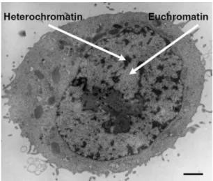
Figure 14: Human HeLa cell observed by electron microscopy. 34 Figure 15: Organization of pericentric heterochromatin into chromocenters in mouse cells. 35 Figure 16 : Schematical representation of pericentric heterochromatin in mammalian cells. 37
Figure 17: Breast cancer tumorigenesis. 40
Figure 18: The Elston & Ellis grading system in breast cancer. 40
Table II: HP1 nomenclature. 44
Figure 19: Constitutively expressed HP1 homologues in various species. 45
Figure 20: The three HP1 isoforms in human cells. 46
Figure 21: Schematical representation of the structural domains of HP1 proteins. 48 Table III : Major interacting partners of Drosophila and mammalian HP1 proteins and functional
implications. 52
Table IV: Phenotypes associated with depletion of HP1 proteins 56 Table V: Subunits of the CAF-‐1 complex in different species 62 Figure 22: Conserved domains within the subunits of CAF-‐1. 63 Figure 23: Cellular functions of the CAF-‐1 protein complex. 65
Table VI: Phenotypes of CAF-‐1 loss of function 67
Figure 24: CAF-‐1 levels are downregulated in quiescence and upregulated in breast cancer cells. 68
Figure 25: Structures of CPD and 6,4-‐PP lesions. 70
Figure 26: Schematical representation of the Nucleotide Excision Repair (NER) pathway. 72
Figure 27: Local UV irradiation and CAF-‐1 recruitment. 73
Table VII: Correlation between expression levels of genes in early-‐stage breast cancers 168 Figure 29: Involvement of HP1 in different nuclear processes. 170
List of abbreviations
6,4-‐PP: 6-‐4 pyrimidine-‐pyrimidone adducts Å: AngströmAsf1: Anti-‐silencing function 1 bp: base pairs
CAF-‐1: Chromatin Assembly Factor 1 CGH: Comparative Genomic Hybridization CHD: Chromodomain
CHO: Chinese Hamster Ovarian ChIP: Chromatin Immunoprecipitation CPD: Cyclobutane Pyrimidine Dimers CpG: Cytosine-‐phospho-‐Guanine CS: Cockayne syndrome CSD: Chromoshadowdomain DNA: Deoxyribonucleic acid DNMT: DNA Methyl Transferase DSB: Double Strand Break dsRNA: double stranded RNA E(var): Enhancer of variegation E2F: E2 promoting Factor ER: Estrogen receptor
EZH2: Enhancer of Zeste Homolog 2 FISH: Fluorescent In Situ Hybridization
FRAP: Fluorescence Recovery After Photobleaching
GGR: Global Genome Repair HAT: Histone Acetyl Transferase HD: Histone Demethylase HDAC: Histone Deacetylase
HER2: Human Epidermal Growth Factor Receptor-‐2
HIRA: Histone Regulator A
HMT: Histone Methyl Transferase HP1: Heterochromatin Protein 1 IF: Immunofluorescence KAP-‐1: KRAB associated protein 1 Kb: Kilobase
KO: Knock-‐out
KRAB: Kruppel-‐associated box M: Molar
Mb: Megabase
MBD: Methyl Binding Domain
MCM: Minichromosome Maintenance mRNA: messenger RNA
Nasp: Nuclear Autoantigenic Sperm Protein NER: Nucleotide Excision Repair
ORC: Origin Recognition Complex PCNA: Proliferating Cell Nuclear Antigen PEV: Position Effect Variegation PR: Progesteron Receptor
PRC: Polycomb Repressor Complex Rb: Retinoblastoma
RNA: Ribonucleic Acid RNAi: RNA interference
SAHF: Senescence Associated Heterochromatin Foci
siRNA: short interfering RNA Su(var): Suppressor of Variegation SV40: Simian Virus 40
TCR: Transcription-‐Coupled Repair TSA: Trichostatin A
TTD: Trichothiodystrophy UV: Ultra-‐Violet
XP: Xeroderma Pigmentosum
Abstract
In cancer cells, the organization of DNA in chromatin is frequently affected. Thus, it is crucial to understand how factors involved in chromatin organization contribute to tumorigenesis. Of particular interest in this context is the Chromatin Assembly Factor 1 (CAF-1). CAF-1 is a complex involved in chromatin assembly coupled to DNA synthesis, both during DNA replication and DNA repair. Two subunits of the CAF-1 complex are downregulated in quiescent cells and can be used as proliferation markers in cancer cells. In addition, CAF-1 has a role in the maintenance of the compact chromatin domains near the centromeres, called pericentric heterochromatin, through its interaction with Heterochromatin Protein 1 (HP1). Three HP1 isoforms (HP1α, β et γ) exist in mammalian cells, among which HP1α associates most specifically with pericentric heterochromatin regions.
During my PhD, I have addressed two main questions: First, how do the three
human HP1 isoforms relate to cell proliferation and tumorigenesis? Using both cell line
models and tumoral and healthy human tissue samples, I showed that the expression of HP1α isoform, but not HP1β ou γ, is proliferation-dependent. Downregulation of HP1α, specifically, results in mitotic defects. In addition, HP1α, but not HP1β ou γ, is overexpressed in several tumoral tissues compared to the corresponding healthy tissues. In breast cancer, the level of HP1α overexpression significantly correlates with global survival and the occurence of metastasis. Our results suggest that HP1α overexpression confers a growth advantage to tumor cells that is related to the organization of pericentric heterochromatin and the passage of mitosis. We put forward HP1α as new prognostic marker in breast cancer and potentially other cancers. This study gave rise to the deposition of a patent and a publication in EMBO Molecular Medecine.
The second main question I have addressed is: how do quiescent cells, which express
CAF-1 at very low levels, deal with chromatin assembly coupled to DNA repair? This
question is of particular interest since a differential repair capacity between tumoral (proliferative) cells and healthy (quiescent) cells will affect the efficiency and the toxicity of genotoxic cancer treatments, such as chemo- and radiotherapy Using ultra-violet (UV) irradiation as a means to induce DNA lesions, I was able to show that CAF-1 expression is not induced upon DNA damage and that the low levels of CAF-1 are sufficient for its recruitment to sites of UV lesions, suggesting that its function is conserved in quiescence. However, we observe a delayed repair of a specific type of UV lesions in quiescence. Our results suggest that the repair of these lesions might require additional chromatin dynamics, which would be the limiting step in quiescence, potentially due to the extremely low levels of CAF-1. These observations gave rise to a manuscript that is currently in preparation.
In these two major projects, I have demonstrated how factors involved in chromatin organization are related to cell proliferation, tumorigenesis and, more generally, genome stability. In addition, we have been able to put forward a new marker of clinical interest for breast cancer prognosis.
Résumé
Dans les cellules cancéreuses, des défauts affectant l’organisation d’ADN en chromatine sont fréquemment observés. L’étude de facteurs impliqués dans cette organisation est donc essentielle pour mieux appréhender leur implication dans la tumorigénèse. Un facteur particulièrement intéressant dans ce contexte est le facteur d’assemblage de la chromatine, le complexe CAF-1 (Chromatin Assembly Factor 1). CAF-1 est impliqué dans l’assemblage en chromatine de l’ADN lors de la réplication et la réparation de l’ADN. Deux sous-unités de CAF-1 sont sous-exprimés dans les cellules non-proliférantes (quiescentes) et constituent des marqueurs de prolifération dans le cancer. De plus, CAF-1 a un rôle au niveau des régions de chromatine dense proches des centromères, l’hétérochromatine péricentrique, par son interaction avec les protéines HP1 (Heterochromatin Protein 1). Il existe trois isoformes de HP1 dans les cellules mammaires (HP1α, β et γ), dont HP1α est le plus spécifiquement associé aux régions d’hétérochromatine péricentrique, impliqués dans la répression des gènes et la ségrégation des chromosomes.
Pendant ma thèse, je me suis penchée sur deux questions majeures: Premièrement,
est-ce que l’expression des isoformes de HP1 est régulée d’une façon dépendante de la prolifération et de la tumorigénèse ? En combinant des modèles de lignées cellulaires et des
échantillons de tissu humain, j’ai pu montrer que l’expression de l’isoforme HP1α, mais pas HP1β ou γ, est dépendante de la prolifération. La déplétion de HP1α, spécifiquement, affecte le passage de la mitose. De plus, HP1α, mais pas HP1β ou γ, est surexprimé dans de nombreux types de cancer comparé aux tissus sains correspondants. La surexpression de HP1α dans le cancer du sein est corrélée de façon significative à la survie des patientes et la formation de métastases. Ces résultats révèlent HP1α comme un marqueur pronostique dans le cancer du sein et potentiellement dans d’autres types de cancer. Nous proposons que la surexpression de HP1α présente un avantage sélectif pour les cellules cancéreuses, liée à l’organisation de l’hétérochromatine péricentrique et le passage de la mitose. Ces résultats ont donné lieu à un brevet et à une publication dans EMBO Molecular Medecine.
La seconde question à laquelle je me suis intéressée est : comment des cellules
quiescentes, qui expriment peu de CAF-1, gèrent l’assemblage de la chromatine couplé à la réparation de l’ADN ? En effet, une capacité de réparation différente entre cellules
proliférantes (tumorales) et quiescentes (saines) aura un impact majeur sur l’efficacité et la toxicité des traitements génotoxiques comme la chimio- et la radiothérapie. J’ai pu montrer que, dans les cellules quiescentes, les irradiations aux ultra-violets (UV) n’induisent pas l’expression de CAF-1. De plus, la faible quantité de CAF-1 est recrutée aux sites des lésions d’UV, suggérant que sa fonction dans la réparation est conservée hors du cycle cellulaire. Cependant, en quiescence, nous observons une réparation retardée d’un type spécifique de lésions, qui reflète potentiellement une difficulté des cellules quiescentes à gérer la prise en charge de ces lésions au sein de la chromatine. Ces résultats font l’objet d’un manuscrit actuellement en préparation.
Dans ces deux projets majeurs, j’ai mis en avant comment des facteurs de l’organisation de la chromatine peuvent être impliqués dans la prolifération, la tumorigénèse et la réparation d’ADN suite à des traitements génotoxiques. De plus, nous avons pu proposer un nouvel outil d’intérêt médical pour le pronostique des cancer du sein.
Preface
How to read beyond the genetic code?
The Human Genome Project (1990 – 2006) had for goal to sequence the human genome in order to reveal the secrets and the complexity of human beings. Although sequencing and analysis is still ongoing, it has become clear that the DNA sequence alone was not able to reveal all secrets of life. A human genome is estimated to contain fewer genes than those of many plants and human genes show very high similarity (up to 98%) with their homologues in apes. So what accounts for the differences that, as we like to think, would make us superior to apes and plants?
Even more intriguing are twins. Monozygotic twins possess the same genome; they are true clones of each other. Yet, they do show differences in phenotype, character and sensitivity to certain diseases. So what accounts for these differences that are not encoded in the genome?
Even within a single organism, most cells contain the same genome, which is formed from a paternal and a maternal set of chromosomes upon fertilization. Yet, many different cells with specialized functions and distinct tissue organizations can be formed out of one genome.
In conclusion, In addition to the genetic code, other sources of information are needed to account for these observations. This extra information would determine how to read the information encoded by DNA.
The term “epigenetics” has been put forward by Conrad Waddington in 1942 to designate “interactions between the genome and the environment that brings the phenotype into being”. It was based on the concept of epigenesis, put forward by Aristotle to explain how the complexity of an organism arises during development. More recently, the definition of epigenetics has been refined and is most often used to denote all those somatically heritable changes in gene expression that do not involve changes in
DNA sequence. The concept of heritability is of major importance and distinguishes
epigenetic changes can be transmitted through cell division and potentially even from one generation to the next.
Until recently, cancer was considered as a genetic disease: an accumulation of mutations, deletions or translocations that affect oncogenes and tumorsuppressor genes and that can ultimately lead to tumorigenesis (Hanahan & Weinberg, 2000). The American Society of Cancer actually still states: “Cells become cancer cells because of damage to DNA” (http://www.cancer.org). Yet, only a few types of cancer harbor mutations in the same genes among different patients (with the possible exception of the tumor suppressor gene p53) and genetic alterations alone cannot predict cancer prevalence or patient outcome. Thus, mechanisms that alter the usage of the genome, without affecting the genetic sequence, are thought to contribute to cancer initiation and progression. As an example, colon cancer is often characterized by a specific series of genetic alterations, affecting among others the gene MLH1. However, at least two cases have been identified in which this gene was not mutated but silenced by epigenetic mechanisms (Suter et al, 2004).
In this thesis, I will focus on epigenetic mechanisms in the context of cell proliferation, DNA damage and tumorigenesis, particularly breast cancer. Breast cancer is a heterogeneous disease, both clinically and genetically. Unlike colon cancer, many different genetic alterations can be found among breast cancer patients, and the correlation with the clinical characteristics are not evident. Several recent studies have compared in a genome-‐wide fashion the genomic alterations and the changes in gene expression in breast cancer samples (Valladares et al, 2006; Vincent-‐Salomon et al, 2008; Yao et al, 2006). They show that relatively few changes in gene expression can be attributed to genomic alterations. The recent development of high-‐throughput DNA sequencing techniques might allow the detection of single nucleotide polymorphisms or point mutations associated with familial breast cancer (reviewed in Stratton et al, 2009). Yet, the vast majority of familial and sporadic breast cancer is not related to mutations in high-‐risk genes and the contribution of epigenetic changes could thus to be of particular importance. How such changes arise and how they contribute to cancer development remains largely unknown.
In order to address these issues in a comprehensive manner, I will first introduce the best-‐known candidate for the transmission of epigenetic information: the
organization of DNA into chromatin. Next, I will discuss how chromatin organization can be affected in breast cancer and what remains to be learnt. I will then present in more detail two factors involved in chromatin organization: Heterochromatin Protein 1 (HP1) and Chromatin assembly factor 1 (CAF-‐1). This background information, provided in the introduction, will bring me to the questions that have intrigued me during my PhD: (i) Are the Heterochromatin Protein 1 isoforms related to cell proliferation and to breast tumorigenesis? and (ii) Are the chromatin dynamics that accompany DNA repair dependent on cell proliferation? Next, I will present published and unpublished results obtained during my PhD, which I will then discuss and put into a broader context in the discussion.
I. Introduction
1. Chromatin: a vector of information
1.1 Organization of DNA into chromatin
Walther Flemming used the term “chromatin” as early as 1882 to describe the structures that he observed in the nuclei of cells stained with basic dyes (Flemming, 1882). However, it wasn’t until the 20th century, with the exciting discovery that DNA
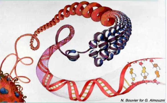
was the genetic material (Avery et al, 1944; Watson & Crick, 1953), that scientific attention focused on the molecular structure of the nucleus and the organization of DNA in chromatin in the nucleus. Chromatin is composed of DNA and a multitude of other molecules, among which are the histone proteins, and serves to organize, compact, and regulate DNA. Chromatin organization, as depicted in Figure 1, comprises multiple steps of compaction, which help to confine 2 meters of DNA in a cell nucleus of about 10 microns. How these different levels of chromatin organization are achieved will be the first focus of this chapter. Secondly, I will discuss the biological implications of this organization in normal cells and the changes encountered in cancer cells.
Figure 1: Artistic representation of chromatin organization. DNA is wrapped around histone
proteins (blue and white) to form the repeated unit of chromatin, the nucleosome. Nucleosomal arrays can adopt higher order levels of organization corresponding to nuclear domains in interphase nuclei (bottom left). Watercolour by N. Bouvier for G. Almouzni.
1.1.1 The nucleosome: basic unit of chromatin
The first level of chromatin organization and genome compaction consists of the formation of the basic repeated subunit of chromatin, the nucleosome (Kornberg, 1977; Oudet et al, 1975). The nucleosome consists of a core particle, in which 146bp of DNA turn 1.6 times around a histone octamer, and of linker DNA, which interconnects two nucleosomes. The length of the linker DNA varies depending on cell type and chromatin compaction levels (Kornberg, 1977).
The octamer of histone proteins is composed of an (H3-‐H4)2 tetramer flanked by two
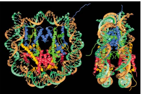
H2A-‐H2B dimers. Histone proteins are small, highly basic proteins with a high affinity for acidic DNA. They are characterized by the presence of histone fold domains that are highly conserved and form α-‐helices (Arents & Moudrianakis, 1995). The minimal histone fold domain comprises three α-‐helices connected by two loops. The 2.8Å resolution of the crystal structure of the core particle (Davey et al, 2002; Luger et al, 1997) revealed the detailed interactions both between different histones proteins and between histones and DNA (Figure 2).
Figure 2: Crystal structure of the nucleosome core particle at a resolution of 2.8Å. A human
repetitive DNA fragment and recombinant core histones were crystallized and their structure analyzed by X-ray crystallography. DNA: brown and turquoise; histone H3: blue; histone H4: green; histone H2A: yellow; histone H2B: red). Left panel: front view. Right panel: side view. Note that unstructured N-terminal tails of the histones protrude from the core particle and are thus accessible for protein interactions. Image from Luger et al, 1997.
In addition to the four histone proteins found in the core particle, a fifth type of histone protein can bind to the linker DNA. This family of linker histones, of which histone H1 is the best-‐known example, seems to bind a stretch of 15 to 20 nucleotides of DNA at the exit of the core particle, but is also in contact with DNA that is located within the nucleosomal core particle (Figure 3) (reviewed in Happel & Doenecke, 2009). The presence of histone H1 has been associated with the formation of a regularly spaced, higher order organization of nucleosomes (see below). Different variants of histone H1 exist, which are differentially regulated throughout differentiation and development (reviewed in Godde & Ura, 2008).
Figure 3: Schematical model of the binding of linker histone H1 to nucleosomal DNA. DNA is
represented in light grey. The 146bp that are part of the core particle are indicated. Histone H1, which binds the linker DNA that exits and enters the core particle, is depicted in dark grey. Image adapted from Thoma et al, 1979.
1.1.2 Higher order chromatin structure
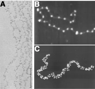
Arrays of nucleosomes, observed as “beads on a string” at low ionic strength using atomic force or electron microscopy (Figure 4) (Olins & Olins, 1974; Olins et al, 1980; Oudet et al, 1975), need to be further compacted for nuclear organization. The first level of compaction is promoted by the binding of linker histones. Indeed, in presence of linker histones, arrays of nucleosomes fold into a fiber with an approximate diameter of 30nm (Figure 4) (Thoma et al, 1979).
Figure 4: Nucleosomal arrays visualized as beads on a string. A. Chromatin fibers from
rat thymus nuclei, stained with
phosphotungstic acid and ethanol, as
observed by electron microscopy. Scale bar: 0.2 µm. Image from Olins & Olins, 1974. B&C. A glutaraldehyde-fixed chromatin fiber prepared from chicken erythrocytes as observed by scanning force microscopy. Linker histones have been removed (B) or not (C) with 0.35M NaCl. Images from Bustamante et al, 1997.
How the nucleosomes are exactly arranged within this fiber is a highly debated subject. Two models have been proposed (Figure 5) (reviewed in Robinson & Rhodes, 2006): (i) the one-‐start model, in which the array of nucleosomes forms a left-‐hand helix (Robinson et al, 2006), and (ii) the two-‐start model, in which the linker DNA connects nucleosomes back and forth in a zigzag manner (Dorigo et al, 2004). Recent studies using reconstituted nucleosome arrays suggest that both structures can be formed, depending on the length of the linker DNA (Kruithof et al, 2009; Routh et al, 2008). Indeed, shorter linker DNA length favors the formation of the more compact two-‐start model, while longer linker DNA lengths seem more compatible with the formation of a one-‐start helix (Kruithof et al, 2009; Routh et al, 2008). However, it should be kept in mind that the structures of reconstituted chromatin fibers as observed by microscopy are variable and highly dependent on the preparation techniques and the fixation method. The extent to which 30nm fibers can be formed in vivo remains unclear. An extreme view is that there may not be any fiber formation and that the nucleosomes instead form a disorganized “sea of nucleosomes” in vivo (Eltsov et al, 2008).
Figure 5: Schematic representation of the two models for the 30nm fiber folding. A.
The one-start model, in which the array of nucleosomes forms a left-handed helix. B. The two-start model, in which the linker DNA connects nucleosomes back and forth in a zigzag manner. Image from Robinson & Rhodes, 2006.
Further compaction levels ultimately lead to the most condensed form of chromatin found in mitotic chromosomes. The existence of long chromatin loops has been put forward in the context of DNA replication (Berezney et al, 2000), gene transcription (van Driel & Fransz, 2004) and DNA recombination (Kantidze & Razin, 2009), in order to explain the observed spatial distribution of these processes. Domains of approximately 1Mb would fold into ~100kb loops and help to compact and organize chromatin (Bode et al, 2003; Kantidze & Razin, 2009). Yet, to what extent such loops exist in vivo and contribute to higher order chromatin organization remains an open issue.
1.1.3 Nuclear chromatin organization
Many organisms display a non-‐random organization of chromatin in the interphase nucleus. Both in mammalian (reviewed in Cremer et al, 2006) and in yeast (Berger et al, 2008) nuclei, each chromosome occupies a distinct location (Figure 6). Although the localization of each chromosome is highly variable from one cell to another and seems to depend on cell type and proliferation status (Bolzer et al, 2005), a general picture is emerging in which chromosomes can be organized based on their size or their gene density. However, this organization is dynamic, and particularly highly expressed genes can be found to protrude outside of their chromosome territory (e.g. Mahy et al, 2002, Chambeyron et al, 2005 and Chambeyron & Bickmore, 2004). Thus, a certain degree of intermingling of chromosomes is thought to occur (Branco & Pombo, 2007). In addition, sub-‐nuclear compartments can be distinguished that contribute to gene
regulation and cell functioning. Cells from several types of cereals show a Rabl-‐ chromosome configuration, with centromeres at one side of the nucleus and telomeres at the other side (Shaw et al, 2002). In yeast, the telomere sequences are also localized at the nuclear envelope, where they form clusters (Gotta et al, 1996). In eukaryotes, the nuclear membrane is often associated with compact, repetitive and transcriptionally silent chromatin, called constitutive heterochromatin. Tethering a chromosomal region to the nuclear periphery can indeed modulate transcriptional regulation, both in terms of gene activation and gene repression (reviewed in Ruault et al, 2008). Thus, a picture emerges in which the nuclear organization seems to impact on transcriptional regulation and vice versa. Whether these effects are direct or indirect, and whether they follow a general rule, is a current subject of investigation.
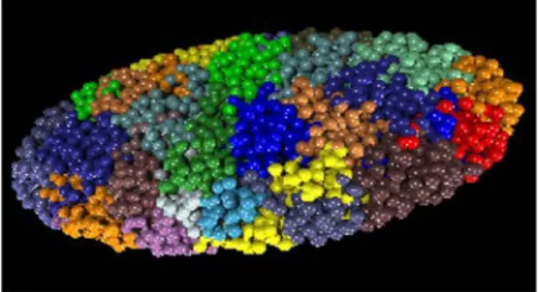
Figure 6: Chromosome territories in a human fibroblast. Computer simulation of the distribution of all
chromosomes in the nucleus of a human quiescent fibroblast, based on a chromatin painting experiment in which all chromosomes were visualized by fluorescent in situ hybridization. Adapted from Bolzer et al, 2005.
1.2 Chromatin dynamics: histone chaperones and histone variants
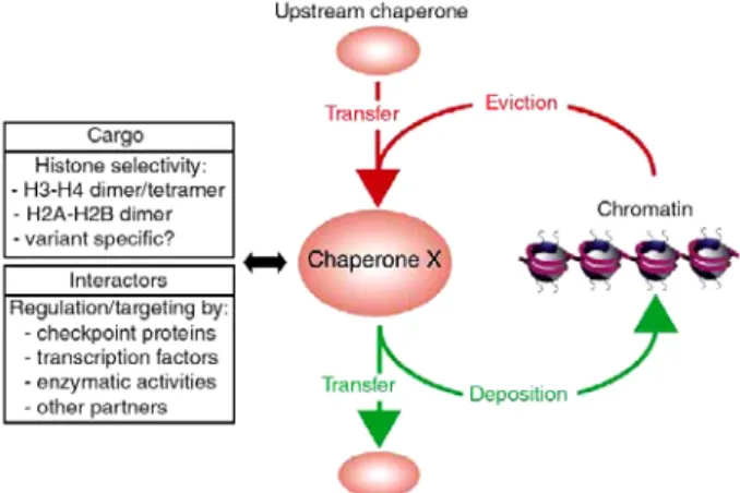
Histones are not permanently associated with DNA. Newly synthesized histones have to be transported to the nucleus and targeted to the required location, whereas old or damaged histones have to be discarded. In addition, cellular processes involving DNA can require transient histone eviction and replacement (Loyola & Almouzni, 2004). As histones are highly basic proteins, their presence in the cell could lead to deleterious effects. Thus, when histones are not in association with DNA, they are bound to dedicated proteins called histone chaperones (De Koning et al, 2007, see appendix 1). These escort proteins, defined as “factors that associate with histones and stimulate a reaction involving histone transfer, without being part of the final product”, help to control histone supply and incorporation into chromatin. This implies that histone chaperones are implicated in all cellular processes that operate on chromatin, like transcription, replication and repair (Corpet & Almouzni, 2009; Groth et al, 2007b; Polo & Almouzni, 2006). They interact with an intricate network of different partners to regulate histone traffic (Figure 7).
Figure 7: Schematic representation of the relationship between a given histone chaperone and its network of interacting partners. A given chaperone X will show
specificity towards certain histone dimers, or potentially tetramers, which it can receive from, or transfer to, other histone chaperones. Alternatively, it can contribute to histone eviction from or deposition on DNA. Its activity can be regulated by interacting partners involved in various cellular processes. Image from De Koning et al, 2007.
To identify factors that can promote histone deposition, cell-‐free systems have been developed. The first system allowing the study of chromatin assembly was derived from Xenopus egg extracts (Laskey et al, 1977). These egg extracts contain all factors needed for chromatin assembly during the rapid rounds of DNA replication that occur in early development. Incubation of these extracts with circular template DNA from the Simian Virus 40 (SV40) (Laskey et al, 1977) or with circular single stranded
bacteriophage DNA (Almouzni & Mechali, 1988a; Almouzni & Mechali, 1988b) results in chromatin assembly independent from or coupled to DNA synthesis, respectively. This model system allowed the identification of the most abundant histone H2A-‐H2B chaperone in Xenopus eggs, nucleoplasmin (Laskey et al, 1978). The further development of a system using a combination of human cytosolic and nuclear extracts and biochemical fractionation (Stillman, 1986) helped to identify Chromatin Assembly Factor 1 (CAF-‐1), a chaperone promoting chromatin assembly coupled to DNA synthesis (Smith & Stillman, 1989).
Histone chaperones can be distinguished based on their specificity towards distinct histones or histone variants. We have proposed a classification of currently known histone chaperones, according to their histone preference and whether they act alone, within a chaperone complex or within an enzymatic complex (De Koning et al, 2007, appendix 1). A first level of specificity is based on whether they bind either to H3-‐ H4 or H2A-‐H2B, with several chaperones preferentially binding to specific isoforms (variants) of histones H3 or H2A. Indeed, among the histone proteins, we can distinguish replicative histone variants, which are expressed and deposited mainly during S-‐phase, and replacement variants, which are constitutively expressed and incorporated in a replication independent fashion. As an example, CAF-‐1 shows specificity towards the replicative variant H3.1, while the histone chaperone histone regulator A (HIRA) shows specificity for the replacement variant H3.3. Elegant studies using Xenopus egg extracts and in vitro chromatin assembly have shown that whilst CAF-‐1 promotes the deposition of H3.1-‐H4 in a manner coupled to DNA synthesis, HIRA does so for H3.3-‐H4 independently of DNA synthesis (Tagami et al, 2004) (figure 8). Anti-‐silencing function 1 (Asf1), a factor that cannot promote histone deposition on its own in vivo (Mello et al, 2002; Ray-‐Gallet et al, 2007) but rather acts as a histone donor for CAF-‐1 and HIRA in histone deposition (Polo & Almouzni, 2006), can interact with both H3.1-‐H4 and H3.3-‐ H4. However, only the Asf1a isoform can interact with HIRA (Tagami et al, 2004; Zhang et al, 2005), thus providing an indirect specificity for H3.3-‐H4 deposition. However, it should be kept in mind that the chaperone-‐histone interaction is not always a strict specific interaction, as exemplified by Nuclear Autoantigenic Sperm Protein (Nasp). Nasp was first reported to co-‐purify with the linker histone H1 (Richardson et al, 2000; Wang et al, 2008a), but was more recently found to interact with H3-‐H4 (Tagami et al, 2004). This may not be so surprising since Nasp is the homologue of the H3-‐H4
chaperone N1/N2 in Xenopus. Thus, histone binding preferences should be taken with caution and may be subject to regulation, depending on the cellular context and binding partners.
Figure 8: Chromatin assembly in a replication-dependent or -independent fashion. CAF-1
deposits H3.1-H4 in a replication-coupled fashion, while HIRA specifically deposits H3.3-H4 in a manner independent from DNA synthesis. Asf1 serves as a histone donor for both CAF-1 and HIRA and can interact with both H3.1 and H3.3. However, only the Asf1a isoform interacts with HIRA, while both Asf1a and b can interact with CAF-1. Image from De Koning et al, 2007.
1.3 Chromatin organization as a source of information
In the previous chapters, we have discussed how DNA is organized into chromatin and how the basic unit of this organization, the nucleosome, is formed. Here, I will address how this organization can be a possible source of epigenetic information, i.e. contribute to a heritable status of gene silencing and expression that contributes to cell identity and behavior.
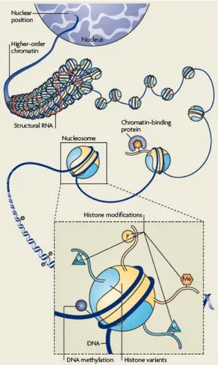
Chromatin organization is dynamic and can show a large repertoire of molecular characteristics, or marks, which contribute to different levels of organization (Figure 9). The combination of these marks, rather than the presence or absence of a single mark, is thought to constitute the motif that can, for example, impact on the accessibility and dynamics of a given part of the genome.
Figure 9: Schematic representation of potential epigenetic marks. At all levels of
chromatin organization, form the DNA up to the nuclear position, marks and variants can change and contribute to the accessibility and dynamics of the genome. Image from Probst et al, 2009.
1.3.1 DNA methylation
First, the DNA component of chromatin can be methylated. In mammalian cells, this methylgroup is almost exclusively found on the symmetrical CpG (cytosine-‐ phospho-‐guanine) dinucleotides. These CpG dinucleotides are not distributed uniformly across the genome, but are grouped into so-‐called CpG islands, which often correspond to gene promoter regions. A high fraction of methylated CpGs within such islands is generally associated with gene silencing. Specific enzymes, named DNA Methyl Transferases (DNMTs) ensure the establishment and maintenance of DNA methylation. Several types of DNMTs are found in mammalian cells. In general, Dnmt3a and b, together with their co-‐factor Dnmt3L, are involved in de novo DNA methylation, while Dnmt1 is involved in the maintenance of the methylation mark through S-‐phase (Figure 10). Indeed, upon DNA replication, the newly synthesized strand is unmethylated, thus resulting in a hemi-‐methylated CpG dinucleotide. For transmission of the mark, the new strand needs to acquire the same marks as the mother strand. Dnmt1 can be recruited to the DNA replication fork through its interaction with the polymerase accessory factor Proliferating Cell Nuclear Antigen (PCNA) (Chuang et al, 1997). However, the dominant manner of Dnmt1 recruitment seems to be through its interaction with UHRF1, a protein that binds specifically hemi-‐methylated DNA and is crucial for the maintenance of methyl marks (reviewed in Prokhortchouk & Defossez, 2008) and (Probst et al, 2009). Since the methyl mark can be transmitted from mother to daughter cells, DNA methylation is considered as a bona fide epigenetic mark.
Figure 10: Schematic representation of DNA methylation in mammalian cells. Dnmt3a and b are
de novo methyl transferases, which can establish a silent chromatin status. DNA replication results in
hemi-methylated DNA. Dnmt1 can restore metylation patterns by methylating the newly synthesized strand. Image from Issa, 2004.
In the absence of Dnmt1 activity, passive DNA demethylation would occur when cells go through multiple cell divisions. For a long time, this was thought to be the only mechanism to erase DNA methyl marks in mammalian cells. Recently, however, mechanisms have been proposed that might enable the removal of DNA methylation in
vivo, which involve the transformation of the methylated cytosine into a mark that can
be recognized and excised by DNA damage repair pathways (Gehring et al, 2009; Ma et al, 2009). Thus, DNA methylation in mammals might be a less stable mark than initially thought. Interestingly, in Drosophila, DNA methylation seems restricted to a short time window in early development and does not necessarily correlate with gene silencing (reviewed in Mandrioli & Borsatti, 2006).
1.3.2 Histone variants
At the level of the nucleosome, different isoforms of histones H3 and H2A, called variants, can be incorporated (reviewed in Loyola & Almouzni, 2007; Polo & Almouzni, 2006; Pusarla & Bhargava, 2005). Two types of histone variants can be distinguished (Table I) (Osley, 1991). Replicative histone variants, of which the synthesis and the deposition peak in S-‐phase, supply the required amount of histones to ensure chromatin
assembly during DNA replication. Replacement variants, which are expressed at steady state levels throughout the cell cycle and in quiescence, provide a constitutive source for histone turnover or dynamics. The centromere specific histone H3 variant CENPA (Centromere Protein A) is an exceptional case, since its expression peaks in G2 (Shelby et al, 2000) and its deposition occurs during a short time window at the exit of mitosis in mammalian cells (Dunleavy et al, 2009; Foltz et al, 2009; Jansen et al, 2007).
Table I: Somatic Mammalian Histone Variants
Histone type Variant Specificity/function
H2A replicative H2A.1
Not determined H2A.2
replacement H2AX DNA repair signaling
H2AZ Regulation of transcription
macroH2A Inactivation of one X chromosome in female
H2ABbd Active chromatin, nulceosome destabilization
H3 replicative H3.1 deposition coupled to DNA synthesis
H3.2 deposition coupled to DNA synthesis, generally
associated with repressive chromatin replacement
H3.3 deposition independent of DNA synthesis, generally
associated with active chromatin
CENPA centromere structure and function
Histone variants often show high similarity in amino acid sequence, as exemplified by histone H3.1 and H3.3, which only differ by 5 amino acids. Nevertheless, the choice of the histone variant is of importance for genome function and structure. Indeed, most histone variants are not distributed uniformly over the genome, but show specificity for certain chromatin regions, as summarized in Table I. For example, H3.3 dynamics are most pronounced at promoters, enhancers and transcriptionally active genes (Mito et al, 2005) and the presence of H3.3 seems to facilitate gene expression by forming unstable nucleosomes that can be easily disrupted (Jin & Felsenfeld, 2007). In addition to the somatic histone variants presented in table I, tissue specific histone variants have been identified, such as the testis-‐specific variant H3.1t (Witt et al, 1996) that differs in four amino acids from canonical H3.1. The presence of such variant might reflect an adaptation to particular cellular demands in terms of histone levels and dynamics during rapid proliferation and/or development.
Interestingly, the presence of multiple histone variants has arisen during evolution. The yeast S. cerevisiae contains only one histone H3 variant, which is most closely related to mammalian H3.3. In organisms such as plants, flies and frogs, an
)@@+-+.*):! '8E:+3)-+.*Q@8E8*@8*-! F)'+)*-\! ;+5+:)'! -.! ?#%"\! 4);! 8F.:F8@%! &*:D! 5)55):;! 8PE'8;;! -V.! @+//8'8*-! '8E:+3)-+.*Q@8E8*@8*-! F)'+)*-;\! ?#%$! )*@! ?#%"%! 748! +*3'8);+*(! *95=8'! ./! 4+;-.*8! F)'+)*-;! -4'.9(4.9-! 8F.:9-+.*! +;! -4.9(4-! -.! E)')::8:! -48! +*3'8);+*(! *93:8)'!3.5E:8P+-D\!'8d9+'8@!-.!.E-+5+,8!)*@!@+F8';+/D!(8*.58!9;)(8%!
!"9"9#61,2+'&#-+8131)021+',#
2*! +5E.'-)*-! ;.9'38! ./! +*/.'5)-+.*! +;! E'.F+@8@! =D! )*! 8P-8*;+F8! E)*8:! ./! E.;-Q -')*;:)-+.*):!5.@+/+3)-+.*;!./!4+;-.*8!E'.-8+*;!gL+(9'8!$$h%!748;8!5.@+/+3)-+.*;!+*3:9@8! )38-D:)-+.*;\! 58-4D:)-+.*;\! E4.;E4.'D:)-+.*;! )*@! 9=+d9+-+*)-+.*;! )*@! 5)+*:D! 3.*38'*! )5+*.! )3+@;! +*! -48! 1Q-8'5+*):! -)+:;! ./! 4+;-.*8! E'.-8+*;\! V4+34! E'.-'9@8! /'.5! -48! *93:8.;.58!)*@!)'8!-48'8/.'8!5.;-!)338;;+=:8%!!
!!
Figure 11: Scheme of histone modifications. Histones modifications include
acetylation (ac), methylation (me), phosphorylation (ph) and ubiquitination (ub1). Most modifications occur on the N-terminal tails of histones, with some exceptions involving the C-terminal histone tails or their globular domains, here depicted as colored ovals. Image from Bhaumik et al, 2007.
Y-!4);!=88*!E'.E.;8@!-4)-!4+;-.*8!5.@+/+3)-+.*;!3.9:@!=8!+*F.:F8@!=.-4!+*!;4.'-Q -8'5! 38::! ;+(*):+*(! )*@! +*! 48'8@+-)'D! :.*(Q-8'5! '8(9:)-+.*;%! &*:D! +*! -48! :)--8'! 3);8\! 4+;-.*8! 5.@+/+3)-+.*;! 3)*! =8! 3.*;+@8'8@! );! -'98! 8E+(8*8-+3! 5)'R;! g79'*8'\! "BB"W! 79'*8'\! "BB>h! gL+(9'8! $"h%! ?.V! ;E83+/+3! 4+;-.*8! 5.@+/+3)-+.*;! 3)*! E.;;+=:D! =8! -')*;5+--8@!-4'.9(4!012!'8E:+3)-+.*!)*@!5+-.;+;!+;!39''8*-:D!)*!+5E.'-)*-!d98;-+.*!+*! -48!/+8:@!gC.'E8-!f!2:5.9,*+\!"BBHW!M'.=;-!8-!):\!"BBHh%!! ! !
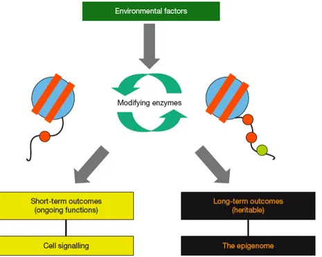
Figure 12: Histone modifications and epigenetics. Signals from the environment may induce
certain histone modifications. These modifications can have short-term outcomes in ongoing processes, such as transcription, and thus serve in cell signalling. Alternatively, these modifications can have long-term, hereditary, effects on chromatin organization and thus contribute to the epigenetic state of a cell. Scheme from Turner, 2007.
While particular combinations of modifications have been associated with activation of gene transcription, others are implicated in gene silencing. Different histone variants are associated with different post-‐translational modifications, of which some are already partially present before histone incorporation into chromatin (reviewed in Loyola & Almouzni, 2007). Importantly, it is the combination of multiple histone modifications within one or several nucleosome(s), rather than one signal modification, that will be decisive for gene activity. This view has been put forward as the “histone code” (Jenuwein & Allis, 2001; Turner, 2000; Turner, 2002). In line with this view, several modifications are known to “cross-‐talk”, either within the same histone or between different histones (Suganuma & Workman, 2008). For example, the presence of phosphorylation on serine 10 of histone H3 (H3S10P) seems to render inaccessible the trimethylation of the neighboring lysine 9 of histone H3 (H3K9me3) (Duan et al, 2008; Fischle et al, 2005; Hirota et al, 2005), while H3K9me3 has been proposed to be required for the establishment of trimethylation on lysine 20 of histone H4 (H4K20me3), at least in established mouse embryonic fibroblasts cell lines (Schotta et al, 2004).