HAL Id: dumas-02952342
https://dumas.ccsd.cnrs.fr/dumas-02952342
Submitted on 29 Sep 2020
HAL is a multi-disciplinary open access
archive for the deposit and dissemination of
sci-entific research documents, whether they are
pub-lished or not. The documents may come from
teaching and research institutions in France or
abroad, or from public or private research centers.
L’archive ouverte pluridisciplinaire HAL, est
destinée au dépôt et à la diffusion de documents
scientifiques de niveau recherche, publiés ou non,
émanant des établissements d’enseignement et de
recherche français ou étrangers, des laboratoires
publics ou privés.
Distributed under a Creative Commons Attribution - NonCommercial - ShareAlike| 4.0
International License
Prognostic value and time course evolution of left
ventricular Global Longitudinal Strain in septic shock:
an exploratory prospective study
Florian Bazalgette
To cite this version:
Florian Bazalgette. Prognostic value and time course evolution of left ventricular Global Longitudinal
Strain in septic shock: an exploratory prospective study. Human health and pathology. 2018.
�dumas-02952342�
UNIVERSITE DE MONTPELLIER
FACULTE DE MEDECINE MONTPELLIER-NIMES
Thèse
Présentée et soutenue publiquement
Le 5 octobre 2018
Par
Florian BAZALGETTE
Pour obtenir le titre de DOCTEUR EN MÉDECINE
Titre
Valeur pronostique et évolution temporelle du strain global longitudinal du
ventricule gauche au cours du choc septique, résultats d'une étude prospective
Prognostic value and time course evolution of left ventricular Global Longitudinal Strain in
septic shock: An exploratory prospective study
Directeur de thèse : Dr Claire ROGER
Président :
Professeur Jean Yves LEFRANT
Professeur des Universités – Praticien Hospitalier
Assesseurs :
Professeur Pascal COLSON
Professeur des Universités – Praticien Hospitalier
Professeur Guillaume CAYLA
Professeur des Universités – Praticien Hospitalier
Docteur Claire ROGER
Praticien Hospitalier
Membre invité :
Professeur Laurent MULLER
3
ANNEE UNIVERSITAIRE 2017 - 2018
PERSONNEL ENSEIGNANT
Professeurs Honoraires ALLIEU Yves ALRIC Robert ARNAUD Bernard ASTRUC Jacques AUSSILLOUX Charles AVEROUS Michel AYRAL Guy BAILLAT Xavier BALDET Pierre BALDY-MOULINIER Michel BALMES Jean-Louis BALMES Pierre BANSARD Nicole BAYLET René BILLIARD Michel BLARD Jean-Marie BLAYAC Jean Pierre BLOTMAN Francis BONNEL François BOUDET CharlesBOURGEOIS Jean-Marie BRUEL Jean Michel BUREAU Jean-Paul BRUNEL Michel CALLIS Albert
DIMEGLIO Alain DU CAILAR Jacques DUBOIS Jean Bernard DUMAS Robert DUMAZER Romain ECHENNE Bernard FABRE Serge
FREREBEAU Philippe GALIFER René Benoît GODLEWSKI Guilhem GRASSET Daniel GROLLEAU-RAOUX Robert GUILHOU Jean-Jacques HERTAULT Jean HUMEAU Claude JAFFIOL Claude JANBON Charles JANBON François JARRY Daniel JOYEUX Henri LAFFARGUE François LALLEMANT Jean Gabriel LAMARQUE Jean-Louis LAPEYRIE Henri LESBROS Daniel MEYNADIER Jean MICHEL François-Bernard MICHEL Henri MION Charles MION Henri MIRO Luis NAVARRO Maurice NAVRATIL Henri OTHONIEL Jacques PAGES Michel PEGURET Claude POUGET Régis PUECH Paul PUJOL Henri PUJOL Rémy RABISCHONG Pierre RAMUZ Michel RIEU Daniel RIOUX Jean-Antoine ROCHEFORT Henri
ROUANET DE VIGNE LAVIT Jean Pierre
SAINT AUBERT Bernard SANCHO-GARNIER Hélène SANY Jacques
4
CANAUD Bernard CASTELNAU Didier CHAPTAL Paul-André CIURANA Albert-Jean CLOT Jacques D’ATHIS Françoise DEMAILLE Jacques DESCOMPS BernardLOPEZ François Michel LORIOT Jean LOUBATIERES Marie Madeleine MAGNAN DE BORNIER Bernard MARY Henri MATHIEU-DAUDE Pierre SERRE Arlette SIMON Lucien SOLASSOL Claude THEVENET André VIDAL Jacques VISIER Jean Pierre
Professeurs Emérites ARTUS Jean-Claude BLANC François BOULENGER Jean-Philippe BOURREL Gérard BRINGER Jacques CLAUSTRES Mireille DAURES Jean-Pierre DAUZAT Michel DEDET Jean-Pierre ELEDJAM Jean-Jacques GUERRIER Bernard JOURDAN Jacques MAURY Michèle MILLAT Bertrand MARES Pierre MONNIER Louis PRAT Dominique PRATLONG Francine PREFAUT Christian PUJOL Rémy ROSSI Michel SULTAN Charles TOUCHON Jacques VOISIN Michel ZANCA Michel
5
Professeurs des Universités - Praticiens Hospitaliers
PU-PH de classe exceptionnelle
ALBAT Bernard - Chirurgie thoracique et cardiovasculaire
ALRIC Pierre - Chirurgie vasculaire ; médecine vasculaire (option chirurgie vasculaire) BACCINO Eric - Médecine légale et droit de la santé
BASTIEN Patrick - Parasitologie et mycologie BONAFE Alain - Radiologie et imagerie médicale CAPDEVILA Xavier - Anesthésiologie-réanimation COMBE Bernard - Rhumatologie
COSTA Pierre - Urologie
COTTALORDA Jérôme - Chirurgie infantile COUBES Philippe - Neurochirurgie
CRAMPETTE Louis - Oto-rhino-laryngologie
CRISTOL Jean Paul - Biochimie et biologie moléculaire DAVY Jean Marc - Cardiologie
DE LA COUSSAYE Jean Emmanuel - Anesthésiologie-réanimation DELAPORTE Eric - Maladies infectieuses ; maladies tropicales
DE WAZIERES Benoît - Médecine interne ; gériatrie et biologie du vieillissement, médecine générale, addictologie
DOMERGUE Jacques - Chirurgie générale DUFFAU Hugues - Neurochirurgie
DUJOLS Pierre - Biostatistiques, informatique médicale et technologies de la communication ELIAOU Jean François - Immunologie
FABRE Jean Michel - Chirurgie générale GUILLOT Bernard - Dermato-vénéréologie
HAMAMAH Samir-Biologie et Médecine du développement et de la reproduction ; gynécologie médicale HEDON Bernard-Gynécologie-obstétrique ; gynécologie médicale
HERISSON Christian-Médecine physique et de réadaptation JABER Samir-Anesthésiologie-réanimation
JEANDEL Claude-Médecine interne ; gériatrie et biologie du vieillissement, médecine générale, addictologie
JONQUET Olivier-Réanimation ; médecine d’urgence
JORGENSEN Christian-Thérapeutique ; médecine d’urgence ; addictologie KOTZKI Pierre Olivier-Biophysique et médecine nucléaire
6
LANDAIS Paul-Epidémiologie, Economie de la santé et Prévention LARREY Dominique-Gastroentérologie ; hépatologie ; addictologie LEFRANT Jean-Yves-Anesthésiologie-réanimation
LE QUELLEC Alain-Médecine interne ; gériatrie et biologie du vieillissement, médecine générale, addictologie
MARTY-ANE Charles - Chirurgie thoracique et cardiovasculaire MAUDELONDE Thierry - Biologie cellulaire
MERCIER Jacques - Physiologie MESSNER Patrick - Cardiologie MOURAD Georges-Néphrologie
PELISSIER Jacques-Médecine physique et de réadaptation
RENARD Eric-Endocrinologie, diabète et maladies métaboliques ; gynécologie médicale REYNES Jacques-Maladies infectieuses, maladies tropicales
RIBSTEIN Jean-Médecine interne ; gériatrie et biologie du vieillissement, médecine générale, addictologie
RIPART Jacques-Anesthésiologie-réanimation ROUANET Philippe-Cancérologie ; radiothérapie SCHVED Jean François-Hématologie; Transfusion TAOUREL Patrice-Radiologie et imagerie médicale UZIEL Alain -Oto-rhino-laryngologie
VANDE PERRE Philippe-Bactériologie-virologie ; hygiène hospitalière YCHOU Marc-Cancérologie ; radiothérapie
PU-PH de 1re classe
AGUILAR MARTINEZ Patricia-Hématologie ; transfusion AVIGNON Antoine-Nutrition
AZRIA David -Cancérologie ; radiothérapie
BAGHDADLI Amaria-Pédopsychiatrie ; addictologie BEREGI Jean-Paul-Radiologie et imagerie médicale
BLAIN Hubert-Médecine interne ; gériatrie et biologie du vieillissement, médecine générale, addictologie
BLANC Pierre-Gastroentérologie ; hépatologie ; addictologie BORIE Frédéric-Chirurgie digestive
BOULOT Pierre-Gynécologie-obstétrique ; gynécologie médicale CAMBONIE Gilles -Pédiatrie
CAMU William-Neurologie CANOVAS François-Anatomie
7
CARTRON Guillaume-Hématologie ; transfusion
CHAMMAS Michel-Chirurgie orthopédique et traumatologique COLSON Pascal-Anesthésiologie-réanimation
CORBEAU Pierre-Immunologie
COSTES Valérie-Anatomie et cytologie pathologiques COURTET Philippe-Psychiatrie d’adultes ; addictologie CYTEVAL Catherine-Radiologie et imagerie médicale DADURE Christophe-Anesthésiologie-réanimation DAUVILLIERS Yves-Physiologie
DE TAYRAC Renaud-Gynécologie-obstétrique, gynécologie médicale DEMARIA Roland-Chirurgie thoracique et cardio-vasculaire
DEMOLY Pascal-Pneumologie ; addictologie DEREURE Olivier-Dermatologie - vénéréologie DROUPY Stéphane -Urologie
DUCROS Anne-Neurologie -
FRAPIER Jean-Marc-Chirurgie thoracique et cardiovasculaire KLOUCHE Kada-Réanimation ; médecine d’urgence
KOENIG Michel-Génétique moléculaire LABAUGE Pierre- Neurologie
LAFFONT Isabelle-Médecine physique et de réadaptation LAVABRE-BERTRAND Thierry-Cytologie et histologie LECLERCQ Florence-Cardiologie
LEHMANN Sylvain-Biochimie et biologie moléculaire LUMBROSO Serge-Biochimie et Biologie moléculaire
MARIANO-GOULART Denis-Biophysique et médecine nucléaire MATECKI Stéfan -Physiologie
MEUNIER Laurent-Dermato-vénéréologie MONDAIN Michel-Oto-rhino-laryngologie MORIN Denis-Pédiatrie
NAVARRO Francis-Chirurgie générale
PAGEAUX Georges-Philippe-Gastroentérologie ; hépatologie ; addictologie PETIT Pierre-Pharmacologie fondamentale ; pharmacologie clinique ; addictologie
PERNEY Pascal-Médecine interne ; gériatrie et biologie du vieillissement, médecine générale, addictologie
PUJOL Jean Louis-Pneumologie ; addictologie PUJOL Pascal-Biologie cellulaire
8
PURPER-OUAKIL Diane-Pédopsychiatrie ; addictologie
QUERE Isabelle-Chirurgie vasculaire ; médecine vasculaire (option médecine vasculaire) SOTTO Albert-Maladies infectieuses ; maladies tropicales
TOUITOU Isabelle-Génétique TRAN Tu-Anh-Pédiatrie
VERNHET Hélène-Radiologie et imagerie médicale
PU-PH de 2ème classe
ASSENAT Éric-Gastroentérologie ; hépatologie ; addictologie BERTHET Jean-Philippe-Chirurgie thoracique et cardiovasculaire BOURDIN Arnaud-Pneumologie ; addictologie
CANAUD Ludovic-Chirurgie vasculaire ; Médecine Vasculaire CAPDEVIELLE Delphine-Psychiatrie d'Adultes ; addictologie CAPTIER Guillaume-Anatomie
CAYLA Guillaume-Cardiologie
CHANQUES Gérald-Anesthésiologie-réanimation
COLOMBO Pierre-Emmanuel-Cancérologie ; radiothérapie COSTALAT Vincent-Radiologie et imagerie médicale
COULET Bertrand-Chirurgie orthopédique et traumatologique CUVILLON Philippe-Anesthésiologie-réanimation
DAIEN Vincent-Ophtalmologie
DE VOS John-Cytologie et histologie DORANDEU Anne-Médecine légale -
DUPEYRON Arnaud-Médecine physique et de réadaptation
FESLER Pierre-Médecine interne ; gériatrie et biologie du vieillissement, médecine générale, addictologie
GARREL Renaud -Oto-rhino-laryngologie GAUJOUX Viala Cécile-Rhumatologie GENEVIEVE David-Génétique
GODREUIL Sylvain-Bactériologie-virologie ; hygiène hospitalière GUILLAUME Sébastien-Urgences et Post urgences psychiatriques -
GUILPAIN Philippe-Médecine Interne, gériatrie et biologie du vieillissement ; addictologie GUIU Boris-Radiologie et imagerie médicale
HAYOT Maurice-Physiologie
9
JACOT William-Cancérologie ; Radiothérapie JUNG Boris-Réanimation ; médecine d'urgence KALFA Nicolas-Chirurgie infantile
KOUYOUMDJIAN Pascal-Chirurgie orthopédique et traumatologique LACHAUD Laurence-Parasitologie et mycologie
LALLEMANT Benjamin-Oto-rhino-laryngologie
LAVIGNE Jean-Philippe-Bactériologie-virologie ; hygiène hospitalière LE MOING Vincent-Maladies infectieuses ; maladies tropicales LETOUZEY Vincent-Gynécologie-obstétrique ; gynécologie médicale LOPEZ CASTROMAN Jorge-Psychiatrie d'Adultes ; addictologie LUKAS Cédric-Rhumatologie
MAURY Philippe-Chirurgie orthopédique et traumatologique MILLET Ingrid-Radiologie et imagerie médicale
MORANNE Olvier-Néphrologie MOREL Jacques -Rhumatologie
NAGOT Nicolas-Biostatistiques, informatique médicale et technologies de la communication NOCCA David-Chirurgie digestive
PANARO Fabrizio-Chirurgie générale
PARIS Françoise-Biologie et médecine du développement et de la reproduction ; gynécologie médicale PASQUIE Jean-Luc-Cardiologie
PEREZ MARTIN Antonia-Physiologie
POUDEROUX Philippe-Gastroentérologie ; hépatologie ; addictologie PRUDHOMME Michel-Anatomie
RIGAU Valérie-Anatomie et cytologie pathologiques RIVIER François-Pédiatrie
ROGER Pascal-Anatomie et cytologie pathologiques ROSSI Jean François-Hématologie ; transfusion ROUBILLE François-Cardiologie
SEBBANE Mustapha-Anesthésiologie-réanimation SEGNARBIEUX François-Neurochirurgie
SIRVENT Nicolas-Pédiatrie
SOLASSOL Jérôme-Biologie cellulaire SULTAN Ariane-Nutrition
THOUVENOT Éric-Neurologie THURET Rodolphe-Urologie
10
VILLAIN Max-Ophtalmologie
VINCENT Denis -Médecine interne ; gériatrie et biologie du vieillissement, médecine générale, addictologie
VINCENT Thierry-Immunologie
WOJTUSCISZYN Anne-Endocrinologie-diabétologie-nutrition
PROFESSEURS DES UNIVERSITES 1re classe :
COLINGE Jacques -Cancérologie, Signalisation cellulaire et systèmes complexes
2ème classe :
LAOUDJ CHENIVESSE Dalila - Biochimie et biologie moléculaire VISIER Laurent - Sociologie, démographie
PROFESSEURS DES UNIVERSITES - Médecine générale 1re classe :
LAMBERT Philippe
2ème classe :
AMOUYAL Michel
PROFESSEURS ASSOCIES - Médecine Générale DAVID Michel
RAMBAUD Jacques
PROFESSEUR ASSOCIE - Médecine BESSIS Didier - Dermato-vénéréologie)
PERRIGAULT Pierre-François -Anesthésiologie-réanimation ; médecine d'urgence ROUBERTIE Agathe – Pédiatrie
Maîtres de Conférences des Universités - Praticiens Hospitaliers
MCU-PH Hors classe
11
CARRIERE Christian-Bactériologie-virologie ; hygiène hospitalière CHARACHON Sylvie-Bactériologie-virologie ; hygiène hospitalière
FABBRO-PERAY Pascale-Epidémiologie, économie de la santé et prévention
HILLAIRE-BUYS Dominique-Pharmacologie fondamentale ; pharmacologie clinique ; addictologie PELLESTOR Franck-Cytologie et histologie
PUJOL Joseph-Anatomie
RAMOS Jeanne-Anatomie et cytologie pathologiques RICHARD Bruno-Thérapeutique ; addictologie RISPAIL Philippe-Parasitologie et mycologie
SEGONDY Michel-Bactériologie-virologie ; hygiène hospitalière STOEBNER Pierre -Dermato-vénéréologie
MCU-PH de 1re classe
ALLARDET-SERVENT Annick-Bactériologie-virologie ; hygiène hospitalière BADIOU Stéphanie-Biochimie et biologie moléculaire
BOUDOUSQ Vincent-Biophysique et médecine nucléaire BOULLE Nathalie-Biologie cellulaire
BOURGIER Céline-Cancérologie ; Radiothérapie BRET Caroline -Hématologie biologique COSSEE Mireille-Génétique Moléculaire GABELLE DELOUSTAL Audrey-Neurologie
GIANSILY-BLAIZOT Muriel-Hématologie ; transfusion GIRARDET-BESSIS Anne-Biochimie et biologie moléculaire LAVIGNE Géraldine-Hématologie ; transfusion
LE QUINTREC Moglie-Néphrologie
MATHIEU Olivier-Pharmacologie fondamentale ; pharmacologie clinique ; addictologie MENJOT de CHAMPFLEUR Nicolas-Neuroradiologie
MOUZAT Kévin-Biochimie et biologie moléculaire PANABIERES Catherine-Biologie cellulaire
PHILIBERT Pascal-Biologie et médecine du développement et de la reproduction RAVEL Christophe - Parasitologie et mycologie
SCHUSTER-BECK Iris-Physiologie
STERKERS Yvon-Parasitologie et mycologie
TUAILLON Edouard-Bactériologie-virologie ; hygiène hospitalière YACHOUH Jacques-Chirurgie maxillo-faciale et stomatologie
12
MCU-PH de 2éme classe
BERTRAND Martin-Anatomie
BRUN Michel-Bactériologie-virologie ; hygiène hospitalière DU THANH Aurélie-Dermato-vénéréologie
GALANAUD Jean Philippe-Médecine Vasculaire GOUZI Farès-Physiologie
JEZIORSKI Éric-Pédiatrie
KUSTER Nils-Biochimie et biologie moléculaire
LESAGE François-Xavier-Médecine et Santé au Travail MAKINSON Alain-Maladies infectieuses, Maladies tropicales
MURA Thibault-Biostatistiques, informatique médicale et technologies de la communication OLIE Emilie-Psychiatrie d'adultes ; addictologie
THEVENIN-RENE Céline-Immunologie
MAITRES DE CONFERENCES DES UNIVERSITES - Médecine Générale COSTA David
FOLCO-LOGNOS Béatrice
MAITRES DE CONFERENCES ASSOCIES - Médecine Générale CLARY Bernard GARCIA Marc MILLION Elodie PAVAGEAU Sylvain REBOUL Marie-Catherine SEGURET Pierre
MAITRES DE CONFERENCES DES UNIVERSITES
Maîtres de Conférences hors classe
BADIA Eric - Sciences biologiques fondamentales et cliniques
Maîtres de Conférences de classe normale BECAMEL Carine - Neurosciences
BERNEX Florence - Physiologie
CHAUMONT-DUBEL Séverine - Sciences du médicament et des autres produits de santé CHAZAL Nathalie - Biologie cellulaire
13
GUGLIELMI Laurence - Sciences biologiques fondamentales et cliniques HENRY Laurent - Sciences biologiques fondamentales et cliniques
LADRET Véronique - Mathématiques appliquées et applications des mathématiques LAINE Sébastien - Sciences du Médicament et autres produits de santé
LE GALLIC Lionel - Sciences du médicament et autres produits de santé
LOZZA Catherine - Sciences physico-chimiques et technologies pharmaceutiques MAIMOUN Laurent - Sciences physico-chimiques et ingénierie appliquée à la santé MOREAUX Jérôme - Science biologiques, fondamentales et cliniques
MORITZ-GASSER Sylvie - Neurosciences MOUTOT Gilles - Philosophie
PASSERIEUX Emilie - Physiologie RAMIREZ Jean-Marie - Histologie TAULAN Magali - Biologie Cellulaire
PRATICIENS HOSPITALIERS UNIVERSITAIRES CLAIRE DAIEN-Rhumatologie
BASTIDE Sophie-Epidémiologie, économie de la santé et prévention FAILLIE Jean-Luc-
Pharmacologie fondamentale ; pharmacologie clinique ; addictologie GATINOIS Vincent-Histologie, embryologie et cytogénétique
HERLIN Christian -Chirurgie plastique ; reconstructrice et esthétique ; brûlologie HERRERO Astrid-Chirurgie générale
PANTEL Alix-Bactériologie-virologie ; hygiène hospitalière
PERS Yves-Marie-Thérapeutique, médecine d’urgence ; addictologie
PINETON DE CHAMBRUN Guillaume-Gastroentérologie ; hépatologie ; addictologie TORRE Antoine-Gynécologie-obstétrique ; gynécologie médicale
14
REMERCIEMENTS
A MES MAITRES
Le Professeur Jean Yves LEFRANT,
Je vous remercie de me faire l’honneur de présider ce jury de thèse. Des premiers jours de mon internat
à ma dernière garde d’interne dont vous étiez le sénior, des conseils scientifiques aux métaphores
sportives, vous avez toujours été encourageant et bienveillant.
Le Professeur Pascal COLSON,
Votre présence dans ce jury de thèse est un grand honneur. Merci de m’avoir supporté et trainé dans les
couloirs du DAR D dès l’entente de l’acronyme E.C.M.O.
Merci de votre aide et votre soutien dans l’élaboration du projet de simulation qui m’est cher. Merci enfin
de vos conseils professionnels. Vous êtes un des rares à nous apprendre la patience et l’humilité.
Le Professeur Guillaume CAYLA,
Votre présence dans ce jury de thèse est un grand honneur. L’insuffisance cardiaque ainsi que les
techniques d’assistance cardiaques ont pour moi un grand intérêt. Après avoir embolisé les couloirs du
DAR D et de l’USIC montpelliérains, j’espère que nous aurons l’occasion de travailler ensemble à ce sujet
avec je l’espère une grande fluidité.
Le Professeur Laurent MULLER,
Merci Laurent, merci pour tout : tes conseils, ton humanité envers les patients, ton aide dans l’élaboration
de ce travail…
Tes cours resteront pour moi des moments qui ont marqué mon internat. Travailler à tes cotés va être un
immense honneur.
Le Docteur Claire ROGER,
Je te dois énormément, des premiers conseils naïfs de début d’internat aux avis téléphoniques plus précis
de remplacements ou de gardes seniorisées. Merci de ton aide dans l’élaboration de la thèse. Merci
également pour le magnifique dévouement dont tu fais preuve chaque jour pour l’équipe de réanimation
chirurgicale.
Merci également au Docteur Aurélien DAURAT pour ton aide dans l’élaboration de ce travail. Il faut
s’arrêter à cinq membres de jury sans quoi cela aurait été un honneur de te compter comme membre.
15
Merci au Docteur Benjamin LOUART pour sa contribution au travail d’analyse statistique de cette thèse.
Merci Professeur JABER pour votre expérience en réanimation vos enseignements.
Merci Professeur CAPDEVILA pour la coordination du DES, pour vos enseignements et pour ces quelques
gardes passées ensemble le lundi.
AU MONDE DE LA CARDIOLOGIE POUR AVOIR ACCUEILLI UN ANESTHESISTE DANS LEUR MUR
Merci à Jean, Etienne et Camille pour m’avoir enseigné les rudiments de l’ETT.
Merci à François ROUBILLE pour m’avoir accueilli 6 mois à l’USIC. Ta rigueur et ta capacité de travail
resteront pour moi impressionnantes.
Merci à Kamila et Myriam, les chefs de cliniques qui ont accompagné ce stage.
Merci aux autres PH : Delphine, Fred, Jean Christophe et Stéphane, merci pour vos visites alliant rigueur
et plaisanterie.
Et un GRAND MERCI à mes co-internes. Jean, ta gentillesse et ta bonne humeur mise à rude épreuve par
cette allergie au téléphone. Leslie dit la Ba_ _ _ K, ton pragmatisme et ta rigueur ont su eux résister au
téléphone (ou presque). Merci à tous les deux pour votre coaching en cardio. David puis Pablo, mes
compagnons non cardiologues suspectés et surveillés comme moi à la moindre modification du Bisoce,
merci pour votre gentillesse et votre bonne humeur.
AUX PERPIGNANAIS
Pastou, IADE emblématique, Celestin BRU pour ton diplôme d’intubation.
Sébastien KOLLEN et Jean Marie QUINTARD, merci de votre gentillesse et de vos conseils. Vous êtes à
l’origine de mon intérêt pour l’ECMO, comme vous le dites, vous m’avez fait « tomber dedans quand
j’étais petit ».
A tous les autres PH qui ont marqué mon passage dans la région : Laurent, Arnaud, Vincent, ODM, JAT,
Florence, Olivier, Philippe, PGD, Frank, Aline.
AUX NIMOIS
Merci aux ARC pour tout ce que vous avez fait pour m’aider.
Guillaume, ton pragmatisme, ta facilité à simplifier les choses… et les conseils brico..
Pascal, désormais à PGS, tu fais partie des quelques personnes à avoir profondément marqué mon
internat.
Pierrot, merci pour cet intérêt commun à l’ECMO, merci pour ta bienveillance.
Robert, merci pour ton expérience transmise chaque jour.
Patrice, en cinq ans d’internat et de multiples lieux de stages, je n’ai pas trouvé meilleur repas le weekend.
Steph, merci pour ton aide dans sepsistrain.
16
Merci à toute l’équipe paramédicale, je ne peux citer tout le monde : Delphine, Jérôme, Didier, Marina,
Pierrette, Babette, Isabelle, Mag, Ursula, Françoise….
Merci aux équipes du Bloc et tout particulièrement la team Mater modifiée depuis par les différents
mercatos : Jean Michel, Mathieu, Cathy, Béa, Laurent.
A L’EQUIPE DE SIMULATION
Laurent, merci de nous avoir bercé tout petit, et de peu à peu nous laisser voler seul. Olivier et Domi,
merci de votre accueil et de votre aide. Encore un grand merci au Pr Colson d’avoir cru en nous et nous
avoir soutenu. Merci à Meriem, Norddine, Fabien, le noyau dur, d’avoir fait en sorte que ce projet de
simulation se concrétise. Merci à tous les autres qui de près ou de loin, de Nîmes et de Montpellier nous
ont aidés et soutenus (Pierre, Geoffroy, Jean Claude, Julien, Fred).
A L’EQUIPE DU DAR D QUI M’A BEAUCOUP EU DANS LES PATTES
Philippe, merci pour ton expérience que tu nous transmets chaque jour.
Jacob, merci pour ta bonne humeur, pour ton énorme expérience en assistance, et …. pour ta participation
sans rechigner aux soirées.
Nordine, je te dois énormément, merci pour ton aide, ton écoute, tes conseils à persévérer pour faire ce
qu’il plait. Et sans oublier ces discussions de gardes qui ont quand même était la base du projet de
simulation.
Marc merci pour ta bonne humeur, pour ton expérience que tu partages avec plaisir.
Meriem, je te dois énormément et sur beaucoup de points, merci pour tout : tes enseignements, la
simulation, les avis mais également ta gentillesse et ta disponibilité.
Gégé, merci pour tes enseignements en échographie, et comme Jacob ta participation indéfectible aux
soirées.
Merci également à tous les autres anesthésistes avec qui j’ai moins partagé mais qui m’ont également
beaucoup apporté : Michelle, Philippe, Denis, Marion, Pierre, Marine.
ET UN GRAND merci aux perfus : Jean Claude, merci pour ta légendaire bonne humeur ta soif de
transmettre et cette passion pour ton métier. Fabien, merci pour ces heures passées à bricoler ce
simulateur, ta gentillesse, ta disponibilité. Fred, merci pour cette bonne humeur que tu nous transmets,
tes conseils et ta disponibilité. Julien, la force tranquille, le dernier arrivé, merci de ton aide pour la
simulation.
17
A LA REANIMATION MEDICALE
Merci à toute l’équipe de la réanimation médicale pour leur accueil, leur écoute … et parfois ces longs
débats, et ces observ infinissables … Professeur JONQUET, Professeur KLOUCHE et Professeur JUNG,
Vincent, Philippe, Laura, Noémie, Delphine, Liliane, Corine, Aurel, Sonia, Valérie.
Merci à mes co-internes qui ont égayé ce stage. Merci à Yassir, Mélanie, Mathieu, Max, Benoit, Ileana,
Fanny, Féie, Cédric.
Merci également à tant d’autres dans les différents DAR qui m’ont beaucoup apporté : Olivier en ALR
Jonathan en traumato, Anne Cha, Christophe, Pablo, Isa, Olivier, Laurent, Christelle, Maud, Christine en
pédiatrie.
A L’EQUIPE DE LA CLINIQUE DES FRANCISCAINES,
Merci pour leur excellent accueil.
Guillaume, merci pour ces longues discussions, tes enseignements, ta disponibilité, ton enthousiasme.
David, merci pour ta gentillesse, ton efficacité dans le travail, et cette foi en l’EBM.
Damien, merci pour cette rigueur, ces conseils et ta disponibilité.
Rick, merci d’avoir accepté de faire cobaye en simulation d’ECMO.
Antoine, les Olivier, Jean François, Isabelle, Monsieur Julien pour votre expérience, votre sens clinique
que vous m’avez transmis.
Toufic, merci pour ta gentillesse, ton humanisme en réanimation.
Et à tous les autres que je ne cite pas.
A MA FAMILLE
A mes parents,
La loyauté, la servitude, la dévotion pour son prochain sont les mots qui vous caractérisent. Je vous admire
tous les jours. Si je vis cette aventure de médecine, c’est intégralement grâces à vous. La ténacité à
toujours m’expliquer que si les autres y arrivent, pourquoi pas moi. Ces valeurs humanistes que vous
m’avez enseignées, je ne les oublierai jamais.
A Elsa, ma chérie,
Parfois je me demande comment tu supportes les inconvénients inhérents à notre métier. D’abord l’ECN
puis l’internat avec ses gardes et maintenant ce rôle de senior avec les responsabilités et les doutes qui
lui incombent, ces repas au silence pesant lorsque l’on ressasse une prise en charge à tort ou à raison
discutable… Merci pour ton soutien, tu me rends plus fort, tu nous rends plus fort. Merci pour ton aide
dans la correction de ce travail.
18
A mes ainés qui ne sont plus là,
Ma grand-mère, je t’ai peu connue mais là encore les valeurs de travail, fierté, robustesse et générosité
été présentes.
Franc et Henriette : je ne saurais jamais si c’est la sagesse de l’âge mais vous avez été d’un soutien
indéfectible ne marquant jamais en ma présence l’ombre d’un doute à la réussite d’un quelconque
examen et par voie de fait ne marquant aucune surprise à l’annonce de sa réussite mais un simple « je le
savais ».
A ma tante et mon oncle,
Je vous dois également beaucoup, soutien de toujours. Des noëls passés en famille, à l’aide dans les
travaux d’aménagement, vous avez toujours été présents. Votre histoire, faite de joies et de tristesses,
les valeurs de famille, de travail et de bienveillance que vous portez sont pour moi des piliers importants.
A mes cousins,
Laurent, que de souvenirs dans l’insouciance de l’adolescence, ces parties de braconnage, ces journées
d’hiver s’entrainant au pilotage sur neige. A Sophie qui partage sa vie et le rend heureux.
Sophie, ma cousine, avec qui je garde le souvenir d’une enfance heureuse, complice et insouciante. La vie
ne nous a malheureusement pas permis de nous connaitre adulte mais tu gardes une place importante
dans mon cœur.
Marine, ma cousine montpelliéraine. Merci pour ces discussions que l’on peut avoir. Tu as connu mes
doutes et mes joies. Je sais que je peux compter sur toi.
A mes petits cousins TITI et Margaux.
A Simone GEMINARD, je te dois énormément. Je n’oublierai jamais ce que tu as fait pour moi. Le concours
de première année, c’est en grande partie à toi que je le dois. Tu as fait preuve de générosité, tu m’as
appris la persévérance, comment canaliser mes doutes, comment aller à l’essentiel. Je crois bien qu’il
s’agit de la clef indispensable de la réussite.
Merci également à tous les autres membres de ma famille qui comptent beaucoup. Je ne peux citer tout
le monde.
A MES AMIS D’ENFANCE
Pierre, Laure, Elena, Christophe, Seb et John. De l’école primaire à ce jour, vous avez été constamment
présents. Vous avez toujours été là pour moi malgré les kilomètres et les inconvénients du métier. Je ne
l’oublierai jamais. Les valeurs de travail, de servitude, d’amitié qui nous ont été enseignées dans la petite
19
enfance et que vous me rappelez à chacun de mes retours aux pays fond partie de mes piliers. Les (rares)
sorties à moto, les soirées sont également des moments de décompression inestimables.
A MES AMIS D’EXTERNATS
Corentin, merci pour ta droiture légendaire, tes valeurs, tes racines campagnardes. Le tableau serait
presque parfait si tu n’étais pas ardéchois.
Peete, merci pour ces délires, ta gentillesse, ta disponibilité et notre complicité. Même si tes racines
catalanes t’on rappelées à l’ordre et éloignées de la maison mère, j’espère que l’on se verra
régulièrement.
Thomas Roland (TR), merci pour ces soirées inoubliables.
Mathieu, Simon, Najib, Arnal, J, Guiz et Greg, merci pour tous ces moments passés ensemble.
Mathilde, Julie et Léa, merci pour votre soutien lors de la D4. Merci de votre générosité.
A MES AMIS D’INTERNAT
Yassir, merci pour ta disponibilité, ces discussions nocturnes interminables…
Merci à tous mes autres copains de promo : Geoffrey, Tim , Lucie , Yoyo, Guigui, Céline, Julien, Simon ,
Severin, Clément , Guillaume, Benjamin , Habib et Maxime. Merci pour ces semestres passés ensemble,
pour ces séminaires et pour votre soutien.
Kevin, Clément, Jerem, Jean Clément et Medhi, pas la même génération mais vous êtes brillamment sortis
du 10ml/kg de vT. Merci les potos, ce stage au DAR D et vos conseils de premiers remplacements, je ne
les oublierai pas.
Et merci à tous les autres qui ont compté pour moi et que je ne peux citer, mes coexternes de Montpellier,
tous mes co-internes dans les différents services, tous mes chefs de cliniques, les infirmiers, secrétaires
et aides-soignants …
20
SOMMAIRE
INTRODUCTION: ... 21 METHODS ... 23 Study design ... 23 Ultrasound protocol ... 23 Statistical Analysis ... 24 RESULTS ... 25 Patient’s characteristics ... 25Global longitudinal strain... 25
Natural history ... 25
Comparison of GLS value in apical 2, 4 chambers views and APLAX view. ... 25
Prognostic value ... 26
DISCUSSION ... 27
Key findings: ... 27
Relationship with previous literature: ... 27
Clinical implications: ... 29 Study limitations: ... 29 CONCLUSION ... 30 REFERENCES ... 31 TABLES ... 34 FIGURES ... 39 PERMIS D’IMPRIMER ... 45 ABSTRACT ... 46
21
Prognostic value and time course evolution
of left ventricular Global Longitudinal Strain in septic shock:
An exploratory prospective study
INTRODUCTION:
Septic cardiomyopathy (SCM) is observed in up to 40% of patients with septic shock (1). This
clinical entity has been firstly described by Parker in the 80’s, who suggested that left ventricle
ejection fraction (LVEF) was paradoxically higher in non-survivors of septic shock (2). Subsequent
studies did not confirm these results and a meta-analysis did not show any link between LVEF
and mortality in septic population(3).
The main mechanism for high LVEF is profound inflammatory-induced vasoplegia, which
decreases cardiac afterload and artificially increases LVEF. Severe vasoplegia and high dose of
vasopressors requirement is associated to
poor outcome, leading to refractory shock (4). Thus,
LVEF seems too dependent on loading conditions to be a reliable marker of intrinsic myocardial
contractility in septic shock (5)
(6). It could be hypothesized that other echocardiographic markers
of intrinsic contractility could be interesting to detect SCM. The use of conventional
echocardiography is now widely recommended at bedside for non-invasive hemodynamic
assessment in intensive care unit (ICU) patients, but the clinical usefulness of myocardial strain
assessment remains unclear(7) (8).
Real-time speckle-tracking imaging is now widely available on echocardiography machine at
bedside. This technology allows to assessing myocardial deformation over the cardiac cycle.
Myocardial deformation is quantified by measuring global longitudinal strain (GLS) as an index of
systolic function. In cardiology, this techniques have been validated after comparison with gold
standard techniques such as magnetic resonance imaging
(9). Myocardial longitudinal strain
assessment is interesting due to a fast learning curve, an automatized measurement and a good
inter and intra observer reproducibility
(10)(11)
(12)
.
Initial experimental and humans data
suggest that initial GLS could be a better and earlier marker of SMC than LVEF (13)
(6). The impact
of GLS alterations on prognosis remains debated. An altered GLS value at admission (> - 13 %)
has been shown to independently predict mortality in septic shock (14) (15)
whereas one study
22
did not confirm the association between GLS and prognosis (16). Two issues remain unclear
about strain in ICU. First, the evolution of GLS over time can be of clinical importance but has
been poorly described
(16). Second, the possibility to simplify strain assessment in ICU patients
seems necessary as previous reports have suggested a feasibility ranging from 42% to 77%
(6)(17).
This variable feasibility is related to difficult assessment of GLS because of limited echogenicity
for anterior wall in 2 and 3 chambers views (18). Theoretically, GLS should be assessed in 2, 3 and
4 chambers views and subsequently averaged. This is difficult and time consuming in ICU
patients, even if automatic real-time measurement simplifies the procedure.
To bring answers to the two last issues, the present study was aimed at:
1) Describing the natural history of left ventricle (LV) GLS during septic shock.
2) Evaluating the correlation between average LV GLS measured in apical two, three or four
chambers and a single value of LV GLS recorded in a single 4 chamber view (Single
Longitudinal Strain, SLS), which is easier and more rapid to assess. We hypothesized that
SCM is a global heart disease and that there would be no difference between average LV
GLS and SLS.
23
METHODS
Study design
A prospective, observational cohort study was conducted in a single medico-surgical ICU of the
Nimes University hospital from February 2016 to march 2018(19).
According to the French law, the study protocol was approved by the local Ethics Committee
(IRB/16.02.07) that waived written inform consent. Written information was given to the patient
or his/her relatives who could refuse to participate.
All patients aged ≥ 18 years admitted for a diagnostic of septic shock were enrolled in the present
study. Septic shock was defined by clinical evidence of sepsis with persisting hypotension
requiring vasopressors to maintain MAP ≥65 mm Hg and a serum lactate level >2 mmol/L
(18mg/dL) despite adequate volume resuscitation, according to current guidelines
(20).
Non-inclusion criteria were age < 18 years, pregnancy, severe valvular disease, ischemic heart
disease with known past history of ischemic sequelae, others cardiomyopathies, atrial fibrillation,
patients in whom LV walls could not be evaluated through standard apical views.
The following variables were recorded: age, gender, body mass index, past medical history, septic
origin, vasopressor doses, mechanical ventilation use, ICU length of stay and 30 days mortality
(21). The severity of organ failure was assessed with the simplified acute physiology score (SAPS)
II at admission (22).
All patients were treated according to international guidelines septic shock management
(23).
Ultrasound protocol
The echocardiographic examination was firstly performed during the first 24 hours after ICU
admission (T1) and repeated once between day 3 and day 5 (T2) then once between day 6 and
day 8 (T3).
All echocardiographic exams were performed by an expert in echocardiography using a Vivid S70
ultrasound device (GE Healthcare). Two-dimensional apical two-chamber, four-chamber and
long-axis views (2C, 4C and ALAX) were recorded. Speckle tracking analysis was performed during
a post treatment analysis after recording the echocardiographic loops. Briefly, analysis of LV
myocardial deformation is performed from 2-dimensional gray-scale loops by automatic tracking
of myocardial speckles after manual selection of landmark points using apical views of the left
ventricle. GLS was calculated as the average of negative peak of longitudinal strain from each of
24
the 18 LV segments from the 2C, 4C and ALAX views. Formula:
GLS = (Ls - Ld) / Ld) (annex 1)
(where Ls= systolic length Ls= diastolic Length)
The main objective of the present study was to report the course of GLS throughout sepsis.
Therefore we compare GLS with LVEF in the overall population and in two distinct population:
LVEF ≥45% and LVEF < 45%.
Single Longitudinal Strain (SLS) was defined as longitudinal strain measured on 4 chambers view
Left ventricle volumes and LVEF were calculated using the Simpson’s method
(24). The E and A
waves velocities and E wave deceleration time were measured using pulsed wave (PW) Doppler
in the mitral inflow at the tip of the valve visualized by an apical 4-chambers view. Early diastolic
tissue wave velocity of the lateral mitral annulus (E’ wave velocity) was measured in the apical
4C view using PW tissue Doppler. Then we calculated the E/E’ ratio. Velocity Time Integral (VTI)
of the left ventricular outflow tract we measured on an apical five-chambers view. For the right
ventricular (RV) evaluation the dilatation is assessed in four chambers with focus on the right
ventricle
(25). The longitudinal contractility was assessed by the tricuspid annular plane systolic
excursion (TAPSE) by using M mode on a 4-chambers view.
At the time of echocardiographic examination, were also recorded clinical parameters: heart
rate, systolic, diastolic, and mean blood pressure, as well as biologic parameters: pH, pO
2, pCO
2,
base excess, bilirubin, lactates, troponins, platelets and creatinine.
Statistical Analysis
Descriptive statistics were reported using median and interquartile range (25th to 75th
percentile) for continuous variables. For categorical variables, frequencies and proportions were
given. To assess the change between T1 and T2 for the average GLS, the LVEF, the PAS, the heart
rate, lactates, mitral E wave, E/Ea ratio and the sub aortic VTI, we used a paired Student's t-test.
To study the relation between several covariates at baseline (average GLS, LVEF, MAP, heart rate,
lactates, SAPSII, sex, age and troponin) and survival we used a Cox proportional hazards model.
The hazard ratio was computed for each variable in an univariate way. Finally, to assess the
agreement between average GLS and four-chamber GLS we used the Bland and Altman method
by computing the mean difference and its 95% confidence interval and limits of agreement
defined as mean difference ± 1.96 SD. A p value of 0.05 was considered as significant. All
statistical analysis were performed using R software (version 3.3.2).
25
RESULTS
Patient’s characteristics
Among 40 initially consecutive patients, 3 patients were excluded: two patients had previous
ischemic cardiomyopathy with anterior sequelae and one patient had an atrial fibrillation. Table
1 summarizes the main clinical characteristics of 37 included patients. All patients reached the
targeted mean arterial blood pressure > 65 mmHg as usually recommended (8). No patient was
lost for follow up. No patient died during study protocol. Nine patients (24.3%) died during the
month after ICU admission. The site of infection is shown in Table 1.
Hemodynamic and biological parameters: (Tables 2 and 3)
Between T1 and T2 exams, a decrease in heart rate over time was observed, and increases in
systolic blood pressure and pulse pressure. Considering the echocardiography parameters, ITV
increased between T1 and T2 whereas others parameters remained stable (Table 2).
Table 3 shows the biological parameters with a decrease in arterial lactate and an increase in
base excess between T1 and T2
Global longitudinal strain
Natural history
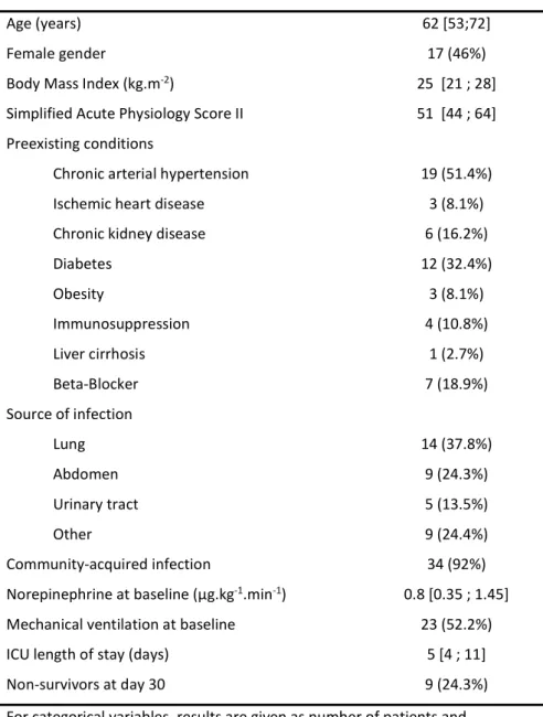
GLS could be calculated in 36, 23 and 9 patients at T1, T2 and T3, respectively. Baseline GLS was
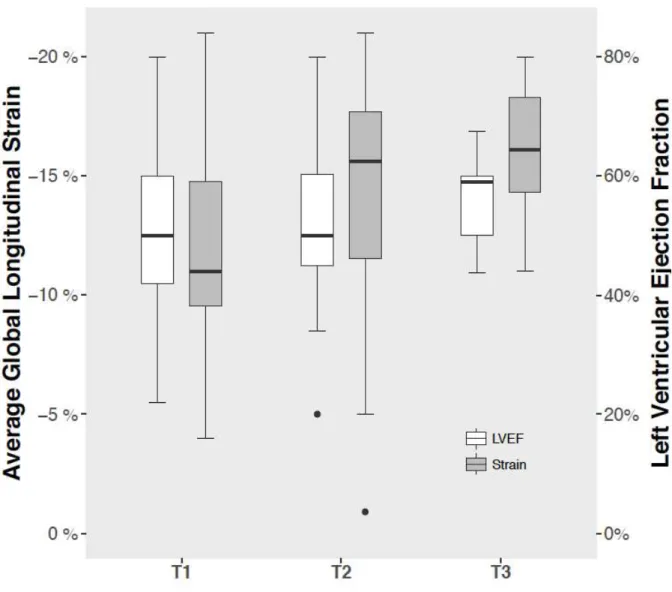
impaired (> -13%) in 23 patients (65.7%). In overall population GLS, improved throughout the
study whereas FEVG did not change (figure1).
In the subgroup of patients with LVEF <45% (7 patients), LVEF and GLS evolution over time was
similar (Figure 2a and 2b). The initial GLS was impaired in low LVEF Group compared to LVEF≥45%
group (-9.6 vs -13.3 %, p = 0.01). In this sub group, both GLS and LVEF significantly improved at
T2 (GLS: -9.6 vs -14.3 % p = 0.008) (LVEF: 29% vs 50 % p=0.01) (Figure 2a 2b).
Comparison of GLS value in apical 2, 4 chambers views and APLAX view.
The comparison between mean GLS values in 2, 3 and 4 chambers view and the averaged GLS
are reported in Figure 3. The Bland and Altman method shows a good agreement between GLS
and SLS: For T1 exams (n= 35) mean of differences -0.04 [-0.7; 0.62]; For T2 exams (n=23) mean
of differences 0.1 [-0.96 ; 1.16 ] Figure4 a et 4b.
26
Prognostic value
The course of GLS in survivors and non-survivors is shown in table 4. Only Initial GLS alteration
was significantly associated with 30-day mortality rate (-9.1 in non-survivors at day 30 vs -11 %
in survivors OR 1,44 [1.13; 1.85], p 0,004).
27
DISCUSSION
Key findings
Global longitudinal strain was altered at day 1 of septic shock and significantly improved over
time, until normalization. On the opposite, conventional methods for LV systolic function
assessment as LVEF and S wave velocity were not affected by the natural course of sepsis. As a
second result, GLS values were comparable whatever the echocardiographic view (2, 4 chambers
and APLAX views) considered, suggesting that a single measurement in 4 chambers view (SLS) is
sufficient at bedside. Finally, our study confirms that GLS alterations at day 1 are associated with
poor outcome.
Relationship with previous literature
Septic cardiomyopathy has been well reported during septic shock(26). Recent data suggest that
diastolic dysfunction is more frequent and more associated with prognosis than systolic
dysfunction (27) (28). Moreover, supranormal systolic function (hyperkinetic state, defined as an
LVEF > 60 %) is associated with worse prognosis (6)
.
Systolic function is usually assessed by
echocardiography, by measuring conventional LVEF. Diastolic function is assessed by E’ wave
velocity recording (16). Global longitudinal strain has two theoretical advantages over
conventional TTE myocardial function assessment. First, GLS is correlated to both systolic and
diastolic function during sepsis (16). Second, GLS can be altered while LVEF remains preserved.
Therefore, GLS could be a better and earlier indicator of systolic dysfunction than LVEF. In an
experimental model of E coli-induced sepsis in anesthetized and ventilated piglets, Hestenes et
al
(13) reported a significant linear alteration of LV GLS after E coli infusion (from -17.2% ± 2.8%
to -12.3% ± 3.2%, p = 0.04) on a study period of 240 minutes. Right ventricle GLS was also
significantly altered (from -24.2% ± 4.1% to -16.9% ± 5.7%, p = 0.02). In this model, LVEF remained
unaltered (from 48% ± 7% to 49% ± 5% over the study period, p = 0.4). The conclusion was that
GLS helps at assessing myocardial dysfunction during sepsis while LVEF assessment may
underestimate this diagnosis. Analogous results have been obtained in humans. The evolution of
strain has been studied in the first day of sepsis and septic shock (29). At baseline, GLS was
abnormal both in sepsis and septic shock patients (-15 %). At 24 hours, GLS was significantly
impaired only in septic shock patients (- 13% at H 24) but unchanged in sepsis group (- 17% at H
24), suggesting a more profound myocardial impairment during septic shock than during sepsis
and a link between severity and GLS. In this study, in contrast to GLS, LVEF remained normal in
both groups. This study showed that rapid and profound change in GLS value can occur during
the first 24 hours of septic shock, without concomitant alterations of LVEF. In our study, patients
28
were mainly analyzed at the end of the first 24 hours of septic shock, explaining why a very poor
value of GLS (- 11.7 %) was reported at day 1, close to value reported by Shahul et al (29). In a
case control study involving 62 septic shock, Ng et al showed that septic patients had a greater
degree of myocardial dysfunction measured by GLS as compared to non-septic patients (-14.5 %
vs -18.3 %, p <0.001, respectively)(30). In this study, LVEF was normal and not different between
the two groups (59 % in the sepsis group vs 61 % in the control group, P = 0.169). In a large study
involving 132 septic patients, GLS was altered in 70% of patients, whatever the value of LVEF (6).
Similarly, our results confirm that GLS is altered during sepsis whereas LVEF remains normal as
well as S wave velocity, a classical marker of systolic function, which is not altered over time in
the present study (Table 2). Additionally, the present results show that, in the whole population,
GLS improves over time while LVEF and S wave velocity remain stable in the natural course of
sepsis. In the sub group of patients with altered LVEF at baseline, both LVEF and strain improved
from T1 to T2. In the sub group of patients with altered LVEF, the clinical interest of strain to
characterize SCM is less clear than for patients with normal LVEF
Because both preload and afterload are decreased at the early phase of sepsis, LVEF can be
artificially high and usually normal in both survivors and non-survivors (27). Theoretically, GLS
should be less affected by loading conditions than LVEF
(31)because it does not depend on
ventricular volume
(32)(16). However, recent studies challenge the concept that myocardial strain
is independent from loading conditions. Nafati et al. recently reported that, in preload dependent
patients, strain can move from pathological to normal value after fluid challenge (- 13.3 ± 3.5 to
- 18.4% ± 4.5 (p < 0.01)(33). This means that the improvement of GLS over time during sepsis
observed in the present report could be partially due to improvement of preload conditions
during fluid resuscitation. We did not observe any changes in classical static echocardiography
preload indicators (E wave velocity, E/E’ ratio, E/A ratio) (6) over time while we report a
significant improvement of sub aortic VTI (reflecting stroke volume) over the study period.
Therefore, we hypothesize that GLS improvement over time is rather due to favorable myocardial
function evolution during sepsis rather than to preload increase. Our results slightly differ from
those obtained by De Geer et al.
in terms of improvement timing (16). This group reported a
significant impairment of GLS from day 1 to day 3-4 and a return to baseline value at the end of
sepsis. Both studies suggest that GLS is affected by sepsis and subsequently improves.
While a new assessment tool becomes available at bedside, it seems crucial to evaluate both its
reliability and its feasibility in order to implement this tool in clinical practice. Thus, the ability for
29
an ICU physician to rapidly and easily perform GLS in ICU patients at bedside is of importance.
Boissier et al. (6)
recently reported a poor feasibility (< 50 %) of GLS during septic shock. Difficult
assessment of GLS is classically due to limited echogenicity for anterior wall in 2 and 3 chambers
views(18)
.
Moreover, GLS assessment in 2 and 4 chambers views and APLAX incidence is time
consuming, even with machines allowing automatized real time analysis. Interestingly, our
results suggest that GLS values are not statistically different in these three incidences. These
findings support the hypothesis that septic cardiomyopathy is a global disease, which equally
affects all LV myocardial segments. Therefore, GLS analysis from a single incidence (4 chambers
view) appears to be good enough at bedside to detect SCM.
Our study shows that only GLS value at the early phase of sepsis is associated with mortality
whereas subsequent values are not related to prognosis. The prognosis value of early GLS
confirms the conclusions of previous reports (14)
(15). In a recent metanalysis, Sanfilippo et al.
demonstrated worse GLS values associated with higher mortality in patients with severe sepsis
or septic shock (34) These results highlight that clinical usefulness of GLS to predict outcome
deserve further investigation.
Clinical implications:
The present report shows that GLS is profoundly depressed at the early phase of septic shock and
subsequently improves. This early value of GLS better reflect SCM than LVEF. The second
implication is that a single GLS measurement on 4 chambers view indirectly reflects the whole
myocardial strain. This can be a simplest method to evaluate GLS at bedside. Finally, our results
support previous results showing that initial (Day 1) GLS is associated with outcomes. This can
help to detect a more severe population, even when LVEF is normal.
Study limitations:
This study has several limitations. First, this is a single centre study. Second, a small number of
subjects were included, limiting the statistical power of the study. The third limitation is the high
number of missing data at day 8.
30
CONCLUSION
Myocardial strain is depressed at the early phase of septic shock and improves over time. Despite
a profound alteration of strain, LVEF remains normal and does not reflect severity of SCM and its
subsequent evolution. A single measurement of GLS in a 4 chambers view appears sufficient at
bedside as compared to complete myocardial evaluation (2, 4 chambers and APLX views). Finally,
our results support the idea that GLS at day 1 (and not subsequent days) is associated with patient
outcome.
31
REFERENCES
1. Ehrman RR, Sullivan AN, Favot MJ, Sherwin RL, Reynolds CA, Abidov A, et al. Pathophysiology,
echocardiographic evaluation, biomarker findings, and prognostic implications of septic cardiomyopathy: a review of the literature. Crit Care Lond Engl. 2018 May 4;22(1):112.
2. Parker MM, Shelhamer JH, Bacharach SL, Green MV, Natanson C, Frederick TM, et al. Profound but reversible myocardial depression in patients with septic shock. Ann Intern Med. 1984 Apr;100(4):483–90. 3. Huang SJ, Nalos M, McLean AS. Is early ventricular dysfunction or dilatation associated with lower mortality
rate in adult severe sepsis and septic shock? A meta-analysis. Crit Care Lond Engl. 2013 May 27;17(3):R96. 4. Martin C, Medam S, Antonini F, Alingrin J, Haddam M, Hammad E, et al. NOREPINEPHRINE: NOT TOO MUCH,
TOO LONG. Shock Augusta Ga. 2015 Oct;44(4):305–9.
5. Vieillard-Baron A. Septic cardiomyopathy. Ann Intensive Care. 2011 Apr 13;1:6.
6. Boissier F, Razazi K, Seemann A, Bedet A, Thille AW, de Prost N, et al. Left ventricular systolic dysfunction during septic shock: the role of loading conditions. Intensive Care Med. 2017 May;43(5):633–42. 7. Seymour CW, Rosengart MR. Septic Shock: Advances in Diagnosis and Treatment. JAMA. 2015 Aug
18;314(7):708–17.
8. Cecconi M, De Backer D, Antonelli M, Beale R, Bakker J, Hofer C, et al. Consensus on circulatory shock and hemodynamic monitoring. Task force of the European Society of Intensive Care Medicine. Intensive Care Med. 2014 Dec;40(12):1795–815.
9. Amundsen BH, Helle-Valle T, Edvardsen T, Torp H, Crosby J, Lyseggen E, et al. Noninvasive myocardial strain measurement by speckle tracking echocardiography: validation against sonomicrometry and tagged magnetic resonance imaging. J Am Coll Cardiol. 2006 Feb 21;47(4):789–93.
10. Negishi T, Negishi K, Thavendiranathan P, Cho G-Y, Popescu BA, Vinereanu D, et al. Effect of Experience and Training on the Concordance and Precision of Strain Measurements. JACC Cardiovasc Imaging. 2017 May;10(5):518–22.
11. Dandel M, Lehmkuhl H, Knosalla C, Suramelashvili N, Hetzer R. Strain and strain rate imaging by echocardiography - basic concepts and clinical applicability. Curr Cardiol Rev. 2009 May;5(2):133–48. 12. Mentz RJ, Khouri MG. Longitudinal Strain in Heart Failure With Preserved Ejection Fraction: Is There a Role
for Prognostication? Circulation. 2015 Aug 4;132(5):368–70.
13. Hestenes SM, Halvorsen PS, Skulstad H, Remme EW, Espinoza A, Hyler S, et al. Advantages of strain echocardiography in assessment of myocardial function in severe sepsis: an experimental study. Crit Care Med. 2014 Jun;42(6):e432-440.
14. Chang W-T, Lee W-H, Lee W-T, Chen P-S, Su Y-R, Liu P-Y, et al. Left ventricular global longitudinal strain is independently associated with mortality in septic shock patients. Intensive Care Med. 2015
Oct;41(10):1791–9.
15. Innocenti F, Palmieri V, Guzzo A, Stefanone VT, Donnini C, Pini R. SOFA score and left ventricular systolic function as predictors of short-term outcome in patients with sepsis. Intern Emerg Med. 2018 Jan;13(1):51– 8.
16. De Geer L, Engvall J, Oscarsson A. Strain echocardiography in septic shock - a comparison with systolic and diastolic function parameters, cardiac biomarkers and outcome. Crit Care Lond Engl. 2015 Mar 26;19:122.
32
17. Nafati C, Lançon V, Blasco V, Zieleskiewicz L, Harti K, Wiramus S, et al. Two-dimensional-strain
echocardiography in intensive care unit patients: A prospective, observational study. J Clin Ultrasound JCU. 2016 Jul 8;44(6):368–74.
18. Marwick TH, Leano RL, Brown J, Sun J-P, Hoffmann R, Lysyansky P, et al. Myocardial strain measurement with 2-dimensional speckle-tracking echocardiography: definition of normal range. JACC Cardiovasc Imaging. 2009 Jan;2(1):80–4.
19. Authors: Marc Leone, Jean-Michel Constantin, Claire Dahyot, Caroline Duracher, Arnaud Friggeri, Olivier Joannes Boyau, Olivier Langeron, Matthieu Legrand, Yazine Mahjoub, Sébastien Mirek, Ségolène Mrozek, Laurent Muller, Jean-Christophe Orban, Christophe Quesnel, Antoine Virat, Xavier Capdevila. French intensive care unit organization [Internet]. [cited 2018 Sep 18]. Available from: Anesthesiology and Intensive Care Medicine
20. Singer M, Deutschman CS, Seymour CW, Shankar-Hari M, Annane D, Bauer M, et al. The Third International Consensus Definitions for Sepsis and Septic Shock (Sepsis-3). JAMA. 2016 Feb 23;315(8):801–10.
21. Nouira H, Ben Abdelaziz A, Kacem M, Ben Sik Ali H, Fekih Hassen M, Ben Abdelaziz A. Which indicators used to assess quality performance in Intensive Care Units? A systematic review of medical literature. Anaesth Crit Care Pain Med. 2018 Jul 25;
22. Le Gall JR, Lemeshow S, Saulnier F. A new Simplified Acute Physiology Score (SAPS II) based on a European/North American multicenter study. JAMA. 1993 Dec 22;270(24):2957–63.
23. Rhodes A, Evans LE, Alhazzani W, Levy MM, Antonelli M, Ferrer R, et al. Surviving Sepsis Campaign: International Guidelines for Management of Sepsis and Septic Shock: 2016. Intensive Care Med. 2017 Mar;43(3):304–77.
24. Lang RM, Bierig M, Devereux RB, Flachskampf FA, Foster E, Pellikka PA, et al. Recommendations for chamber quantification: a report from the American Society of Echocardiography’s Guidelines and Standards
Committee and the Chamber Quantification Writing Group, developed in conjunction with the European Association of Echocardiography, a branch of the European Society of Cardiology. J Am Soc Echocardiogr Off Publ Am Soc Echocardiogr. 2005 Dec;18(12):1440–63.
25. Harjola V-P, Mebazaa A, Čelutkienė J, Bettex D, Bueno H, Chioncel O, et al. Contemporary management of acute right ventricular failure: a statement from the Heart Failure Association and the Working Group on Pulmonary Circulation and Right Ventricular Function of the European Society of Cardiology. Eur J Heart Fail. 2016 Mar;18(3):226–41.
26. Beesley SJ, Weber G, Sarge T, Nikravan S, Grissom CK, Lanspa MJ, et al. Septic Cardiomyopathy. Crit Care Med. 2018 Apr;46(4):625–34.
27. Sanfilippo F, Corredor C, Fletcher N, Landesberg G, Benedetto U, Foex P, et al. Diastolic dysfunction and mortality in septic patients: a systematic review and meta-analysis. Intensive Care Med. 2015
Jun;41(6):1004–13.
28. Sanfilippo F, Corredor C, Arcadipane A, Landesberg G, Vieillard-Baron A, Cecconi M, et al. Tissue Doppler assessment of diastolic function and relationship with mortality in critically ill septic patients: a systematic review and meta-analysis. Br J Anaesth. 2017 Oct 1;119(4):583–94.
29. Shahul S, Gulati G, Hacker MR, Mahmood F, Canelli R, Nizamuddin J, et al. Detection of Myocardial Dysfunction in Septic Shock: A Speckle-Tracking Echocardiography Study. Anesth Analg. 2015 Dec;121(6):1547–54.
30. Ng PY, Sin WC, Ng AK-Y, Chan WM. Speckle tracking echocardiography in patients with septic shock: a case control study (SPECKSS). Crit Care Lond Engl. 2016 May 14;20(1):145.
33
31. Pulido JN, Afessa B, Masaki M, Yuasa T, Gillespie S, Herasevich V, et al. Clinical Spectrum, Frequency, and Significance of Myocardial Dysfunction in Severe Sepsis and Septic Shock. Mayo Clin Proc. 2012
Jul;87(7):620–8.
32. Russo C, Jin Z, Homma S, Rundek T, Elkind MSV, Sacco RL, et al. Relationship of multidirectional myocardial strain with radial thickening and ejection fraction and impact of left ventricular hypertrophy: a study in a community-based cohort. Echocardiogr Mt Kisco N. 2013 Aug;30(7):794–802.
33. Nafati C, Gardette M, Leone M, Reydellet L, Blasco V, Lannelongue A, et al. Use of speckle-tracking strain in preload-dependent patients, need for cautious interpretation! Ann Intensive Care. 2018 Feb 21;8(1):29. 34. Sanfilippo F, Corredor C, Fletcher N, Tritapepe L, Lorini FL, Arcadipane A, et al. Left ventricular systolic
function evaluated by strain echocardiography and relationship with mortality in patients with severe sepsis or septic shock: a systematic review and meta-analysis. Crit Care Lond Engl. 2018 Aug 4;22(1):183.
34
Tables
Table 1. Population characteristics (n=37)
Age (years) 62 [53;72]
Female gender 17 (46%)
Body Mass Index (kg.m-2) 25 [21 ; 28]
Simplified Acute Physiology Score II 51 [44 ; 64]
Preexisting conditions
Chronic arterial hypertension 19 (51.4%)
Ischemic heart disease 3 (8.1%)
Chronic kidney disease 6 (16.2%)
Diabetes 12 (32.4%) Obesity 3 (8.1%) Immunosuppression 4 (10.8%) Liver cirrhosis 1 (2.7%) Beta-Blocker 7 (18.9%) Source of infection Lung 14 (37.8%) Abdomen 9 (24.3%) Urinary tract 5 (13.5%) Other 9 (24.4%) Community-acquired infection 34 (92%)
Norepinephrine at baseline (µg.kg-1.min-1) 0.8 [0.35 ; 1.45]
Mechanical ventilation at baseline 23 (52.2%)
ICU length of stay (days) 5 [4 ; 11]
Non-survivors at day 30 9 (24.3%)
For categorical variables, results are given as number of patients and
percentages while for continuous variables as median and interquartile range (25th;75th percentiles). ICU = Intensive Care Unit, min=minutes.
35
Table 2. Hemodynamic and echocardiographic characteristics.
Mean Arterial Pressure (mmHg)
T1 (n=37) 82 [73 ; 90]
T2 (n=24) 83 [75.75 ; 92.25]
T3 (n=9) 81 [76 ; 97]
Systolic Arterial Pressure (mmHg)*
T1 (n=37) 123 [104 ; 142]
T2 (n=24) 134 [119.75 ; 152.25]
T3 (n=9) 143 [126 ; 145]
Diastolic Arterial Pressure (mmHg)
T1 (n=37) 61 [58 ; 70]
T2 (n=24) 63.5 [54 ; 74.25]
T3 (n=9) 63 [53 ; 66]
Heart rate (beats.min-1)
T1 (n=37) 100 [84 ; 115] T2 (n=24) 92.5 [76 ; 101.25] T3 (n=9) 85 [80 ; 96] LVEF (%) T1 (n=33) 50 [42 ; 60] T2 (n=24) 50 [45 ; 60.25] T3 (n=9) 59 [50 ; 60]
Peak mitral E-wave velocity (m.s-1)
T1 (n=37) 0.8 [0.61 ; 0.92]
T2 (n=24) 0.8 [0.7 ; 0.91]
T3 (n=9) 0.8 [0.7 ; 1.2]
Peak mitral A-wave velocity (m.s-1)
T1 (n=35) 0.75 [0.54 ; 0.98]
T2 (n=24) 0.78 [0.55 ; 1]
T3 (n=8) 0.8 [0.69 ; 1]
Lateral E/Ea ratio
T1 (n=37) 7.7 [6.7;10]
T2 (n=24) 8.3 [6.8;10.2]
T3 (n=9) 8 [6.4;10.9]
Peak mitral S-wave velocity (m.s-1)
T1 (n=34) 0.1 [0.08 ; 0.12] T2 (n=23) 0.09 [0.08 ; 0.12] T3 (n=9) 0.12 [0.09 ; 0.16] LVEDV (ml) T1 (n=34) 68.5 [56.25 ; 81.25] T2 (n=21) 75.7 [52 ; 96] T3 (n=8) 96.85 [90 ; 107.67]
Sub Aortic VTI (cm)*
T1 (n=37) 17 [12.45 ; 21]
T2 (n=24) 19.35 [15.57 ; 22.65]
36
TAPSE (mm) T1 (n=35) 18 [15 ; 22] T2 (n=23) 19 [16 ; 22.6] T3 (n=9) 22 [21 ; 26] Tricuspid S wave (m.s-1) T1 (n=36) 0.14 [0.11 ; 0.18] T2 (n=23) 0.16 [0.12 ; 0.22] T3 (n=9) 0.18 [0.17 ; 0.21]Right Ventricular dilatation
T1 (n=37) 3 (8%)
T2 (n=24) 0
T3 (n=9) 0
Results are given as median and interquartile range (25th-75th percentiles) for continuous
variables and as number of patients and percentages for categorical variables. For each time and variable, the number of patients for which data were available is specified in brackets (n=). Ea= tissue doppler mitral annular early diastolic velocity, LVEF = Left Ventricular Ejection Fraction, LVED = Left Ventricular End-Diastolic Volume, TAPSE = Tricuspid Annular Plane Systolic Excursion. *: p < 0.05 between T1 and T2
37
Table 3. Biological data Arterial pH T1 (n=36) 7.36 [7.28 ; 7.41] T2 (n=24) 7.40 [7.33 ; 7.46] T3 (n=9) 7.44 [7.35 ; 7.46] P02 (mmHg) T1 (n=36) 86.45 [74.25 ; 127] T2 (n=24) 72.7 [66 ; 94.75] T3 (n=9) 77 [67.9 ; 83.9] PaO2/FiO2 (mmHg) T1 (n=21) 202 [152 ; 275] T2 (n=24) 257 [186 ; 287] T3 (n=2) 180 [173 ; 187] pCO2 (mmHg) T1 (n=36) 37.95 [32.75 ; 43.25] T2 (n=24) 39.5 [35.75 ; 46.62] T3 (n=9) 39 [35 ; 49] Base Excess * T1 (n=26) -5 [-7.47 ; -1.1] T2 (n=21) -0.9 [-3.4 ; 4.5] T3 (n=6) 1.65 [-1.65 ; 7.2]
Arterial Lactate (mmol.l-1)*
T1 (n=36) 2.7 [1.98 ; 3.42] T2 (n=24) 1.6 [1.37 ; 2.08] T3 (n=9) 1.3 [1.3 ; 2.6] Troponin (UI) T1 (n=28) 42.3 [19.17 ; 122.62] T2 (n=3) 70.6 [68.3 ; 645.8] T3 (n=2) 1731.5 [868.75 ; 2594.25] Bilirubin (µmol.l-1) T1 (n=27) 15.9 [11.65 ; 23.75] T2 (n=12) 15.85 [11.97 ; 54.92] T3 (n=7) 64 [16.9 ; 105.4] p ?
Plasma Creatinine (µmol.l-1)
T1 (n=37) 133 [81 ; 178] T2 (n=23) 153 [83.5 ; 217] T3 (n=9) 95 [47 ; 144] Platelets (.cm-3) T1 (n=37) 170 [91 ; 329] T2 (n=23) 158 [51.5 ; 350.5] T3 (n=9) 112 [49 ; 333]
Results are given as median and interquartile range (25th-75th percentiles). For each time and variable, the number of patients for which data were available is specified in brackets (n=).