Alteration of Th17 and Treg cell subpopulations
co-exist in patients affected with systemic sclerosis
Daniela Fenoglio
a,b, Florinda Battaglia
a, Alessia Parodi
a, Silvia Stringara
a,
Simone Negrini
a,b, Nicoletta Panico
b, Marta Rizzi
a, Francesca Kalli
a,
Giuseppina Conteduca
a, Massimo Ghio
b, Raffaele De Palma
c,
Francesco Indiveri
a,b, Gilberto Filaci
a,b,⁎
aCentre of Excellence for Biomedical Research (CEBR), University of Genoa, Viale Benedetto XV n. 7, Genoa, Italy b
Department of Internal Medicine (DIMI), University of Genoa, Viale Bendetto XV n. 6, Genoa, Italy c
Department of Clinical and Experimental Medicine, Second University of Naples, c/o II Policlinico, via Pansini n. 5, Napoli, Italy
Received 5 October 2010; accepted with revision 22 January 2011 Available online 2 February 2011
KEYWORDS
Systemic sclerosis; Th17 cells; Treg
Abstract Aim of the study has been to understand the relationship between TH17 and Treg cell subsets in patients affected with systemic sclerosis (SSc). Phenotypes and functions of Th17 and Treg cell subsets were analyzed in a series of 36 SSc patients. Th17 cell concentration in the peripheral blood was found to be increased in SSc patients with respect to healthy controls independently from type or stage of disease. After PBMC stimulation with a polyclonal stimulus or Candida albicans antigens the frequency of Th17 T cell clones was significantly higher in SSc patients with respect to controls suggesting the skewing of immune response in SSc patients toward Th17 cell generation/expansion. Concerning the Treg compartment, both CD4+CD25+ and CD8+CD28− Treg subsets showed quantitative and qualitative alteration in the peripheral blood of SSc patients. Collectively, these data highlight the existence of an imbalanced ratio between Th17 and Treg cell subsets in SSc patients.
© 2011 Elsevier Inc. All rights reserved.
1. Introduction
Systemic sclerosis (SSc) is a systemic connective tissue disease characterized by chronic inflammation leading to tissue
fibrosis. Although SSc pathogenesis is still unclear, immune mediated phenomena are admitted to have a role based on the presence of circulating autoantibodies and of activated T cells infiltrating diseased tissues[1]. Among the immune derived factors sustaining the inflammatory process and its evolution in fibrosis in SSc, TGFβ, interleukin (IL) 6, and IL1 might have a central role due to their elevated concentrations in tissues/ sera and their capacity to induce fibroblast activation, proliferation and collagen production [2–5]. Recently, the same cytokines have been found to be involved in the ⁎ Corresponding author at: Department of Internal Medicine (DIMI),
University of Genoa, Viale Benedetto XV n. 6, 16132 Genova, Italy. Fax: +39 010 353025.
E-mail address:[email protected](G. Filaci).
1521-6616/$– see front matter © 2011 Elsevier Inc. All rights reserved. doi:10.1016/j.clim.2011.01.013
a v a i l a b l e a t w w w . s c i e n c e d i r e c t . c o m
C l i n i c a l I m m u n o l o g y
differentiation/expansion of Th17 cells, a pro-inflammatory T cell subset secreting IL17[6–8]. This finding could relate the inflammatory phenomena and the immune response peculiar for SSc to the generation of Th17 cells and IL17 secretion. Indeed, studies assessing IL17 serum levels in SSc patients have demonstrated the presence of increased IL17 concentrations in the sera of SSc patients with respect to healthy subjects [9,10]; moreover, increased frequencies of Th17 cells in both CD45Ro+ and CD45Ra+CD4+ T cell subsets of SSc patients have been reported[11].
Interestingly, TGFβ is also involved in the generation of peripheral regulatory T lymphocytes (Treg)[12]. Treg are considered of great relevance in inducing peripheral tolerance of potentially pathogenic autoreactive T cells [13]. Indeed, Treg alterations have been identified in several autoimmune diseases including nuclear anti-bodies (ANA) positive diseases such as rheumatoid arthritis, systemic lupus erythematosus and SSc itself [14–19]. Hence, the same cytokine, TGFβ, is strictly related to the generation of functionally opposite T cell subpopulations, effectors (Th17) and regulatory (Treg) ones, and the functional fate of TGFβ-exposed T cells is mainly depen-dent on the co-presence or not of pro-inflammatory cytokines such as IL6 and IL1. In this context, the co-existence in the same inflammatory milieu of TGFβ and IL6 in SSc lesions could favor the generation of effectors cells at expenses of Treg, thus profoundly altering the homeo-static equilibrium. This consideration prompted us to perform a study aimed at evaluating phenotype and function of both Th17 and Treg subsets in SSc patients. Since several Treg subsets have been so far identified belonging to both CD4+CD25+ and CD8+CD28− T cell subpopulations, our analysis targeted both CD4+ and CD8+ Treg cell compartments. Collectively our study allowed the achievement of a wide and comprehensive picture of T cell immune responses in single SSc patients unveiling unprec-edented findings related to the existence of an unbalanced relationship between effector (mainly Th17-mediated) and Treg activities.
2. Patients and methods
2.1. Patients
Thirty-six patients, who attended the Division of Internal Medicine and Clinical Immunology of the Department of Internal Medicine, University of Genoa, were included in the study after giving their informed consent. The study was carried out in compliance with Helsinki Declaration and approved by the Ethical Committee of San Martino Hospital in Genoa. All the patients met the American College of Rheumatology preliminary criteria for the classification of SSc. Patients were subdivided as having limited cutaneous SSc (lSSc, 24 patients) or diffuse cutaneous SSc (dSSc, 12 patients) on the basis of the extent of their skin involvement. Active and stable diseases were identified taking into account disease behavior according to Eustar disease score. Clinical features of patients as well as treatments adminis-tered are shown inTable 1.
Ten healthy subjects, matched for age and sex with SSc patients, were studied as controls.
2.2. Monoclonal antibodies (mAb)
For immunostaining and analysis by flow cytometer (FACS), the following mAbs were used: allophycocianin (APC)-cyanin 7 conjugated anti-CD3, APC conjugated anti-CD25, CD8, CD45R0, CD127, CD49d, phycoerythrin (PE) conjugated anti-CD25, IL-4, CCR6, CCR7, Pe-cyanin 7 conjugated anti-IFN-γ, CD8, CD25, fluorescein isothiocynate (FITC) conjugated anti-IFN-γ, CD45RA (Becton Dickinson (BD) Biosciences); FITC, Alexa-Fluor 647 conjugated anti-IL-17A (eBioscience); APC conjugated anti-CD161 (Miltenyi Biotec); PerCP-cyanin 5 conjugated anti-CD8, CD4, CD28 (Biolegend); FITC conjugat-ed anti-CD39 (Serotec) and PE conjugatconjugat-ed anti-GITR (R&D Systems). Fluorochrome-conjugated isotype matched Abs were used as controls (BD Biosciences).
2.3. Measurement of intracellular cytokines
Peripheral blood mononuclear cells (PBMC) were isolated from heparinized venous blood by using density-gradient centrifugation over Ficoll-Hypaque (Biochrom AG). The intracellular expression of IFNγ, IL-4, IL-17A by PBMC was analyzed as follows: the cells (resuspended in culture medium conditioned with 10% autologous plasma at the concentration of 1 × 10⁷/ml) were stimulated with phorbol-12-myristate-13-acetate (PMA 50 ng/ml, Sigma) and ionomi-cine (2μg/ml, Sigma) for 6 hours at 37 °C. Brefeldin (BFA 10μg/ml, Sigma) was added to the cells for the last 4 hours of incubation. After washings, the samples were stained with fluorochrome-conjugated antibodies specific for surface markers, before fixing and permeabilizing the lymphocytes with the Cytofix/Cytoperm kit (BD Bioscience) following manufacturer's instructions. The cells were washed in Perm-Wash buffer (BD Bioscience) and incubated with the fluorochrome-conjugated antibodies anti-IL4, IFN-γ, or IL-17A. Thereafter the samples were washed in Perm-Wash buffer, fixed with FACS-Lysing solution (BD Bioscience) and stored at 4 °C. The cytokine profile was analyzed using a FACSCanto flow cytometer (BD Bioscience) by a FACSDiva software.
2.4. Generation of antigen-specific T-cell lines
Antigen-specific CD4+ T-cell lines were generated by stimulation of PBMC (6 × 10⁶ cells), resuspended in culture medium constituted by RPMI 1640 (Invitrogen) and 5% autologous plasma, with autoclaved ifae and bodies from Candida albicans (Ca). The cells were plated (2 × 10⁶cells/ml) in a 24-well cluster plate (Costar, Cambridge, MA, USA) at 3 × 10⁶ cells per well. Four days later IL-2 (Proleukin, Eurocetus) was added to the wells at 30 U/ml final concentration. After two weeks the cells were collected and their cytokine profile was analyzed. To this end 3 × 10⁵ cells/well were stimulated with PMA and ionomicyne in the presence of BFA using the above described protocol. At this stage the lines containedN95% CD4+ T-cells as assessed by direct immunofluorescence. C. albicans organisms (ca-organisms) were grown in RPMI 1640 medium for 2 days, then washed twice in PBS, autoclaved and used at 2 × 10⁶ bodies/ml final concentration in culture. This concentration,
determined in preliminary titrations experiments, was optimal for PBMC stimulation.
2.5. Purification of CD4+CD25+ Treg
CD4+CD25+ Treg were purified from PBMC of SSc patients and healthy subjects by magnetic bead cell sorting using the CD4+CD25+ regulatory T cell isolation kit, human (Miltenyi Biotech). In brief the approach comprises a negative selection of CD4+ T cells from PBMC by a cocktail of biotinylated antibodies and anti-biotin microbeads for depletion of non-CD4+ T cells, followed by subsequent positive selection using CD25 microbeads. The suppressive
capability of CD4+CD25+ Treg and the phenotype were evaluated ex-vivo.
2.6. In vitro generation of CD8+CD28− Treg from the peripheral blood of healthy subjects
CD8+CD28− T cells were isolated from PBMC by repeated run of magnetic bead cell sorting using microbeads conjugated with mAb specific for the CD8 and the CD28 antigens (Dynabeads, Invitrogen).
Purified CD8+CD28− T lymphocytes (1×10⁵ cells/well), resuspended in culture medium constituted by RPMI 1640 culture medium (Invitrogen) added with 10% fetal calf serum
Table 1 Clinical data of SSc patients enrolled in the study.
Patient no. Age Sex Type of disease Years from diagnosis Active disease Autoantibodies Treatment
1 73 f lSSc 5 no Anti-centromer Hydroxychloroquine 2 65 f lSSc 12 no Anti-centromer 3 64 f lSSc 20 no Anti-centromer Hydroxychloroquine, bosentan 4 70 f lSSc 17 no Anti-centromer 5 64 f lSSc 10 no Anti-centromer 6 61 m lSSc 5 no Anti-centromer Cyclosporin A 7 73 f lSSc 15 no Anti-centromer 8 68 m lSSc 14 yes Anti-centromer 9 62 m lSSc 8 no Anti-centromer Hydroxychloroquine
10 75 f lSSc 7 yes Anti-centromer Bosentan
11 65 m lSSc 10 yes Anti-centromer Cyclosporin A
12 61 f dSSc 15 yes Anti-Scl70 Bosentan
13 57 f lSSc 23 yes Anti-centromer
14 77 f lSSc 12 yes Anti-centromer Hydroxychloroquine
15 65 f lSSc 9 yes Anti-centromer
16 78 f lSSc 29 yes Anti-centromer Bosentan
17 77 f lSSc 6 no Anti-centromer Hydroxychloroquine
18 50 f lSSc 15 no Anti-centromer
19 65 f lSSc 10 no Anti-centromer
20 62 f lSSc 5 no Anti-centromer Hydroxychloroquine 21 47 f dSSc 11 yes Anti-Scl70 Hydroxychloroquine
22 63 f lSSc 6 no Anti-centromer
23 79 m lSSc 2 no ANA speckled,
anti-Ro/La
24 57 f dSSc 10 no ANA speckled,
anti-Ro/La
25 68 m dSSc 14 yes ANA speckled,
anti-Ro/La
26 66 f dSSc 9 no ANA speckled
27 68 m dSSc 10 yes Anti-Scl70
28 60 f dSSc 3 yes Anti-Scl70 Cyclosporin A
29 64 f lSSc 9 no Anti-centromer
30 60 f dSSc 8 yes Anti-Scl70
31 61 f dSSc 10 yes Anti-centromer Hydroxychloroquine 32 60 f dSSc 2 yes Anti-Scl70 Hydroxychloroquine,
bosentan
33 58 f lSSc 12 yes Anti-centromer
34 76 f lSSc 12 yes Anti-centromer Bosentan
35 62 m dSSc 8 no Anti-Scl70 Bosentan
(Invitrogen), 2% glutamine (Invitrogen), were incubated with 20 U/ml of IL-2 (Proleukin, Eurocetus) and 40 ng/ml of IL-10 (R&D System) in 96 well flat bottomed plates (Sardsted) at 37 °C for 7 days.
The regulatory activity and the membrane antigen study were performed on both freshly ex-vivo purified CD8+CD28− T cells and on in vitro generated CD8+CD28− Treg.
2.7. Proliferation suppression assay
The suppressive activity of Treg was evaluated by monitoring the inhibition of dye dilution in PBMC stained with CFDA-SE (5μM) (Molecular Probes, Invitrogen) before the test. Thereafter, the cells were pulsed with the anti-CD3 UCTH-1 mAb (5μg/ml, BD Bioscience) and cultured for 5 days in a 96 well flat bottomed plate (1 × 10⁵ cells/well) in the presence (or not) of CD8+CD28− or CD4+CD25+ T lympho-cytes (1 × 10⁵ cells/well). Then the sample were washed in PBS and acquired by flow cytometer. The dead cells were excluded from analysis by adding 7AAD (BD Bioscience) before acquisition.
2.8. Statistical analyses
Statistically significant differences between mean percent concentrations of different T cell subsets, as well as between mean percent suppressor activities of Treg cells from SSc patients and controls were analyzed by Mann–Whitney test for nonparametric values. Statistically significant correlation be-tween variables was searched by Spearman test for nonpara-metric data. Contingency analysis on frequency of SSc patients and controls showing ex vivo freshly isolated CD8+ Treg was performed using Fisher's exact test. Calculations were per-formed using the GraphPad Prism, Version 4.00 software.
3. Results
3.1. Increased Th17 cell concentration in the peripheral blood of SSc patients
In order to achieve comprehensive information on the composition of the effector T cell compartment in SSc patients, circulating CD4+ T cells were analyzed for their
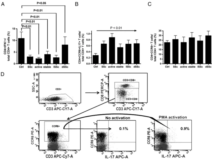
Figure 1 Analysis of frequencies of CD4+IFNγ+ (panel A), CD4+CCR6+IL17+ (panel B) and CD4+CCR6+ (panel C) T cell subpopulations in the circulation of SSc patients and healthy controls. Panel D: flow cytometry analysis of CD4+CCR6+IL17+ T cells in PBMC from representative patients no. 8.
intracellular content of IFNγ, IL4 and IL17 cytokines characterizing Th1, Th2 and Th17 T cell subsets, respective-ly. Decreased concentration of CD4+IFNγ+ T cells was observed in SSc patients with respect to healthy controls, independently from type (diffuse or limited) and stage (activated or stable) of disease (Fig. 1A). No differences of CD4+IL4+ T cell concentration was detected between patients and controls (not shown). Since Th17 have been reported to be phenotypically characterized by the expres-sion of the CCR6 chemokine receptor[20], the frequency of CD4+CCR6+IL17+ T cells among total CD4+ T cells was analyzed. Although no significant differences in the frequen-cy of CD4+CCR6+ T cells were present between SSc patients and controls (Fig. 1C), the analysis of CD4+CCR6+IL17+ T cells demonstrated a statistically significant increase of their frequency in SSc patients with respect to controls indepen-dently from type or stage of disease (Fig. 1B).Fig. 1D shows the results of the representative analysis performed on PBMC from patient no. 8.
3.2. The immune response in SSc patients is skewed toward Th17 cell generation/expansion
In order to dynamically analyze the immune response of SSc patients, PBMC were stimulated using a combination of anti-CD3 and anti-CD28 specific mAbs, and subsequently the frequencies of CD4+IFNγ+, CD4+IL4+, and CD4+CCR6+IL17+ cell subpopulations were analyzed. Upon these experimental conditions, the frequency of CD4+IFNγ+ T cells was lower in SSc patients than in controls (Fig. 2A), the frequency of CD4 +CCR6+IL17+ cells was significantly higher in SSc patients with respect to healthy subjects (in particular, in patients
with active disease) (Fig. 2B), and no detectable IL4-secreting T cells were generated (not shown).
To verify the consistency of these findings in an antigen-dependent setting, a new array of experiments was performed in which PBMC were stimulated with ifae and bodies of C. albicans (Ca). This choice originates from the knowledge that protection against Ca infection is mainly Th17-related [21] and that Ca preferentially induces IL17 secretion through the interaction between the Ca mannans and the macrophage mannose receptor [22]. Upon Ca stimulation, unchanged or reduced (in patients with active disease) frequency of CD4+IFNγ+ T cells was detected (Fig. 3A), while higher frequency of CD4+CCR6+IL17+ T cells, independently from type or stage of disease, was observed in SSc patients with respect to healthy controls (Fig. 3B).
3.3. CD4+CD25+ Treg in SSc patients
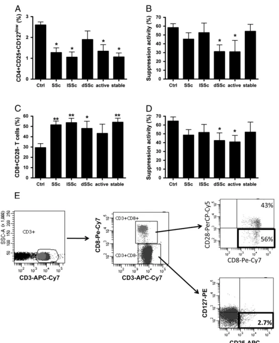
The finding of a spontaneously skewed T cell response toward the pro-inflammatory Th17 cell type might suggest that the healthy balance between effector and regulatory immunity is broken and that a decrease of Treg functions could parallel the effector immune responses in SSc, leading to immune-mediated lesions. With this in mind, phenotypic and functional features of CD4+CD25+highCD127lowTreg were examined. Reduced frequency of these cells was observed in the circulation of SSc patients with respect to controls (Fig. 4A and E). When the function of CD4+CD25+highCD127low Treg was analyzed, decreased levels of suppressive activity were observed in patients with diffuse and active disease with respect to healthy controls (Fig. 4B). Hence, in SSc
Figure 2 Analysis of frequencies of CD4+IFNγ+ (panel A) and CD4+CCR6+IL17+ (panel B) T cell subpopulations in PBMC from SSc patients and controls stimulated by an anti-CD3 mAb and an anti-CD28 mAb.
Figure 3 Analysis of frequencies of CD4+IFNγ+ (panel A) and CD4+CCR6+IL17+ (panel B) T cell subpopulations in PBMC from SSc patients and controls stimulated by bodies of Ca.
patients circulating CD4+CD25+highCD127low Treg show nu-merical and/or functional defects.
3.4. CD8+CD28− Treg in SSc patients
At variance with CD4+CD25+ Treg cells, no specific markers for CD8+ Treg subsets have been so far identified: hence, a precise phenotypic enumeration of these cells is not feasible at the moment. However, since we had previously shown that these cells (and their precursors as well) do not express the CD28 molecule at their cell surface[23], the frequency of CD8+CD28− T cells was analyzed in both SSc patients and
healthy subjects. SSc patients showed increased levels of circulating CD8+CD28− T cells independently from the type (limited or diffuse) of disease (Fig. 4C). In order to functionally analyze CD8+ Treg, the suppressive function was tested on both ex vivo freshly isolated CD8+CD28− T cells and in vitro generated CD8+ Treg. Concerning ex vivo freshly isolated CD8+CD28− T cells, in our experimental setting cells derived from healthy subjects never showed an efficient suppressive activity (≥ 25%). In contrast, 11 out of 21 (52%) of tested SSc patients (9 affected with diffuse and 12 with limited disease, 8 with active and 13 with stable disease) presented a suppressor activity≥25% exerted by ex vivo freshly isolated CD8+CD28− T cells (P=0.005). When the
Figure 4 Analysis of frequency and function of Treg subpopulations from SSc patients and healthy controls. Panel A: frequency of circulating CD4+CD25+highCD127− Treg cells; panel B: suppressive activity of CD4+CD25+highCD127− Treg cells; panel C: frequency of circulating CD8+CD28− T cells; panel D: suppressor activity exerted by in vitro generated CD8+ Treg; panel E: flow cytometry analysis of CD4+CCR6+IL17+ and CD8+CD28− T cells in PBMC from representative patients no. 8. *Pb0.01 vs controls; **Pb0.05 vs controls.
suppressor activity of in vitro generated CD8+ Treg was analyzed, significantly reduced values were detected in patients affected with diffuse or active disease (Fig. 4D).
3.5. Co-existence of alterations concerning both Th17 and Treg compartments in single patients
The results so far exposed highlight the skewing toward Th17 cell function and the presence of Treg alterations as immunological stigmata of SSc patients. In order to verify whether the two phenomena tend to associate in single patients, a correlation analysis was performed. The presence of increased concentra-tion of circulating Th17 cells, with respect to the range of concentrations detected in our series of control subjects (0.1–0.3%), was considered as the variable related to the Th17 cell compartment. The existence of any alteration of the Treg compartment, taking into to consideration (a) lowered concen-tration of circulating CD4+CD25+CD127− Treg with respect to the normal range detected in our series of control subjects (2–3.5%), (b) functional deficiency of CD4+CD25+CD127− Treg and (c) functional deficiency of CD8+ Treg, was considered as the variable related to the Treg compartment.Table 2shows that a statistically significant correlation (P=0.02) was detected between the two variables, likely suggesting the preferential association of increased Th17 immune responses with altered Treg functions in single SSc patients.
4. Discussion
The results of our studies show that: a) CD4+CCR6+ Th17 cells are increased in the circulation of SSc patients; b) T cell immune response in SSc patients is polarized toward the Th17 cell compartment; c) numerical and functional altera-tions of Treg, affecting both CD4+CD25+ and CD8+ Treg subpopulations, are consistently detected in SSc patients; d) alterations of CD4+CCR6+ Th17 and of Treg co-exist and correlate in single SSc patients.
The discovery in the 1980s/1990s of the Th1/Th2 dichotomy [24,25] led to the classification of immune-mediated diseases into predominantly Th1 or Th2 polarized diseases[26]. SSc was then classified as Th2-linked disease on the basis of the predominance of Th2 cells in patient skin and of the increased serum concentration of Th2-related markers (such as CD30 and IL4)[26–28].
The recent unveiling of a new effector T cell subset mainly characterized by its secretion of IL17 (Th17) opened the way for the achievement of new pathogenic insights regarding chronic inflammatory diseases and autoimmune diseases in particular[29–32]. Indeed, several autoimmune diseases have been already found associated with abnormal Th17-driven immune responses[33–36]. In particular, Th17 have been linked to psoriasis, rheumatoid arthritis, Crohn disease. The importance of IL-17 in SSc is known from a long time [9], and more recently the importance of a possible Th17 mediated mechanism(s) in the development of this disease is under debate[10,11,37,38].
Analysis of Th17 cells in the circulation it is not an easy task, due to the very low number of these cells in periphery that makes them hardly detectable. However, the finding that Th17 cells are mainly characterized by the expression of the CCR6 chemokine receptor[39]provides a useful tool for restricting and targeting the search of IL17-secreting cells within a more defined subpopulation.
Here, the analysis of CD4+CCR6+IL17+ T cells in SSc patients allowed us to show for the first time that Th17 cells are increased in the peripheral blood of SSc patients in comparison to healthy subjects.
Moreover, we also observed that stimulation of T cells from SSc individuals with anti-CD3 plus anti-CD28 mAbs expanded preferentially Th17 cells. Similar results were observed when T cells from SSc patients were activated by a complex antigen known to expand IL-17 producing cells (such as C. albicans)[21]. Indeed, in both cases, we found that SSc patients expanded Th17 cells at a significantly higher frequency in comparison to healthy control subjects.
Taken together, these two findings strongly suggest that Th17 cells play a pivotal role in SSc. On the other hand, it is well known that SSc is characterized by elevated production of TGFβ, IL6 and IL1 that are all biological agents strictly related to Th17 differentiation and/or expansion) [2–8], thus further supporting the hypothesis that this disease may be strongly related to a polarization of the immune response toward the Th17 pathway. This is even more intriguing in light of the recent findings showing that human fibroblasts may per sè promote Th17 response [40], thus unveiling a possible role of fibroblasts in determining the kind of T cell polarization in SSc.
Th17 is an effector T cell: one leading hypothesis on the pathogenesis of autoimmune diseases proposes the develop-ment of an unbalanced relationship between effector and
Table 2 Correlation between the percentage of Th17 cells in the peripheral blood and the presence of alterations in the Treg compartment in single patients.
Patient no.
1 2 3 4 5 6 7 8 9 10 11 12 13 14 15 16 17 18 19 20 21 22 23 24 Th17* 1 1 1 0 0 0 0 0 0 1 1 1 1 1 0 1 0 1 1 1 0 0 1 1 Treg* 1 1 1 0 1 0 1 1 1 1 1 1 1 1 1 1 1 1 1 1 0 1 1 1
Th17: 0 was attributed to SSc patients showing Th17 percentage concentrations comprised in the range detected in healthy controls (0.1– 0.3%) and 1 was attributed to patients with increased values.
Treg: 0 was attributed to SSc patients without alterations of Treg (considering both percentage concentration in the circulation of CD4+CD25+ Treg and function of CD4+ or CD8+ Treg) and 1 was attributed to patients with any type of Treg alteration.
regulatory arms of the immune system as a relevant event [12,13].
An unbalanced ratio between effector and regulatory T cell responses as pathogenic mechanism leading to the onset of autoimmune phenomena would imply that abnormal activation of effector cells may associate with deficiencies in the regulatory compartment.
Accordingly with this hypothesis, a complex of alterations was found in our series of SSc patients affecting frequency and function of both CD4+CD25+ and CD8+ Treg subsets. In particular, reduced frequency and impaired function of CD4+CD25+ Treg were observed in our series of SSc patients. This is partially in contrast with what was reported by Radstake et al. [41] who found increased frequency of CD4+CD25+ Treg in their SSc patients. Although it is difficult to identify a clear explanation for such discrepancy, one possibility could be the occurrence of periodical variations of Treg frequency in the circulation of SSc patients in response to unknown factors. Accordingly, ex vivo analyses of freshly isolated cells showed the presence of effective CD8+ Treg in the circulation of 52% of SSc patients, but not in that of healthy controls, an observation suggesting relevant recir-culation phenomena involving specifically this Treg subset in SSc patients. Future studies aimed at describing in detail the kinetics of Treg subpopulations in the blood of SSc patients could help solve this issue.
Altogether these findings, underlining the co-existence of divergent and pleiotropic alterations concerning both effec-tor and regulaeffec-tory immune responses, support the concept that SSc could, at least in part, originate from an altered relationship and an uncorrect interplay between functionally different T cell compartments more than from structural/ molecular deficiencies targeting one specific T cell subset. To this regard, it is of particular interest to note that Th17 and Treg seem to be closely linked and their origin regulated by co-evolutionary processes [reviewed in 42]. It is well known that SSc fibroblasts are able to produce several cytokines critical to regulate T cell polarization and function, as we already mentioned. On this basis, we may hypothesize that the over production of given cytokines, as for instance TGF-beta, from SSc fibroblast could promote Th17 response while inhibiting Treg differentiation, thus leading to a chronic inflammation.
In conclusion here we show for the first time that SSc patients display a T cell response characterized by expansion of Th17 cells and a preferential polarization toward Th17 upon specific and non specific stimulation. This feature is counterbalanced by a diminished capacity to develop T cell regulation and it may be worth to explore the possibility that these phenomena are, to some extent, regulated by the cytokine milieu characterizing SSc. These results could therefore bring new lines of investigations on the role of T cell response in SSc and, in case, lead to new therapeutical approaches.
Acknowledgments
The manuscript has been supported by a grant from Compagnia di San Paolo, Torino,“Tolerogenic gene immu-nization and adoptive suppressor cell transfer as therapies for systemic lupus erythematosus.”
References
[1] A. Gabrielli, E.V. Avvedimento, T. Krieg, Scleroderma, N. Engl. J. Med. 360 (2009) 1989–2003.
[2] Y. Kawaguchi, S.A. McCarthy, S.C. Watkins, T.M. Wright, Autocrine activation by interleukin 1α induces the fibrogenic phenotype of systemic sclerosis fibroblasts, J. Rheumatol. 31 (2004) 1946–1954.
[3] C.P. Denton, D.J. Abraham, Transforming growth factor-β and connective tissue growth factor: key cytokines in scleroderma pathogenesis, Curr. Opin. Rheumatol. 13 (2001) 505–511. [4] A.E. Koch, L.B. Kronfeld-Harrington, Z. Szekanecz, M.M. Cho,
G.K. Haines, L.A. Harlow, R.M. Strieter, S.L. Kunkel, M.C. Massa, W.G. Barr, S.A. Jimenez, In situ expression of cytokines and cellular adhesion molecules in the skin of patients with systemic sclerosis. Their role in early and late disease, Pathobiology 61 (1993) 239–246.
[5] Y.S. Gu, J. Kong, G.S. Cheema, C.L. Keen, G. Wick, M.E. Gershwin, The immunobiology of systemic sclerosis, Semin. Arthritis Rheum. 38 (2008) 132–160.
[6] M. Veldhoen, R.J. Hocking, C.J. Atkins, R.M. Locksley, B. Stockinger, TGFbeta in the context of an inflammatory cytokine milieu supports de novo differentiation of IL-17-producing T cells, Immunity 24 (2006) 179–189.
[7] E. Bettelli, Y. Carrier, W. Gao, T. Korn, T.B. Strom, M. Oukka, H.L. Weiner, V.K. Kuchroo, Reciprocal developmental path-ways for the generation of pathogenic effector TH17 and regulatory T cells, Nature 441 (2006) 235–248.
[8] R.K. Benwell, D.R. Lee, Essential and synergistic roles of IL1 and IL6 in human Th17 differentiation directed by TLR ligand-activated dendritic cells, Clin. Immunol. 134 (2010) 178–187. [9] K. Kurasawa, K. Hirose, H. Sano, H. Endo, H. Shinkai, Y.
Nawata, K. Takabayashi, I. Iwamoto, Increased interleukin-17 production in patients with systemic sclerosis, Arthritis Rheum. 43 (2000) 2455–2463.
[10] M. Murata, M. Fujimoto, T. Matsushita, Y. Hamaguchi, M. Hasegawa, K. Takehara, K. Komura, S. Sato, Clinical associa-tion of serum interleukin-17 levels in systemic sclerosis: is systemic sclerosis a Th17 disease? J. Dermatol. Sci. 50 (2008) 240–242.
[11] T.R. Radstake, L. van Bon, J. Broen, A. Hussiani, R. Hesselstrand, D.M. Wuttge, Y. Deng, R. Simms, E. Lubberts, R. Lafyatis, The pronounced Th17 profile in systemic sclerosis (SSc) together with intracellular expression of TGFbeta and IFNgamma distinguishes SSc phenotypes, PLoS ONE 4 (2009) e5903.
[12] W. Chen, J.E. Konkel, TGF-β and ‘adaptive’ Foxp3+ regulatory T cells, J. Mol. Cell. Biol. 2 (2010) 30–36.
[13] S. Sakaguchi, T. Yamaguchi, T. Nomura, M. Ono, Regulatory T cells and immune tolerance, Cell 133 (2008) 775–787. [14] T.M. Brusko, A.L. Putnam, J.A. Bluestone, Human regulatory T
cells: role in autoimmune disease and therapeutic opportuni-ties, Immunol. Rev. 223 (2008) 371–390.
[15] J.M. van Amelsfort, K.M. Jacobs, J.W. Bijlsma, F.P. Lafeber, L.S. Taams, CD4(+)CD25(+) regulatory T cells in rheumatoid arthritis: differences in the presence, phenotype, and function between peripheral blood and synovial fluid, Arthritis Rheum. 50 (2004) 2775–2785.
[16] E.Y. Lyssuk, A.V. Torgashina, S.K. Soloviev, E.L. Nassonov, S.N. Bykovskaia, Reduced number and function of CD4+CD25highFoxP3+ regulatory T cells in patients with systemic lupus erythematosus, Adv. Exp. Med. Biol. 601 (2007) 113–119.
[17] M.F. Liu, C.R. Wang, L.L. Fung, C.R.Wu. Decreased, CD4+CD25+ T cells in peripheral blood of patients with systemic lupus erythematosus, Scand. J. Immunol. 59 (2004) 198–202. [18] W. Hsu, W. Zhang, K. Tsuneyama, Y. Moritoki, W.M. Ridgway,
A.A. Ansari, R.L. Coppel, Z.X. Lian, I. Mackay, M.E. Gershwin, Differential mechanisms in the pathogenesis of autoimmune
cholangitis versus inflammatory bowel disease in interleukin-2Ralpha(−/−) mice, Hepatology 49 (2009) 133–140.
[19] G. Filaci, M. Rizzi, M. Setti, D. Fenoglio, M. Fravega, M. Basso, G. Ansaldo, P. Ceppa, G. Borgonovo, G. Murdaca, F. Ferrera, A. Picciotto, R. Fiocca, G. Torre, F. Indiveri, Non-antigen-specific CD8(+) T suppressor lymphocytes in diseases characterized by chronic immune responses and inflammation, Ann. NY Acad. Sci. 1050 (2005) 115–123.
[20] H. Liu, C. Rohowsky-Kochan, Regulation of IL-17 in human CCR6+ effector memory T cells, J. Immunol. 180 (2008) 7948–7957. [21] H.R. Conti, F. Shen, N. Nayyar, E. Stocum, J.N. Sun, M.J.
Lindemann, A.W. Ho, J.H. Hai, J.J. Yu, J.W. Jung, S.G. Filler, P. Masso-Welch, M. Edgerton, S.L. Gaffen, Th17 cells and IL-17 receptor signaling are essential for mucosal host defense against oral candidiasis, J. Exp. Med. 206 (2009) 299–311. [22] F.L. van de Veerdonk, R.J. Marijnissen, B.J. Kullberg, H.J.
Koenen, S.C. Cheng, I. Joosten, W.B. van den Berg, D.L. Williams, J.W. van der Meer, L.A. Joosten, M.G. Netea, et al., The macrophage mannose receptor induces IL-17 in response to Candida albicans, Cell Host Microbe 5 (2009) 329–340. [23] G. Filaci, M. Fravega, S. Negrini, F. Procopio, D. Fenoglio, M.
Rizzi, S. Brenci, P. Contini, D. Olive, M. Ghio, M. Setti, R.S. Accolla, F. Puppo, F. Indiveri, Nonantigen specific CD8+ T suppressor lymphocytes originate from CD8 + CD28- T cells and inhibit both T-cell proliferation and CTL function, Hum. Immunol. 65 (2004) 142–156.
[24] S. Romagnani, Human TH1 and TH2 subsets: doubt no more, Immunol. Today 12 (1991) 256–257.
[25] S. Romagnani, The Th1/Th2 paradigm, Immunol. Today 18 (1997) 263–266.
[26] V.K. Singh, S. Mehrotra, S.S. Agarwal, The paradigm of Th1 and Th2 cytokines: its relevance to autoimmunity and allergy, Immunol. Res. 20 (1999) 147–161.
[27] M.M. D'Elios, P. Romagnani, C. Scaletti, F. Annunziato, M. Manghetti, C. Mavilia, P. Parronchi, C. Pupilli, G. Pizzolo, E. Maggi, G.F. Del Prete, S. Romagnani, In vivo CD30 expression in human diseases with predominant activation of Th2-like T cells, J. Leukoc. Biol. 61 (1997) 539–544.
[28] R. White Needelman, F.M. Wigley, R.W. Stair, IL-1, IL-2, IL-4, lL-6, TNF-alpha, IFN-gamma levels in sera from patients with seleroderma, Arthritis Rheum. 35 (1992) 1402–1403.
[29] L.E. Harrington, R.D. Hatton, P.R. Mangan, H. Turner, T.L. Murphy, K.M. Murphy, C.T. Weaver, Interleukin 17-producing CD4+ effector T cells develop via a lineage distinct from the T helper type 1 and 2 lineages, Nat. Immunol. 6 (2005) 1123–1132.
[30] C.L. Langrish, Y. Chen, W.M. Blumenschein, J. Mattson, B. Basham, J.D. Sedgwick, T. McClanahan, R.A. Kastelein, D.J.
Cua, IL-23 drives a pathogenic T cell population that induces autoimmune inflammation, J. Exp. Med. 201 (2005) 233–240. [31] M. Veldhoen, R.J. Hocking, C.J. Atkins, R.M. Locksley, B.
Stockinger, TGFbeta in the context of an inflammatory cytokine milieu supports de novo differentiation of IL-17-producing T cells, Immunity 24 (2006) 179–189.
[32] Y. Iwakura, H. Ishigame, The IL-23/IL-17 axis in inflammation, J. Clin. Invest. 116 (2006) 1218–1222.
[33] M. El-Behi, A. Rostami, B. Ciric, Current views on the roles of Th1 and Th17 cells in experimental autoimmune encephalo-myelitis, J. Neuroimmune Pharmacol. 5 (2010) 189–197. [34] Z.J. Liu, P.K. Yadav, J.L. Su, J.S. Wang, K. Fei, Potential role of
Th17 cells in the pathogenesis of inflammatory bowel disease, World J. Gastroenterol. 15 (2009) 5784–5788.
[35] S.L. Gaffen, The role of interleukin-17 in the pathogenesis of rheumatoid arthritis, Curr. Rheumatol. Rep. 11 (2009) 365–370.
[36] A. Nalbandian, J.C. Crispín, G.C. Tsokos, Interleukin-17 and systemic lupus erythematosus: current concepts, Clin. Exp. Immunol. 157 (2009) 209–215.
[37] B. Deleuran, D.J. Abraham, Possible implication of the effector CD4+ T-cell subpopulation TH17 in the pathogenesis of systemic scleroderma, Nat. Clin. Pract. Rheumatol. 3 (2007) 682–683. [38] B. Rueda, J. Broen, O. Torres, C. Simeon, N. Ortego-Centeno,
M.M. Schrijvenaars, M.C. Vonk, V. Fonollosa, F.H. van den Hoogen, M.J. Coenen, J. Sanchez-Román, M.A. Aguirre-Zamorano, R. García-Portales, A. Pros, M.T. Camps, M.A. Gonzalez-Gay, J. Martin, T.R. Radstake, The interleukin 23 receptor gene does not confer risk to systemic sclerosis and is not associated with systemic sclerosis disease phenotype, Ann. Rheum. Dis. 68 (2009) 253–256.
[39] E.V. Acosta-Rodriguez, L. Rivino, J. Geginat, D. Jarrossay, M. Gattorno, A. Lanzavecchia, F. Sallusto, G. Napolitani, Surface phenotype and antigenic specificity of human interleukin 17-producing T helper memory cells, Nat. Immunol. 8 (2007) 639–646.
[40] C. Schirmer, C. Klein, M. von Bergen, J.C. Simon, A. Saalbach, Human fibroblasts support the expansion of IL-17 producing T-cells via up-regulation of IL-23 production by dendritic T-cells, Blood 116 (2010) 1715–1725.
[41] T.R. Radstake, L. van Bon, J. Broen, M. Wenink, K. Santegoets, Y. Deng, A. Hussaini, R. Simms, W.W. Cruikshank, R. Lafyatis, Increased frequency and compromised function of T regulatory cells in systemic sclerosis (SSc) is related to a diminished CD69 and TGFbeta expression, PLoS ONE 4 (2009) e5981.
[42] C.T. Weaver, R.D. Hatton, Interplay between the TH17 and TReg cell lineages: a (co-)evolutionary perspective, Nat. Rev. Immunol. 9 (2009) 883–889.