Université de Montréal
Studies of the role of MAP kinase-activated protein
kinase-5 (MK5) in reactive and reparative fibrosis in the
murine heart
parSherin Ali Nawaito
Département de Pharmacologie et Physiologie, Faculté de Médecine
Thèse présentéeà la Faculté des études supérieures en vue de l’obtention du grade de PhD
en Physiologie
Décembre, 2017
Université de Montréal
Facult
éde
MédecineCette thèse intitulée:
Studies of the role of MAP kinase-activated protein
kinase-5 (MK5) in reactive and reparative fibrosis in the
murine heart
Présentée par:
Sherin Ali Nawaito
A été évaluée par un jury composé des personnes suivantes:
Dr. René Cardinal
Président-rapporteur
Dr. Bruce Gordon Allen
Directeur de recherche
Dr. Guy Rousseau
Membre du jury
Dr. Ian M.C. Dixon
Examinateur externe
Dr. Denis Deblois
Représentant du doyen
Résumé
MK5 est une sérine/thréonine kinase, identifiée à l'origine comme une protéine kinase régulée/activée par p38 (PRAK), activée par les p38 MAPK et MAPK atypiques ERK3 et ERK4. Bien que MK5 soit exprimée dans le cœur, sa fonction physiologique commence seulement à être étudiée. Nous avons étudié les effets de la surcharge de pression chronique induite par la constriction aortique transversale (TAC) et les effets de l'infarctus du myocarde (IM) induit par la ligature permanente de l'artère coronaire descendante antérieure gauche (LADG) chez des souris hétérozygotes (MK5+/-). Aussi, nous avons étudié in vitro le rôle de MK5 dans la fonction de fibroblaste cardiaque en utilisant des fibroblastes de génotype MK5+/+, MK5+/-, MK5-/- et des fibroblastes avec un knockdown de MK5 par siRNA (MK5-kd).
À l'âge de douze semaines, les souris MK5+/- étaient plus petites que les souris MK5+/+. La fonction systolique était diminuée chez les souris MK5+/-. Deux semaines après TAC, les poids cardiaque/longueur du tibia (PC/LT) ont augmenté de manière similaire et significative dans les 2 groupes.L’augmentation de l'ARNm du collagène de type 1α1 (COL1A1) MK5+/+ était atténuée de manière significative chez les souris MK5+/-. Huit semaines après TAC, PC/LT était significativement moins chez les souris TAC-MK5+/-. La progression de l'hypertrophie était atténuée dans les cœurs MK5+/-. L’immunoréactivité de MK5 a été détectée dans les fibroblastes cardiaques mais pas dans les myocytes.Ces données suggèrent que MK5 dans les fibroblastes cardiaques joue un rôle pro-fibrotique et pro-hypertrophique important dans le remodelage cardiaque pathologique.
Nous avons examiné l'effet de MK5+/- dans la fibrose réparatrice après un IM. Les taux de mortalité chez les 2 groupes avec LADGne différaient pas 7 jours suivant l'IM mais ilsétaient plus élevés chez les souris MK5+/- sur une période de 21 jours. La principale cause de décès était la rupture cardiaque. Les fonctions systolique et diastolique sont également altérées chez les 2 groupes avec LADG. La zone de cicatrice et le collagène dedans la cicatrice ont diminué dans les cœurs de MK5+/-8 jours après l'IM.L’infiltration des cellules inflammatoires était similaire dans les deux groupes avec LADG. L’angiogenèse était significativement élevéedans la zone péri-infarctus de cœursduMK5+/-.
Ces données suggèrent que MK5 peut jouer un rôle dans la régulation de la fonction des fibroblastes cardiaques. C’est pourquoi, nous avons examiné la mobilité et la prolifération du fibroblastes qui étaient diminuées chez les fibroblasts MK5-/-. Le transcriptome pour les protéines impliquées dans le remodelage de la matrice extracellulaire différait selon le génotype des fibroblastes. La sécrétion de COL1A1 et fibronectine étaient significativement augmentée dans les fibroblastes MK5-/- et MK5-kd. La contraction du myofibroblaste a été diminuée dans les fibroblastes MK5-kd. Ces données suggèrent que MK5 est impliqué dans la régulation de multiples aspects de la fonction des fibroblasts cardiaques.
En conclusion, nous avons montré un rôle de MK5 dans le remodelage cardiaquepathologique ainsi qu’un rôle de MK5 dans la fonction des fibroblastes cardiaques.
Mots-clés : MK5/PRAK, hypertrophie cardiaque, fibroblaste, surcharge de pression chronique, fibrose réparatrice, infarctus du myocarde, matrice extracellulaire, p38 MAPK
Abstract
MK5 is a serine/threonine kinase, originally identified as a p38 Regulated/Activated Protein Kinase (PRAK), activated by p38 MAPK and the atypical MAPKs ERK3 and ERK4. Although MK5 is expressed in the heart, its physiological function is just beginning to be studied. Herein, we studied the effects of chronic pressure overload induced by transverse aortic constriction (TAC) and the effects of myocardial infarction (MI) induced by permanent ligation of the left anterior descending coronary artery (LADL) in heterozygous mice for a functional knockout of MK5 (MK5+/-). We also studied the role of MK5 in cardiac fibroblast function and in extracellular remodeling in healthy heart in vitro using MK5 wild type (MK5+/+), haplodeficient (MK5+/-), deficient (MK5-/-), and siRNA-mediated knockdown (MK5-kd) fibroblasts.
At twelve weeks of age, MK5+/- mice were smaller than age-matched wild-type littermates (MK5+/+). Left ventricular end-diastolic diameter and systolic function were reduced in MK5+/- mice. Two weeks post-TAC, heart weight/tibia length ratios (HW/TL) were similarly and significantly increased in both MK5+/+ and MK5+/- hearts. However, eight weeks post-TAC, HW/TL ratios were significantly lower in TAC-MK5+/- mice compared to TAC-MK5+/+ mice. Thus, the progression of hypertrophy in response to chronic pressure overload was attenuated in MK5+/- hearts. Furthermore, two weeks of pressure overload induced increase in collagen type 1 α 1 (COL1A1) mRNA in MK5+/+ mice and this increase was significantly attenuated in MK5+/- mice. As MK5 immunoreactivity was detected in cardiac fibroblasts but not myocytes, these findings suggest that MK5 within cardiac fibroblasts plays an important pro-fibrotic and pro-hypertrophic role in cardiac remodeling during chronic pressure overload.
We then examined the effect of reduced MK5 expression on reparative fibrosis following MI. Mortality rates for MK5+/+ and MK5 +/- did not differ significantly over seven days post-MI. In contrast, mortality was higher in MK5+/- mice over twenty-one days. Systolic and diastolic functions were similarly impaired in both MK5+/+ and MK5+/- mice post-MI. Scar area and scar collagen content were reduced in MK5+/- hearts eight days post-MI. Inflammatory cell infiltration was similar in both ligated groups whereas angiogenesis was
significantly greater in the peri-infarct zone in LADL-MK5+/- hearts. These results suggest that MK5 may play a role in regulating cardiac fibroblast function.
Finally, we examined the effect of reduced MK5 expression on fibroblast function. Motility and proliferation were reduced in MK5-/- fibroblasts compared to MK5+/+ and MK5+/- fibroblasts. The transcriptome for proteins involved in extracellular matrix remodeling (ECM) differed depending on fibroblast genotype. Similarly, collagen 1-α1 and fibronectin secretion was increased in MK5-/- and MK5-kd fibroblasts compared with MK5+/+. In addition, knocking down MK5 decreased myofibroblast contraction. Taken together, these data suggest that MK5 is involved in regulating multiple aspects of cardiac fibroblast function.
In conclusion, we have shown a role for MK5 in cardiac remodeling during chronic pressure overload and myocardial infarction. Moreover, we have demonstrated a role for MK5 in cardiac fibroblast function and extracellular matrix remodeling.
Keywords : MK5/PRAK,cardiac hypertrophy, fibroblast, chronic pressure overload, reparative fibrosis, myocardial infarction, extracellular matrix, p38MAPK
Table of contents
Résumé ... i Abstract ... iii Table of contents ... v List of tables ... ix List of figures ... xList of abbreviations ... xii
Acknowledgement ... xvi
1. Introduction ... 1
1.1 Left ventricular (LV) hypertrophy ... 2
1.1.1 Physiological cardiac hypertrophy ... 3
1.1.2 Pathological cardiac hypertrophy ... 5
1.2 Myocardial infarction... 7
1.2.1 Epidemiology, risk factors, and principal causes of myocardial infarction ... 7
1.2.2 Pathophysiology of MI ... 7
1.2.3 Infarct healing following MI ... 8
1.2.3.1 Cardiomyocyte death via necrosis and apoptosis ... 9
1.2.3.2 The inflammatory phase of infarct healing ... 11
1.2.3.3 The proliferative and reparative phase of infarct healing (scar formation) ... 14
1.2.3.3.1 Myofibroblasts during the proliferative phase ... 16
1.2.3.4 The maturation phase of infarct healing ... 20
1.2.4 Remote non-infarcted part of myocardium after MI ... 21
1.2.4.1 Reactive fibrosis... 21
1.2.5 Pathophysiologic consequences of MI at the cellular level ... 22
1.2.6 Complications associated with MI ... 22
1.3 Cardiac (myo)fibroblasts ... 24
1.3.1 Origin, development and activation ... 24
1.3.2.1 ECM production, maintenance, and reabsorption as well as cardiac
homeostasis ... 26
1.3.2.2 Production of bioactive molecules ... 26
1.3.2.3 Cardiac vessel homeostasis ... 27
1.3.2.4 Proliferation of cardiomyocytes ... 27
1.3.2.5 Electrophysiological function of cardiac fibroblasts ... 27
1.3.3 Fibroblast-myocyte coupling ... 28
1.3.4 Signalling in cardiac fibroblasts... 30
1.3.4.1 Mechanical signalling ... 30
1.3.4.2 Chemical signalling ... 30
1.4 Cardiac extracellular matrix (ECM) ... 31
1.4.1 Cardiac ECM structure and function ... 31
1.4.1.1 Collagen ... 33
1.4.1.1.1 Collagen structure and function ... 33
1.4.1.1.2 Collagen biosynthesis and degradation ... 34
1.4.1.2 Fibronectin (FN) ... 36
1.4.2 ECM remodeling during heart diseases ... 37
1.4.2.1 ECM remodeling following pressure overload ... 37
1.4.2.2 ECM remodeling following MI ... 38
1.4.3 ECM Regulation by microRNA... 39
1.5 Mitogen-activated protein kinase-activated protein kinase 5 (MK5) ... 41
1.5.1 Overview ... 41
1.5.2 Identification of MK5 ... 42
1.5.3 MK5 Gene ... 42
1.5.4 MK5 mRNA... 43
1.5.5 MK5 protein ... 43
1.5.6 Subcellular localization and regulation of MK5 activity ... 45
1.5.6.1 Regulation by p38 MAPK ... 45
1.5.6.2 Regulation by ERK3 and ERK4 ... 45
1.5.6.3 Regulation by PKA ... 46
1.5.7 Activation of MK5 ... 46
1.5.8 MK5 substrates ... 49
1.5.9 Biological Functions of MK5 ... 49
1.5.9.1 MK5 and F-actin remodeling and cell migration ... 50
1.5.9.2 MK5 and cell proliferation and malignant tumors ... 51
1.5.9.3 MK5 and cell growth and metabolism ... 52
1.5.9.4 MK5 and Autophagy... 53
1.5.9.5 MK5 and neurological function ... 53
1.5.9.6 MK5 and cardiac function ... 54
1.5.10 Pharmacological inhibitors of MK5... 54
2 Hypothesis and Objectives ... 55
2.1 Hypothesis... 55
2.2 Objectives ... 55
3 Article-1 ... 56
3.1 Abstract ... 58
3.2 Introduction ... 60
3.3 Material and Methods ... 62
3.4 Results ... 68 3.5 Discussion ... 74 3.6 Conclusions ... 77 3.7 References ... 79 4 Article-2 ... 101 4.1 Abstract ... 104 4.2 Introduction ... 106
4.3 Material and Methods ... 108
4.4 Results ... 115 4.5 Discussion ... 121 4.6 Conclusions ... 125 4.7 References ... 126 5 Article-3 ... 147 5.1 Abstract ... 149
5.2 Introduction ... 151
5.3 Materials and Methods ... 152
5.4 Results ... 157 5.5 Discussion ... 161 5.6 Conclusion ... 165 5.7 References ... 167 Discussion ... 189 Conclusion ... 196
Original contribution to the literature ... 197
List of tables
INTRODUCTION TABLE1………32 ARTICLE-1 TABLE 1. ... 88 TABLE 2. ... 89 TABLE 3.. ... 91 TABLE 4.. ... 92 ARTICLE-2 TABLE 1. ... 139 ARTICLE-3 TABLE 1. ... 176 TABLE 2. ... 177 TABLE 3. ... 179 TABLE 4. ... 180 TABLE 5. ... 181 TABLE 6. ... 182Listoffigures
Introduction Figure 1………5 Figure 2………9 Figure 3………...14 Figure 4………...20 Figure 5………..…………...26 Figure 6………...………...29 Figure 7………...………...36 Figure 8………...………...42 Figure 9………...………...44 Article -1 FIGURE 1 ... 94 FIGURE 2 ... 95 FIGURE 3 ... 96 FIGURE 4 ... 97 FIGURE 5 ... 98 FIGURE 6 ... 99 FIGURE 7 ... 100 ARTICLE-2 FIGURE 1 ... 141 FIGURE 2 ... 141 FIGURE 3 ... 142 FIGURE 4 ... 143 FIGURE 5 ... 144 FIGURE 6 ... 145 FIGURE 7 ... 146 ARTICLE-3 FIGURE 1 ... 183 FIGURE 2 ... 184FIGURE 3 ... 185
FIGURE 4 ... 186
FIGURE 5 ... 187
Listof abbreviations
α-MHC α-SMA β-MHC ACE AD AGEs AMPK Ang-II ANP ATP AT1R BNP CAMK CD CFs COL1A1 CREB CTGF Cx DAMPs DDR-2 DMBA DNA DUSP2 ECM EGCG EMT EndoMT EPDCs ERKs ET-1α-myosin heavy chain α-smooth muscle actin β-myosin heavy chain
Angiotensin-converting enzyme Alzheimer’s disease
Advanced glycation end-products AMP-activated protein kinase Angiotensin II
Atrial natriuretic peptitde Adenosine triphosphate Angiotensin 1 receptor B-type natriuretic peptide
Calcium/calmodulin-dependent protein kinase Docking motif
Cardiac fibroblasts Collagen type 1 alpha 1
cAMP- binding response element-binding protein Connective tissue growth factor
Connexin
Danger-associated molecular patterns Discoidin domain receptor 2
Dimethylbenzanthracene Deoxyribonucleic acid
Dual specificity protein phosphatase 2 Extracellular matrix
Epithelial –to-mesenchymal transition Endothelial-to-mesenchymal transition Epicardial-derived cells
Extracellular signal-regulated kinases Epigallocatechin gallate Endothelin 1 FAK FGF FN FSP-1 GAGs
Focal adhesion kinase Fibroblast growth factor Fibronectin
Fibroblast-specific protein 1 Glycosaminoglycans
GAPDH GPCR IGF2BP1 IHD IL I/R HA HAT HEK293 HMGB1 HSP hTid-1s HUVECs IGF1 JNK KIM LADL LAP LDL LOX LTBP LV M1 M2 MAPK MCP-1 MEFs MFG-E8 MI miRNAs MK5 MMP mPTP mRNA MRTF mTOR NES NF
Glyceraldehyde 3-phosphate dehydrogenase G protein-coupled receptors
Insulin-like growth factor 2 binding protein Ischemic heart disease
Interleukin
Ischemia/reperfusion injury Hyaluronan
Histone acetyltransferase Human embryonic kidney 293 High mobility group box 1 Heat shock proteins
Human tumorous imaginal disc1sInsulin-like growth factor 1 Human umbilical vein endothelial cells
Insulin-like growth factor 1 c-jun amino-terminal kinases 1-3 Kinase-interacting motif
Ligation of left anterior descending coronary artery Latency associated peptide
Low Density lipoproteins Lysyl oxidases enzymes Latent TGF-β binding protein Left ventricular
Proinflammatory type I macrophage Proreparative type II macrophage Mitogen activated protein kinase Monocyte chemoattractant protein 1 Mouse embryonic fibroblasts
Milk fat globule-epidermal growth factor 8 Myocardial infarction
MicroRNAs
Mitogen-activated protein kinase-activated protein kinase5 Matrix metalloproteinase
Mitochondrial permeability transition pore Messenger RNA
Myocardin-related transcription factor Mammalian target of rapamycin Nuclear export signal
NLS NLRs OMM PAI-1 PAKs PDGFBB PDGFR PGs PI3K PKA PKC PRAK PRRs RAAS RAGE Reg3β revD ROS RNA RSK SAPK SERCA2a SR TAC TβRI TβRII Tcf21 TGF-Β
Nuclear localization signal
Cytosolic nucleotide-binding oligomerization domain-like receptors Outer mitochondrial membrane
Plasminogen activator inhibitor-1 Group 1 p21-activated kinases Platelet derived growth factor-BB Platelet derived growth factor receptor Proteoglycans
Phosphoinositide 3-kinase Protien kinase A
Protein kinase C
p38-regulated/activated protein kinase Pattern recognition receptors
Renin angiotensin aldosterone system
Receptor for advanced glycation end-products Regenerating islet-derived 3 beta
Reverse D motif
Reactive oxygen species Ribonucleic acid
P90 ribosomal-S6-kinase Stress-activated protein kinase
Sarcoplasmic/ endoplasmic reticulum Ca(2+)ATPase 2a Sarcoplasmic reticulum
Transverse aortic constriction TGF-β receptor type 1
TGF-β receptor type 2 Transcription factor 21
Transforming growth factor-β Thr-182
TIMPs TLRs
Threonine residue 182
Tissue inhibitor of metalloproteinases Toll-like receptors
TNF-α Tumor necrosis factor α TSP
VEGF VSMC
Thrombospondin
Vascular endothelial growth factor Vascular smooth muscle cells
Dedication
To my late husband.
To my mother and father.
Acknowledgement
I would like to thank all the peoples that contribute to this work and make this thesis as it is.
Dr. Bruce Allen, my research supervisor, and advisor; I do not know where I can start. Thanks Bruce for providing me the opportunity to join your laboratory. I joined your laboratory like a baby who was crawling and you stood by me and still standing by me till I grew up and learned how to walk, run, and think. Without your precious continuous support, guidance, patience, and motivation, it would not be possible to conduct this research. The door of your office was always open whenever I had a trouble or a question about my research or writing; you were always available. Working with you, exposed me to a new way of thinking that gave me the opportunity to be a better thinker. I sincerely appreciate my experience in your laboratory. Bruce, you are not only a supervisor, you are also a friend to whom I can discuss life events and take his advice and who supported me in my critical events of my life “my husband death and my high-risk pregnancy”. I will never forget your joke about all joined females candidates at your laboratory that got pregnant after “we will change the name of the lab from cellular biochemistry lab to fertility lab”☺.
Beside my advisor, I would like to thank my members of the jury for this thesis: Dr. René Cardinal, Dr. Ian Dixon, and Dr. Guy Rousseau for accepting with interest to spend their time for reading and commenting on my work, for their insightful and valuable comments and I really appreciate them.
I am very grateful for the guidance and support of Dr. Angelo Calderone. I do appreciate your help. I would like to express my sincere gratitude to my laboratory members for all the fun we have had together, for being a family. I am thankful for all the help I got during my Ph.D. studies from my colleagues; Ana Branco, George Vaniotis, Clemence Merlen, Rahma Boulayah, Artak Tadevosyan, Dharmendra Dingar, Matthieu Ruiz and Rabah Dabouz. Special thanks to Pramod Sahadevan for always being so helpful, without your help; my project would not have gone smoothly as it did. You were always so kind, patient, smiley, hard worker. It is always inspiring working with you. Thank you, Fatiha Sahmi for all your help you did in order to finish this work. You were always being there for me at any time I
need help. You are a wonderful friend. Thanks, Bahira Hussein, it was a great pleasure to work with you. You were in my place completing animal’s surgery when I was admitted to the hospital. You are an organized, attentive and smart girl. I greatly appreciate your help. I am also so grateful for Laurie Caland (trainee) and Joelle Trepanier (master student) that give me the chance to learn how to share my knowledge.
Also, I would like to thank my friend Nassiba Merabet for pushing me to keep going and for her psychological support and encouragement to finish this work, and for being with me in good as well as bad times.
I would like to acknowledge Maya Mamarbachi, Marc-Antoine Gillis, Karine Bouthillier and Robert Parent for all the technical help you supported me with. I would like also to thank Dr.Jean-Claude Tardif, Yanfen Shi and Danielle Gelinas for echocardiography support. Thanks, Marie-Elaine Clavet-Lanthier for your histological advice and troubleshooting.
I would like to thank the funding agencies, the Canadian Institutes of Health Research (CIHR), Heart and Stroke Foundation of Canada, and Montreal Heart Institute Foundation (FICM) for providing funds to accomplish my research project.
Last but not least, I would like to thank my mother Sohir for providing me with moral and physical support throughout my years of Ph.D. and through my thesis writing. I love you and I thank you for encouraging me to continue in my difficult times. Without your presence in my life, your warm, your caring about me and my kids, I would not achieve what I achieved today and this accomplishment would not have been possible.
1.1 Left ventricular (LV) hypertrophy
The heart is made up of cardiomyocytes (muscle cells), non-myocytes (e.g. fibroblasts, endothelial cells, vascular smooth muscle cells, inflammatory cells), and the extracellular matrix. Cardiomyocytes account for only one third of the total cell number but 70-80% of the heart’s mass 8, 11. The fundamental contractile units of the cardiac myocyte are the sarcomeres, which are arranged into myofilaments comprising ‘thin’ actin and ‘thick’ myosin filaments. Myofilaments are, in turn, bundled into myofibrils. Cardiac myocytes are connected to each other via intercalated discs, which maintain cell-cell adhesion in order to transmit the contractile force between adjacent myocytes 8, 12. In addition, cardiomyocytes may possess an intrinsic mechano-sensing mechanism, as their plasma membrane contains stretch sensitive ion channels as well as structural proteins (such as integrins) that couple the extracellular matrix (ECM) with the cytoskeleton, sarcomere, calcium handling proteins, and nucleus 8, 13, 14.
Cardiac hypertrophy refers to an increase in heart mass due to increased cardiomyocyte size 15, 16. In response to an increase in load, cardiac growth is initiated by activation of signaling pathways, enhanced protein synthesis, changes in gene expression and contractile protein organization into sarcomeres (sarcomere assembly). The hypertrophic response is also associated with an increase in the release of autocrine and paracrine humoral factors such as angiotensin II (Ang II), insulin-like growth factor 1 (IGF1), transforming growth factor-β (TGF-β), and endothelin 1 (ET-1). These factors bind to receptors on cardiac cells, triggering the activation of intracellular signaling pathways, leading to hypertrophic growth of cardiomyocytes 8, 17. Furthermore, in response to progressive or chronic increases in load (volume or pressure overload), cardiomyocytes undergo an initial compensatory hypertrophy to normalize wall stress and to maintain normal cardiovascular function; however, persistence of the increase in wall stress leads to decompensation associated with LV dilatation and contractile dysfunction promoting heart failure 8, 18.
Left ventricular hypertrophy can occur in healthy individuals as a physiological adaptation to physical exercise (athlete’s heart), pregnancy (physiological), post-natal development of the heart until adulthood (developmental), or in disease as a pathologic response, which is either genetic or secondary to increased cardiac load (pathological) 15, 18, 19. Both physiological and pathological cardiac hypertrophies have been subdivided into either concentric or eccentric
hypertrophy (Figure 1) depending on the type of overload, which has a great influence on sarcomere assembly, cardiac geometry, and re-expression of fetal genes. Pressure overload (e.g. hypertension, aortic stenosis) results in concentric hypertrophy that involves an increase in cardiac mass and in relative wall thickness, with reduced or unchanged chamber volume, due to the addition of sarcomeres in a parallel pattern resulting in an increase in cardiomyocytes width. In contrast, volume overload (e.g. aortic regurgitation, arteriovenous fistula) results in eccentric hypertrophy, which is characterized by an increase in both cardiac mass and chamber volume (dilated chambers) with normal, decreased, or increased relative wall thickness as a result of the addition of sarcomeres in series and hence increased myocyte length (Figure 1) 8, 20. Pathological and physiological cardiac hypertrophies are accompanied by distinct structural and molecular phenotypes 8, 18.
1.1.1 Physiological cardiac hypertrophy
Physiological hypertrophy, resulting from pregnancy or exercise, is reversible and associated with enhanced or preserved cardiac function 18, 21.However, recent studies have demonstrated thatintense endurance exercise resulted inthe development of artial fibrosis and the induction of atrial dilatation and fibrillation(reviewed in22). In the case of athlete’s heart, intense training results in an increase in LV mass, chamber enlargement, and proportional change in wall thickness that is considered a physiological adaptation. Depending on the type of training, the heart changes in shape: endurance exercise (e.g. running, swimming) increase venous return to heart leading to increase in volume overload and eccentric hypertrophy while isometric exercise (e.g. weight lifting) results in pressure overload and concentric hypertrophy 15. At cellular and molecular levels, physiological cardiac hypertrophy is characterized by the induction of angiogenesis, absence of cardiac fibrosis and cardiomyocyte apoptosis, and a proper balance of increased glycolytic and fatty acid oxidation for generating ATP in the heart. Moreover, during physiological hypertrophy the fetal gene programme is not induced and genes encoding calcium-handling proteins (e.g. sarcoplasmic reticulum (SR) Ca2+-ATPase 2a (SERCA2a)) remain unaffected 21. Furthermore, smooth muscle α-actin, a marker of fibroblast to myofibroblast activation in pathological remodeling, is not detected in exercised rat hearts 21, 23. Finally, in physiological hypertrophy there is an increase in myosin ATPase activity and enhanced contractility. Insulin-like growth factor 1 is considered to be the main initiator of physiological growth signaling and activates the
phosphoinositide 3-kinase [PI3K, (p110α)]-Akt pathway; however, other signaling pathways are also involved 8. Although athlete’s heart is a physiological condition, exercise can induce cardiac changes that not involve only the left ventricle, but all heart chambers such asatrial dilatation and arrythmias22. Long-term endurance exercise has been shown to induce myocardial damage, right ventriclar inflammation and fibrosisin athletes 24, 25. In rat model of prolonged and vigorous exercise, the atria and right ventricle showed eccentric hypertrophy and diastolic dysfunction with atrial dilatation and fibrosis26. Moreover, excessive physiological hypertrophy may be accompanied with poor angiogenesis resulting in heart failure27. In conclusion, despite the beneficial effects of the exercise on the heart, a high exercise load was recently emerged as a potential cause of atrial fibrillation.
1.1.2 Pathological cardiac hypertrophy
In contrast to physiological hypertrophy, pathological hypertrophy is induced by conditions such as hypertension, valve disease, myocardial infarction, coronary artery disease, genetic mutations, or diabetic cardiomyopathy that impose either pressure or volume overload on the heart. Remodeling caused by hypertension or myocardial infarction is a major risk factor for heart failure. Pathological growth is associated with cardiomyocyte apoptosis and necrosis, interstitial fibrosis, decreased systolic and diastolic function, and increased risk of heart failure and sudden death. Increased collagen deposition in the remodeling heart stiffens the ventricles resulting in impaired
Figure 1. Diagram showing concentric and eccentric hypertrophy. Pressure overload produces LV wall thickness as sarcomeres are added in parallel without change in chamber volume resulting in concentric hypertrophy, while volume overload leads to an increase in cardiac mass along with increase in chamber volume due to addition of sarcomeres in series resulting in eccentric hypertrophy (adapted from Bernardo et al, 2010) 8.
contraction and relaxation, impairment of cardiomyocytes electrical coupling, and reduced capillary density. Each of these changes contribute to the transition to heart failure 8, 18. Furthermore, the substrate profile of the hypertrophied myocardium for ATP production is also affected, as there is a shift from fatty acid oxidation to glucose metabolism 21. In addition, an important molecular feature of pathological hypertrophy is the reactivation of the fetal gene programme. There is upregulation of atrial natriuretic peptide (ANP), B-type natriuretic peptide expression as well as the fetal isoforms of contractile proteins suchas skeletal α-actin and β-myosin heavy chain (β-MHC) that is accompanied by a decrease in expression of α-myosin heavy chain (α-MHC) and SERCA2a 8. However, a retrospective study has found no correlation between fetal gene expression and increased heart weight, leading the authors to propose that increased fetal gene expression may be a protective rather than pathological response28. The increased expression of ANP and BNP prevent cardiac hypertrophy 29, 30and fibrosis 31. As the ATPase activity and contractile velocity for α-MHCare higher than those ofβ-MHC, the shift in expression of myosin heavy chain isoform represents an adaptive response to preserving energy despite the increased oxygen demand 32. Reduced SERCA2a expression, which is responsible for calcium re-uptake into the sarcoplasmic reticulum, impedes cardiac relaxation 28. Pathological cardiac hypertrophy is partly induced by humoral cardiovascular regulating factors such as angiotensin-II (Ang-II) and endothelin 1 that activate GTP-binding protein (Gq), resulting in cardiac hypertrophy. Overexpression of Gq resulted in cardiac hypertrophy and contractile dysfunction that lead to the induction of heart failure 33. G
q regulate several intracellular signaling pathways including mitogen-activated protein kinase (MAPK) signaling cascades. Here we will focus on MAPK-activated protein kinase 5 (MK5) which is downstream of MAPK signaling pathway 8.
1.2 Myocardial infarction
1.2.1 Epidemiology, risk factors, and principal causes of myocardial infarction
Despite the reduction in rates of mortality and hospitalization for ischemic heart disease since the 1970s, it remains the principal cause of death and disability worldwide 34. In developed countries, cardiovascular diseases are responsible for 38.5% of deaths in which coronary artery disease counts for 45% of all cardiovascular deaths 35. Data from the Public Health Agency of Canada’s Canadian Chronic Disease Surveillance System for the year 2012/2013 indicates that about 2.4 million Canadians aged 20 years and older have been diagnosed with ischemic heart disease (www.canada.ca). The prevalence of MI increases with age and more so in men than women 35. There are multiple risk factors for cardiovascular disease, with hypertension being a principal risk factor. High blood levels of low density lipoproteins (LDLs) is considered as second risk factor, counting for 56% of ischemic cardiomyopathy worldwide. Moreover, smoking, physical inactivity, obesity and inappropriate diet constitute other risk factors for cardiovascular disease. Finally, diabetes is an important risk factor, as diabetic patients are 2 to 4 times more susceptible to develop cardiovascular insults 35.
Myocardial ischemia is a result of an imbalance between myocardial oxygen supply and demand 6. The main cause of MI is coronary artery atherosclerosis 6. Although even with more than 75% luminal narrowing there is no decrease in blood flow at rest, in response to increased myocardial demand (due to exercise, anemia, etc.), the atherosclerotic plaque impedes an increase in oxygen supply, causing the development of ischemia and angina pectoris. Plaque rupture is the most common cause of thrombosis 6. In addition to atherosclerosis, there non-atherosclerotic forms of MI, such as coronary embolism, congenital anomaly of the coronary artery, increased myocardial oxygen demand, hematological troubles, and arteritis due to autoimmune or infectious diseases, which are less frequent 36.
1.2.2 Pathophysiology of MI
Myocardial infarction can occur as a result of partial or complete coronary artery occlusion. The myocardial zone supplied by the occluded coronary artery undergoes cessation of its blood supply, resulting in cardiomyocyte ischemia and death 37, 38. If reperfusion fails to occur within less
than 15-20 minutes, large numbers of cardiomyocytes undergo necrosis and death in the ischemic zone 6. This cardiomyocyte death triggers an acute inflammatory response that helps to clear the infarct of dead cells and matrix debris, promoting replacement of damaged tissue with a non-contractile collagen-based scar that maintains the structural integrity of the ventricular wall 37-39. Moreover, this inflammatory response elicits ventricular remodeling that is associated with molecular and cellular changes in the ischemic zone, the border zone, and the non-ischemic zone, and it is manifested clinically as ventricular chamber dilatation, cardiac hypertrophy, modification of the three-dimensional structure of the heart from spherical to ellipsoidal shape, and deterioration of cardiac function 38, 39. Ventricular remodeling may last for weeks or months, continuing until the tensile strength of the collagen scar is able to compensate for the increasing forces. This compensation is dependent on the size, location, and duration of the ischemia, the patency of the occluded artery, the presence of existing collaterals, and whether the ischemia is transmural or not 40.
Two phases of post-infarction remodeling have been determined; an early phase (within 72 hours) and a late phase (after 72 hours) 40. The early phase involves the acute cardiomyocyte loss through apoptosis and necrosis, which triggers an inflammatory response, and contributes to infarct expansion resulting in wall thinning, ventricular dilatation, and ventricular rupture or aneurysm formation 38, 40. The late phase of remodeling involves structural changes to the left ventricle, such as alterations in shape and compensatory hypertrophy of non-ischemic myocardium, to normalize cardiac function 38, 40. However, if the wall stress is not normalized, progressive ventricular dilatation occurs, leading to deterioration of contractile function 40, ventricular arrhythmias, and heart failure 38.
1.2.3 Infarct healing following MI
As the myocardium has negligible endogenous regenerative capacity, loss of large numbers of cardiomyocytes leads to collagen-based scar formation. Following MI, many events occur in the ischemic zone that contribute to ventricular remodeling. The process of wound healing starts with necrosis and apoptosis of the damaged cardiomyocytes that initiates an inflammatory response (inflammatory phase), which is responsible for the clearance of dead cells and matrix debris from the infarcted region (≈ 3-4 days post MI in mice) 38-40. Reparative fibrosis (reparative and proliferative phases) follows the inflammatory response, which helps in the recruitment of cardiac
fibroblasts from the non-infarcted area (≈ 7 days post MI in mice). Cardiac fibroblasts migrate into the infarcted area and become activated into myofibroblasts, which then proliferate and secrete collagen and other extracellular matrix proteins to form the mature scar (maturation phase) (Figure 2) 39. Finally, successful scar formation is associated with neovascularization in the healing infarct, providing oxygen and nutrients to the reparative cells 39, 41.
1.2.3.1 Cardiomyocyte death via necrosis and apoptosis
Myocardial ischemia is a result of animbalance between oxygen supply and demand of the myocardium by the occluded coronary artery 6. Prolonged coronary ischemia results in cardiomyocyte death via necrosis, apoptosis, and autophagy in the ischemic zone 42. As the mammalian heart is incapable of producing sufficient amounts of energy to maintain cellular processes in an anaerobic environment, a constant supply of oxygen is essential for proper cardiac contractility and viability 38. Occlusion of coronary artery results in reduced oxygen tension and ischemia of cardiomyocytes, which prevents oxidative phosphorylation, and decreases the
Figure 2. The phases of repair following myocardial infarction. Infarct repair is divided into three different, but overlapping phase: 1- the inflammatory phase; initiated by alarmins released by necrotic cardiomyocytes, serves to clear the infarct from dead cells and matrix debris. 2- the proliferative and reparative phase, associated with proinflammatory signaling suppression, fibroblasts activation into myofibroblast and deposition of extracellular matrix proteins. 3- the maturation phase, in which the extracellular matrix is cross-linked and fibroblasts become quiescent (adapted from Frangogiannis, 2015) 6.
production of adenosine triphosphate (ATP), which is required to drive the Na+/K+-ATPase 38. Decreased sodium-potassium pump activity results in the loss of sodium efflux and therefore water accumulation and cardiomyocytes swelling. Furthermore, this pump failure also leads to depletion of the intracellular potassium 38. Moreover, within 10 seconds of occlusion, aerobic metabolism stops, ATP reserves are depleted, and lactic acid accumulates via anaerobic metabolism 6, 38. In parallel, there is a progressive loss of cardiac contractility that is obvious within 1 minute of ischemia 38. Minutes after the onset of ischemia, reversible ultrastructural modifications occur, in the form of depletion of glycogen reserves, distortion of the transverse tubular system, and cellular and mitochondrial swelling 38. However, sustained ischemia with no reperfusion within 20-30 minutes results in irreversible cardiomyocyte injury in the subendocardial area, including sarcolemmal disruption and the presence of small amorphous densities in the mitochondria 6, 38. The main mechanisms of cell death in the infarcted myocardium are necrosis and apoptosis 6. Necrosis is a passive process characterized histologically by a loss of membrane integrity, cell swelling, karyolysis, vacuolar degeneration, and hypereosinophilia. Necrosis is defined by opening of the mitochondrial permeability transition pore (mPTP) in response to Ca2+ entry into the mitochondria, which encourages water influx, resulting in mitochondrial swelling and cardiomyocyte necrosis 6, 43. Necrosis is first observed in the subendocardium followed by a “wavefront of necrosis” moving towards the subepicardium as the duration of ischemia increases 6, 44. Within 24 hours of coronary occlusion, the necrotic phase is completed. Cardiomyocyte necrosis triggers an intense inflammatory response due to the release of ‘danger’ signals 6, 45. In contrast to necrosis, apoptosis does not provoke inflammation 6. Apoptosis is a programmed process that contributes to cardiomyocyte death through cell shrinkage, condensation of the chromatin and fragmentation of DNA into apoptotic bodies, and removal by phagocytes 6. Apoptosis is induced by the binding of death ligands to cell surface death receptors and activation of mitochondrial signaling by permeabilization of the outer mitochondrial membrane (OMM). This permeabilization triggers the release of mitochondrial apoptogens into the cytoplasm causing apoptotic death 6, 46. Furthermore, in response to ischemia, autophagy in cardiomyocytes is stimulated through an AMP-activated protein kinase (AMPK)-dependent pathway 6, 47. Autophagy is a process by which unnecessary constituents of the cell are transported to lysosomes for degradation and provides energy and nutrients in response to stress. There is controversy on the role of autophagy in the infarcted myocardium; both protective and detrimental effects have been shown 6, 48, 49.
1.2.3.2 The inflammatory phase of infarct healing
In the infarcted heart, the inflammatory phase is characterized by an intense inflammatory response and immune cell infiltration that clears the wound from dead cells 42. Cardiomyocyte death via necrosis results in the release of endogenous molecules “alarmins” that rapidly activate innate immune system by binding to pattern recognition receptors (PRRs) such as toll-like receptors (TLRs), receptor for advanced glycation end-products (RAGE) and cytosolic nucleotide-binding oligomerization domain-like receptors (NLRs). Alarmins act as “danger signals” initiating an intense, but transient, inflammatory reaction 6, 50, 51. Alarmins belong to a large family of mediators known as danger-associated molecular patterns (DAMPs) including high mobility group box 1 (HMGB1), RNA, nucleotides, heat shock proteins (HSPs), members of the S100 family, and interleukin (IL)-1α6, 42, 52. The proinflammatory effects of HMGB1, one of the best characterized alarmins following myocardial ischemia, are mediated through activation of the RAGE signaling pathway 6, 53, 54. Activation of the innate immune response is also initiated by damage to the extracellular matrix and release of matrix protein fragments such as hyaluronan and fibronectin fragments, which activate TLRs expressed on leukocytes 6, 52. Moreover, activation of the complement system in the infarcted heart is involved in the inflammatory and immune responses by accentuating cardiomyocyte necrosis and promoting leukocyte recruitment in the injured myocardium 6, 55. Furthermore, following myocardial infarction, there is generation of reactive oxygen species (ROS) that activate immune cells as the antioxidant defenses are overwhelmed 52. ROS induce leukocyte infiltration and chemotaxis in the infarcted heart by upregulating chemokine and cytokine expression, promoting the expression of adhesion molecules such as P-selectin, and activating integrin adhesion receptors on leukocytes 6, 52. Thus, activation of the innate immune system by alarmins activates nuclear factor (NF)-κB resulting in the activation of genes encoding cytokines, chemokines, and adhesion molecules 38, 39, 56-58. NF-κB can also be activated by tumor necrosis factor α (TNFα), IL-1β, and ROS 38. The proinflammatory cytokines IL-1β, IL-6 and TNFα promote synthesis of adhesion molecules and activation of leukocyte integrins, leading to extravasation of inflammatory cells into the infarct region 6, 52. The release of proinflammatory chemokines such as monocyte chemoattractant protein 1 (MCP-1) lead to recruitment of leukocytes (neutrophils and monocytes) to the infarcted region in order to phagocytose the necrotic cells and ECM debris 6, 52. In addition, activated macrophages release cytokines and growth factors, which
promote granulation tissue formation to replace damaged cells 38, 56.
Many cell types that are normally present in the heart (cardiomyocytes, vascular cells, and fibroblasts), as well as newly recruited immune cells, are involved in the postinfarction inflammatory response 6, 42, 52. Dead and surviving cardiomyocytes play important roles in myocardial infarction. The release of alarmins from necrotic cardiomyocytes results in the activation of inflammatory signaling in fibroblasts, immune, and vascular cells, and the stimulation of the surviving cardiomyocytes in the infarct border zone to synthesize and secrete a variety of cytokines such as IL-6 6, 59, 60. Also, viable cardiomyocytes in the infarct border zone act as a barrier to protect the non-ischemic part of myocardium from the inflammatory injury, and they secrete Reg3β protein that mediates recruitment of macrophages involved in the clearance of neutrophil infiltrate and matrix preservation61, 62. Activated vascular endothelial cells promote neutrophil infiltration and leucocyte extravasation into the infarcted area, by the expression of adhesion molecules on their surface, promoting their adhesion with circulating leucocytes 6, 42. Moreover, activated endothelial cells act as a main source of proinflammatory chemokines 6, 63, 64. Fibroblasts are the most abundant non-muscle cells, they remain quiescent in the absence of injury, and are suited as sentinel cells sensing myocardial injury to initiate the inflammatory response 6, 42, 65. In contrast to cardiomyocytes, cardiac fibroblasts are more resistant to oxidative stress and less susceptible to ischemic injury 39, 66. In response to cardiac injury (in the first 24-72 hours), fibroblasts are activated via DAMPs, ROS, IL-1, TNF-α, and oncostatin-Mand secrete inflammatory cytokines and chemokines and show matrix-degrading properties (Figure 3) 3, 6, 42. A recent study by Nakaya et al. revealed a role of myofibroblasts in the phagocytosis of dead cells following MI 67. Another study demonstrated a role of cardiac fibroblasts in the recruitment of monocytes and their differentiation to M1 or M2 macrophages 68. In parallel, activation of fibroblasts by IL-1 promotes matrix metalloproteinase expression, inhibition of α-smooth muscle actin (α-SMA) expression, and delay fibroblast activation into myofibroblasts until the dead cells and ECM debris has been removed from the infarct (Figure 3) 42, 69.In addition, the normal adult myocardium contains small populations of resident macrophages, dendritic cells, and mast cells 52, 70, 71. These mast cells contain preformed proinflammatory cytokines and, upon cardiac injury, they release their proinflammatory granules and initiate the inflammatory pathway 52, 72. Neutrophils are the first leukocytes to infiltrate the myocardium, peaking within 24 hours of injury. Neutrophil
recruitment and migration to the site of injury is initiated by activation of the complement cascade as well as the release of chemokines and leukotrienes. Once recruited, neutrophils release proteolytic enzymes, thus contributing to the clearance of dead cells and ECM debris 6, 42. Furthermore, proinflammatory Ly6Chi monocytes and lymphocytes are recruited to the infarcted myocardium soon after neutrophil infiltration. At later stages of inflammation, anti-inflammatory monocytes are recruited, contributing to resolution of the inflammatory response post MI 42.
B-lymphocytes play a key role in mobilizing and activating Ly6Chi monocytes following acute myocardial infarction 73. Moreover, in the infarcted heart, monocytes have the ability to undergo phenotypic changes and give rise to mature macrophages in response to the dynamic alterations in cytokine expression. Macrophages are characterized by plasticity, they can differentiate to proinflammatory type I (M1) or anti-inflammatory or proreparative type II macrophages (M2). They contribute to wound healing in different ways: 1) clear the infarct of dead cells and matrix debris, 2) release cytokines and growth factor, thus regulating inflammatory cascade, 3) contribute to new blood vessel formation, and 4) release matrix metalloproteinases (MMPs) and their inhibitors. Thus, inflammatory cells play a significant role in ECM remodeling within the infarct 6.
The ECM not only serves as a structural support but also plays a pivotal role following cardiac injury by regulating the phenotype and the function of multiple cell types that are surrounded by matrix proteins. Ten minutes after coronary occlusion, interstitial MMPs are activated, resulting in the degradation and fragmentation of structural matrix proteins 6, 74. These protein fragments are involved in leukocyte recruitment 6, 75. Matrix degradation is accompanied by the deposition of a newly formed fibrin-based provisional matrix that creates a scaffold for infiltration, migration and proliferation of inflammatory and reparative cells 6, 76.
Many studies have targeted the inflammatory phase of myocardial infarction, as inflammation is thought to play a crucial role in proper myocardial healing 77. Some studies have shown that cardiac rupture is due to an excessive inflammatory response through upregulation of MMP and the degradation of ECM after MI 78-80. In contrast, Matsui et al. have suggested that reduction of the inflammatory response leads to impairment of the formation of granulation tissue, improper reparative response, and cardiac rupture 81. However, clinical trials targeting the complement system and CD18 integrins in order to reduce the recruitment of leukocytes and their
cytotoxic effects in patients with myocardial infarction did not have the expected effects 6, 82-84. Mortality and the composite end points of death, shock, or heart failure were unaffected by administration of a humanized-anti-C5 monoclonal antibody (pexelizumab) to patients with acute ST-elevation MI (STEMI) undergoing percutaneous transluminal coronary intervention 84. Moreover, administration of a CD18 integrin receptor inhibitor in patients with acute MI prior to angioplasty did not reduce infarct size 82. Thus, clinical trials revealed inhibiting the complement system provided no beneficial effect in the treatment of acute MI.
1.2.3.3 The proliferative and reparative phase of infarct healing (scar formation)
After the clearance of dead cells and matrix debris from the infarcted area, a transition from Figure 3. Resident cardiac fibroblasts in ischemic myocardiun contribute to inflammatory response after injury. Cardiac fibroblasts are sensors of cardiac injury. They are activated by ROS, proinflammatory cytokines (TNFα, IL-1β) and ECM fragments and acting as inflammatory cells. Activated fibroblasts initiate the inflammatory response and secrete cytokines, chemokines and matrix metalloproteinases (MMPs) promoting a matrix-degrading phenotype (adapted from Shinde and Frangogiannis, 2014) 3.
the inflammatory to the proliferative and reparative phase is initiated by the activation of intracellular STOP signals that inhibit the inflammatory response and promote the formation of scar tissue and angiogenesis 41. Neutrophils undergo apoptosis and release mediators such as annexin A1 and lactoferrin, which inhibit further neutrophil recruitment and secrete neutrophil gelatinase-associated lipocalin that promotes macrophages conversion into an M2 reparative phenotype 6, 42, 85. M2 macrophages then phagocytose apoptotic neutrophils and secrete anti-inflammatory and profibrotic cytokines, such as IL-10 and TGF-β, and proresolving lipid mediatorsthat are produced from polyunsaturated fatty acid precursors including as lipoxins and resolvins. Lipoxins and resolvins in turn, inhibit neutrophil entry to the site of inflammation, but promote monocyte migration6, 86. TGF-β initially plays a role in inhibiting the inflammatory response by suppressing cytokine expression in the infarcted area. Then, later, TGF-β stimulates 1) the recruitment of cardiac fibroblasts from the non-ischemic region to the ischemic zone, 2) the activation of cardiac fibroblasts to myofibroblasts, 3) the synthesis and deposition of collagen types I and III and other ECM molecules that are necessary for scar formation, and 4) the synthesis of MMPs and tissue inhibitor of metalloproteinases (TIMPs) 37, 39, 87.
The replacement of the inflammatory infiltrate with granulation tissue is accompanied by an extensive angiogenic response, leading to the formation of neovessels that provide oxygen and nutrients to metabolically active mesenchymal cells in the healing myocardium 6, 56. Early neovessels in the infarcted heart lack a pericyte coat, rendering them hyperpermeable and proinflammatory 6, 56, 88. As a collagen-based scar is formed, maturation of the infarct vasculature occurs, playing a role in wound stabilization 6, 56, 88. Matured neovessels acquire pericyte and smooth muscle cell coats, whereas uncoated endothelial cells undergo apoptosis. This mural coat decreases vascular permeability and the angiogenic potential of the vessels, thus contributing to stable scar formation. Platelet derived growth factor (PDGF)-BB-PDGFR-β signaling is involved in the interactions between mural cells and endothelial cells that results in vascular coating 6, 56, 89. Inhibition of PDGFR-β in a mouse model of reperfused ischemia leads to defective vascular maturation, decreased collagen content in the infarcted heart, and uncontrolled inflammation 6, 56, 89. Accordingly, pericyte coating plays an important role in suppressing granulation tissue formation after MI, promoting the resolution of inflammation, and scar stabilization 6, 56.
Phagocytosis of the matrix fragments is followed by formation of a fibrin- and fibronectin-based provisional matrix and deposition of matricellular proteins (macromolecules) 42, 90. These macromolecules do not provide structural support, but bind to cell surface receptors, modifying cell-cell and cell-matrix interactions 42, 91. Osteopontin, periostin, and thrombospondin (TSP) are examples of matricellular proteins that modulate fibroblast phenotype and growth factor signals 3, 91.
Scar formation is a result of reparative fibrosis that is considered as an important physiological response. The objective of reparative fibrosis is to maintain the structural integrity of the ischemic ventricle by replacing the necrosed cardiomyocytes with a collagen-based scar, thus preventing cardiac rupture and LV dilatation. However, if reparative fibrosis is altered, due to inadequate collagen deposition, decrease myofibroblast proliferation, or insufficient angiogenesis, scar formation will be impaired resulting in increased ventricular dilatation and adverse cardiac remodeling 37-39.
Many cell types are implicated in scar formation. Immune cells and vascular endothelial cells act indirectly by secreting fibrogenic mediators whereas fibroblasts act directly by producing ECM proteins 41. Myofibroblasts play a central role in the proliferative phase of wound healing. 1.2.3.3.1 Myofibroblasts during the proliferative phase
The hallmarks of the proliferative phase of infarct healing are the expansion of cardiac fibroblasts, their activation into synthetic myofibroblast, and infiltration of the infarct border zone with activated myofibroblasts to facilitate scar formation 42, 92-94. Myofibroblasts are abundant in the infarct region but not present in healthy myocardium 95 and their main characteristics include the expression of contractile proteins such as α-SMA and formation of contractile stress fibers, the synthesis and secretion of large amounts of extracellular matrix proteins such as type I and III fibrillar collagens, and a proliferative phenotype 3, 6, 41, 96, 97. Early myofibroblasts (proto-myofibroblasts)do not express α-SMA but have stress fibers composed of cytoplasmic actin and focal adhesions, containing β- and γ-actin microfilaments, connected with nonmuscle myosin. They represent the intermediate cell type during fibroblast-myofibroblast transition, but whether they are present in infarcted myocardium remains unknown 3, 6, 98.
Endothelial- and bone marrow-derived cells have also been proposed to be important sources of myofibroblasts in cardiac fibrosis 10, 95, 99, 101, 102; however, recent studies by Kanisicak et al. and Nakaya et al. reported that resident fibroblasts are the only source of myofibroblasts in the heart 67, 103. Thus, resident cardiac fibroblasts remain the major source of myofibroblasts.
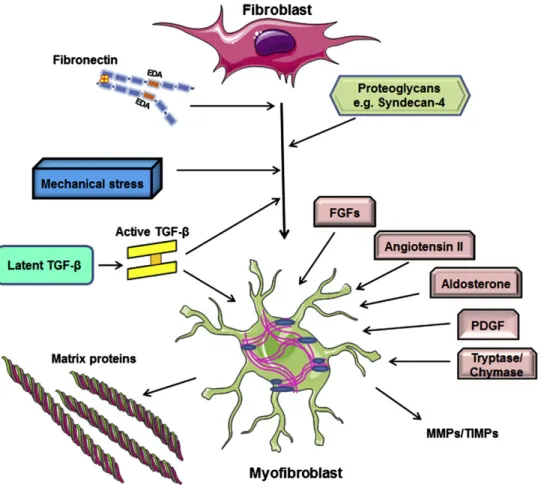
Many pathways are involved in cardiac fibroblast activationinto myofibroblast in the infarct border zone (Figure 4) 99. First, TGF-β is the main stimulus for cardiac fibrosis. There are three TGF-β isoforms: TGF-β1, TGF-β2, TGF-β3. TGF-β is synthesized by many cell types as an inactive “latent propeptide” form that is covalently bound to the latency associated peptide (LAP) 39after release from cells. In the ECM, this inactive complex is bound by latent TGF-β binding protein (LTBP), which is essential for the secretion and storage of TGF-β. Latent TGF-β then becomes activated by proteolytic cleavage (MMP-2, MMP-9, and plasmin) and matricellular proteins such as thrombospondin (TSP)-1 39, 104, 105. Integrins also play a role in TGF-β activation by binding both the latent TGF-β complex and proteases, altering the conformation of the latent TGF-β complex and facilitating its proteolytic cleavage to active TGF-β 106. Once released, activated TGF-β binds to its cell surface receptor type 2 (TβRII), which phosphorylates TGF-β receptor type 1 (TβRI), forming an active receptor complex that activates intracellular signaling through Smad proteins 39, 107. Unphosphorylated R-Smad (Smad2 and Smad3) proteins exist primarily as monomers. After binding to the TGF-β receptor complex and phosphorylation, the activated R-Smads form homomers (homo-dimer and homo-trimer) and can also form heteromers (dimers) with each other. Thereafter, phosphorylated R-Smad form a hetero-oligomericcomplexes with CO-Smad (Smad 4) leadig to dimers or trimers, whichtranslocates to the nucleus to regulate the transcription of target genes 108-111. Inhibitory Smad 7 binds to TβRI and both prevents phosphorylation of Smad 2 or Smad 3 and promotes TβRI degradation through proteosomal pathways 112. TGF-β induces α-SMA transcription in fibroblasts via Smad3-mediated signaling 3, 99, 113, by promoting the translocation of myocardin-related transcription factor (MRTF)-A into the nucleus 3, 99, 114, and by activation of non-canonical pathways such as p38 mitogen-activated protein kinase (MAPK), thus inducing fibroblast activation3, 115, 116. Second, alterations in the matrix environment also modulate myofibroblast activation. Upregulation of the ED-A fibronectin splice variant in the infarcted heart 39, 117 induces TGF-β-mediated myofibroblast activation3, 98, 118-120. Moreover, deposition of collagen VI in the infarcted heart may promote
myofibroblast activation3, 121. Third, expression of proteoglycans such as syndecan 4 and integrins may be important for growth factor-mediated signal transduction in cardiac fibroblasts that leads to myofibroblast activation3, 81. Finally, mechanical stress directly stimulates α-SMA transcription in fibroblasts through Rho kinase and MAPK signaling in the presence of TGF-β 3, 99, 122-124. Following cardiac injury, the structural integrity of the myocardium is disrupted, leading to exposure of fibroblasts to mechanical stress, which significantly contributes to their activation3, 98, 99.
Neurohumoral pathways and several growth factors also play a role in fibroblast activation. Angiotensin II promotes ECM protein synthesis and stimulates fibroblast proliferation within the infarct via AT1 receptors. Some of the effects of angiotensin II in cardiac fibroblasts may be mediated through TGF-β activation 42, 125, 126. Aldosterone also has strong profibrotic actions in the infarcted heart and promotes cardiac fibroblast proliferation 6, 42, 127. Platelet derived growth factor (PDGF), fibroblast growth factor (FGF)-2, and the mast cell-derived proteases tryptase and chymase are potent fibrogenic mediators that are released in the infarcted heart and may be involved in stimulating fibroblasts proliferation and matrix deposition 3, 42, 128-130.
Activated myofibroblasts play a pivotal role in wound healing. Migration of myofibroblasts to the areas devoid of cardiomyocytes is crucial for scar formation. Although the molecular signals implicated in fibroblast migration remains unknown, growth factors such as TGF-β and FGF 113, leukotrienes 131, cytokines such as IL-1 132and chemokines such as monocyte chemoattractant protein (MCP)-1/CCL2 133may promote fibroblast migration to site of injury 3, 39. The migration of fibroblasts through the matrix involves integrin activation, production of matrix-degrading proteases, and deposition of matricellular proteins into the cardiac matrix, favoring a “de-adhesive” state and thereby enhancing migration 3, 39, 91. Inhibitory signals that reduce fibroblast migration are also activated in the infarcted heart in order to limit the excessive fibrotic response and induce wound contraction 3, 39, 63. Moreover, myofibroblasts become highly proliferative 3, 95, 99, 134. In addition, myofibroblasts possess contractile properties as they express high levels of contractile proteins such as α-SMA, vimentin, and focal adhesion proteins that allow scar contraction and reduce scar size by bringing wound margins closer after their perpendicular alignment to the wound rim, thus enabling effective wound healing 37, 135. Myofibroblasts are the primary source of ECM proteins in the healing infarcted heart 3, 136, 137. They synthesize and secrete large amounts of
structural ECM proteins, mainly fibrillar collagen types I and III that provide tensile strength, deposit matricellular proteins, and express MMPs and their inhibitors to promote scar formation, replace the areas of cardiomyocyte loss, and prevent cardiac rupture 3, 37, 95. Unbalanced ECM remodeling may lead to either systolic dysfunction in response to LV dilatation due to inadequate strength of the myocardium or diastolic dysfunction due to heart stiffness resulting from excessive or prolonged ECM accumulation 37.
1.2.3.4 The maturation phase of infarct healing
The proliferative phase ends with the formation of collagen-based scar and is followed by the maturation phase of cardiac wound healing in which the myofibroblasts return to their quiescent state 6, 99. Maturation of the scar is characterized by increased expression of lysyl-oxidase, which catalyzes matrix cross-linking, decreased expression of matricellular proteins, deactivation of the
Figure 4. Activation of cardiac fibroblasts into myofibroblasts. Activation of TGF-β signalling pathway, expression of ED-A fibronectin splice variant, and the degradation of ECM that induce mechanical stress stimulate the activation of cardaic fibroblasts. Many growth factors such as angiotensin II, TGF-β, fibroblast growth factor (FGF), and platelet-derived growth factor (PDGF) stimulate synthesis of collagen and other ECM proteins by myofibroblasts. Finally, myofibroblasts secrete matrix metalloproteinases (MMPs) and tissue inhibitor of metalloproteinases (TIMPs) (adapted from Shinde and Frangogiannis, 2014) 3.
reparative cells, inhibition of angiogenesis, and vascular regression 6, 88, 99, 138, 139. The mechanism of scar maturation remains poorly understood. Apoptosis has been identified as a major mechanism for the elimination of most myofibroblasts from the mature scar 99, 140. Depletion of growth factors, clearance of matricellular proteins, and activation of intracellular inhibitory STOP signals may deprive scar fibroblasts of key signals that promote their survival and activation, resulting in their deactivation and apoptosis 6, 99. Moreover, alterations in matrix composition and the stable mechanical environment of the mature cross-linked scar may play a role in the deactivation of infarct myofibroblasts and their transition to quiescent state 3, 141. However, myofibroblasts in the viable remodeling myocardium may become chronically activated in response to increased hemodynamic load and wall stress, which in turn may contribute to chronic chamber remodeling and ventricular dysfunction 39, 118.
1.2.4 Remote non-infarcted part of myocardium after MI
Following an MI, cardiomyocyte death in the infarcted heart leads to a sudden increase in LV filling pressure that induces a remodeling in both the infarct border zone and non-infarcted myocardium 40. The remodeling of the non-infarct myocardium is an adaptive process that involves structural and functional modifications characterized by interstitial reactive fibrosis and cardiac hypertrophy 6, 40.
1.2.4.1 Reactive fibrosis
An imbalance in synthesis of ECM and/or inhibition of MMPs can lead to cardiac fibrosis. Two types of fibrosis have been identified: reparative and reactive fibrosis 142, 143. Reparative (replacement) fibrosis is an essential physiological response intended to preserve the structural integrity of the heart by the deposition of collagen in the infarcted area resulting in scar formation to replace the dead cardiomyocytes 135, 137, 142, 143. In contrast, reactive (interstitial) fibrosis is a pathologic response that occurs in the non-infarcted myocardial region, which impairs ventricular function and stiffens the ventricles as fibrillar collagen is deposited in intermuscular spaces and within the adventitia of intramyocardial coronary arteries and arterioles (perivascular fibrosis) 135, 142, 144. Interstitial fibrosis can also alter the electrical conduction of cardiomyocytes by isolating them from one another with ECM proteins 142, 145. Moreover, perivascular fibrosis can promote cardiomyocyte hypoxia by increasing the oxygen diffusion distance and decreasing coronary
capillary density 142, 146. Thisdeposition of collagen is secondary to the release of many neurohumoral substances, such as angiotensin II and TGFβ-1, resulting in the activation of fibroblasts in non-infarcted regions that then synthesize and deposit collagen types I and III 135, 144, 147.
1.2.5 Pathophysiologic consequences of MI at the cellular level
Ischemicsystolic and diastolic dysfunctions are the noticeable functional consequences of MI. As action potentials and calcium transients are well maintained during the early stages of ischemia, ischemic systolic dysfunction is mainly linked to inhibition of contractile protein function as a result of intracellular acidosis, which reduces calcium binding to contractile proteins, and the inorganic phosphate that is produced from the breakdown of creatine phosphate reserves 6, 148, 149. Diastolic dysfunction is caused by the production of metabolic products, such as lactate, leading to hyperosmolarity, increased interstitial water, and the reduction of ventricular compliance. In addition, there is impairment of relaxation in MI due to an energetic imbalance 6, 150. Cardiomyocytes normally generate adenosine triphosphate (ATP) through oxidative phosphorylation whereas the ischemic heart employs anaerobic glycolysis to generate ATP from intracellular glycogen stores leading to rapid accumulation of lactate and intracellular acidosis, which in turn affects the rate of anaerobic glycolysis, resulting in marked reduction in ATP levels 6, 151. ATP depletion leads to failure of sodium-potassium (Na+/K+) pump resulting in accumulation of Na+ within the myocytes and depletion of their K+ content. This ionic perturbations cause failure of cardiomyocyte repolarization resulting in conduction block and loss of excitability 6, 152. In parallel, the cytosolic free calcium concentration (Ca2+) is increased during ischemia. All these perturbations can result in an arrhythmogenic response 6.
1.2.6 Complications associated with MI
Rupture of the LV free wall is a drastic and fatal complication of MI, representing about 5-31% of post-MI-related deaths 153, 154. Rupture may occur in the LV free wall, the interventricular septum, or the papillary muscles causing hemopericardium and cardiac tamponade, ventricular septal defect, and acute mitral regurgitation, respectively 6, 155. In murine heart, cardiac rupture occurs 2-6 days post-MI in response to decreased tensile strength of the infarcted myocardium 155. Several factors have been implicated in the induction of cardiac rupture, including a large
transmural infarct, excessive inflammatory response, increased MMP activity, and delayed reparative wound healing 6, 153, 156-158. Infarct expansion and ventricular aneurysm, two possible consequences of adverse postinfarction remodeling, are more prominent in the apex and associated with higher rates of death 6, 159, 160. In mouse models, infarct expansion is characterized by decreased infarct volume, as the scar undergoes maturation and contraction, and not by the progressive increase in the infarct size 6, 161whereas ventricular aneurysm is an area of the infarcted LV wall that bulges with each contraction when a thinned area of noncontractile scar is stretched 6. Another common complication of MI is cardiogenic shock, which represents a significant cause of early mortality following MI due to pump failure 6. Moreover, pericardial disease is also common with acute MI, as in clinical studies the incidence of postinfarction pericarditis and pericardial effusion account for 7-41% and 43% of deaths post-MI, respectively 6, 162, 163. Finally, ventricular arrhythmias are a major cause of mortality during both early and late phase post-MI. Several processes may explain the generation of arrhythmias. In the early phase, ischemia leads to ionic perturbations resulting in the creation of a pathophysiologic substrate that enhances arrhythmias. In the late phase, fibrosis forms inexcitable barriers and reentry circuits 6. Furthermore, oxidative stress and neurohumoral, sympathetic, and activation of the renin angiotensin aldosterone system (RAAS) have been implicated in generation of arrhythmias 6, 164.