Université du Québec
Institut National de la Recherche Scientifique Institut Armand-Frappier
DIINDOLYLMETHANE (DIM) AND RING-SUBSTITUTED
HALOGENATED DERIVATIVES OF DIM (RING-DIMS): THEIR ROLE IN
PROSTATE CANCER PREVENTION
Par
HOSSAM DRAZ
Thèse présentée pour l’obtention du grade de Philosophiae doctor (Ph.D.)
en Biologie
Jury d’évaluation
Président du jury et Prof. Yves St-Pierre
examinateur interne INRS-Institut Armand-Frappier Examinateur externe Prof. Diana Averill-Bates
Département des sciences biologiques Université du Québec à Montréal
Examinateur externe Prof. François Yu
Faculté de médecine Université de Montréal Directeur de recherche Prof. Thomas Sanderson
INRS-Institut Armand-Frappier
ACKNOWLEDGEMENTS
It is a pleasure to express my deepest gratitude and indebtedness to those who generously devoted much effort and time in helping me to accomplish this thesis. First and foremost, I thank my academic advisor, Professor Thomas Sanderson, for his caring guidance, patience, extraordinary support, giving me intellectual freedom in my research work, supporting my attendance at various conferences, engaging me in new ideas, and demanding a high quality of work in all my endeavors. I am very proud and fortunate to be his student.
Additionally, I would like to thank my committee members Professors Yves St-Pierre, Diana Averill-Bates and François Yu for their interest in my work and for their contribution to the evaluation of my thesis. Every result described in this thesis was accomplished with the help and support of labmates, colleagues and collaborators. I would like to thank all my present and former lab members Dr. Alex Goldberg, Dr. Elyse Caron-Beaudoin, Dr. Helene Clabault, Andree-Anne Thibeault, Rachel Viau and Debbie Yancu for helping and supporting me during my research. Dr. Alex Goldberg and I worked together on several different phases of the CIHR funded ring-DIM project, and without his efforts, my job would have undoubtedly been more difficult.
I am also indebted to Professor Stephen H. Safe of Texas A&M University who deserves the credit for providing us with the synthetic ring-DIMs and for his profound reading of my three manuscripts. I am very grateful to Professor Emma Guns of the University of British Colombia who supervised the tissue microarray analyses.
I gratefully acknowledge the funding sources that made my Ph.D. work possible. I was awarded a Fondation Armand-Frappier scholarship for my first 2 years and was honored to hold a prestigious Fonds de Recherche du Québec–Santé (FRQS) doctoral scholarship for the last 3 years of my doctoral program. My profound gratitude stands toward the Canadian Institutes of Health Research (CIHR) for financing the ring-DIM project.
I would like to thank my family, especially my loving and caring wife, for always being extremely compassionate, encouraging and a driving force behind the successful
completion of my research work. She has been my inspiration and motivation for continuing to improve my scientific knowledge and move my career forward. I would like to thank my parents who raised me with a love of science and supported me in all my pursuits. I would like also to thank my sister for her support, love and encouragement.
I would like to show my gratitude to all my friends and colleagues at the INRS-IAF, especially Ahmed Fahmy, Lucas Sagrillo-Fagundes, Elham Dianati, Guillermo-Arango Duque, Ahmed Soliman, Slimane Dridi, Ghislain Paka, Akil Hammami, Monzer Khidiri, Andree-Anne Hudon Thibeault, Hélène Clabault, Marc Fraser, Mohamed Haddad, Edward Kwarting, and Mustapha Iddir for their support and providing a stimulating and fun environment during my studies.
RÉSUMÉ
Cette étude a pour objectif d'examiner les propriétés antiprolifératives du un dérivé de légumes crucifères; le diindolylméthane (DIM), ainsi que certains de ces dérivés synthétiques (ring-DIMs) dans des cellules cancéreuses humaines de la prostate androgènes-dépendantes (AD) et androgènes-indépendants (AI). Ces cellules ont été utilisées pour étudier plusieurs mécanismes moléculaires de l'action anticancéreuse des ring-DIMs. Par ailleurs, des souris porteuses de tumeurs cancéreuses humaines de la prostate bioluminescentes ont été utilisées pour évaluer l'activité anticancéreuse de ring-DIMs in vivo, en utilisant une technique d'imagerie non invasive et en temps réel.
En effet, le DIM est un métabolite de l'indole-3-carbinol (I3C), un composant important des légumes crucifères de la famille des Brassica, tels que le brocoli et les choux de Bruxelles. Selon les études épidémiologiques, la consommation élevée de ces légumes est associée à une réduction des risques du développement de divers cancers. L'I3C et le DIM sont capables d’inhiber la croissance de diverses cellules cancéreuses in
vitro ainsi qu’in vivo incluant le cancer de la prostate, le cancer le plus fréquemment
diagnostiqué chez l'homme occidental. Le DIM inhibe la prolifération des cellules cancéreuses prostatiques AD (LNCaP) qui est stimulée préalablement par la dihydrotestostérone (DHT). De plus, le DIM induit l'apoptose cellulaire et inhibe la croissance des tumeurs de la prostate in vivo. Dans ce même contexte, nos études antérieures ont montré que le DIM et les ring-DIMs diminuent l'expression génique des récepteurs aux androgènes (AR), réduisent les taux de protéine AR et l'expression de l'antigène prostatique spécifique (PSA) médiée par l'AR. Les ring-DIMs disubstitués par des halogènes (Br2 ou Cl2) en positions 4- et 4'- de la molécule DIM bloquent l'accumulation de l'AR dans le noyau des cellules LNCaP stimulées par la DHT. Cependant, nos connaissances concernant la capacité du DIM et ses dérivés à affecter les mécanismes de protection cellulaire tels que l'autophagie ou le stress du réticulum endoplasmique (ER) sont faibles. Les ring-DIMs ont été décrits à agir comme des inhibiteurs de la prolifération des cellules tumorales plus puissants que DIM. En effet, ils sont efficaces contre les cellules cancéreuses de la prostate hormono-dépendantes, ainsi
qu’indépendantes. De ce fait, ils ont un potentiel supplémentaire pour le traitement du cancer de la prostate résistant au traitement par les antiandrogènes. Dans la recherche sur de nouveaux médicaments efficaces contre le cancer de la prostate et l'hyperplasie bénigne de la prostate, ces composés seraient d'excellents candidats pour des études actives sur leurs mécanismes d'action moléculaires et leurs activités biologiques in vivo. Ainsi, notre hypothèse de travail est que les ring-DIMs possèdent des activités anticancéreuses prostatiques et qu'ils sont plus puissants que le DIM. Cela dans le but de développer des médicaments efficaces dans le traitement du cancer de la prostate. Le premier objectif de recherche était de déterminer les événements précoces entraînant la mort cellulaire induite par DIM et les ring-DIMs. Nous avons étudié leurs effets sur la stabilité mitochondriale, le stress du ER et l'autophagie en fonction de la concentration et le temps d’exposition. Nous avons ainsi démontré que le DIM et les ring-DIMs induisent la mort des cellules cancéreuses de la prostate LNCaP positives et AD), C4-2B (AR-positives et AI) et DU145 (AR-négatives, AI et déficientes à l'autophagie), contrairement à leur manque de toxicité chez les cellules épithéliales humaines de la prostate normales immortalisées (RWPE-1). Nous montrons également que les ring-DIMs causent la perte précoce du potentiel de la membrane mitochondriale et la diminution de la production mitochondriale d'ATP dans les cellules cancéreuses de la prostate. Nos observations mettent en évidence une perturbation de la fonction mitochondriale en tant qu'événement déterminant dans les actions cytotoxiques du DIM et tous les ring-DIMs. Cependant, le salubrinal, un inhibiteur du stress ER, inhibait seulement la mort cellulaire médiée par les 4,4'-dihaloDIMs, en revanche, le salubrinal exacerbait la toxicité des 7,7'-dihaloDIMs. En utilisant une analyse in silico de l'affinité par docking en 3-D, nous avons identifié la calmoduline-kinase II dépendante du Ca+2 (CaMKII) en tant que cible potentielle pour le ring-DIM le plus toxique, le 4,4'-dibromoDIM. Nos résultats ont montré que l'inhibiteur de CaMKII (KN93) abrogait complètement la toxicité de ce ring-DIM, mais pas la toxicité du 7,7'-Cl2DIM. L'implication des pores de transition de perméabilité mitochondriale dans la toxicité des ring-DIMs est suggérée par un traitement avec la cyclosporine A (inhibitrice des pores de transition de perméabilité mitochondriale), qui abrogait la toxicité de tous les ring-DIMs, mais pas cela du DIM. L'un des principaux résultats de cette étude était que le DIM et les ring-DIMs induisaient l'autophagie dans les cellules cancéreuses de la
prostate. L'inhibition de l'autophagie avec la bafilomycine A1, la 3-méthyladénine ou par le silençage génique de LC3B sensibilisait les cellules LNCaP et C4-2B, mais pas les cellules DU145 déficientes en ATG5 à la mort cellulaire induite par le DIM et les ring-DIMs. Nous proposons que l'autophagie induite par le DIM et les ring-DIMs ait une fonction cytoprotectrice dans les cellules cancéreuses de la prostate.
Notre deuxième objectif de recherche était d'étudier les effets mécanistiques des ring-DIMs sur les voies de signalisation impliquées dans la mort cellulaire des cellules cancéreuses de la prostate. Nous avons trouvé que l'autophagie induite par le DIM et les ring-DIMs s'accompagnait d'une augmentation de la formation des vacuoles autophagiques et de la conversion de LC3BI en LC3BII dans les cellules cancéreuses humaines de la prostate LNCaP et C4-2B. Le DIM et les ring-DIMs induisaient également la phosphorylation de l'AMPK (protéine kinase activée par l'AMP), de l'ULK-1 (kinase d'activation de l'autophagie de type unc-51 type 1, ATG1) et de l'acétyl-CoA carboxylase (ACC). De plus, le DIM et les ring-DIMs induisaient le gène 1 des protéines astrocytaires (AEG-1). La régulation à la baisse d'AEG-1 ou d'AMPK inhibe l'autophagie induite par le DIM et le ring-DIMs. Un prétraitement avec des siRNA d'AEG-1 ou AMPK potentialisait la cytotoxicité du DIM et des ring-DIMs. Également, la régulation à la baisse d'AEG-1 induisait la sénescence dans des cellules traitées avec des concentrations de DIM ou ring-DIMs ouvertement cytotoxiques et inhibait l'initiation de l'apoptose en réponse à ces composés. En effet, nous avons identifié un nouveau mécanisme pour l'autophagie protectrice induite par le DIM et les ring-DIMs via l'induction de l'AEG-1 et l'activation subséquente de l'AMPK.
Notre troisième objectif de recherche était d'étudier l'effet de DIM et le ring-DIM le plus puissant (4,4'-Br2DIM) sur le développement tumoral dans un modèle de xénogreffe murine du cancer de la prostate humaine. L'utilisation de cellules PC-3 bioluminescentes pour surveiller la croissance tumorale et les effets du traitement chimique avec les nouveaux ring-DIMs constitute l'un des principaux aspects novateurs de la présente étude. La surveillance in vivo en temps réel réduit le nombre d'animaux requis pour les expériences proposées, car ce n'est pas nécessaire de sacrifier les souris à des intervalles pendant la progression de la tumeur. Nous avons trouvé que l'inhibition de l'autophagie par la chloroquine (CQ) sensibilisait significativement les cellules PC-3 à la
mort en présence de concentrations du DIM ou 4,4'-Br2DIM qui étaient sous-toxiques in
vitro. De plus, une combinaison de DIM (10 mg/kg) et CQ (60 mg/kg), 3x par semaine,
réduisait significativement la croissance des tumeurs PC-3 in vivo après 3 et 4 semaines de traitement. De plus, le 4,4'-Br2DIM (10 mg/kg, 3x par semaine) inhibait significativement la croissance tumorale après 4 semaines de traitement. L'analyse de micromatrices tissulaires préparer à partir des tumeurs excisées a montré que le DIM seul ou en co-traitement avec la CQ induisait le marqueur d'apoptose TUNEL, et inhibait significativement le marqueur de prolifération cellulaire Ki67.
En conclusion, nos résultats de recherche fournissent des informations importantes sur les mécanismes d'action potentiels du DIM et les ring-DIMs qui ont des activités biologiques divergentes de DIM. Nous avons également identifié un nouveau mécanisme pour l'autophagie protectrice médiée par le DIM et les ring-DIMs via l'activation de l'AMPK et l'induction de l'AEG-1. De plus, nos découvertes pourrait faciliter le développement de nouvelles pharmacothérapies contre le cancer de la prostate qui comprennent des inhibiteurs sélectifs de l'autophagie en tant qu'adjuvants.
ENGLISH SUMMARY
The objective of this research study is to investigate the antiproliferative properties of the cruciferous vegetable-derived compound diindolylmethane (DIM), and several synthetic disubstituted halogenated DIM-derivatives (ring-DIMs) in androgen-dependent (AD) and androgen-independent (AI) human prostate cancer cells. We aimed to investigate several molecular mechanisms of anticancer action of the ring-DIMs in vitro. In addition, mice carrying bioluminescent human prostate cancer tumors were used to assess the anticancer activity of these compounds in vivo, using a non-invasive, real-time imaging technique.
DIM is formed as a metabolite of indole-3-carbinol (I3C), an important component of cruciferous vegetables of the Brassica family, such as broccoli and Brussels sprouts. Epidemiological studies show that high consumption of these vegetables is associated with decreased risks of various cancers. Both I3C and DIM inhibit the growth of various cancers in vitro and in vivo, including cancer of the prostate, which is the most common malignancy in Western men. DIM also inhibits dihydrotestosterone (DHT)-mediated proliferation of the AD LNCaP prostate cancer cells, induces apoptosis, and inhibits prostate tumor growth in vivo. Our previous studies show that DIM and ring-DIMs down-regulate androgen receptor (AR) expression; reduce AR protein levels and AR-mediated prostate specific antigen (PSA) expression. In addition, 4,4'-dihalo-substituted ring-DIMs, block accumulation of AR in the nucleus of androgen-stimulated LNCaP cells. Less is known about the ability of DIM or its derivatives to affect cell protective mechanisms such as autophagy and endoplasmic reticulum (ER) stress. Further results show that ring-DIMs are considerably more potent inhibitors of tumor cell proliferation than DIM, and are effective against hormone-dependent, as well as -independent prostate cancer cells, indicating their additional potential in the treatment of androgen-refractory prostate cancer. In the quest for novel drugs effective against prostate cancer and benign prostate hyperplasia these compounds are excellent candidates for detailed research concerning their molecular mechanisms of action and biological activity in vivo.
Our working hypothesis is that ring-DIMs exhibit anti-prostate cancer activities and are considerably more potent than DIM. Thus making them suitable candidates for development as drugs effective in the treatment of prostate cancer. Our first research objective was to determine the early events that result in cell death induced by DIM and ring-DIMs by determining their concentration- and time-dependent effects on mitochondrial stability, ER stress and autophagy. We demonstrated that DIM and ring-DIMs induced the death of LNCaP (AR-positives and AD), C4-2B (AR-positives and AI) and DU145 (AR-negatives, AI and autophagy deficient) prostate cancer cells, but not that of immortalized normal human prostate epithelial (RWPE-1) cells. We also showed that ring-DIMs caused the early loss of mitochondrial membrane potential (MMP) and decreased mitochondrial ATP generation in prostate cancer cells. Our evidence points at disruption of mitochondrial function as the defining event in the cytotoxic actions of all ring-DIMs. However, salubrinal, an inhibitor of ER stress, inhibited cell death mediated only by 4,4’-dihaloDIMs by contrast, it exacerbated the toxicity of the 7,7'-dihaloDIMs. Using in silico 3-D docking affinity analysis, we identified Ca+2/calmodulin-dependent kinase II (CaMKII) as a potential direct target for the most toxic ring-DIM, 4,4’-dibromoDIM. Our results showed that CaMKII inhibitor KN93 completely abrogatated the toxicity of this ring-DIM, but not the toxicity of 7,7'-Cl2DIM. Involvement of the mitochondrial permeability transition pore in the toxicity of ring-DIMs is suggested by treatment with the mitochondrial permeability transition pore-inhibitor cyclosporin A, which abrogatated the toxicity of all ring-DIMs, although not that of DIM. One of the main findings of the current study was that DIM and ring-DIMs induced autophagy in prostate cancer cells. Inhibition of autophagy with bafilomycin A1, 3-methyladenine or by LC3B gene silencing sensitized LNCaP and C4-2B, but not ATG5-deficient DU145 cells to DIM- and DIM-mediated cell death. We propose that autophagy induced by DIM and ring-DIMs has a cytoprotective function in prostate cancer cells.
Our second research objective was to study the mechanistic effects of ring-DIMs on signalling pathways involved in prostate cancer cell death. We found that DIM- and ring-DIM-induced autophagy was accompanied by increased autophagic vacuole formation and conversion of LC3BI to LC3BII in LNCaP and C4-2B human prostate cancer cells.
DIM and ring-DIMs also induced the phosphorylation of AMP-activated protein kinase (AMPK), ULK-1 (unc-51-like autophagy activating kinase 1; ATG1) and acetyl-CoA carboxylase (ACC). Interestingly, DIM and ring-DIMs induced the oncogenic protein astrocyte-elevated gene 1 (AEG-1). Downregulation of AEG-1 or AMPK inhibited DIM and ring-DIM-induced autophagy. Pretreatment with either AEG-1 or AMPK siRNAs potentiated the cytotoxicity of DIM and ring-DIMs. Interestingly, downregulation of AEG-1 induced senescence in cells treated with overtly cytotoxic concentrations of DIM or ring-DIMs and inhibited the onset of apoptosis in response to these compounds. Indeed, we have identified a novel mechanism for DIM- and ring-DIM-induced protective autophagy, via induction of AEG-1 and subsequent activation of AMPK.
Our third research objective was to study the effect of DIM and the most potent ring-DIM (4,4'-Br2ring-DIM) on tumor development in a murine xenograft model of human prostate cancer. The use of bioluminescent PC-3 cells to monitor tumor growth and the effects of chemical treatment with the new ring-DIMs is one of the novel aspects of this study. The real-time monitoring of tumor growth in vivo reduces the number of animals required for the proposed experiments, since they do not have to be sacrificed at intervals during tumor progression. We found that the autophagy inhibitor chloroquine (CQ) significantly sensitized PC-3 cells to death in the presence of concentrations of DIM or 4,4'-Br2DIM that were sub-toxic in vitro. Moreover, a combination of DIM (10 mg/kg) and CQ (60 mg/kg), 3 x per week, significantly decreased PC-3 tumor growth in vivo after 3 and 4 weeks of treatment. Furthermore, 4,4'-Br2DIM at 10 mg/kg (3 x per week) significantly inhibited tumor growth after 4 weeks of treatment. Tissues microarray analysis of excised tumors showed that DIM alone or combined with CQ induced apoptosis marker TUNEL, and significantly inhibited the cell proliferation marker Ki67.
Taken together, our research findings provide important information on the potential mechanisms of action of DIM and the ring-DIMs, which have divergent biological activities from DIM. We also identified a novel mechanism for DIM and ring-DIMs mediated protective autophagy via the activation of AMPK and induction of AEG-1. Moreover, our findings may facilitate the development of novel drug therapies against prostate cancer that include selective autophagy inhibitors as adjuvants.
TABLE OF CONTENTS
ACKNOWLEDGEMENTS... I
RÉSUMÉ ... III
ENGLISH SUMMARY ... VII
TABLE OF CONTENTS ... XI LIST OF TABLES... XIII
LIST OF FIGURES ... XV
LIST OF ABBREVIATIONS ... XVI
CHAPTER 1: INTRODUCTION ... 1
1.1 PROSTATECANCER: ... 2
1.1.1 Causes and development of cancer ... 2
1.1.2 Incidence and diagnosis of prostate cancer... 3
1.1.3 Staging of prostate cancer ... 4
1.1.4 Grading of prostate cancer ... 5
1.1.5 Risk factors ... 6
1.1.6 Chemoprevention of prostate cancer ... 7
1.1.7 Hormone-dependent mechanisms of prostate cancer ... 8
1.1.8 Hormone-independent mechanisms of prostate cancer ... 13
1.1.9 Current treatments for prostate cancer ... 18
1.1.10 Causes of castration resistant prostate cancer ... 21
1.1.11 TP53 mutations and prostate cancer ... 21
1.2 DIINDOLYLMETHANE ... 22
1.2.1 Cruciferous vegetables and prostate cancer ... 23
1.2.2 Anti-prostate cancer activity of DIM ... 24
1.2.3 Anticancer effects of DIM in vivo ... 26
1.2.4 DIM clinical trials in treatment of prostate cancer ... 27
1.2.5 Anti-cancer properties of ring-DIMs ... 28
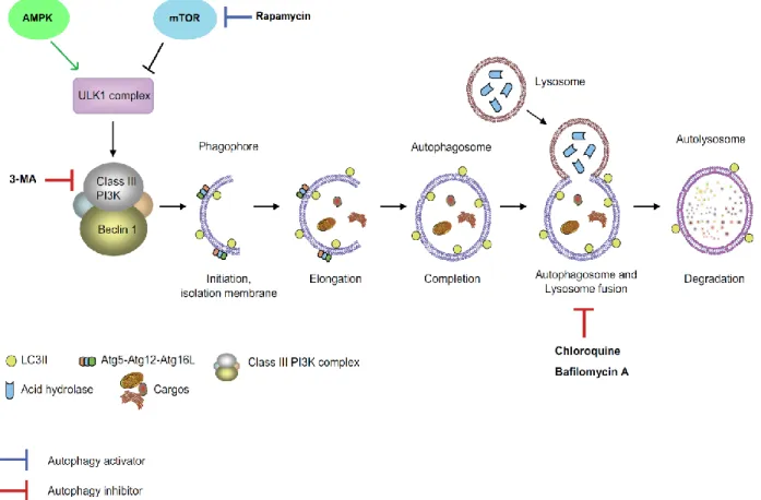
1.3 AUTOPHAGY ... 29
1.3.1 Types of autophagy ... 30
1.3.2 Autophagy mechanism ... 30
1.3.3 Dual Role of autophagy in cancer... 32
1.3.5 Regulation of autophagy by DIM in cancer cells ... 36
1.3.6 The role of AMPK in autophagy ... 37
1.3.7 AEG-1 role in cancer and autophagy ... 38
1.4 MOLECULARPATHWAYSOFAPOPTOSIS,NECROSIS,ENDOPLASMIC RETICULUM(ER)STRESSANDSENESCENCEINCANCER... 39
1.4.1 Apoptosis ... 39
1.4.2 Necrosis and necroptosis ... 41
1.4.3 ER stress ... 43
1.4.4 Senescence ... 46
1.5 STUDYMODELS ... 49
1.5.1 Prostate cancer cell lines ... 49
1.5.2 Xenograft mouse model of prostate cancer ... 50
1.6 HYPOTHESISANDOBJECTIVES ... 51
1.6.1 Hypothesis ... 51
1.6.2 Objectives ... 51
1.7 PROJECTSIGNIFICANCE ... 52
CHAPTER 2: PUBLICATIONS ... 53
2.1 3,3’-DIINDOLYLMETHANE (DIM) AND ITS RING-SUBSTITUTED HALOGENATED ANALOGS (RING -DIMS) INDUCE DIFFERENTIAL MECHANISMS OF SURVIVAL AND DEATH IN ANDROGEN-DEPENDENT AND –INDEPENDENT PROSTATE CANCER CELLS ... 54
2.2 DIINDOLYLMETHANE AND ITS HALOGENATED DERIVATIVES INDUCE PROTECTIVE AUTOPHAGY IN HUMAN PROSTATE CANCER CELLS VIA INDUCTION OF THE ONCOGENIC PROTEIN AEG-1 AND ACTIVATION OF AMP-DEPENDENT KINASE (AMPK) ... 75
2.3 AUTOPHAGY INHIBITION IMPROVES THE CHEMOTHERAPEUTIC EFFICACY OF CRUCIFEROUS VEGETABLE-DERIVED DIINDOLYMETHANE IN A MURINE PROSTATE CANCER XENOGRAFT MODEL ... 90
CHAPTER 3: DISCUSSION ... 107
CONCLUSION AND PERSPECTIVE ... 119
REFERENCES ... 123
APPENDIX I ... 148
LIST OF TABLES
TABLE 1: TNM STAGING OF PROSTATE CANCER... 5 TABLE 2: CHARACTERISTICS OF COMMON PROSTATE CANCER AND IMMORTALIZED CELL
LIST OF FIGURES
FIGURE 1: PHASES OF CARCINOGENESIS. ... 3
FIGURE 2: THE CLASSICAL REPRESENTATION OF THE GLEASON GRADES 1-5. ... 6
FIGURE 3: STERIODOGENESIS. ... 10
FIGURE 4: AR SIGNALING PATHWAY. ... 11
FIGURE 5: THE STAGES OF THE CELL CYCLE ... 14
FIGURE 6: AKT/MTOR SIGNALING PATHWAY. ... 16
FIGURE 7: ARYL HYDROCARBON RECEPTOR (AHR) SIGNALING PATHWAY ... 18
FIGURE 8: FORMATION OF DIINDOLYLMETHANE (DIM) FROM INDOLE-3-CARBINOL (I3C). ... 23
FIGURE 9: CHEMICAL STRUCTURES OF RING-DIMS. ... 24
FIGURE 10: AUTOPHAGY MECHANISM. ... 32
FIGURE 11: DUAL ROLE OF AUTOPHAGY IN CANCER. ... 35
FIGURE 12: INTRINSIC AND EXTRINSIC APOPTOSIS SIGNALING PATHWAYS ... 41
FIGURE 13: NECROPTOSIS PATHWAY ... 43
FIGURE 14: ER STRESS PATHWAY ... 46
FIGURE 15: SENESCENCE MECHANISM. ... 48
LIST OF ABBREVIATIONS
µXBP-1 Unspliced form of X box-binding protein-1
3-MA 3 methyladenine
3βHSDs 3β-hydroxysteroid dehydrogenase 4E-BP1 4E-binding protein 1
4E-BP1 4E-binding protein 1 AD
ADT
Androgen-dependent
Androgen deprivation therapy AEG-1 Astrocyte elevated gene-1
AhR Aryl hydrocarbon receptor
AI Androgen-independent
AIF Apoptosis-inducing factor
AIP AhR interacting protein
AJCC/UICC American joint committee on cancer/international union against cancer AMPK AMP-activated protein kinase
AN Androstenedione
AR Androgen receptor
AREs Androgen response elements
ARF Alternate-reading frame protein ARNT AhR nuclear translocator protein ATF4 Activating transcriptional factor 4 ATF6 Activating transcription factor 6
ATG Autophagy-related genes
B-DIM BioResponse DIM
BECN1 Beclin 1 bHLH BLI Ca2+ Basic helix-loop-helix Bioluminescent imaging Calcium ion CaMKII CSCs
Ca2+/calmodulin-dependent protein kinase Cancer stem cells
C-DIM Methylene-substituted diindolylmethane
CDKs Cyclin-dependent kinases
C-FLIP Cellular FLICE inhibitory protein
CHOP C/EBP homologous protein
cIAP Cellular inhibitors of apoptosis
CQ Chloroquine
CRPC Castration resistant prostate cancer
CsA Cyclosporin A
ctDNA Circulating DNA
CYP cytochrome P450scc
CYP17 Cytochrome P450 steroid 17α-hydroxylase/20,22-lyase
DDR DNA damage response
DHEA DHT
Dehydroepiandrosterone Dihydrotestosterone
DIM Diindolylmethane
DISC Death-inducing signalling complex
DR4 Death receptors 4
DRE Digital rectal examination
eIF2α Eukaryotic translation initiation factor 2
EGF Epidermal growth factor
ER Endoplasmic reticulum
ERAD ER associated degradation
ERK Extracellular signal-regulated kinase
EsR Estrogen receptor
FADD Fas-associated protein with death domain
FasL Fas ligand
FDA Food and drug administration
GADD34 DNA damage-inducible 34
HAHs Halogenated aromatic hydrocarbons
HCC Hepatocellular carcinoma
HIF-1α Hypoxia-inducible factor 1-alpha
HBV Hepatitis B virus
HSPs Heat shock proteins
I3C Indole-3-carbinol
IC50 Half maximal inhibitory concentration
IM-PHFA Immortalized primary human fetal astrocyte IRE1α Inositol-requiring protein-1α
LC3 Microtubule-associated protein 1 light chain 3 LEF1 Lymphoid-enhancing factor 1
LH Luteinizing hormone
LHRH Luteinizing hormone-releasing hormone MAPK P38 mitogen-activated protein kinase MDR1 Multidrug resistance gene 1
MLKL Mixed lineage kinase domain like protein
MOMP Mitochondrial outer membrane permeabilization mPTP
MRT 67307
Mitochondrial permeability transition pore
(N-[3-[[5-Cyclopropyl-2-[[3-(4-morpholinylmethyl)phenyl]amino]-4-pyrimidinyl]amino]propyl] cyclobutanecarboxamide)
MTDH Metadherin
mTOR Mammalian target of rapamycin
mTORC1 mTOR complex 1
MW Molecular weight
NF-κB Nuclear factor kappa-B
NRF2 Nuclear erythroid 2 p45-related factor 2 p70S6K 70S ribosomal protein S6 kinase
PAHs Polyaromatic hydrocarbons
PDGF-D Platelet-derived growth factor-D
PE Phosphotidylethanolamine
PERK Protein kinase R-like ER kinase PIN Prostatic intraepithelial neoplasia
RB Retinoblastoma tumor suppressor protein
ring-DIMs Ring-substituted analogs of 3,3′-diindolylmethane RIP1 Receptor interacting protein kinase-1
ROS Reactive oxygen species
S1P Serine protease site-1
S2P Proteases and metalloprotease site-2 protease
SRD5A Steroid 5α-reductase
sXBP-1 Spliced form of X box-binding protein-1
T-Bid Truncated BID
TCDD TGF-β
2,3,7,8-tetrachlorodibenzo- p-dioxin Transforming growth factor beta 1
TIS Therapy-induced senescence
TNM Tumor-node-metastasis
TRADD TNFR1-associated death domain protein TRAF2 TNF-receptor-associated factor 2
TRAMP Transgenic adenocarcinoma of the mouse prostate TRUS Trans-rectal ultrasound
TUNEL Terminal deoxynucleotidyl transferase dUTP nick end labeling ULK1 Uunc-51-like autophagy activating kinase 1
UPR Unfolded protein response
1.1 PROSTATE CANCER:
Prostate cancer is a major health problem worldwide, ranking as the most common cancer in males and the third leading cause of cancer-related deaths among American and Canadian men (Siegel et al. 2017). Estimated number of cancer related death in 2017 in the United States was 318,420. However, in 2017, 26,730 men died from prostate cancer in the United States (about 8.5% of all cancer deaths in men) (Siegel et al. 2017). Prostate cancer accounts for approximately 20% of all newly diagnosed cancer in Canadian men (21,300 diagnosed with prostate cancer out of 103,100 of all cancers). In 2017, out of 42,600 Canadian men died from cancer, 4100 men died from prostate cancer (about 10% of all cancer deaths in men)(Smith et al. 2018a).
1.1.1 Causes and development of cancer
Cancer is a complex disease with high rate of incidence and mortality, a recent report estimated that around 8 million died from cancer in 2012, which is a relatively high mortality rate compared to the mortality rates of other chronic diseases (Ferlay et al. 2015). Many causes can lead to the development of cancer. Genetic factors are one of the causes. In addition, bad dietary habit and stagnant life style may cause cancer (Anand et al. 2008). Chronic exposure to carcinogens such as toxins, and pollutants are considered as high risk factors (Cao 2015)
All types of cancer including that of the prostate develop in three main carcinogenesis phases: (a) initiation, (b) promotion and progression and finally (c) metastasis (Figure 1). Initiation of cancer is mainly caused by gene mutation after exposure to carcinogens (Berenblum 1954). Normal cells are transformed into cancer cells if there is a failure in the cellular mechanisms that either check and repair or destroy mutated cells through apoptosis (Sinicrope 2010). The initiation step is followed by promotion and progression of cancer cells. Transformed cancer cells acquire the ability to divide (uncontrolled cell proliferation) through their exposure to cancer promoters such as growth factors and hormones. Cancer promoters differ from carcinogens in the fact that they can promote cancer cell growth but cannot initiate the transformation of normal
several hallmarks of cancer. Hallmarks of cancer include sustaining proliferative signaling, evading growth suppressors, resisting cell death, enabling replicative immortality, inducing angiogenesis, reprogramming of energy metabolism and activating invasion and metastasis (Hanahan and Weinberg 2011). Metastasis is the process where cancer cells migrate from their primary site to invade another organ (metastatic site) (Sethi and Kang 2011). The progression of metastatic and primary cancers are dependent on the ability of cancer cells to escape the immune system (Leber and Efferth 2009). Hence, evading immune destruction was introduced as an emerging hallmark (Hanahan and Weinberg 2011). Metastatic cancers are more aggressive and difficult to treat. The rate of mortality in cancer patient with one or more metastatic tumor are much higher than in patients without metastasis (Mehlen and Puisieux 2006).
Figure 1: Phases of carcinogenesis.
Three phases of carcinogenesis include (a) initiation, (b) promotion, and progression, (c) metastasis. Adapted from (Siddiqui et al. 2015)
1.1.2 Incidence and diagnosis of prostate cancer
The incidence of certain cancers is increasing in the West, including various hormone-dependent cancers such as those of the prostate, testis and breast (Ferlay et al. 2015). Prostate cancer is expected to be the leading cancer for American and Canadian men (approximately 20% of all expected newly diagnosed male cases) (Siegel et al. 2017; Smith et al. 2018a). The burden of human suffering and cost to society are expected to increase significantly in the coming decades due to increased life-expectancy.
The age-standardized incidence of prostate cancer has not been stable over the last 30 years. Incidence of prostate cancer has peaked in 1993 and 2001. These peaks are due to the introduction of two waves of intensified screening activity using the prostate specific antigen (PSA) test. PSA, an androgen-dependent (AD) expressed protein, is used to screen for prostate cancer. Increased screening during these periods detected prevalent cancers that otherwise would not be clinically apparent until more advanced stages and temporarily increasing the incidence of prostate cancer (Leland W. K. Chung 2007). Between 1984 and 1995, the mortality rate for prostate cancer increased slowly, and then it is significantly declined from 2001 to 2009 (3.9 % per year). This decline reflects improvement in treatment strategies following the introduction of hormonal therapy (Cooperberg et al. 2003) and advances in radiation therapy (Kuban et al. 2003). Prostate cancer is diagnosed by elevated plasma PSA levels. Prostate cancer is also diagnosed via a digital rectal examination (DRE). Diagnosis of prostate cancer is usually confirmed by a trans-rectal ultrasound (TRUS)-guided biopsy (Ukimura et al. 2015). Liquid biopsy is now introduced as non-invasive blood test that detect circulating tumor cells or circulating tumor DNA (ctDNA) that shed into blood (Hegemann et al. 2016). Recent studies showed that liquid biopsy provided potential biomarkers for prostate cancer diagnoses and may help in guiding treatment for patients with advanced prostate cancer (Pantel and Alix-Panabieres 2012; Scher et al. 2017).
1.1.3 Staging of prostate cancer
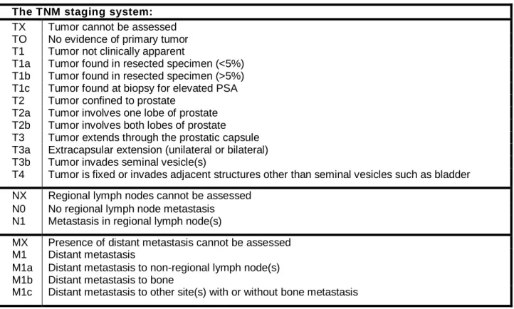
Clinical staging of prostate cancer is very important for the risk assessment of developing the disease, treatment recommendations and follow-up of disease progression. The tumor-node-metastasis (TNM) staging of malignant tumors is the most commonly used classification system that defines the cancer stages based on the following criteria: ‘T’ used to assess the extent of the primary tumor, ‘N’ describes the involvement of lymph nodes, ‘M’ refers to distant metastasis. The TNM staging was established by the American joint committee on cancer/international union against cancer (AJCC/UICC) (Wallace et al. 1975). The classification of prostate cancer based on TNM staging is shown in Table 1.
Table 1: TNM staging of prostate cancer.
TNM describes the tumor stage based on the size of the tumor and where it has spread, ‘T’ defines the extent of the primary tumor, ‘N’ describes the involvement of lymph nodes, and ‘M’ refers to distant metastatic site (Greene 2002).
The TNM staging system:
TX Tumor cannot be assessed
TO No evidence of primary tumor
T1 Tumor not clinically apparent
T1a Tumor found in resected specimen (<5%) T1b Tumor found in resected specimen (>5%) T1c Tumor found at biopsy for elevated PSA
T2 Tumor confined to prostate
T2a Tumor involves one lobe of prostate T2b Tumor involves both lobes of prostate T3 Tumor extends through the prostatic capsule T3a Extracapsular extension (unilateral or bilateral) T3b Tumor invades seminal vesicle(s)
T4 Tumor is fixed or invades adjacent structures other than seminal vesicles such as bladder
NX Regional lymph nodes cannot be assessed
N0 No regional lymph node metastasis
N1 Metastasis in regional lymph node(s)
MX Presence of distant metastasis cannot be assessed
M1 Distant metastasis
M1a Distant metastasis to non-regional lymph node(s) M1b Distant metastasis to bone
M1c Distant metastasis to other site(s) with or without bone metastasis
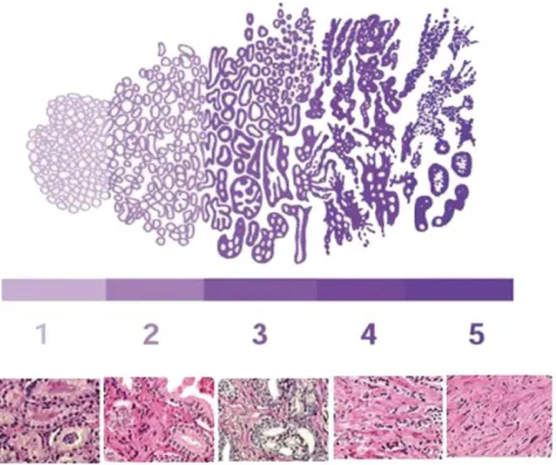
1.1.4 Grading of prostate cancer
Gleason score is the most commonly used prognostic marker for prostate cancer grading. It was introduced by Donald Gleason in 1960 (Gleason 1966). This scoring method is based on staining of tumor tissue with Haematoxylin and Eosin (H&E). The most prevalent and second most prevalent grade is assigned as the grading score. Five grades are defined based on the frequently occurring cells pattern (Harnden et al. 2007). Grade 1 represents well differentiated cells with no signs of stromal infiltration. Grade 2 signifies well differentiated cells with some evidence of stromal infiltration. Grade 3 represents moderately differentiated carcinoma, where cells had some size and shape variations. The poorly differentiated grade 4 carcinoma occurs after fusion of the gland to an anastomosing network. Grade 5 represents undifferentiated carcinoma, and is characterised by presence of clusters or sheets of cells and no evidence of gland formation (Figure 2).
Figure 2: The classical representation of the Gleason grades 1-5.
Cells progress from grade 1 well differentiated carcinoma to grade 5 undifferentiated carcinoma (Harnden et al. 2007)
1.1.5 Risk factors
The risk of prostate cancer increases with age. Prostate cancer is rarely diagnosed in men aged below 40, with risk increasing significantly in men aged above 65 (representing approximately 85% of all cases diagnosed) (Hayat et al. 2007). Risk of prostate cancer is highly correlated with a positive family history (Zeegers et al. 2003). Race and ethnicity are also considered important risk factors for emerging prostate cancer. African-Americans having a higher incidence of prostate cancer compared to other ethnic groups (Robbins et al. 1998). Other risk factors include obesity and diet (MacInnis and English 2006; Kim and Park 2009b; Vykhovanets et al. 2011; Markozannes et al. 2016).
1.1.6
Chemoprevention of prostate cancer
Chemoprevention is the use of specific agents to block the process of tumorigenesis, thus preventing the development of invasive cancer. Cancer chemoprevention involves the chronic administration of a natural or synthetic agent to reduce or prevent the occurrence of malignancy (Steward and Brown 2013). In the past few decades, chemoprevention involving naturally occurring compounds has emerged as a promising and cost-effective approach to diminish prostate cancer incidence. Chemotherapeutic agents are designed to destroy cancer after it occurs (Aggarwal et al. 2004). However, chemopreventative agents had the advantage of inhibiting the precancerous events even before the occurrence of clinical disease (Surh 2003). Phytochemicals are plant chemicals that has chemopreventive activities (Craig 1997; Surh 2003). These phytochemicals include genistein, resveratrol, glucosinolate, indoles, lycopene, curcumin, 6-gingerol, silymarin, and catechins (Surh 2003; Baena Ruiz and Salinas Hernandez 2016). Phytochemicals are derived from natural sources, so they are generally thought to be pharmacologically safe (Aggarwal et al. 2004). However, for many natural product supplements on the market insufficient scientific data on their safety and toxicological profile exist (Pariyani et al. 2015). Moreover, many natural compounds such as capsaisin and ptaquiloside (found in chili pepper and bracken fern, respectively) are potential carcinogens and should be avoided (Bode and Dong 2015). On the other hand, most chemotherapeutic agents are known to have toxic side effects. Their toxic side effects in multiple organs and drug resistance are the major problems for successful clinical use (Sak 2012). Another advantage of chemopreventive agents that phytochemicals are naturally occurring antioxidants, whereas most chemotherapeutic agents destroy cancer cells through the production of reactive oxygen species (Somasundaram et al. 2002; Aggarwal et al. 2004).
Recently, immunotherapy has attracted a great deal of attention for cancer treatment or prevention (Umar et al. 2012; Morrison et al. 2018). A prophylactic or preventative vaccine introduces specific antigen(s) to stimulate the immune system to create antibodies selective against those antigens, which are found on the surfaces of viruses or tumour cells, thus immunizing the body against those viruses/cells, by
triggering their death. Notably, vaccines that target cancer-causing viruses, such as hepatitis B virus (HBV), have been used for hepatocellular carcinoma (HCC) prevention (Schiller and Lowy 2010). However, sipuleucel-T is the only approved autologous vaccine for prostate cancer (Mulders et al. 2015). This vaccine is based on the stimulation of the patient’s T cells using autologous dendritic cells loaded with the prostate tumor antigen PA2024. Therefore, sipuleucel-T is considered to be a therapeutic autologous vaccine and not a prophylactic vaccine.
Prostate cancer, due to its high incidence and long latency, is an ideal candidate for chemoprevention as it provides a very wide window of opportunity for intervention to avert or slow tumour progression (Steward and Brown 2013). Therefore, the development of agents that offer protection against the development of this disease is highly desirable. Such chemopreventive compounds could have an important impact on disease morbidity and mortality for an important segment of the population (Powolny et al. 2011). The identification of agents and their molecular targets for prostate cancer chemoprevention is guided by data derived from a variety of sources including epidemiological, clinical and pre-clinical studies (Adhami and Mukhtar 2007). Prostate cancer develops via modifications in various molecular events, thus inhibiting only one event may not be enough to avert or delay the disease onset. Therefore, identification of chemopreventive agents that target multiple anticancer pathways is essential. Future directions for prostate cancer prevention include genetic, proteomic and immunological approaches for identifying and targeting pathways that are associated with cancer initiation and development (Umar et al. 2012; Turnbull et al. 2018).
1.1.7 Hormone-dependent mechanisms of prostate cancer
Most prostate cancers initiates with AD state and rely on cellular androgen levels. Hence, the biosynthetic pathways, which contribute to androgen production, are crucial for prostate cancer progression.
1.1.7.1 Androgen receptor signaling pathway in prostate cancer
The androgen receptor (AR) signaling pathway is triggered by binding of androgens to AR. There is a large body of evidence supporting a role for androgens in prostate
cancer development and progression (Bladou et al. 1996; Rove and Crawford 2014). Androgen production is initiated by the release of luteinizing hormone-releasing hormone (LHRH) from the hypothalamus, which stimulates the pituitary gland to secrete luteinizing hormone (LH). LH then binds to LH receptors in Leydig cells to initiate the production of androgens.
1.1.7.2 Steroidogenesis
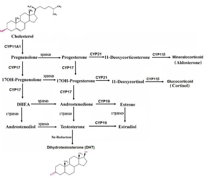
Endocrine tissues such as the gonads and the adrenals are tissues that are able to produce active steroid hormones from cholesterol by a process known as steroidogenesis. These steroid hormones then enter the blood circulation to exert their action at sites distant from where they are produced. Steroidogenesis begins with the irreversible cleavage of a 6-carbon group from cholesterol, producing pregnenolone, catalysed by cytochrome P450scc (side chain cleavage enzyme, CYP11A1). A small repertoire of cytochrome P450 and non-P450 enzymes then convert pregnenolone to other 21-carbon steroids (including glucocorticoids, and mineralocorticoids), 19-carbon steroids (androgens) and 18-carbon steroids (estrogens) (Sharifi and Auchus 2012). The transformations catalyzed by the cytochrome P450s, 5α-reductases, and individual isoforms of 3β-hydroxysteroid dehydrogenase (3βHSDs) are all irreversible reactions, giving rise to the general pathways of steroidogenesis (Figure 3).
Cytochrome P450 steroid 17α-hydroxylase/20,22-lyase (CYP17) is the key enzyme in the synthesis of 19-carbon sex steroid precursors from 21-carbon pregnanes. CYP17 is unique due to its ability to catalyze two independent reactions in the same active center, CYP17 catalyzes both the 17α-hydroxylation of pregnenolone and progesterone and the subsequent 17,20-lyase cleavage (side chain cleavage from 17α-hydroxypregnenolone and 17α-hydroxyprogesterone ) (Lee-Robichaud et al. 1995) to produce dehydroepiandrosterone (DHEA) and androstenedione (AN), respectively (Yamaoka et al. 2010). CYP17A1 is expressed in testicular, adrenal and prostatic tumor tissues; the expression of CYP17A1 in metastatic prostate cancer was 16.9-fold higher (p = 0.0005) than that in primary prostate tumors (Montgomery et al. 2008). Although small amounts of AN, testosterone and other 19-carbon steroid metabolites can be directly produced by the adrenal glands, most androgens in the castrated male are produced in peripheral
tissues, where 3βHSD converts DHEA to AN and androstenediol to testosterone, respectively. Testosterone is then irreversibly 5α-reduced to the higher affinity ligand DHT by steroid 5α-reductase (SRD5A) isoenzymes. Healthy prostate contains mainly SRD5A2, whereas in malignancy SRD5A1 protein becomes over-expressed. Androgens are important for the maintenance of SRD5A expression in healthy prostate, and anti-androgens (and possibly CYP17 inhibitors) cause downregulation of SRD5A expression (Nishiyama et al. 2004). SRD5A gene regulation is dependent on the transcription factor Sp1 (Blanchard et al. 2007).
Figure 3: Steriodogenesis.
The cytochrome P450 enzyme CYP11A1 cleaves cholesterol to pregnenolone. pregnenolone is converted to other 21-carbon steroids (including glucocorticoids, and mineralocorticoids), 19-carbon steroids (androgens) and 18-19-carbon steroids (estrogens). Adapted from (Miller and
1.1.7.3 Activation of AR signaling
The AR is a member of the steroid–thyroid–retinoid nuclear receptor superfamily (Quigley et al. 1995). In the absence of androgens, the AR is located in the cytoplasm, in a complex with heat-shock proteins (HSPs). Androgen binding to its receptor induces conformational changes that facilitate the formation of AR homodimer complexes, which can then bind to androgen response elements (AREs) in the promoter regions of target genes (Figure 4). The activated DNA-bound AR homodimer complex recruits regulatory proteins, activators or repressors, to the AR complex. The co-activators/repression allow interaction of the AR complex with the general transcription machinery to stimulate or inhibit target gene transcription (McKenna et al. 1999).
Figure 4: AR signaling pathway.
Testosterone enters prostate cells and is then converted to dihydrotestosterone (DHT) by the enzyme steroid 5α-reductase. Binding of DHT to the AR induces receptor phosphorylation and dissociation from HSPs. The AR dimerizes then bind to androgen-response elements. Activation of target genes leads to biological responses including growth, survival (Feldman and Feldman 2001).
1.1.7.4 Activation of estrogen receptor signaling
In addition to androgens, estrogens are implicated in AD prostate tumor growth (Bosland 2000). Although considered to be growth inhibitory in normal prostate (Taplin and Ho 2001), estrogens appear to have direct proliferative effects in malignancy (Risbridger et al. 2003) and cause malignant prostate metaplasia at pharmacological doses (Levine et al. 1991). Various mechanisms have been suggested for the carcinogenic effects of endogenous estrogens in prostate, including metabolism to genotoxic 4-hydroxyestrogens (Cavalieri et al. 2002), stimulation of estrogen receptor-(EsR)-mediated cell proliferation, or increased affinity of AR for estrogens (as in LNCaP cells) due to mutations in AR protein (Culig et al. 1997). EsR status also appears important as increased estrogen concentrations in aging prostate cause upregulation of EsRα (cell-proliferative) and downregulation of EsRβ (anti-proliferative) (Chang and Prins 1999). Loss of EsRβ occurs as prostate cancer progresses, which may be associated with increased local estrogen production (Ellem and Risbridger 2006). The key enzyme responsible for the final step in estrogen biosynthesis is aromatase (CYP19). CYP19 expression and catalytic activity has been detected in human prostate and is upregulated in benign prostate hyperplasia and prostate cancer. Importantly, in healthy prostate
CYP19 is expressed at low levels almost exclusively in stromal cells, whereas in
malignancy increased epithelial expression occurs, with altered promoter usage (Ellem and Risbridger 2006). This runs remarkably parallel to what happens in hormone-dependent breast cancer, where CYP19 activity is increased in malignancy, and normal promoter usage switches from glucocorticoid-responsive I.4 to more aggressive cAMP-responsive I.3 and pII promoters (Zhou et al. 2001). However, little is known about aromatase regulation in human prostate, the signalling pathways involved, or modulation by xenobiotics. Aromatase inhibitors used clinically for the treatment of estrogen-dependent breast cancer also appear effective in the treatment of prostate cancers (Miller and Jackson 2003).
1.1.8 Hormone-independent mechanisms of prostate cancer
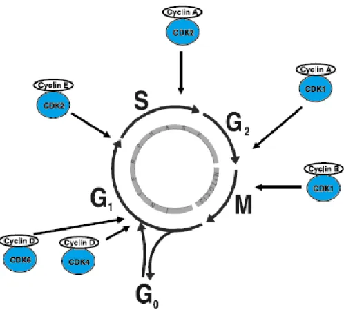
1.1.8.1 Cell cycle progressionThe cell cycle is a complex cellular process, which controls cells growth. It relies on DNA duplication (S phase) and chromosomal separation into daughter cells (M phase). These key events are spaced by gaps of growth and reorganization (G1 and G2 phases) After M phase, cell can either enter G1 or the GO quiescent phase (Figure 5). Cell cycle progression is regulated by cyclin-dependent kinases (CDKs) (Murray 2004). Activation of CDKs will lead to transition into the mitotic cell cycle, where association of CDKs with cyclins induces the kinases catalytic activity. Activation of CDKs is triggered by P38 mitogen-activated protein kinase (MAPK)/extracellular signal-regulated kinase (ERK)-mediated mitogenic signaling. Induction of mitogenic signaling pathways is associated with various cellular responses, such as proliferation, differentiation, survival, and transformation (McCubrey et al. 2006). Epidermal growth factor (EGF) and transforming growth factor beta 1 (TGF-β1) induce mitogenic signaling and tumor progression through ERK pathway activation (Thakur et al. 2009; Wilson et al. 2009). Previous studies have shown that these mitogenic factors are overexpressed in prostate cancer tissues and linked to advanced malignancy (Leav et al. 1998; Cardillo et al. 2000). IL-6 is overexpressed in prostate cancer tissues (Wegiel et al. 2008). A previous study of PC-3 xenografts treated with an anti-IL-6 antibody led to tumor suppression through inhibition of ERK phosphorylation (Steiner et al. 2003).
Cell cycle arrest occurs in response to cellular stress through activation of signal transduction pathways commonly known as checkpoints (Hartwell and Weinert 1989). The checkpoints are activated in the G1/S phase, which prevent replication of damaged DNA or in the G2/M phase to prevent damaged cells from entry to mitosis. Cell cycle checkpoints ensure the accuracy of DNA replication and division (Hartwell and Weinert 1989). Progression of cells from G2 into the M phase of the cell cycle requires activation of Cdk1/cyclin B kinase complex (Peng et al. 1997).
Figure 5: The stages of the cell cycle
Cell division is divided into two stages: a nuclear division stage, which is known as mitosis (M), and the interlude between two M phases, which is known as interphase. The interphase includes G1, S and G2 phases. DNA replication occurs in S phase. S phase is preceded by a gap called G1 during which the cell prepares for DNA synthesis and is followed by G2 gap during which the cell is preparing for mitosis. G1, S, G2 and M phases are the traditional subdivisions of cell cycle. Before commitment to DNA replication, cells in G1 can enter a resting state called G0. The site of activity of regulatory CDK/cyclin complexes is indicated in arrows (Vermeulen et al. 2003).
Cells can exit the cell cycle before mitogenic stimulation and entre G0, where several gatekeepers for cell cycle transitions are activated to avoid cell cycle progression. Retinoblastoma tumor suppressor protein (RB) is one of the key gatekeepers that inhibits target genes involved in DNA replication (e.g. cyclin A) (Knudsen and Knudsen 2006). Cancer cells deficient in RB are resistant to cell cycle arrest mediated by CDK inhibition (Lukas et al. 1995).
1.1.8.2 PI3K/Akt/mTOR signalling pathway
The PI3K/Akt/mTOR signalling pathway is a cascade of events that plays a key role in cancer (Figure 6). It is one of the most frequently targeted pathways in all human cancers. It has been estimated that mutations in the individual components of this pathway account for as much as 30% of all known human cancers (Shaw and Cantley 2006). This complex pathway modulates AR signaling and androgen responsiveness (Liao et al. 2004; Salas et al. 2004), but is also critical for cell adhesion, growth and apoptosis (Polakis 2000). Akt activates various factors through phosphorylation, which in turn regulate four broad processes: cell-cycle progression, cell survival, cell proliferation and cell metabolism. Activation of Akt is correlated with prostate cancer progression. It has been shown that AR is upregulated by Akt in prostate cancer cells (Ha et al. 2011). The mammalian target of rapamycin (mTOR) kinase is the catalytic subunit of two complexes, mTOR complex 1 and 2 (mTORC1/2) that regulate growth and are often dysregulated in disease (Zoncu et al. 2011). The mTORC1 is the target of the well-known drug rapamycin, and includes the mTOR catalytic subunit, which phosphorylates 70S ribosomal protein S6 kinase (p70S6K) and eukaryotic translation initiation factor 4E-binding protein 1 (4E-BP1). The rapamycin-insensitive mTORC2 activates Akt by phosphorylating serine 473 (Zoncu et al. 2011). Once the PI3K/Akt/mTOR pathway is activated, it plays a crucial role in cell division and metabolism, thus influencing the metastasis, invasion and aggressiveness of cancer cells. Targeting this pathway is an important therapeutic option for better prognosis in cancer patients (Falasca et al. 2011).
Figure 6: AKT/mTOR signaling pathway.
The PI3K/Akt pathway functions downstream of receptor tyrosine kinases (RTKs). Upon recruitment to the cell membrane, Akt is phosphorylated by phosphoinositide-dependent kinase 1 (PDK1), a reaction catalyzed by PIP3. Activated Akt is a kinase which in turn phosphorylates and activates many growth and survival pathways (Toren and Zoubeidi 2014)
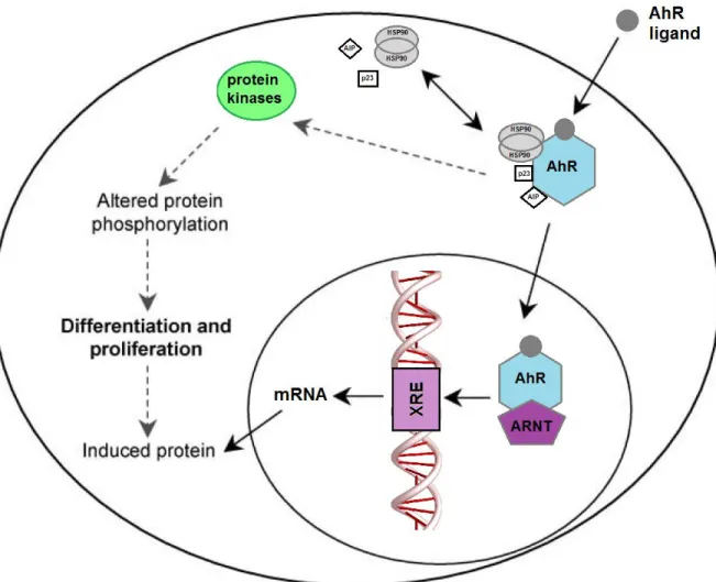
1.1.8.3 Aryl hydrocarbon receptor (AhR) signaling pathway
AhR is a basic helix-loop-helix (bHLH) protein located in the cytosol. The AhR is the only ligand-activated member of the bHLH family; it is activated by the binding of a wide range of environmental hydrocarbons (Safe 2001). In the cytosol, AhR is found in a complex that consist of two molecules of heat shock protein 90 (HSP90), co-chaperone p23, immunophilin-like AhR-interacting protein (AIP) and tyrosine kinase c-Src (Perdew
and Bradfield 1996). This protein complex is designed to maintain the inactive conformation and prevent nuclear translocation. Upon binding by polyaromatic hydrocarbons (PAHs) or halogenated aromatic hydrocarbons (HAHs), AhR dissociates from its chaperone proteins and translocated to the nucleus where the receptor heterodimerizes with the AhR nuclear translocator protein (ARNT) (Pollenz 1996) (Figure 7). The nuclear AhR complex interacts with xenobiotic response elements (XRE) in the promoter region of responsive genes, such as cytochrome P450 1A1 and 1B1 (CYP1A1 and CYP1B1) (Bacsi et al. 1995). Although extensively studied as a key regulator of xenobiotic metabolism, AhR has been shown to influence a number of cellular processes, including differentiation, proliferation and cell cycle progression. Activation of the AhR by exogenous ligands has been reported to antagonize AR signaling. AhR ligands are a structurally diverse group of natural and synthetic compounds that elicit a broad range of biological effects. AhR ligand such as the most potent HAHs, 2,3,7,8-tetrachlorodibenzo-p-dioxin (TCDD) inhibited testosterone-dependent transcriptional activity and testosterone-regulated PSA expression in a dose dependent manner (Jana et al. 1999). TCDD blocks androgen-dependent proliferation of prostate cancer cells (Barnes-Ellerbe et al. 2004; Richmond et al. 2014). TCDD itself is carcinogenic (Enan and Matsumura 1996) and, therefore, is not suitable as anti-cancer treatment. Previous studies showed that AhR is constitutively active in AI prostate cancer (DU145 and PC-3 cells) but not in AD prostate cancer cells (LNCaP) (Richmond et al. 2014). Recently, activation of AhR using an AhR agonist in LNCaP cells promoted its invasiveness as measured by cell migration on matrigel (Ide et al. 2017).
Figure 7: Aryl hydrocarbon receptor (AhR) signaling pathway
Binding of the ligand to the AhR results in the release of AhR-associated proteins and translocation of AhR to the nucleus and then dimerizes with ARNT. The AhR–ARNT complex binds the XRE promoting target gene transcription. Ligands can also exert their effects in the cytoplasm through AhR-associated protein kinases to alter the function of a variety of proteins through a cascade of protein phosphorylation (Pocar et al. 2005).
1.1.9 Current treatments for prostate cancer
Current therapies for prostate cancer include hormonal therapy, radiotherapy, surgery, chemotherapy, and immunotherapy. The most common hormonal therapies used to treat prostate cancer include chemical castration and treatment with AR antagonists, such as (hydroxy)flutamide and bicalutamide, or inhibitors of steroidogenesis. Inhibition of androgen synthesis by blocking the catalytic activity of SRD5A is another effective treatment against androgen dependent prostate tumors.
SRD5A inhibitors such as finasteride block synthesis of dihydrotestosterone (DHT), the major and most potent androgen produced by the prostate (Miyamoto et al. 2004). Finasteride and dutasteride are used clinically to inhibit SRD5A1/2 (Xu et al. 2006). Also, CYP17 inhibitors, such as abiraterone acetate, which block androgen synthesis improved overall survival in advanced prostate cancer patients (Fizazi et al. 2012).
Two phase I trials were conducted using abiraterone acetate in patients with progressive prostate cancer who were receiving androgen deprivation therapy (ADT), but had not received previous chemotherapy. In the first clinical trial, treatment with oral abiraterone acetate 250–2000 mg once daily, decreased serum testosterone levels to undetectable or near undetectable levels. Moreover, treatment with abiraterone acetate reduced PSA levels by ≥30% in 66% of recruited patients. Levels of other components of the androgen synthesis pathway were also decreased with abiraterone acetate therapy, including androstenedione and DHEA. In the second clinical trial, treatment with abiraterone acetate 250-1000 mg daily, reduced PSA levels by ≥50% in 55% of recruited patients; in addition, circulating cortisol and androgens levels were substantially decreased (Attard et al. 2008; Ryan et al. 2010).
Ironically, prostate cancers become apparent at an age when androgen levels in males have decreased and estrogen-to-androgen ratios have increased significantly, at least in serum (Ellem and Risbridger 2006). Estrogens play a role in prostate cancer progression, and aromatase (CYP19) inhibitors (which block estrogen synthesis) appear to offer protection (Narashimamurthy et al. 2004).
Over time, prostate cancers become refractory to hormone therapy and develop castration resistant phenotype. The poor prognosis of castration resistant prostate cancer (CRPC) has focused current research on drug therapies that delay progression to AI, or the use of cocktails of drugs with alternate targets for anticancer action (docetaxel/prednisone chemotherapy, selenium adjuvant therapy). Immunotherapy using the cancer vaccine sipuleucel-T has been approved by the US Food and Drug Administration (FDA) for the treatment of metastatic CRPC (Groves-Kirkby 2010; Drake et al. 2014; Mulders et al. 2015). Immunotherapy using immune checkpoint inhibitors has been effective in treating cancers such as metastatic melanoma (Hodi et al. 2010).
Immune checkpoint inhibitors block checkpoint proteins that prevent the immune system from attacking cancer cells. Checkpoint proteins include CTLA-4, PD-L1, (expressed on T cells), and PD-1 (expressed on tumor cells)(Venturini and Drake 2018). In recent years, immunotherapy has resulted in successful immune response to several types of tumors, such as metastatic melanoma, lung cancer and advanced renal cancer. Ipilimumab is the first antibody-based checkpoint inhibitor granted approval by FDA in 2011. Ipilimumab improved the survival of patients with metastatic melanoma (Hodi et al. 2010). However, immune checkpoint inhibitors appear to be less effective in treating prostate cancer (Goswami et al. 2016; Venturini and Drake 2018). In a recent phase 3 clinical trial, Ipilimumab (anti CTLA-4) did not improve survival of metastatic CRPC patients (Beer et al. 2017). Combination therapy is one alternative to overcome the lack of responsiveness to immune checkpoint monotherapy. Preclinical studies in mouse models of prostate cancer using immune checkpoint inhibitors combined with different cancer treatments such as surgical resection of primary tumor, cryoablation or tumor vaccines resulted in effective tumor immune response (Kwon et al. 1999; Curran and Allison 2009; Waitz et al. 2012).
A phase 3 clinical trial conducted using patients with CRPC showed that the CYP17 inhibitor abiraterone prolonged the survival time of patients by 4 months (de Bono et al. 2011). Recently, a phase 3 trial showed a prolonged survival with apalutamide (also a CYP17 inhibitor) in non-metastatic CRPC (Smith et al. 2018b). Another drug that is attracting attention for prostate cancer treatment is enzalutamide (an AR antagonist)(Nadal and Bellmunt 2016). Enzalutamide decreased the risk of metastasis and death in nonmetastatic CRPC patients (Hussain et al. 2018). A phase 3 trial conducted using 1199 men with CRPC after previous chemotherapy showed that enzalutamide prolonged the survival of metastatic CRPC patients after chemotherapy (Scher et al. 2012). Enzalutamide and abiraterone acetate have shown promising efficacies in clinical trials and the FDA has approved both drugs for treatment of CRPC. However, resistance to enzalutamide and abiraterone acetate treatment is still a challenge. Enzalutamide and abiraterone acetate resistance are associated with the presence of AR splice variants (AR-V7) (Antonarakis et al. 2014). Novel potential drugs,
such as phytochemicals, which are less toxic to healthy tissues, need to be evaluated to improve the clinical care of patients suffering from prostate cancer.
1.1.10 Causes of castration resistant prostate cancer
Various mechanisms contribute to CRPC, including (1) mutations in AR resulting in expression of constitutively active AR splice variant such as ARv7 that lacks the ligand binding domain (Gelmann 2002; Recouvreux et al. 2017), (2) overexpression of AR increasing its sensitivity to low androgen levels, thus impairing effectiveness of antiandrogen treatment (Linja et al. 2001), (3) induction of bypass pathways independent of AR such glucocorticoid receptor signaling (Kassi and Moutsatsou 2011), (4) increased intraprostatic androgen biosynthesis (Stanbrough et al. 2006) or (5) constitutive activation of cell proliferative pathways with gradual loss of AR (Gioeli 2010).
1.1.10.1
Cancer stem cells (CSCs) and CRPC
CSCs are a small subpopulation of cells within a tumor capable of self-renewal, tumor initiation and differentiation into various cell types that constitute the tumor (Yu et al. 2012). A great challenge for current therapies of advanced prostate cancer is the development of drug resistance. Accumulated experimental evidence has demonstrated differences in drug resistance among different clones of a tumor (Carreira et al. 2014; Tereshchenko et al. 2014; Bansal et al. 2016). Previous studies suggest that a subset of such clones represent resistant CSCs (Ojha et al. 2014; Leao et al. 2017; Howard et al. 2018). The cellular origin of CRPC is still controversial, but several studies have found CSCs in CRPC (Vander Griend et al. 2008; Qin et al. 2012). In addition, CSCs appear to contribute substantially to drug resistance in multiple cancer types including that of prostate (Ni et al. 2014). Therefore, targeting CSCs represent a potential strategy for prostate cancer treatment (Yun et al. 2016).
1.1.11 TP53 mutations and prostate cancer
The TP53 gene is a tumor suppressor gene. p53 protein regulates cell division by preventing uncontrolled cell division and prevents the formation of tumor. P53 suppress tumor formation through 3 main mechanisms: cell cycle arrest, triggering apoptosis,
activation of DNA repair (Brady and Attardi 2010).The TP53 gene is mutated in approximately 30% of prostate tumors (Ecke et al. 2007). Analysis of the genomically aberrant pathways in metastatic CRPC revealed that the frequency of TP53 mutations was significantly increased among metastatic CRPC samples (53.3%) compared to primary prostate cancer (Robinson et al. 2015). In addition, mutations in TP53 contribute to tumor progression and drug resistance, and is often associated with increased risk of recurrence (Olivier et al. 2010). Moreover, p53 mutated proteins acquire new activities referred to as gain of function that contribute to tumor progression and increased resistance to cancer therapeutics (Santoro et al. 2014).
Mutations in TP53 influence multiple cellular process including cell proliferation, apoptosis, autophagy, DNA damage and senescence (Aubrey et al. 2016).
P53 mutants with gain of function inhibit the formation of autophagic vesicles through repression of some key autophagy genes (BECN1 and ATG12) (Cordani et al. 2016). In vitro and in vivo studies demonstrated that mutant p53 promoted cell proliferation, and inhibited apoptosis and senescence (Murphy et al. 2000; Matas et al. 2001; Lehmann et al. 2007; Duan et al. 2008). It has been shown that two TP53 mutants (p53-223Leu and p53-274Phe) inhibit Fas expression in DU145 cells, which contributes to apoptosis resistance in prostate carcinoma cells (Gurova et al. 2003). In addition, a previous study demonstrated that targeting mutant p53 using RNA interference induced apoptosis in DU145 cells (Zhu et al. 2011).
1.2 DIINDOLYLMETHANE
Cruciferous vegetables contain large amounts of indole-3-carbinol (I3C), which in the stomach undergoes acid-catalyzed condensation reactions to produce various metabolites, of which a major condensation product is diindolylmethane (DIM) (Figure 8) (De Kruif et al. 1991b). Particularly DIM is considered to be the most abundant biologically active metabolite formed in the stomach (Reed et al. 2006).Oral administration of I3C in mice showed that I3C is not detected in plasma or tissues within 1 hour of treatment. However, DIM reached a maximum concentration in plasma after 2 hours of treatment and remained detectable as a major bioactive compound in plasma after 6 hours