HAL Id: tel-01195989
https://tel.archives-ouvertes.fr/tel-01195989
Submitted on 9 Sep 2015
HAL is a multi-disciplinary open access archive for the deposit and dissemination of sci-entific research documents, whether they are pub-lished or not. The documents may come from teaching and research institutions in France or abroad, or from public or private research centers.
L’archive ouverte pluridisciplinaire HAL, est destinée au dépôt et à la diffusion de documents scientifiques de niveau recherche, publiés ou non, émanant des établissements d’enseignement et de recherche français ou étrangers, des laboratoires publics ou privés.
Weihua Chen
To cite this version:
Weihua Chen. Role of RhoA/ROCK pathway in angiogenesis and their potential values in prostate cancer treatment. Tissues and Organs [q-bio.TO]. Université René Descartes - Paris V; Tongji uni-versity (Shanghai, Chine), 2014. English. �NNT : 2014PA05T047�. �tel-01195989�
UNIVERSITÉ PARIS DESCARTES
FACULTÉ DE MÉDECINE PARIS DESCARTES
THÈSE
Pour obtenir le grade de
DOCTEUR
en Sciences de la Vie et de la Santé
Ecole Doctorale : Génétique Cellulaire, Immunologie, Infectiologie et
Développement (GC2ID)
Spécialité : Physiopathologie
présenté et soutenu par
Weihua CHEN
Le 20 octobre 2014
Role of RhoA/ROCK pathway in angiogenesis and
their potential values in prostate cancer treatment
Jury
Directeur de thèse : Pr Anh Tuan DINH-XUAN
Directeur de thèse : Pr Zhongmin LIU
Rapporteur :
Pr Morgan ROUPRÊT
Rapporteur :
Pr Zhichen GUAN
PARIS DESCARTES UNIVERSITY
PARIS DESCARTES FACULTY OF MEDECINE
THESIS
For the doctoral degree
on Life Sciences and Health
Doctoral School: GC2ID
Specialty: Pathophysiology
Presented and defensed by
Weihua CHEN
October 20, 2014
Role of RhoA/ROCK pathway in angiogenesis and
their potential values in prostate cancer treatment
Jury
Thesis Director:
Prof Anh Tuan DINH-XUAN
Thesis Director:
Prof Zhongmin LIU
Repportor :
Prof Morgan ROUPRÊT
Repportor :
Prof Zhichen GUAN
3
This is a cotutorial thesis between Paris Descartes University and Tongji University. This thesis has been carried out jointly with the following laboratory: UPRES EA2511(Paris Descartes University), Sino-french Physiology and Pathophysiology Laboratory
(Shanghai East Hospital, Tongji University) and Clinical and Translational Research Centre (Shanghai East Hospital, Tongji University).
4
ACKNOWLEDGEMENT
First and foremost, I would like to express my sincere gratitude to my research
supervisors Prof. Anh-Tuan DINH XUAN and Prof. Zhongmin LIU, for their continuous support of my Ph.D study and research, for their patience, motivation, enthusiasm, and immense knowledge. Their guidance helped me in all the time of research and writing of this thesis. Without their assistance and dedicated involvement in every step throughout the process, this paper would have never been accomplished. I could not have imagined having a better director and mentor for my Ph.D study.
Besides my directors, I would like to thank the rest of my thesis committee: Prof. Morgan ROUPRÊT, Prof. GUAN Zhichen and Prof. Michaël PEYROMAURE for their attendance to the jury of my defence, to their encouragement, insightful comments and questions. I would like to express the special appreciation to Pr. Bernard DEBRE, who has set up sino-french urological centre in Shanghai with his cooperators and introduced me to study in France.
I would like to express the appreciation to Dr. Kaili MAO, who brought me to Paris 3 years ago. He and his cooperators built the sino-french scientific team and research platform for this project. He was always supporting and encouraging me with his scientific ideas and administrative ability in these 3 years.
I am grateful for Thong Hua-Huy, for his help almost all the time during my three years study, for so many precious advices he has given on my experiments, for analyzing so many results with me together, for the week-ends and holidays we were working together.
5
I also thank the director of our laboratory Pr Isabelle Fajac, and the rest of my fellow labmates in our laboratory: Dr Clémence Martin, Dr Yihua BEI, Dr Justine Frija-Masson, Muriel Dambo, Guiti Thevenot, Van-ha Hoang for the stimulating discussions, and for all the fun we have had in the past three years.
I would like to express the deepest appreciation to the colleagues in the Urology Department and Pathology Department of Paris COCHIN Hospital, Dr. Nicolas BARRY DELONGCHAMP, Dr. Frédéric BEUVON for their deeply participation in the project and analysis of the clinical and pathological data, for their help and support constantly during the clinical part of the project.
I would like to offer my special thanks to my colleagues in our urology department of Shanghai East Hospital for their support during my thesis research.
Last but not the least, I would like to thank my parents Yirong CHEN and Huolian YOU, for supporting me spiritually throughout my life. I would like to thank my wife Yinghong SHEN, my daughter Huizhe CHEN for their encouragement and support during the past three years.
Also my sincerely thank to all the persons who has given me so many helps during my thesis study.
This project has been supported by a grant from the Program of International Science & Technology Cooperation (Grant Number: 2012DFG31440), awarded by the Ministry of Science and Technology, People s Republic of China.
6
ABSTRACT
Prostate cancer remains a major cause of mortality among males in western countries. Treatment options for metastatic castration-resistant disease remain limited. There is a continuing unmet need for new systemic interventions in patients with progressive prostate cancer.
RhoA/Rho-associated protein kinases (ROCK) are key regulators of the cytoskeleton and have been implicated in PCa angiogenesis and tumour invasion. In the first study (Part I), we
investigated the anti-angiogenic effects of fasudil, a ROCK inhibitor, on PCa-induced angiogenesis in vitro. Proliferation of PCa-conditioned human umbilical vein endothelial cells (HUVECs) was assessed using a bromodeoxyuridine (BrdU) assay, and migration was assessed with a wound healing assay. In vitro angiogenesis of PCa-conditioned HUVECs was evaluated by tube formation and a spheroid sprouting assay. Fasudil inhibited PCa-induced endothelial cell proliferation, and also decreased PCa-induced endothelial cell migration. In the in vitro angiogenesis assay, tube formation and spheroid sprouts were significantly inhibited by fasudil in a dose dependent manner. Western blotting results showed that expression of phosphorylated myosin phosphatase target subunit 1 (MYPT-1) was significantly lower after fasudil treatment, confirming that fasudil inhibited ROCK activity in these model systems. In the second study (Part II & III), we evaluated RhoA/ROCK expression and RhoA activity in a total of 34 paraffin embedded and 20 frozen prostate specimens, respectively, obtained from 45 patients treated with radical prostatectomy for clinically localized cancer. The expression patterns of RhoA/ROCK were tested by
immunohistochemical staining and Western blotting, and further compared between the tumour centre, tumour front and distant peritumoral tissue. RhoA activity was assessed by G-LISA. Our results showed an increasing gradient of expression from the centre to the periphery of index tumour foci. RhoA expression was indeed significantly higher at the tumour front compared to tumour centre, using immunohistochemistry (p=0.001). Also, Gleason scores were significantly higher in patients with higher RhoA expression in both tumour front and tumour centre (p=0.044 and 0.039, respectively). After a median follow-up of 52 months, the rate of PSA relapse was higher in patients with a higher RhoA expression at the tumour front (62.5% vs 35%), although the difference was not significant (p=0.089). There was no association between RhoA expression and PSA, pathological stage. We also found ROCK2 expression, but not ROCK1 expression, was significantly higher in the prostate cancer tumor front. In conclusion, we found fasudil
significantly inhibits the key steps of endothelial cell angiogenesis, including proliferation, migration, capillary tube formation and spheroid sprouting, in a dose-dependent manner. These effects may due to inhibition of ROCK activity induced by PCa cell secretions. We also identified higher RhoA and ROCK2 expression in human prostate tumour front. The correlation of higher RhoA expression with higher Gleason score and higher rate of cancer relapse. This indicated the association of RhoA/ROCK2 pathway with aggressiveness of prostate cancer. The insights described here may provide the foundation for novel therapeutic approaches targeting
RhoA/ROCK pathway to inhibit angiogenesis and clinically aggressiveness of PCa. Fasudil may be a useful anti-angiogenic agent and should be investigated further for its potential role in PCa treatment.
7 TABLE OF CONTENTS
Acknowledgement ... 4
Abstract... 6
Table of Contents ... 7
List of Figures ... 9
Abreviations ... 11
Introduction ... 13
1 Epidermiology of prostate cancer ... 14
2 Current status of prostate cancer treatment ... 14
3 Hallmarks in cancer development ... 16
4 Angiogenesis in cancer development ... 17
5 Angiogenesis in prostate cancer ... 21
6 Anti-angiogenic therapy in prostate cancer ... 30
7 Invasion in cancer development ... 31
8 Invasion in prostate cancer ... 33
9 RhoA/ROCK signaling pathway ... 34
10 RhoA/ROCK pathway in cancer angiogenesis... 52
11 RhoA/ROCK pathway in prostate cancer angiogenesis ... 56
12 RhoA/ROCK pathway in cancer invasion ... 57
13 RhoA/ROCK pathway in prostate cancer invasion ... 59
Aims of the work ... 61
Results ... 62
PART I Article I Fasudil inhibits prostate cancer-induced angiogenesis in vitro... 63
PART II Article II High RhoA expression at the tumour front but not in the center of prostate cancer ... 72
PART III Other results ... 80
Discussion ... 87
8
Targeting RhoA/ROCK pathway inhibited prostate cancer angiogenesis ... 90
RhoA/ROCK pathway involved in prostate cancer invasion ... 91
Targeting RhoA/ROCK pathway in the treatment of prostate cancer ... 95
Conclusions and perspectives ... 96
References ... 99
9
LIST OF FIGURES
Figure 1 The hallmarks of cancer (Hanahan and Weinberg 2011). ... 16
Figure 2 The balance of the angiogenesis control. ... 18
Figure 3 Mechanisms regulating angiogenesis in PCa metastasis(Li and Cozzi 2010). ... 23
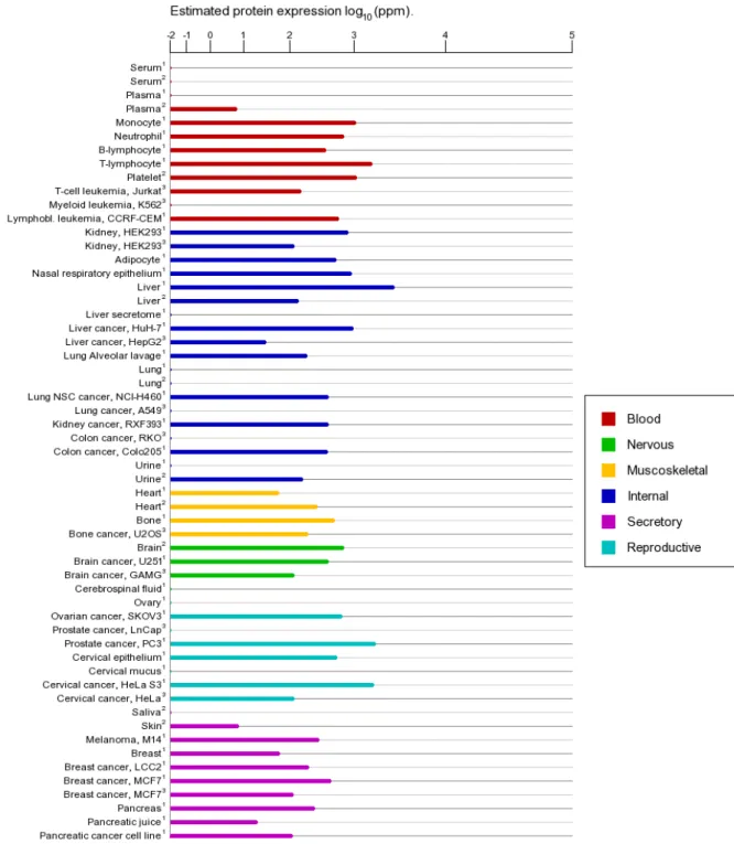
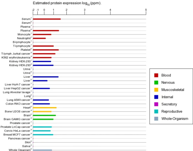
Figure 4 Expression of RhoA in various tissues and cells in human (BioGPS. 2014). ... 36
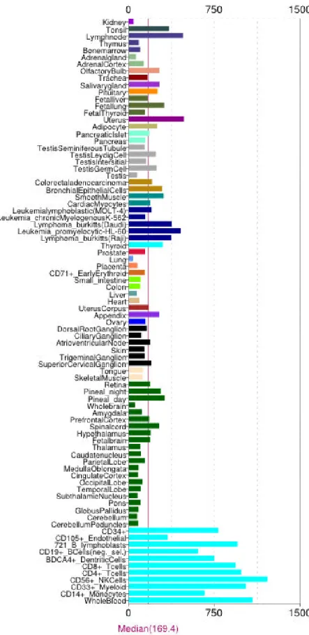
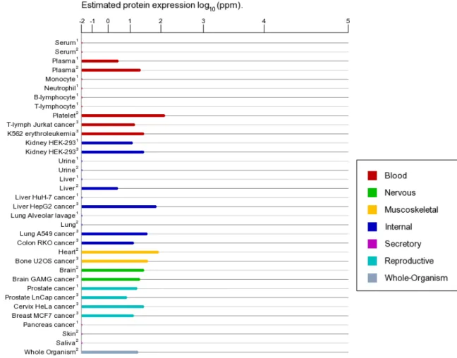
Figure 5 Expression of RhoA in human cancer cells and tissues (GeneCards. 2014). ... 37
Figure 6 Regulation of RhoA activity. ... 38
Figure 7 Illustraction of RhoA functioin and the main signaling molecules involved. ... 41
Figure 8 ROCK structure. ... 43
Figure 9 The molecular structure of ROCK1 and ROCK2. ... 43
Figure 10 ROCK1 expression in human tissues (BioGPS. 2014) ... 44
Figure 11 ROCK2 expression in human tissues (BioGPS. 2014) ... 45
Figure 12 ROCK1 expression in human cancer (GeneCards. 2014) ... 46
Figure 13 ROCK2 expression in human cancer (GeneCards. 2014) ... 47
Figure 14 The mechanism of positive regulation of ROCKs activity(Duong-Quy, Bei et al. 2013). ... 49
Figure 15 Role of RhoA/ROCK pathway in the actin–myosin-based contractile force generation ... 51
Figure 16 Illustration of the RhoA/ROCK pathway in the angiogenesis. ... 55
Figure 17 ROCK signaling and the leading edge(Schofield and Bernard 2013). ... 59
Figure 23 RhoA expression in the capillary endothelial cells in human PCa tissue. ... 82
Figure 24 ROCK1 expression in the capillary endothelial cells in human PCa tissues. ... 82
Figure 25 ROCK2 expression in the capillary endothelial cells in human PCa tissues. ... 83
10
11
ABREVIATIONS
AA arachidonic acid
ADT androgen deprivation therapy bFGF basic fibroblast growth factor BPH benign prostate hyperplasia CALGB Cancer and Leukemia Group B Cdc42 cell division control protein 42
CRD cystein-rich domain
CRIK citron kinase
CRPC castration-resistant PCa DMPK myotonic dystrophy kinase DRFs diaphanous-related formins
EC endothelial cell
EMT epithelial-mesenchymal transition
ERM ezrin/radixin/moesin
FERM N-terminal 4.1/ezrin/radixin/moesin GAP GTPase-activating proteins
GDI guanine nucleotide dissociation inhibitor
GDP guanosine diphosphate
GEF guanine exchange factor
GTP guanosine triphosphate
GTPase guanosine triphosphate hydrolase HGF hepatocyte growth factor
LIMK LIM-Kinase
MAP mitogen-activated protein
MBS myosin binding subunit
MCP-1 monocyte chemoattractant protein 1
MLC myosin light chain
MLCK MLC kinase
MLCP MLC phosphatase
MRCK
DMPK-related cell division control protein 42 binding kinase
MVD microvessel density
MYPT-1 myosin phosphatase target subunit 1 PC-3 human prostate cancer cell line
PC3CM PCa cell line PC3-conditioned medium
12
PDGF platelet-derived endothelial cell growth factor PDGF-a platelet-derived growth factor alpha
PH pleckstrin-homology
PKN protein kinase N
pMYPT-1
phosphorylated myosin phosphatase target subunit 1
PRK protein kinase C-related kinase PSMA prostate specific membrane antigen
RBD Rho-binding domain
Rho Ras homology
RhoA Ras homolog gene family, member A ROCK Rho associated coiled-coil kinase
RP radical prostatectomy
SPC sphingosine phosphorylcholine
SRF serum response factor
TNF-a tumour necrosis factor alpha
TRAMP transgenic adenocarcinoma of the mouse prostate
VE vascular endothelial
VE-Cadherin vascular endothelial (VE) cadherin VEGF vascular endothelial growth factor
14
1 EPIDERMIOLOGY OF PROSTATE CANCER
Prostate cancer (PCa) is the most prevalent malignancy in men in Western countries (Siegel, Naishadham et al. 2013). In France, 56800 PCa are newly diagnosed in the year of 2012, with age standardized incidence about 100 per 100,000 (Invs 2013). While in the other western European countries the incidence rate varies between 73.1 and 104.2 per 100,000 in the year of 2008 (UK 2012). It is the second leading cause of
cancer-related death in this area. 8900 cases of PCa related death are recorded in France in 2012. Several autopsy studies have shown that 60-70% of older men have histological PCa(Heidenreich, Bastian et al. 2014). PCa is diagnosed in 15-20% of men during their lifetime (Heidenreich, Bastian et al. 2014). In China, the incidence of PCa increased rapidly during the past decades, partly because of the application of PSA screening in the elder male patients. 994 cases of prostate cancer were diagnosed in Shanghai in 2009, with an incidence of 32.23 per 100,000, fifth cause of tumour in men ("Incidence of malignancy". 2012).
2 CURRENT STATUS OF PROSTATE CANCER TREATMENT
Data from autopsy showed that advanced and metastatic stages of the disease are found in 35% of patients with PCa (Bubendorf, Schöpfer et al. 2000). With the widely
application of PSA screening, the diagnosis of PCa becomes earlier. But till now, there are still a rather large portion of patients are diagnosis as advanced stage, as reported 11.7% in USA and 31.2 in Europe in 2013. A multicenter report in China showed at least 36.2% of PCa patients were diagnosed at advanced stage (Peyromaure, Debre et al. 2005).
15
For PCa patients who are diagnosed with early stage, therapeutic options include radical prostatectomy (RP), radiotherapy, and active surveillance. Active surveillance has become more accepted by the patients and doctors. But it is still strictly limited to some of the low risk patients who don t want to receive the operation immediately and who can comply with regular follow-up under surveillance. Although there is some
controversy on the treatment choice between RP and radiotherapy, RP is still considered as the best treatment for localized early stage patients. It has also been reported to have a little benefit on overall survival as compared with radiotherapy in a 15 years long term cohort study (Sooriakumaran, Nyberg et al. 2014). Thus, RP is the most common treatment choice for these patients. Even for patients who are eligible for radical
prostatectomy, approximately 35% will develop recurrence (metastatic disease) within 10 years of surgery (Hull, Rabbani et al. 2002, Roehl, Han et al. 2004).
For most of the patients who present with or progress to advanced or metastatic disease, hormone therapy can effectively palliate the symptoms. This is also referred to androgen deprivation therapy (ADT), including castration with or without anti-androgens.
However, there is currently no conclusive evidence to show that it extends life.
Furthermore, the median duration of response to ADT is limited to between 8 months and 3 years (Daneshgari and Crawford 1993) and these patients will eventually become castration resistant. When the patients progress to CRPC, there are few effective
alternative treatments for them, such as chemotherapy. Chemotherapy regime including docetaxel is effective treatment for castration-resistant PCa. Docetaxel was shown to mildly prolong survival in patients with CRPC. Despite demonstrating an improvement in overall survival, responses are not durable and eventual progression of the disease is inevitable. The median duration of response is only 10.3 months (Eymard, Oudard et al. 2010). As regard to the high incidence rate and large portion of progressive stage
16
patients, there is clearly an urgent need to develop additional systemic interventions for these patients.
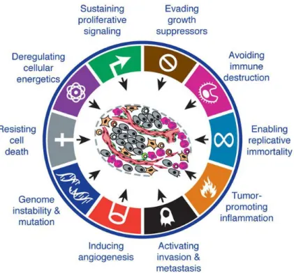
3 HALLMARKS IN CANCER DEVELOPMENT
There are ten hallmarks which represent the crucial biological capabilities acquired for the multistep development of human tumours, including sustaining genome instability & mutation, escaping growth suppressors, evading cell death, enabling replicative
immortality, proliferative signaling, deregulating cellular energetics, promoting inflammation, inducing angiogenesis, avoiding immune destruction and activating invasion and metastasis. Many new therapies are developing targeting these hallmarks. Many studies have been conducted to interfere with these acquired capabilities in the tumour growth and progress. Researches targeting angiogenesis and tumour metastasis are among those which are actively being pursued.
17
4 ANGIOGENESIS IN CANCER DEVELOPMENT
The tumour-associated neovasculature is a prerequisite for the expansion of solid tumours beyond 1-3 mm3(Folkman, Cole et al. 1966). During the very early stage of the
tumour, these lesions may stay dormant by reaching a steady state between growth and apoptosis. Dormant tumours have been discovered in autopsies of patients who died of causes other than cancer (Black and Welch 1993). The tumour-associated
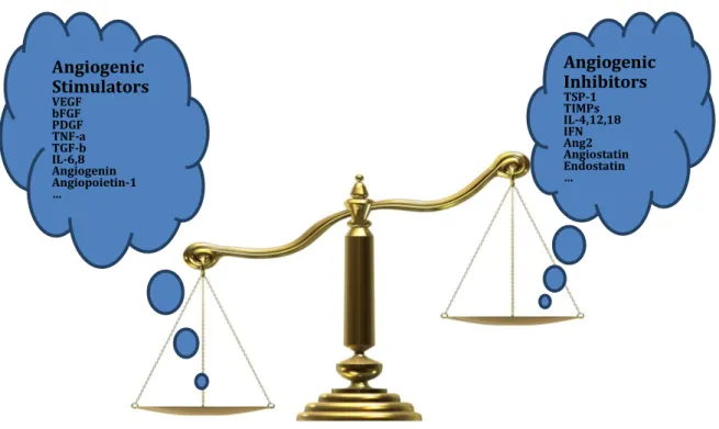
neovasculature mainly generated by the process of angiogenesis. During embryogenesis, neovasculature includes the birth of new endothelial cells and their assembly into new vessels (vasculogenesis) as well as the sprouting (angiogenesis) of new vessels from existing ones. In the adult, the normal vasculature becomes largely quiescent. This
balance of angiogenesis is sophistically governed by the balance of counteracting factors: pro-angiogenic and anti-angiogenic factors. Only some of physiologic processes such as female reproductive cycling or wound healing, pro-angiogenetic factors are only
transiently elevated, resulting in transiently turn on of angiogenesis. These are strictly controlled by the balance of angiogenic stimulators or inhibitors. In contrast, during solid tumour progression, in which consistently elevated pro-angiogenetic factors are always found, angiogenesis is nearly always activated and remains on, causing
continually sprouting of new vessels into the expanding neoplasm (Hanahan and Folkman 996 . This step has been termed as angiogenic switch Fig. . Tumours acquire the ability to recruit their own blood supply is one of the important hallmarks in the progress of the tumour (Fig. 1).
18
Figure 2 The balance of the angiogenesis control.
4.1 CELLS INVOLVED IN TUMOUR ANGIOGENESIS
Angiogenesis is a multiple step process involving a complex series of events. Firstly, an increase in the permeability of the basement membrane allows a new capillary to sprout. Next, endothelial cells(ECs) activated by angiogenic factors migrate through the
basement membrane into the extracellular matrix, towards the angiogenic stimulus. The leading front of migrating cells is driven by enhanced proliferation of ECs. This is then followed by re-organization of ECs to form tubules with a central lumen, together with the recruitment of peri-endothelial cells (pericytes) and vascular smooth muscle cells for new capillary stabilization(Sakamoto, Ryan et al. 2008). Besides cancer cells, many other cells in the cancer microenvironments including, fibroblast, inflammatory cells, pericytes and immunocytes take part in this process.
Tumours growth results in the increasing of diffusion distances from the existing vascular and decreasing oxygen supply. Tumour hypoxia can also arise as a result to
Angiogenic Inhibitors TSP-1 TIMPs IL-4,12,18 IFN Ang2 Angiostatin Endostatin … Angiogenic Stimulators VEGF bFGF PDGF TNF-a TGF-b IL-6,8 Angiogenin Angiopoietin-1 …
19
increased metabolic activity and oxygen consumption by rapidly proliferating tumour cells. As a reaction to hypoxia, the transcription factor hypoxia inducible factor (HIF) -1 is activated. Among which, HIF- is the most ubiquitously expressed, and functions as the master regulator of oxygen homeostasis, which is also the key regulator of
hypoxia-induced angiogenesis (Krock, Skuli et al. 2011). HIF-1 can directly activate many gene transcription by HRE binding (Manalo, Rowan et al. 2005). Multiple HIF-1 target genes have been demonstrated to modulate angiogenesis. These lead to induction of VEGF RNA stability, transcription and translation. It also reduces the levels of the endogenous anti-angiogenesis factor TSP1. Some other pro-angiogenic factor expression also increase due to HIF-1 activation. Tumour cell gene instability and mutation may be another reason for the elevated other pro-angiogenic factor expression of tumour cells. There are abundant reports on elevated pro-angiogenic factors secretion by tumour cells (Santin, Hermonat et al. 1999).
Stromal fibroblasts in tumour tissue also plays an important role in tumour angiogenesis. During tumour growth, fibroblasts become activated, secreting various ECM
components, such as collagens, fibronectin, heparan sulfate proteoglycans, which are very important to provide a support to neovasculature. These cells also secrete soluble angiogenic growth factors such as VEGF (Kellouche, Mourah et al. 2007), transforming growth factor- (TGF- ) (Paunescu, Bojin et al. 2011), and platelet-derived growth factor (PDGF) (Paunescu, Bojin et al. 2011), Several studies have reported that these
fibroblasts, also called carcinoma-associated fibroblasts (CAFs), promote tumour growth partially through promotion of angiogenesis (Newman, Nakatsu et al. 2011).
Since long time, there have been many researches on the relation between inflammation and cancer. The inflammation in some organs may increase the risk of tumour. In the tumour microenvironment. Inflammation also contributes to tumour angiogenesis, as
20
well as metastasis etc. It is known as cancer-related inflammation (CRI). Characteristics of cancer-related inflammation include the infiltration of white blood cells, prominently tumour-associated macrophages (TAMs), the presence of inflammation factors such as cytokines (TNF, IL-1, IL-6), which can promote angiogenesis in the tumour stroma. Recently, more and more evidences were shown that one subset of monocytes
expressing the Tie2 receptor (TEM) has a crucial role in tumour angiogenesis (De Palma, Murdoch et al. 2007) (De Palma, Venneri et al. 2005). These peri-tumoural inflammatory cells, as well as inflammatory factors which they have secreted, help to trigger the
angiogenic switch in previously quiescent tumour tissue and to maintain ongoing angiogenesis along with tumour growth.
4.2 OTHER MECHANISM OF THE FORMATION OF TUMOUR-ASSOCIATED VASCULATURE
Besides angiogenesis, there are several other mechanisms also involved in tumour vessel formation, including vascular co-option, mosaicism, vasculogenic mimicry and postnatal vasculogenesis.
4.2.1 VASCULAR CO-OPTION
Some tumours can grow and metastasize without angiogenesis. Studies have found an alternative way for the tumour to gain its blood supply, vessel co-option. It means tumour cells migrate to and along the preexisting blood vessels of the host organ. Vessel co-option occur in many malignancies, and may be more important in some highly vascularized tissues such as lung, brain, and liver. It may also be important as a potential mechanism of anti-angiogenic drug resistance, as it has been reported that
anti-angiogenic therapy promoted tumour invasion and metastasis by facilitating the tumour cells migration towards the less hypoxia region.
21
4.2.2 VASCULOGENIC MIMICRY
Vasculogenic mimicry (VM) is a newly-defined pattern that tumour gains ts blood supply. Cancer stem cells (CSCs) are found to have the capacity of self-renewal and multipotent differentiation, and may contribute to the new vessels formation in the tumour tissue. It has been known for many years that tumour endothelia has differences in molecular markers and the phenotype with normal endothelium. While some of them can be found in malignant cells (Hida, Hida et al. 2004). A fraction of the tumour vascular endothelial cells has been proven to be of neoplastic origin (Pezzolo, Parodi et al. 2007). CSCs have been proved to be capable of forming functional blood vessels de novo by
trans-differentiating into EPCs, ECs, or vascular smooth muscle-like cells. 4.2.3 VASCULOGENESIS
Tumour vasculogenesis means that new vasculatures are constituted via recruitment of endothelial progenitor cells (EPCs) from the bone marrow. Peripheral tissue hypoxia results in increased secretion of EPC-mobilizing factors (for example granulocyte colony–stimulating factor (G-CSF), vascular endothelial growth factor(VEGF), basic fibroblast growth factor (bFGF), placental growth factor, erythropoietin or SDF-1) which can mobilize EPC in the BM, leading to an increase of EPC in blood (De Falco, Porcelli et al. 2004). Once in circulation, EPCs response to chemokine signaling in the tumour tissue and home to the tumour site. They cross the endothelial monolayer, migrate through the blood vessel basement membrane and through the interstitial extracellular matrix (ECM) alonging the gradient of chemokines to exert their functions.
5 ANGIOGENESIS IN PROSTATE CANCER
Angiogenesis plays a critical role in cancer progression and metastasis, and its
22
was more prominent in carcinomas than in benign prostatic hyperplasia (BPH) and normal tissue (Bigler, Deering et al. 1993, Stefanou, Batistatou et al. 2004). It has been reported that MVD increases with increased Gleason's score, especially in poorly differentiated PCa (Weidner, Carroll et al. 1993). Increased MVD was significantly associated with high-grade carcinomas (Stefanou, Batistatou et al. 2004). Weidner showed that MVD was significantly increased in prostate cancer tissues of those patients with metastatic disease as compared with those without metastasis (Weidner, Carroll et al. 1993). Borre et al reported that MVD of PCa samples was significantly correlated with stage, grade and disease specific survival in 221 prostate cancer patients with a median followup of 15 years (Borre, Offersen et al. 1998).
5.1 REACTIVE STROMA
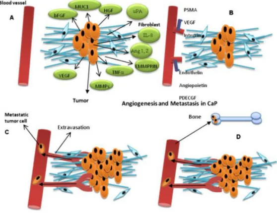
Till now, mechanism of angiogenesis in PCa has not yet been well elucidated. Although there are several mechanisms of tumour neovascularization, only angiogenesis was well explored in PCa. Microenvironment of PCa comprises a reactive stroma, characterized by an increasing number and abnormal function in myofibroblasts and fibroblasts, a corresponding augmentation in extracellular matrix components production, and an increase in microvessel density (Tuxhorn, Ayala et al. 2002). Prostate cancer cells, together with cells in their reactive stroma, secreted increasing level of angiogenic stimulator and decreasing level of angiogenic inhibitor, resulting in the angiogenic switch in local foci. Many stimulators were found to be significantly higher in the PCa tissue, including HIF-1, VEGF, bFGF, PDGF, IL-8 etc.
23
Figure 3 Mechanisms regulating angiogenesis in PCa metastasis(Li and Cozzi 2010).
5.2 ANGIOGENIC STIMULATORS IN PROSTATE CANCER 5.2.1 HIF
Huss et al. demonstrated that the increased expression of HIF-1 and VEGFR-1 in the early stage of angiogenic switch in transgenic adenocarcinoma of the mouse prostate (TRAMP) model, resulting in the increase in neovasculature in high grade prostatic intraepithelial neoplasm and prostate cancer lesions (Huss, Hanrahan et al. 2001) . In PCa patients, the accumulation of HIF-1a protein has also been reported to be an early event in PCa as well as in HGPIN (Hao, Chen et al. 2004) (Zhong, Semenza et al. 2004). Increase expression of HIF-1a directly lead to increasing transcription of the angiogenic factor VEGF. The over expression of VEGF in PCa is associated with the extent of
24
tumour hypoxia, with according overexpressed HIF-1a in PCa tissues in comparison with benign prostatic tissues (Cvetkovic, Movsas et al. 2001).
5.2.2 IGF-1
IGF-1 is a stimulator of cellular proliferation and cell survival as well as a stimulator of angiogenesis. It has been reported that local levels of IGF-1 and its receptor (IGF-1R) are both increasing during progression of PCa in TRAMP model (Kaplan, Mohan et al. 1999). In this model, serum IGF-I levels correlate with the increase in MVD associated with the development of PCa lesions, indicating the role of IGF-I in the induction of prostatic neovascularization. Clinical study also found increased levels of IGF-I in men with PCa compared with controls (Chokkalingam, Pollak et al. 2001, Woodson, Tangrea et al. 2003). Higher levels of serum IGF-1 have been proven to be a strong risk factor of developing PCa in the aged population (Chan, Stampfer et al. 1998). IGF-1 can
upregulate the expression of modulators of endothelial cell function such as VEGF, which has been postulated to be the main downstream events (Akagi, Liu et al. 1998) (Fukuda, Hirota et al. 2002).
5.2.3 ANDROGENS
Androgens have been proven to regulate the vasculature both in PCa animal model and PCa patients. Androgens have been shown to be involved in the up-regulation of HIF-1 expression in PCa (Mabjeesh, Willard et al. 2003). Androgens have also been reported to regulate VEGF expression thus influenced angiogenesis (Cheng, Zhang et al. 2004). Both in the glandular epithelial cells and in smooth muscle cells of the prostate, the secretion of VEGF is regulated by androgens. Richard et al demonstrated an androgen-mediated mechanism of VEGF modulation in a PCa mouse model (Richard, Kim et al. 2002). Androgen deprivation resulted in decrease angiogenesis in PCa in animals models
25
(Cheng, Zhang et al. 2004). These results indicated that androgen has an important role in the formation of PCa neovsculature and this takes effects mainly through VEGF pathway.
5.2.4 ENDOTHELINS
The endothelin (ET) pathway axis, which includes the biological functions of ETs and their receptors (ETAR and ETBR), acts as a modulator of vasomotor tone, tissue differentiation and development, cell proliferation and hormone production. They are also linked to angiogenesis. Three isoforms of endothelins, ET-1, 2, and 3 are secreted by epithelial cells(Kopetz, Nelson et al. 2002). They interact with two membrane-bound receptors, endothelin A receptor (ETAR) and endothelin B receptor (ETBR). ET-1 stimulates ETAR to induce VEGF release leading to an angiogenesis response
(Goligorsky, Budzikowski et al. 1999, Salani, Taraboletti et al. 2000). ET-3 interacts with ETBR to promote endothelial cell growth and new blood vessel formation(Lara, Twardowski et al. 2004). In PCa, key molecules of the ET-1 clearance pathway, neutral endopeptidase, are impaired, resulting in an accumulation in local ET-1
concentrations(Bagnato and Rosanò 2008). It has been reported that ET-1 secreted by prostate cancer cells promotes neovascularization via ETAR and ETBR(Kopetz, Nelson et al. 2002). The various mechanisms by which ET-1 regulates angiogenesis include
endothelial cell proliferation, migration, invasion, protease production, tube formation, and the production of VEGF.
5.2.5 FIBROBLAST GROWTH FACTOR (FGF)
FGF is another important pro-angiogenic factor which has been shown to mediate angiogenesis bypassing blockage of therapy targeting VEGF and VEGF receptor. FGF has
26
been found to be overexpressed in prostate cancer(Kwabi-Addo, Ozen et al. 2004). Immunohistochemical analysis on prostate cancer samples revealed FGF2 receptor protein expression was elevated in prostate cancer cells and in endothelial cells. This increase in the expression of FGF receptor was correlated with androgen-independent prostate cancer. Expression of FGF2 and its receptors were significantly higher in androgen-independent and more aggressive prostate cancer cells(Kwabi-Addo, Ozen et al. 2004).
5.2.6 TGF
In benign prostate epithelium TGF is expressed in a very low quantity to maintain epithelial homeostasis in a paracrine mode(Russell, Bennett et al. 1998). It has been found to be overexpressed in prostate cancer cells, increasing extracellular matrix production and inducing angiogenesis. It can also inhibit host immune function and promote tumour cells to escape from host immune surveillance. TGF has also been shown to favour osteoblastic bone metastases in an animal model. TGF has three isoforms, TGF , TGF , and TGF , with related receptors TGF R), TGF R)), and TGF R))) respectively. In prostate cancer, high-grade tumours and advanced clinical tumour stage have higher expression of TGF R). Additionally, TGF R) expression correlates with an increase of tumour vascularity and metastasis (Wikstrom, Damber et al. 2001) . On the contrary, TGF R))) expression is decreased or lost in human prostate cancer. This loss correlates with advancing tumour stage and a higher probability of recurrence, suggesting TGF R))) expression to play an important negative role in prostate cancer progression.
27
5.2.5 VASCULAR ENDOTHELIAL GROWTH FACTOR (VEGF) VEGF is the most prominent and important regulator of physiological
angiogenesis(Lonser, Glenn et al. 2003). It plays a significant role in angiogenesis. Cells in tumour tissue including cancer cells, fibroblasts and macrophages secrete VEGF to stimulate the formation of new vessels in response to hypoxia (Dvorak, Detmar et al. 1995, Byrne, Bouchier-Hayes et al. 2005). Several studies have shown that PCa cells highly express VEGF in vitro and in vivo (Ferrer, Miller et al. 1997, Ferrer, Miller et al. 1998). VEGF expression by PCa specimens and PCa cell lines (such as LNCaP, PC-3, and DU 145) is far more greater than that by stromal cells of the normal prostate (Harper, Glynne-Jones et al. 1996, Ferrer, Miller et al. 1997, Jackson, Bentel et al. 1997). Besides its critical role in stimulating neovasculature, VEGF is also a survival factor for tumour cells by protecting them from hypoxia, chemotherapy, and radiotherapy (Byrne, Bouchier-Hayes et al. 2005). Thus, VEGF and VEGF pathway have been chosen as the first and the most important target in anti-angiogenic therapy.
Clinical studies comparing PCa with benign prostate hyperplasia (BPH) revealed that VEGF expression was higher in PCa, especially in high grade PCa, and correlated with increased levels of angiogenesis (Stefanou, Batistatou et al. 2004). In prostate cancer, serum levels of the humoral ligand VEGF were found to be significantly higher in those patients with metastatic disease (Duque, Loughlin et al. 1999). Plasma VEGF levels have also been reported to be an independent prognostic factor in patients with metastatic prostate cancer (George, Halabi et al. 2001). Peyromaure et al compared 17 patients who developed bone metastases after RP with 23 patients who remained disease free and found the expression of VEGF was significantly higher in those who developed bone metastases after RP(Peyromaure, Camparo et al. 2007). The levels of VEGF in serum, plasma, or urine are correlated with patient outcome in both localized as well as
28
disseminated PCa (Bok, Halabi et al. 2001). In a study of 50 patients with locally advanced disease treated with radical radiotherapy, Green and co-workers reported a significant correlation between higher VEGF expression and worse disease-specific survival (Green, Hiley et al. 2007). In addition, levels of the VEGFR were correlated with a poorer grade of tumour differentiation and prognosis in PCa(Huss, Hanrahan et al. 2001). Based on these findings, VEGF and VEGF pathway have been targeted as a hot strategy to treat prostate cancer.
5.2.6 OTHERS
There are many other angiogenic stimulators also have been implicated in PCa
angiogenesis, including Basic fibroblast growth factor (bFGF), epidermal growth factor (EGF) (Trojan, Thomas et al. 2004), Transforming growth factor-beta (TGF-b) (Zhang, Lee et al. 2005), Platelet-derived endothelial cell growth factor (PD-ECGF) (Okada, Yokoyama et al. 2001, Sivridis, Giatromanolaki et al. 2002), Hepatocyte growth factor (HGF) (Zhu and Humphrey 2000, Davies, Mason et al. 2003), Interleukin-8 (IL-8) (Inoue, Slaton et al. 2000, Kim, Uehara et al. 2001, Murphy, McGurk et al. 2005) etc.
Role of vasculogenic mimicry has also been explored in prostate cancer. An in vitro study to assess vasculogenic mimicry by prostatic tumour cell lines has been performed (Sharma, Seftor et al. 2002). It revealed that prostate tumour cells can form perfusable vasculogenic-like networks and express various vascular markers in vitro. Prostatic tumour cell-lined channels were also observed in vivo in high grade tumours in PCa patients.
29
Angiogenic inhibitors, together with angiogenic stimulators, control and maintain the balance of angiogenesis in physical conditions. While in cancer micro-envirement, there are always abundant angiogenic stimulators either secreted by cancer cells or the other cells such as fibroblasts, inflammatory cells in the reactive stroma, overtake the effects of angiogenic inhibitors, leading to neovasculature in tumour. They seem less significant in tumour angiogenesis.
5.3.1 THROMBOSPONDIN-1 (TSP1)
TSP1, the family of extracellular matrix proteins, is the first anti-angiogenic molecule that has been characterized (Good, Polverini et al. 1990). TSP1 inhibits the migration of endothelial cells and induces their apoptosis in vitro (Jimenez, Volpert et al. 2000). TSP1 expression is inhibited in many tumours, including prostate cancer. In androgen
dependent prostate cancer, TSP1 expression is negatively correlated with MVD (Kwak, Jin et al. 2002). But in CRPC, TSP1 was no longer correlated with vascularization (Colombel, Filleur et al. 2005).
5.3.2 TISSUE INHIBITOR OF METALLOPROTEINASES (TIMPS) Tissue inhibitors of metalloproteinases(TIMPs) negatively regulate the activities of MMPs. MMPs are key proteins in remodelling the ECM during angiogenesis as well as tumour invasion and metastasis. They can activate many pro-angiogenic factors by cleavage of their precursor proteins to increase their bioactivities(Shuman Moss, Jensen-Taubman et al. 2012). Escaff et al shown that both MMPs and TIMPs are
significantly elevated in PCa samples (Escaff, Fernandez et al. 2010). But clinical trials of a variety of MMP inhibitors showed significant adverse effects with no therapeutic benefit for cancer patients (Coussens, Fingleton et al. 2002).
30
6 ANTI-ANGIOGENIC THERAPY IN PROSTATE CANCER
The growth of PCa, as with the growth of the other solid tumours, depends on angiogenesis. Therapeutic strategies aimed at preventing the growth of new blood vessels to supply tumours have yielded clinical benefits for patients with many different types of cancers, most notably renal cell carcinoma. There is a strong preclinical basis for studying inhibitors of angiogenesis in prostate cancer, as this process appears to play an important role in prostate carcinogenesis and maintenance. However, it has become increasingly apparent that current anti-angiogenic therapy targeting angiogenesis has only a modest effect in the clinical setting.
As VEGF pathway is the most important one in cancer angiogenesis, many drugs that inhibit VEGF signaling have been tested in prostate cancer. Antibody against VEGF has been developed and tested in PCa, including preclinical and clinical studies.
Bevacizumab is the most well-known one and has been approved as a treatment for several cancer since 2008. It is a humanized MAb that binds the VEGF ligand and prevents receptor binding and signal transduction. Recently a phase-III Cancer and Leukemia Group B (CALGB) 90401 trial of bevasizumab in combination with docetaxel and prednisone failed to show significant improvement in OS in men with mCRPC(Kelly, Halabi et al. 2012). Isayeva et al demonstrated that inhibitors of the VEGFR-2 delayed tumour progression only when administered in the early stages of PCa in a TRAMP model, while it was ineffective if administered during the late stages of PCa(Isayeva, Chanda et al. 2007). The other strategy targeting angiogenesis has also been explored extensively, including small molecule inhibitors for VEGFR tyrosine kinase activity, Platelet-Derived Growth Factor Alpha (PDGF-a) and Endogenous Angiogenesis
Inhibitors(Wozney and Antonarakis 2014). Only few of them entered phase III clinical trial.
31
The antitumour effects of anti-angiogenic therapy are rapidly become resistant, notably due to increased invasiveness and accelerated metastasis. These treatments, mostly targeting the VEGF pathway, can lead to disease stabilization and longer periods of progression free survival, but did not lead to prolonged overall survival (Bergers and Hanahan 2008). Some studies in preclinical models have revealed that anti-VEGF therapies promote tumour invasiveness and metastasis (Ebos, Lee et al. 2009, Paez-Ribes, Allen et al. 2009). Thus the anti-angiogenic therapy combined with anti-invasion and anti-metastasis therapy can be a prosperous strategy.
7 INVASION IN CANCER DEVELOPMENT
Cell migration and invasion are critical parameters of tumour progress and the characteristics of higher pathological grades cancer. Migratory cancer cells undergo profound molecular and cellular changes in their cell-cell and cell-matrix adhesion and their actin cytoskeleton, molecular processes that involve the activity of various
signalling networks. In most epithelial cancers, alteration of cell adhesion molecules and adherent junctions, conversion from a polygonal to a spindly morphology, expression of matrix-degrading enzymes, increase of cell motility and higher resistance to apoptosis take place. These alterations are characterized by loss of the cell-cell adhesion molecule E-cadherin and obtain of mesenchymal markers N-cadherin and the conversion of epithelial cells to mesenchymal, migratory and invasive cells, a process called the epithelial-mesenchymal transition (EMT). Several studies of solid tumours have proved the association of increased EMT with the capability of cancer cells migration, invasion and metastasis (Gotzmann, Mikula et al. 2004, Brabletz, Hlubek et al. 2005, Huber, Kraut et al. 2005).
32
Normal benign epithelial cells express epithelial adhesion molecules, such as E-cadherin, which helps them to adhere to nearby cells and extracellular matrix. This makes them fix in the constant position and helps to keep their normal shape. It is also considered to be an inhibitor of oncogenesis. Loss of their expression may lead to the alteration of cell shape and mobility, thus the cells are less confined by surrounding compartments and easier to move, which have been frequently observed in human cancer(Cavallaro and Christofori 2004). Epithelial mesenchymal transition also
associates with induction of mesenchymal cadherins, such as N-cadherin(Berx and van Roy 2009). This type of adhesion molecules normally expressed in mesenchymal cells during organogenesis. They may help cancer cells to form temporary adhesion to extracellular matrix while migrating.
Under most circumstances, cancer cells mimic morphogenic developmental programs to migrate and invade. The reorganization of the actin cytoskeleton are involved in these processes, which lead to the formation of membrane protrusions for cell motility in the tumour micro-environment, including lamellipodia, filopodia, podosomes and invadopodia(Wang, Eddy et al. 2007) (Gupton and Gertler 2007) (Vignjevic and
Montagnac 2008) (Olson and Sahai 2009) . Members of the Rho GTPase family play a critical role in transmitting signals from growth factor and cell adhesion receptors to effector proteins of actin cytoskeleton remodelling(Sahai and Marshall 2002,
Valderrama and Ridley 2008, Vega and Ridley 2008, Narumiya, Tanji et al. 2009). New imaging technologies facilitated the visualization of the activities of Rho GTPases and the homeostasis of adherent junctions in live cells in a temporal and spatial manner, and have provided new insights into the coordinating control of actin cytoskeleton assembly and disassembly during cell mobility(Sahai 2007, Cavey, Rauzi et al. 2008).
33
In mesenchymal migration, cells have high levels of focal adhesions and cytoskeletal contractility, interact with focalized matrix and move in a fibroblast-like
manner(Grinnell 2008). Besides this individually movement, there is also another mode of cell migration, collectively movement. The tumour cells migrate in sheets, tubes, and clusters, either still connecting with their original tissue or separating as independently migrating clusters (Friedl and Gilmour 2009). This procedure may contribute to local cancer invasion (Alexander, Koehl et al. 2008).
Cells migrating in collective mode also form membrane protrusions, ruffles and pseudopods. They connect cell-matrix adhesion receptors to the actin cytoskeleton to form focal adhesions. In this case, the maintenance of cell-cell adhesions result in partial or complete silence of migration activity inside the mass yet supports cytoskeletal activity at outside edges (Friedl, Noble et al. 1995, Farooqui and Fenteany 2005).They pass through the matrix scaffold by directly proteolyse the extracellular matrix. Actin-myosin contraction is involved in local contraction and cell movement.
8 INVASION IN PROSTATE CANCER
EMT is the most important mechanism of invasion and metastasis in many cancers. Its importance has also been proved in PCa. Several studies have revealed that
epithelial-mesenchymal transition markers, E-Cadherin, was down-regulated in prostate cancer. Its expression was negatively correlation with Gleason score and prognosis (Umbas, Isaacs et al. 1994, Contreras, Ledezma et al. 2010) (Putzke, Ventura et al. 2011). Another EMT marker, N-cadherin, which is normally absent in benign prostate gland cells, had also been found to express in prostate cancer and its expression was
34
2000, Jaggi, Nazemi et al. 2006). Monoclonal antibody targeting of N-cadherin inhibited prostate cancer growth, metastasis (Tanaka, Kono et al. 2010).
In prostate cancer of high and intermediate differentiation, collective-cell movement is also a putative mechanism. In highly differentiated prostate cancer, glandular structures are retained in the invading foci. With the loss of apical-basal polarity and
dedifferentiation, amorphous cell strands and clusters that with or without an inner lumen extend into the surrounding tissue.
9 RHOA/ROCK SIGNALING PATHWAY
9.1 MOLECULAR STRUCTURE AND EXPRESSION OF RHOA
RhoA is a small guanosine triphosphate hydrolase (GTPase) belonging to the Ras homology (Rho) family. The Rho family of GTPases comprise at least 23 members, including Cdc42, Rac1, and RhoA, which are the three most well studied members. (Wennerberg, Rossman et al. 2005, Bustelo, Sauzeau et al. 2007).
In humans, RhoA protein is encoded by the gene RhoA. This gene locates at Chromosome 3 in the cytogenetic band of 3p21.31. Its sequence contains 5 exons. The transcript length is 1919 bps, which translated to a protein of 193 residues.
Rho protein structures comprise of a single domain with eight helices and six-stranded beta-sheet. Rho proteins contain three insertions and one deletion. The 13-residue insertion (Asp 124 to Gln 136 in RhoA) is the most distinctive feature as compared with other Ras-related GTPases. Different Rho proteins have distinctive amino acid sequence in this insert, resulting in different functional features. RhoA have two specific regions,
35
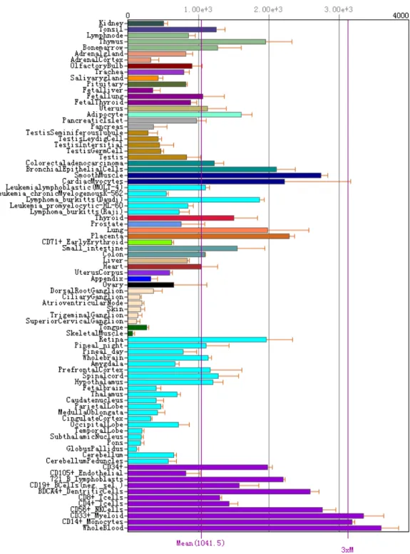
called switch I and switch II, distinguishing the conformational differences between RhoA-GDP and RhoA-GTP. Another important difference between the GDP-bound and GTP-bound RhoA is the mode of Mg ion binding. The removal of Mg2+ can be caused by GEFs. GEFs can also cause a displacement in the switch I region, which makes GDP binding site open for GEF interaction, replacing GDP with GTP. A small displacement between at Pro36 and Phe39 is induced in switch I when RhoA bound to GTP instead of GDP. This cause three hydrophobic residues, Val35, Val38, and Phe39, become exposed, which involve in target binding after activation. Both switch I and switch II are involved in the binding with its substrates (eg. ROCKs). RhoA has a ubiquitous tissue
distribution(Fig. 4), indicating its versatile function in cell biology. It also has been found to increase in many malignant tumour(Fig. 5).
36
37
38
9.2 REGULATION OF RHOA ACTIVITY
Figure 6 Regulation of RhoA activity.
Rho GTPases function as molecular switches, cycling between an active GTP-bound conformation and an inactive guanosine diphosphate (GDP)-bound conformation by the exchange of GDP to GTP(Bourne, Sanders et al. 1990, Bourne, Sanders et al. 1991). These switches are conducted via guanine nucleotide exchange factors (GEFs) and GTPase activating proteins(GAPs)(Fig. 6). Intrinsic phosphatase activity hydrolyzes GTP to GDP, deactivating RhoA function, and this process is accelerated by interaction with GTPase-activating proteins. Conversely, interaction with guanine-nucleotide exchange factors facilitates the exchange of GDP to GTP, which restores the activation of RhoA. A large number of GEFs are discovered to enable this activation and involved in various specific signalling pathways. The relative affinity difference of the effector molecules between the GTP- and GDP-bound states of the Rho GTPase can be as much as 100-fold, resulting in a highly-specific interaction only with the GTP-bound activated state. When binding with GTP, they interact with downstream effectors to propagate signal
transduction (Spiering and Hodgson 2011). In addition, Rho proteins are also regulated by guanine nucleotide dissociation inhibitors (GDIs), which can inhibit both the
exchange of GDP to GTP and the hydrolysis of bound GTP. In most cases, Rho proteins are post-translationally modified at their C-termini by prenylation of a conserved
RhoA RhoA GDP GTP GAPs GEFs RhoA GDP GDI
39
cysteine, facilitating their attachment to cell membranes, which is prerequisite to its activation and essential for its role in cytoskeleton modulation.
9.3 FUNCTION OF RHOA
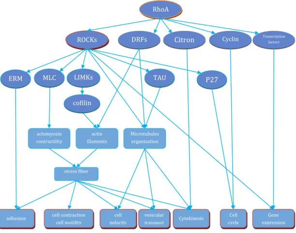
Several downstream effectors of RhoA have been discovered, such as ROCKs, diaphanous-related formins (DRFs), citron kinase, PRK1/PKN etc. These effectors
conduct versatile function in cell biological function, including cytoskeleton organization, cytokinesis, cell cycle regulation, embryotic development, transcriptional control etc. RhoA plays a central role in modulating cell shape, polarity and motility by its effects on actin polymerization, actomyosin contractility, cell adhesion, and microtubule dynamics. RhoA activates its downstream effector DRFs to promote actin polymerization by
addition of actin monomers to the growing end of actin filaments. Meanwhile activation LIMK by ROCKs and consequent inhibition of cofilin, an actin-binding proteins
regulating disassembly of actin filaments, contributes to the increase in actin filaments in response to RhoA. Thus DRFs act together with ROCKs to regulate Rho-induced stress fiber formation. In addition, RhoA activates ROCKs and subsequent phosphorylate several proteins involved in regulating myosins and other actin-binding proteins to induce actomyosin-based contractility, which is crucial in migrating cells. Microtubules are critical for determining cell polarity and for vesicular locomotion and intracellular transport. The coordinating action of ROCKs and DRFs is essential in the organization of microtubules. ROCK phosphorylates TAU and MAP2, proteins that regulate microtubule stability. DRFs directly participate in generating and orienting stable microtubules. RhoA also plays a key role in modulating the adhesion between neighbouring cells and
40
between cell and extracellular matrix. ROCKs and DRFs act reciprocally to regulate the migration of epithelial cells by reorganizing these adhesions.
Cytokinesis requires actomyosin-based contraction. RhoA s effectors ROCKs, citron kinase and DRFs are all implicated in this process. These effectors were found to localize in the cleavage furrow during cytokinesis. Their concerted actions stimulate local actin polymerization and coordinate microtubules with actin filaments at the contractile ring. RhoA also plays an important role in G1 cell cycle progression, primarily through
regulation of the expressions of cyclin D1 and cyclin-dependent kinase inhibitors p21 and p27. Multiple pathways were found to be involved in RhoA-dependent cyclin D1 expression, by activation of downstream protein kinases, resulting in the subsequent modulation of transcription factor activity. RhoA suppresses p21 expression in multiple normal and cancer cell lines, by other transcriptional regulators independent of p53. RhoA also regulates the levels of p27 in a ROCKs dependent manner.
RhoA also has a role in processes involving cell migration in development, such as neurite growth, dorsal closure, bone formation, and myogenesis. Rho-loss of function is lethal in mouse embryo development, resulting from failure in gastrulation and an inability of cell migration.
Many other transcriptional regulations mediated by RhoA has also been described, such as SRF , NF-kappaB, c/EBPb , Stat3 , Stat5 , FHL-2 , PAX6 , GATA-4 , E2F, Oestrogen Receptor alpha , Oestrogen Receptor beta , CREB , and transcription factors that depend on the JNK and p38 MAP kinase pathways. Substrates to these kinases include c-Jun, ELK, PEA3, ATF2, MEF2A, Max and CHOP/2GADD153.
41
Figure 7 Illustraction of RhoA functioin and the main signaling molecules involved.
9.4 MOLECULAR STRUCTURE AND EXPRESSION OF ROCK
ROCK( also called Rho-associated kinase) is a serine/threonine kinase with a molecular mass of about 160 kDa, which has been shown to be the principle downstream target of RhoA(Ishizaki, Maekawa et al. 1996). It is identified in 1996 as one of the effectors of Rho families (Ishizaki, Maekawa et al. 1996). ROCK displays the greatest homology to other downstream effectors of Rho kinase families such as myotonic dystrophy kinase (DMPK), DMPK-related cell division control protein 42 (Cdc42) binding kinase (MRCK), and citron kinase (CRIK).
RhoA
ROCKs DRFs Transcription factors
actin filaments assembly actomyosin contractility stress fiber cell polarity Microtubules organisation Gene expression Cytokinesis cell contraction cell motility vesicular transport adhesion Cell cycle P27 Cyclin D1 ERM MLC LIMKs Citron TAU cofilin
42
There are two ROCK isoforms: ROCK1 (also known as p160ROCK and ROCK and ROCK2 (also known as Rho-kinase and ROCK . )n human, ROCK-1 and ROCK-2 are encoded by two different genes localizing on chromosome 18 (18q11.1) and
chromosome 2 (2p24), respectively. Two isoforms of Rho-kinase in human have the homologue structure with about 65% of amino acid and 58% of RBD. The highest similarity (92%) is presented at kinase domain. These kinases are formed by parallel homodimers including a kinase domain in its amino-terminus (N-terminal domain), a coiled-coil in its middle dimerization portion, and a putative Pleckstrin-homology (PH) domain in its cysteine-riche domain (C-terminal domain, CRD) (Fig. 8) (Ishizaki,
Maekawa et al. 1996, Matsui, Amano et al. 1996). These carboxyl terminal domains constitute an autoinhibitory region that reduces the kinase activity of ROCKs (Amano, Chihara et al. 1999). The Rho-binding domain of ROCKs is localized in the C-terminal portion of the coiled-coil region, and it shows sequence homology to the Rho-interaction domain of kinectin which is a regulating protein of microtubule-based organelle motility (Alberts, Bouquin et al. 1998) (Fig. 9). The coiled-coil region of ROCKs is showed to interact with other -helical proteins, whereas the PH domain is involved in protein localization (Riento and Ridley 2003).
In human, ROCKs are also widely expressed in various normal and pathological tissues (Fig. 10), including vascular endothelial cells (ECs)(Ming, Barandier et al. 2004,
Horowitz, Binion et al. 2007) and many cancer cells (Morgan-Fisher, Wewer et al. 2013). Both ROCK-1 and ROCK-2 are expressed in vascular smooth muscle and in heart.
Cell-fractionation studies show that ROCKs are mainly distributed in the cytoplasm fraction but a small amount of ROCKs is also found in the membrane fraction. However, ROCKs are also found in subcellular localization at the vimentin intermediate-filament network and at actin stress fibers.
43
Figure 8 ROCK structure.
Figure 9 The molecular structure of ROCK1 and ROCK2.
The kinase domain is located at the amino terminus (N-terminus) of the protein, followed by coiled-coil region containing the Rho-binding domain (RBD). The Pleckstrin-homology (PH) with an internal cysteine-rich domain (CRD) is situated in the carboxyl terminus
44
45
46
47
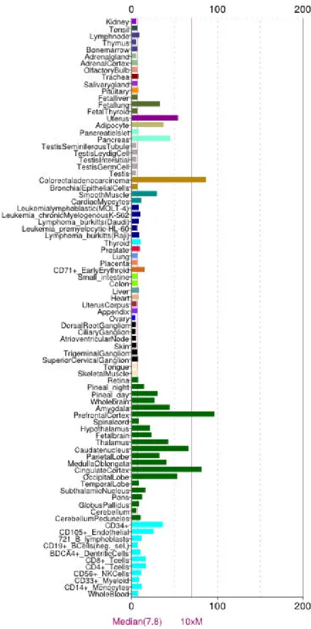
Figure 13 ROCK2 expression in human cancer (GeneCards. 2014)
9.5 REGULATION OF ROCK ACTIVITY
In the structure of ROCKs, the C-terminus (Rho-binding domain and PH domain) plays a role as a dominant-negative autoinhibitor (Fig. 14) due to its independent interaction with the catalytic domain (N-terminus). Lacking of the C-terminus of ROCKs (truncated forms) are constitutively formed the active form kinase. Beside of the self-associative autoinhibition, the activity of ROCKs is also influenced by its affinity for ATP which is regulated by the dimerization of kinases. The relative affinity difference of ROCK
between the GTP- and GDP-bound states of the Rho GTPase can be as much as 100-fold, resulting in a highly-specific interaction only with the GTP-bound activated state. Binding of GTPase-RhoA to ROCKs at Rho-binding domain induces conformational
48
changes of ROCKs, resulting in relieve of autoinhibitory blockage of kinase activity. This binding is believed to stimulate the phosphor-transferase activity of ROCKs (positive regulation) by disrupting the interaction between catalytic and the C-terminal region of proteins, which thereby frees the kinase activity (Fig.14). Independently of RhoA, ROCK activity might be activated by other stimulators such as arachidonic acid (AA),
sphingosine phosphorylcholine (SPC), caspase-3 or granzyme B. AA and SPC interact with the negative regulatory region at PH domain, thus disrupting its inhibitory property on the catalytic activity of ROCKs. Caspase-3 cleaves ROCK-1 at the cleavage site DETD1113 whereas granzyme B cleaves the ROCK-2 at the C-terminus at IGLD1131, thus removing an inhibitory region (Fig.14). The activity of ROCKs is also negatively regulated by other small G-binding proteins such as RhoE, Gem, and Rad. RhoE binds to the N-terminal region of kinase domain of ROCK-1 (amino acids 1–420) and therefore interferes with the kinase activity and prevents GTPase-RhoA binding to Rho-binding domain (Fig.14). Overexpression of Gem and Rad might inhibit respectively the downstream responses of ROCKs, but the mechanism is not clearly demonstrated. In addition, the negative regulation of ROCK-mediated target effects by these small G-binding proteins are localized at the different cellular structure, such as RhoE in the Golgi and Gem and Rad in the cytoskeleton.
49
Figure 14 The mechanism of positive regulation of ROCKs activity(Duong-Quy, Bei et al. 2013).
9.6 FUNCTION OF ROCKS
ROCKs phosphorylate a large numbers of targets and mediate a broad range of cellular
responses. The most well-studied and important role is a key regulator of actin
cytoskeleton, involving in actin filament assembly and contraction via several substrates such as MBS, LIM Kinases, MLCK, ERM proteins.
ROCK is the key molecule required for actin–myosin-based contractile force generation. When bound and activated by RhoA, ROCKs translocate from the cytoplasm to the cell membrane where it increases phosphorylation of the myosin light chain (MLC) of myosin II. This is achieved either by direct phosphorylation, or by phosphorylation of the regulatory myosin-binding subunit of myosin phosphatase (also known as the
50
phosphatase-targeting subunit), which inhibits the phosphatase activity of this molecule (Somlyo and Somlyo 2003). This in turn, enhances actin binding and the actin-induced adenosine triphosphatase activity of myosin, facilitating the interaction of myosin with F-actin, and ultimately cell contractility and mobility. Moreover, ROCK phosphorylates LIM kinase-1 and 2 (LIMK1/2) (Riento and Ridley 2003). The phosphorylation of LIMK1/2 promotes their activity and subsequently phosphorylates and inhibits cofilin protein s activity to disassemble actin filaments.Thus it facilitates the organization of F-actin into stress fibers and re-arrangement of the actin cytoskeleton (Spiering and Hodgson 2011). The net effect of increased ROCK activity is to elevate force generation, and then facilitate cell adhesion, motility, and invasion. Thus, they play an important role in the regulation of smooth muscle contraction, neurite retraction.
ROCKs phosphorylate ERM to mediate anchoring of actin filaments to integral proteins of the plasma membrane (Matsui, Maeda et al. 1998). ERM proteins contain both a C-terminal actin-binding subunit and an N-terminal 4.1/ezrin/radixin/moesin (FERM) domain, which interacts with plasma membrane proteins. Phosphorylation of ERM leads to the disruption of the head-to-tail association of ERM proteins and actin cytoskeletal reorganization, thus dissociate the cell-cell and cell-matrix adhesion and facilitate cell mobility.
Also, they show scaffolding properties that function to polymerize actin and affect the formation of microtubules, which is required for centrosome positioning and
centrosome-dependent exit from mitosis.
Other downstream targets of the Rho kinases include signal transduction molecules and transcription factors such as IRS-1 and PI 3-K/AKT, PTEN c-Ras, SRF, resulting in a variety modulation of gene expression and cell biology.
51
Figure 15 Role of RhoA/ROCK pathway in the actin–myosin-based contractile force generation
9.7 DIFFERENCE BETWEEN ROCK1 AND ROCK2
The differences between ROCK1 and ROCK2 are still poorly understood. Their different tissue distribution and several different roles have been found. ROCK1 and ROCK2 both have ubiquitous tissue distributions. There are still some different patterns of tissue expressions of ROCK1 and ROCK2. A preferential protein expression of ROCK2 can be found in brain and muscle, and that of ROCK1 in blood cells. Subcellularly, ROCK2 is
RhoA ROCKs MLC-p MLC MLC cofilin-p cofilin Actomyosin contraction Actin polymerization Stress fiber LIMK Focal adhesion assembly/stabilit y Cell migration invasion
52
found primarily in the cytoplasm (Leung, Manser et al. 1995, Matsui, Amano et al. 1996) in association with vimentin (Sin, Chen et al. 1998) and actin stress fibers (Chen, Tan et al. 2002). It also localizes to the plasma membrane (Kimura, Fukata et al. 1998) with an association with its C-terminal region (Kher and Worthylake 2011). It can also be found at the cleavage furrow during late mitosis (Inada, Togashi et al. 1999). In contrast, the intra-cellular distribution of ROCK1 is still not well established. ROCK1 may
predominantly associate with the plasma membrane at the apical junctions in
endothelial cells (Ishiuchi and Takeichi 2011). Specifically, it indirectly associates with epithelial-cadherin (E-cadherin) complexes through its interaction with the E-cadherin scaffold protein p120-catenin (Smith, Dohn et al. 2012). ROCK1 additionally localizes to the microtubule organizing centre (Chevrier, Piel et al. 2002).
10 RHOA/ROCK PATHWAY IN CANCER ANGIOGENESIS
10.1 ANGIOGENESIS MECHANISM
Angiogenesis is a five-step process involving a complex series of events. Firstly, an
increase in the permeability of the basement membrane allows a new capillary to sprout. Next, ECs activated by angiogenic factors migrate through the basement membrane into the extracellular matrix, towards the angiogenic stimulus. The leading front of migrating cells is driven by enhanced proliferation of ECs. This is then followed by re-organization of ECs to form tubules with a central lumen, together with the recruitment of
peri-endothelial cells (pericytes) and vascular smooth muscle cells for new capillary stabilization (Sakamoto, Ryan et al. 2008). The RhoA/ROCK pathway plays a role in each of these key steps.
53
10.2 PERMEABILITY
The endothelium is a semi-permeable barrier that lines the vasculature, comprising ECs that are connected to each other by inter-endothelial junctions, consisting of protein complexes organized as tight junctions and adherent junctions. The latter are in the majority(Mehta and Malik 2006), and are composed of vascular endothelial (VE)
cadherin that associates homotypically with VE-cadherin on adjacent cells. VE-cadherin binds to the actin cytoskeleton. Actin-mediated EC contraction occurs as a result of MLC phosphorylation, and this can cause dysfunction of the endothelial barrier by inducing the formation of small gaps between neighbouring cells.(Dudek and Garcia 2001). RhoA, through its downstream effector ROCK, plays a role in endothelial barrier dysfunction by potentiating MLC phosphorylation via inhibition of MLC phosphatase activity. Studies have also confirmed that RhoA contributes to VEGF-induced hyper-permeability in the endothelium(Sun, Breslin et al. 2006).
10.3 MIGRATION
The formation of stress fibers and cellular contraction is essential for EC migration, and these processes are mediated by Rho GTPases(Kiosses, Daniels et al. 1999). Van Nieuw Amerongen et al demonstrated in vitro that VEGF induces the activation of RhoA and this increase in RhoA activity is necessary for VEGF-induced reorganization of the F-actin cytoskeleton. This process can be inhibited by transfection of ECs with a RhoA dominant-negative mutant vector or by a RhoA inhibitor C3 (van Nieuw Amerongen 2002). Zhao et al. showed that increased expression of RhoA in human umbilical vein ECs significantly enhanced cytoskeletal reorganization of transfected cells, cell migration and angiogenic capacity, which suggests that RhoA plays a key part in these processes in vitro(Zhao, Xu et al. 2006).
54
10.4 PROLIFERATION
Several lines of evidence suggest that Rho proteins play an important role in normal and cancerous cell growth processes, including G1 phase cell cycle progression and
mitogenesis(Van Aelst and D'Souza-Schorey 1997). Cytokinesis is a step in mitogenesis which is critical within the cell cycle. In eukaryotic cells, cytokinesis requires an actin and myosin contractile ring, which constricts and cleaves the cell, forming two daughter cells. Inhibition of Rho GTPases prevents the assembly of this contractile ring in a
variety of mammalian cells. Expression of constitutively activated Rho GTPases also blocks cytokinesis, suggesting that cycling between the active and inactive forms is required for its function (Glotzer 2001).
The role of RhoA signalling in cell survival has been evaluated in several non-EC cell types. Results showed that inhibition of Rho signalling leads to apoptosis via alterations in cell adhesion and the induction of p53 and other pro-apoptotic proteins, or via
ceramide up-regulation leading to caspase cleavage and subsequent activation(Bobak, Moorman et al. 1997, Petrache, Crow et al. 2003). Studies have shown that the ROCK inhibitors, fasudil and Y-27632, not only inhibit VEGF-induced cell proliferation, but also reverse the protective effect of VEGF on apoptosis, which results in a decrease in
viability of VEGF-stimulated ECs(Yin, Morishige et al. 2007, Bryan, Dennstedt et al. 2010). Data obtained with these inhibitors have revealed the important role of the RhoA/ROCK pathway in EC proliferation and cell viability.
10.5 MORPHOGENESIS
Cultured ECs can undergo marked changes in shape and tube formation that closely imitate pre-capillary cord formation in vivo(Montesano, Orci et al. 1983). In vitro