2
UNIVERSITÉ DE SHERBROOKE
Faculté des sciences de l’activité physique
Département de kinanthropologie
L’influence de l'anticipation sur les modulations de puissance dans la
bande de fréquence bêta durant la préparation du mouvement
et
L'effet de la variance dans les rétroactions sensorielles sur la rétention à
court terme
Par
Canaveral, Cesar Augusto B.Sc.
Mémoire par article présenté à la Faculté des sciences de l’activité
physique
En vue de l’obtention du grade de maîtrise en sciences (M.Sc.)
M.Sc. en sciences de l’activité physique
Créneau de recherche en neuromécanique et ergonomie
Dépôt final effectué en
septembre 2019
© Cesar Augusto Canaveral, 2019
3
UNIVERSITÉ DE SHERBROOKE
Faculté des sciences de l’activité physique
Département de kinanthropologie
The influence of anticipation on power modulations in the beta-band
during movement planning
and
The effect of variance in sensory feedback on short-term retention
Par
Canaveral, Cesar Canaveral B.Sc.
A été évalué par un jury composé des personnes suivantes :
Félix Berrigan
Président du jury
Pierre-Michel Bernier
Directeur de recherche
Maxime Trempe
Membre interne du jury
Erin Cressman
Membre externe du jury
4
RÉSUMÉ
La production du mouvement est un aspect primordial de la vie qui permet aux organismes vivants d'interagir avec l'environnement. En ce sens, pour être efficaces, tous les mouvements doivent être planifiés et mis à jour en fonction de la complexité et de la variabilité de l'environnement. Des chercheurs du domaine du contrôle moteur ont étudié de manière approfondie les processus de planification et d’adaptation motrice. Puisque les processus de planification et d'adaptation motrice sont influencés par la variabilité de l'environnement, le présent mémoire cherche à fournir une compréhension plus profonde de ces deux processus moteurs à cet égard.
La première contribution scientifique présentée ici tire parti du fait que les temps de réaction (TR) sont réduits lorsqu'il est possible d'anticiper l’objectif moteur, afin de déterminer si les modulations de TR associées à l'anticipation spatiale et temporelle sont sous-tendues par une activité préparatoire similaire. Cela a été fait en utilisant l'électroencéphalographie (EEG) de surface pour analyser l'activité oscillatoire dans la bande de fréquence bêta (13 - 30 Hz) au cours de la période de planification du mouvement. Les résultats ont révélé que l'anticipation temporelle était associée à la désynchronisation de la bande bêta au-dessus des régions sensorimotrices controlatérales à la main effectrice, en particulier autour du moment prévu de l'apparition de la cible. L’ampleur de ces modulations était corrélée aux modulations de TR à travers les participants. En revanche, l'anticipation spatiale a augmenté de manière sélective la puissance de la bande bêta au-dessus des régions pariéto-occipitales bilatérales pendant toute la période de planification. Ces résultats suggèrent des états de préparation distinct en fonction de l’anticipation temporelle et spatiale.
D’un autre côté, le deuxième projet traite de la façon dont la variabilité de la rétroaction sensorielle interfère avec la rétention à court terme dans l’étude de l’adaptation motrice. Plus précisément, une tâche d'adaptation visuomotrice a été utilisée au cours de laquelle la variance des rotations a été manipulée de manière paramétrique à travers trois groupes, et ce, tout au long de la période d’acquisition. Par la suite, la rétention de cette nouvelle relation visuomotrice a été évaluée. Les résultats
5 ont révélé que, même si le processus d'adaptation était robuste à la manipulation de la variance, la rétention à court terme était altérée par des plus hauts niveaux de variance. Finalement, la discussion a d'abord cherché à intégrer ces deux contributions en revisitant l'interprétation des résultats sous un angle centré sur l'incertitude et en fournissant un aperçu des potentielles représentations internes de l'incertitude susceptibles de sous-tendre les résultats expérimentaux observés. Par la suite, une partie de la discussion a été réservée à la manière dont le champ du contrôle moteur migre de plus en plus vers l’utilisation de tâches et d’approches expérimentales plus complexes, mais écologiques aux dépends des tâches simples, mais quelque peu dénaturées que l’on retrouve dans les laboratoires du domaine. La discussion a été couronnée par une brève proposition allant dans ce sens.
6
ABSTRACT
Motor behavior is a paramount aspect of life that enables the living to interact with the environment through the production of movement. In order to be efficient, movements need to be planned and updated according to the complexity and the ever-changing nature of the environment. Motor control experts have extensively investigated the planning and adaptation processes. Since both motor planning and motor adaptation processes are influenced by variability in the environment, the present thesis seeks to provide a deeper understanding of both these motor processes in this regard.
More specifically, the first scientific contribution presented herein leverages the fact that reaction times (RTs) are reduced when the anticipation of the motor goal is possible to elucidate whether the RT modulations associated with temporal and spatial anticipation are subtended by similar preparatory activity. This was done by using scalp electroencephalography (EEG) to analyze the oscillatory activity in the beta frequency band (13 – 30 Hz) during the planning period. Results revealed that temporal anticipation was associated with beta-band desynchronization over contralateral sensorimotor regions, specifically around the expected moment of target onset, the magnitude of which was correlated with RT modulations across participants. In contrast, spatial anticipation selectively increased beta-band power over bilateral parieto-occipital regions during the entire planning period, suggesting that distinct states of preparation are incurred by temporal and spatial anticipation.
Additionally, the second project addressed how variance in the sensory feedback interferes with short-term retention of motor adaptation. Specifically, a visuomotor adaptation task was used during which the variance of exposed rotation was parametrically manipulated across three groups, and retention of the adapted visuomotor relationship was assessed. Results revealed that, although the adaptation process was robust to the manipulation of variance, the short-term retention was impaired.
7 The discussion first sought to integrate these two projects by revisiting the interpretation of both projects under the scope of uncertainty and by providing an overview of the internal representation of uncertainty that might subtend the experimental results. Subsequently, a part of the discussion was reserved to allude how the motor control field is transitioning from laboratory-based tasks to more naturalistic paradigms by using approaches to move motor control research toward real-world conditions. The discussion culminates with a brief scientific proposal along those lines.
8
TABLE OF CONTENT
RÉSUMÉ ... 4
ABSTRACT ... 6
1. PREFACE: THE STUDY OF MOTOR BEHAVIOR ... 10
1.1. INTRODUCTION ... 10
1.2. HOW TO STUDY MOTOR BEHAVIOR ... 10
1.3. INVESTIGATING MOTOR BEHAVIOR THROUGH MOTOR PLANNING AND MOTOR ADAPTATION ... 12
2. PART I: INVESTIGATING MOTOR BEHAVIOR THROUGH MOTOR PLANNING ... 14
2.1. OPERATIONAL FRAMEWORK OF MOVEMENT PLANNING ... 14
2.2. OPERATIONAL DEFINITION OF MOVEMENT PLANNING ... 15
2.3. HOW TO STUDY MOVEMENT PLANNING ... 15
2.4. FACTOR INFLUENCING REACTION TIMES ... 17
2.5. NEURAL OSCILLATIONS TO INVESTIGATE MOVEMENT PLANNING ... 19
2.6. RESEARCH PROBLEM ... 23
2.7. RESEARCH QUESTION ... 24
2.8. SUBMITTED ARTICLE IN JOURNAL OF COGNITIVE NEUROSCIENCE ... 25
2.9. DISSOCIATION BETWEEN TEMPORAL AND SPATIAL ANTICIPATION IN THE NEURAL DYNAMICS OF GOAL -DIRECTED MOVEMENT PREPARATION ... 26
3. PART II: INVESTIGATING MOTOR BEHAVIOR THROUGH MOTOR ADAPTATION... 56
3.1. OPERATIONAL FRAMEWORK OF MOTOR ADAPTATION: THE THEORY OF INTERNAL MODELS ... 56
3.2. OPERATIONAL DEFINITION OF MOTOR ADAPTATION ... 57
3.3. HOW TO STUDY MOTOR ADAPTATION ... 58
3.4. FACTORS INFLUENCING MOTOR ADAPTATION ... 60
3.5. RESEARCH PROBLEM ... 62
3.6. RESEARCH QUESTION ... 64
3.7. PUBLISHED ARTICLE IN JOURNAL OF NEUROPHYSIOLOGY ... 65
3.8. VARIANCE IN EXPOSED PERTURBATIONS IMPAIRS RETENTION OF VISUOMOTOR ADAPTATION... 66
4. DISCUSSION ... 99
4.1. INTEGRATING THE TWO SCIENTIFIC CONTRIBUTIONS ... 99
4.2. GOING FURTHER ... 107
5. CONCLUSION ... 113
6. REFERENCES ... 114
7. ANNEXES ... 132
7.1. AUTHORIZATION TO INTEGRATE THE ARTICLE SUBMITTED TO THE JOURNAL OF COGNITIVE NEUROSCIENCE TO THE PRESENT THESIS ... 132
7.2. AUTHORIZATION TO INTEGRATE THE ARTICLE PUBLISHED IN THE JOURNAL OF NEUROPHYSIOLOGY TO THE PRESENT THESIS ... 133
9
LIST OF FIGURES
Figure 1
10
1. PREFACE: THE STUDY OF MOTOR BEHAVIOR
1.1. Introduction
Movement is a paramount aspect of life, without which no interaction with the environment would be possible. The ability to generate movements is more than just a convenience that enables us to do basic things, like walking or manipulating objects; it is a critical component of the evolutionary development of living species. Some experts argue that movement is the main reason we have a nervous system (Wolpert, 2013; Wolpert, Ghahramani, & Flanagan, 2001).
Behavior results from a constant interaction between the living species and a complex environment (Cisek & Kalaska, 2010). For such interaction to take place, the brain is subject to a constant gathering of information about the environment, which affords a large variety of possibilities for action (Gibson, 2014). Since the environment is complex and susceptible to changes, the brain is compelled to the constant adaptation of behaviors depending on the constraints imposed by the outside world. From an evolutionary perspective, both humans and animals have developed the capacity to remain flexible enough to control and update their motor behavior within an inherently uncertain environment (Friston, Mattout, & Kilner, 2011; Sterling & Laughlin, 2015; Todorov & Jordan, 2002).
Broadly, studying motor behavior translates into investigating how the motor system controls its movements to interact with the outside world. Motor control experts have been interested in unraveling the intricacies by which the brain gathers external sensory information to execute and to update goal-directed movements in response to changes in the environment (Elliott et al., 2010; Schmidt & Lee, 2011; Scott, 2012). For this, motor planning and motor adaptation can be view as fundamental mechanisms that allow efficient interaction between the motor system and the world. 1.2. How to study motor behavior
Since the turn of the 19th century, motor control experts have developed distinct procedures and approaches that led to the increasing understanding of how humans and
11 animals behave (Rosenbaum, 1991; Schmidt & Lee, 2011; Scott, 2012; Woodworth, 1899). For that matter, two distinct levels of analysis commonly used in the field of motor control will be presented in the following lines.
1.2.1. Behavioral level of analysis
The first level alludes to the behavioral analysis and it can be understood as the quantification of the extent to which a movement is achieved related to an intended behavioral goal. Such quantification is often used in the assignment of scores in sports. For instance, an archer is evaluated based on the distance between his shots and the bullseye and a sprinter is qualified based on how fast he reacts and finishes his dash. These examples introduce two fundamental ways to assess behavior: the quantification of the 1) spatial accuracy and 2) timing of performance. Namely, end-point errors are often used to assess the spatial accuracy of a movement, whereas reaction times (RT) are often used to assess movement readiness (Schmidt & Lee, 2011). The topics of errors and RTs will be extensively discussed in the following parts of this manuscript.
1.2.2. Neurophysiological level of analysis
All behaviors are governed by the brain. Based on that simple premise, another way to further understand motor behavior is by analyzing the neurophysiological basis and mechanisms that subtend behavior. Historically, lesional studies allowed scientists to ascribe specific brain functions to specific brain regions. A classic case was the one of Phineas P. Gage (1823 - 1861), an American railroad foreman whose left frontal lobe was severely injured by an iron rod that pierced through his head. Reportedly, Gage’s personality and social behavior drastically changed after the injury, which led scientists to suggest that damage to specific parts of the brain might induce specific changes in behavior (Harlow, 1848; Macmillan, 2000). Fortunately, today’s neuroimaging modalities allow scientists to investigate the underlying neural processes of behavior without exclusively resorting to patients with brain lesions or diseases.
Amongst a plethora of approaches, electroencephalography (i.e., EEG) allows investigating the electrical activity of cortical neuronal populations using surface electrodes positioned all over the head (Britton et al., 2016; Olejniczak, 2006). Under
12 controlled conditions, EEG can be used to record the neural activity associated with movement planning and execution with high temporal resolution (Berger, 1930; Buzsaki & Draguhn, 2004; Perfetti et al., 2011; Pfurtscheller & Lopes da Silva, 1999). The functional bases of EEG recording will be further explained in the second part of this document (cf. Neural oscillations to investigate movement planning).
1.3. Investigating motor behavior through motor planning and motor adaptation
Considering the variety of possible interactions with the outside world, it is crucial to understand how the various processes of motor control are influenced by the sensory information incoming from the environment. The present thesis presents two scientific contributions that lay their bases on the frameworks of motor planning and motor adaptation. Within their respective framework, the scientific projects presented here address how the motor system can be influenced by the reliability in the sensory information, using neurophysiological and behavioral levels of analysis. For a matter of concision, only the functional mechanisms of visually guided movements will be addressed in this manuscript.
The first part of this thesis will be dedicated to the concept of motor planning, referring to the process by which the key parameters of an upcoming movement are specified and readied for action execution (Gallivan, Chapman, Wolpert, & Flanagan, 2018). Here, we investigated how the simultaneous manipulation of spatial and temporal information of the behavioral goal, which are known to affect RTs in isolation (Hick, 1952; Niemi & Näätänen, 1981), influences the EEG dynamics during the preparatory period of reaching movements. The second part will be dedicated to the concept of motor adaptation, alluding to the behavioral processes that characterize the way that the motor system adapts its movements. Here, we investigated how motor adaptation is affected by the statistical characteristics of the sensory information that leads to adapted behavior. In conclusion, the last portion of the present work will be partly dedicated to a unifying discussion of the two scientific contributions. It will be
13 followed by a discussion of how the field of motor control is currently transitioning from laboratory-based tasks to more naturalistic paradigms.
14
2.
PART I: INVESTIGATING MOTOR BEHAVIOR
THROUGH MOTOR PLANNING
2.1. Operational framework of movement planning
Purposeful actions are often generated to interact with external objects in order to achieve a behavioral goal (Gallivan et al., 2018; Scott, 2016; Wong, Haith, & Krakauer, 2014). These actions are often referred to as goal-directed movements. In order to generate these types of movements, the brain needs to solve (at least) two fundamental problems. For one, the brain needs to specify the spatial parameters that determine how the movement will be accomplished in relation to the behavioral goal (Scott, 2016; Wong et al., 2014). Also, depending on a variety of action contingencies, the brain needs to determine when to execute its movement (Brass & Haggard, 2008; Haith, Pakpoor, & Krakauer, 2016).
Consider a simple movement such as reaching towards a visual target. For the brain to achieve that behavioral goal, it needs to specify where to produce the reach (Cisek & Kalaska, 2010; Scott, 2016; Wong et al., 2014). To do so, spatial information about the position of the target, which is acquired in a sensory format, needs to be reinterpreted in a motor format according to the position of the reaching hand. This process allows the motor system to define a directional vector from the hand to the target’s location in space. This process is called sensorimotor transformation1 (Andersen & Buneo, 2003; Andersen, Snyder, Bradley, & Xing, 1997; Kakei, Hoffman, & Strick, 2003; Kandel, 2013). As the sensorimotor transformation occurs along the dorsal processing pathways (i.e., from dorsal parietal to premotor; Goodale & Milner, 1992; Ungerleider & Mishkin, 1982), neural representation of the motor plan that will allow to achieve the goal (i.e. neuronal activity of directionally tuned neurons) begin to emerge in the sensorimotor regions (Cisek, 2006; Cisek & Kalaska, 2010; Cui & Andersen, 2007; Nakayama, Yamagata, Tanji, & Hoshi, 2008).
1This refers to the series of processes by which extrinsic information about the state of world (i.e. spatial
location of objects) and intrinsic information about our body (i.e. kinematic and kinetic information about our body) are transformed into potential motor plans (Kandel, 2013).
15 Simultaneously, the brain needs to gather evidence that will instruct the initiation of the reach (Klaus, Alves da Silva, & Costa, 2019). The reach onset can be triggered by an external stimulus, such as a go-cue, or it can be done endogenously, in the case of self-initiated movement (Brass & Haggard, 2008). The role of action initiation has long been conferred to the activity in basal ganglia neurons (Cui et al., 2013; Klaus et al., 2019). In the case of stimulus-triggered movements (i.e., signaled by a go-cue), sensory information about that imperative signal is sufficient to elicit dopaminergic activity in the basal ganglia-cortical network, which will ensue in the initiation of the motor command (Jenkinson & Brown, 2011; Schultz, 1986; Schultz & Romo, 1990; see however Klaus et al., 2019). Specifically, the basal ganglia are known to “gate” movement (Brown, Bullock, & Grossberg, 2004; Grossberg, 2016; Zold et al., 2012) and the release of such gating, which is potentially mediated through the suppression of inhibitory cells that refrain existing motor plans (Thura & Cisek, 2017), results in action initiation.
2.2. Operational definition of movement planning
Although the example presented above is nothing but an oversimplification of the kind of situations that take place in real-life, it reliably depicts the following basic idea: Generation of purposeful movements involves simultaneous processes of action specification (Cisek & Kalaska, 2010; Wong et al., 2014) and action initiation (Brass & Haggard, 2008; Haith et al., 2016; Klaus et al., 2019). The framework presented above allows to broadly define goal-directed movement planning as the processes by which the brain transforms sensory information from the environment to specify the potential actions and to determine when to initiate the movement. 2.3. How to study movement planning
Classically, goal-directed movements can be conceptualized by two distinctive phases: a preparatory period and an execution period. Motor control researchers have extensively studied movement planning using laboratory tasks in which a precue
16 marks the start of a delay period2 after which an imperative go-cue instructs the participants to initiate their movement. The use of delay periods enables to distinguish between preparatory-related from execution-related processes. Although preparatory- or execution-related processes are encoded within similar sensorimotor brain regions, there is evidence that some neurons are activated only during the delay period, others discharge during the execution period (Churchland, Cunningham, Kaufman, Ryu, & Shenoy, 2010; Crammond & Kalaska, 2000; Kaufman et al., 2016; Riehle & Requin, 1989), and some other neurons are activated during both planning and execution (Crammond & Kalaska, 2000). Neural activity during the delay period is referred to as preparatory activity (Svoboda & Li, 2018), during which the parameters of the upcoming movement are specified for it to be executed.
2.3.1. The use of reaction time to study motor planning
From a behavioral standpoint, the amount of task-relevant information that the precue provided (i.e., location of a potential target or duration of the delay period) can influence movement performance (Rosenbaum, 1980, 1991; Schmidt & Lee, 2011; Woodworth, 1899), which can be quantified through measurements of RTs (cf. Preface
– How to study motor behavior – behavioral level of analysis).
RTs have been used since the 19th century to investigate motor behavior (Cattell, 1886; Deary, Der, & Ford, 2001; Deary, Liewald, & Nissan, 2011; Jensen & Munro, 1979; Niemi & Näätänen, 1981; Posner, 1980). It can be conceptually defined as the time it takes to initiate a movement in response to a stimulus in the environment. RTs are considered a critical variable to study motor planning since they allow to infer the timing of all the ongoing neural processes that take place from the moment go-cue is delivered until its associated motor response is initiated (Marin & Danion, 2005; Schmidt & Lee, 2011). Interestingly, RTs can be highly influenced if the go-cue can be anticipated (Marin & Danion, 2005).
17 2.4. Factor influencing reaction times
2.4.1. Anticipation effects on RTs
RTs can be modulated according to the context in which a goal-directed reaching task is performed. One of the biggest influences over RTs is the ability to anticipate a sensory event that yields movement generation. Specifically, two forms of anticipation can be distinguished: spatial anticipation and temporal anticipation (Marin & Danion, 2005; Schmidt & Lee, 2011).
Spatial anticipation. Anticipating the spatial position of a target before reaching towards it (i.e., where to move) can incur a significant reduction in RTs. In support, a classical experiment conducted by Rosenbaum (1980) demonstrated that RTs are faster (i.e., reduced) when the direction of an upcoming target is precued as compare to when it is not (Rosenbaum, 1980). This demonstrates that providing directional information as to where to reach reduces RTs. Psychophysicists have established two main kinds of reaction time experiments based on this notion: simple reaction time (SRT) and choice reaction time (CRT) tasks (Luce, 1986; Welford, 1980).
Basically, in SRT reaching tasks, there is one target and consequently, one associated response, whereas in CRT, there are multiple targets, each requiring a different response. It has been well established that CRT is slower than SRT since there is no possibility to anticipate the spatial location of the reaching target before it has been specified by the imperative go-cue (Donders, 1868; O'Shea & Bashore, 2012). The relation between the number of targets and the RTs has been well established by the famous “Hick’s Law” (Hick, 1952).
Although very insightful, this relationship has been amended by subsequent work reporting that it is not the number of cues that determines RTs but rather the spatial separation that they subtend (Bock & Eversheim, 2000). Specifically, Bock and Eversheim (2000) showed that RTs are similar, whether two or more targets are precued within the same angular span. However, if two targets are placed closer together, then the RT is faster than if those two targets are far apart (Bock & Eversheim, 2000). These results have been interpreted using the action selection and specification
18 model (Cisek, 2006), suggesting that RTs are determined by the level of competition between motor plans to reach the potential targets. In other words, the farther apart the targets, the greater the competition between the distributed neural population encoding for each direction, thus requiring more time to select amongst opposing motor plans (Cisek, 2006), thus resulting in an inherent increase in RTs.
All in all, it is well established that anticipating the spatial position of a target leads to faster RTs, and that, in contrast, increasing the spatial separation between multiple targets leads to slower RTs.
Temporal anticipation. Anticipating the temporal occurrence of a go-cue instructing movement initiation (i.e., when to move) can also incur a significant reduction in RTs. The length of the delay period can be used to manipulate temporal anticipation. In support, behavioral evidence has shown that a fixed delay period between the precue and the go-cue provides a temporal frame of reference that enables participants to promptly initiate their response (Niemi & Näätänen, 1981). Throughout several trials, participants can reliably estimate the length of the delay period, and likely anticipate the time at which a go-cue will be delivered, thus reducing their RTs (McMorris, 2014; Quesada & Schmidt, 1970; Rohenkohl, Cravo, Wyart, & Nobre, 2012). In contrast, for blocks of trials with variable delay periods (i.e., delay periods for which the duration is inconsistent across trials), participants cannot successfully anticipate the go-cue, resulting in increased RTs (Niemi & Näätänen, 1981). These evidences suggest that the movement initiation can be anticipated if the participants can estimate the length of the delay period.
Estimating the length of the delay period implies that a representation of time needs to be internalized by the brain. In support for this assumption, there is evidence that the implementation of internal timing can be assured by internal models in the cerebellum (Ivry & Spencer, 2004; Perrett, Ruiz, & Mauk, 1993; Spencer, Zelaznik, Diedrichsen, & Ivry, 2003; Yamazaki & Tanaka, 2009) and then relayed to task-specific brain regions (Ivry, 1996; Ivry & Spencer, 2004; Leon & Shadlen, 2003), which include the cortico-basal ganglia network (Ferrandez et al., 2003; Klaus et al.,
19 2019; Thura & Cisek, 2017). This provides evidence highlighting the existence of neural processes dedicated to the internal representation of time, that might mediate the selection of when to initiate a movement.
In sum, temporal anticipation of the go-cue can facilitate movement initiation through the internalized representation of time in task-specific regions, resulting in reduced RTs.
2.5. Neural oscillations to investigate movement planning
From a neurophysiological standpoint, the preparatory processes that take place during the delay period can be recorded using brain imaging techniques such as EEG. Brain activity during the delay period has been associated with stereotyped patterns of neural oscillations (Adhikari, Shrestha, Mishra, Singh, & Timalsina, 2018; Perfetti et al., 2011; Pfurtscheller & Lopes da Silva, 1999; Shibasaki & Hallett, 2006). The following section will address the physiological bases of neural oscillations and how EEG recordings can be used to investigate motor planning.
2.5.1. Physiological bases of neural oscillations
The fundamental basis of neural oscillations resides in the coordinated spiking activity of a large number of neurons within the neuronal network (i.e., neuronal population), which presumably guide functional activity and the evolution of the processes that are being encoded (Shenoy, Sahani, & Churchland, 2013). The synchronous activity of neuronal populations allows using non-invasive scalp EEG to investigate brain activity during movement preparation.
EEG indirectly captures extracellular fields resulting from superposed ionic contributions from all active cellular processes within a given brain region (Buzsáki, Anastassiou, & Koch, 2012). The contribution of cellular activity to EEG recordings is dependent on the distance between the source (i.e., the cellular activity) and the sensor (i.e., the EEG electrodes), as well as the structural arrangement of the neuronal population being recorded (Cohen, 2014). Thereby, the activity that is generated in the cortex contributes to a greater extent to the EEG signal than the activity generated in deeper structures of the brain (Murakami & Okada, 2006).
20 EEG reflects the rhythmic changes in the extracellular electrical fields that arise mainly (but not exclusively) from synaptic activity (Buzsáki et al., 2012). At the cellular level, synaptic activity refers to the dynamics of the ionic charges in the cellular membranes (Cohen, 2014). The passage of cation from extracellular to intracellular space generates an unbalanced charge that needs to be mediated by an opposing ionic flux from the intracellular to the extracellular space in order to preserve the electroneutrality of the cells (Cohen, 2014).
Unfortunately, scalp EEG cannot measure individual (i.e., small-scale) synaptic events. However, it can indirectly measure the influence of small-scale events over meso- and macroscopic populations that produce large field potentials (Cohen, 2014). Indeed, when the spiking activity of a large neuronal population becomes synchronous, the sum of all electrical fields generated by individual neurons provides a signal powerful enough to be measured. At the systems level, rhythmicity can be observed as neural oscillations. The term oscillation refers to the rhythmic fluctuations in the excitability of neuronal populations, which can be extracted from raw EEG recordings through time-frequency decomposition (Cohen, 2014).
Although there are multiple mechanisms by which oscillations emerge from the brain, it is well accepted that oscillatory activity results from the constant interaction between excitatory pyramidal neurons and inhibitory GABAergic interneurons (Buzsaki, 2006; Cohen, 2014). Specifically, when a pyramidal cell becomes activated (e.g., from inputs provided from other brain areas), its increased excitation also influences cells in its vicinity, including inhibitory interneurons. As the activity of inhibitory interneurons increases, pyramidal neurons progressively become inhibited, which interactively decreases the activity of the inhibitory interneurons, allowing the activity of excitatory pyramidal cells to ramp up again. This “push-pull” between excitatory and inhibitory activity generates rhythmic fluctuations that are manifested as neural oscillations (Cohen, 2014).
Oscillatory activity measured by scalp EEG can be considered as the reflection of synchronous activity amongst underlying cortical structures (Adhikari et al., 2018;
21 Palva & Palva, 2012; Pfurtscheller & Lopes da Silva, 1999; Singer, 2001; Varela, Lachaux, Rodriguez, & Martinerie, 2001). Prevailing theories propose that oscillations might be the mechanism by which the motor system regulates local and network-wide neural communications (Buzsaki & Draguhn, 2004; Engel, Fries, & Singer, 2001; Palva & Palva, 2012). Importantly, neural oscillations seem to be ubiquitous across species, which highlights their evolutionary relevance (Buzsáki et al., 2012; Buzsaki & Draguhn, 2004).
2.5.2. Event-related spectral perturbations in the beta-band
A way to analyses EEG activity is by investigating event-related spectral perturbation (ERSP). ERSPs are defined as changes in the neural oscillations that are time-locked to a specific event, such as a precue stimulus or a go-cue signal. In the motor control domain, patterns of ERSP can be observed within distinct frequencies (i.e., a measure of the number oscillatory cycles per second; Cohen [2014]) and can be quantified by the measurement of the amplitude of the oscillations, commonly expressed in terms of spectral power.
One of the frequency bands that has been classically associated with movement-related sensorimotor activity is the beta-band (Pfurtscheller, Stancak, & Neuper, 1996), reflecting oscillations occurring between 13 and 30 Hz (i.e., cycles per second). When analyzing the oscillatory activity over sensorimotor regions contralateral to the moving arm, beta-band power is known to display a distinctive pattern of modulations respective to baseline activity (Baker, 2007; Kilavik, Zaepffel, Brovelli, MacKay, & Riehle, 2013; Leocani, Toro, Manganotti, Zhuang, & Hallett, 1997; Pfurtscheller et al., 1996). Specifically, during the delay period (i.e., prior to movement initiation), the levels of beta-band activity gradually decline under baseline levels as a function of time (Pfurtscheller & Lopes da Silva, 1999). Upon movement initiation, a larger reduction in beta-band power can be observed, lingering until movement termination (Stancak & Pfurtscheller, 1996; Wheaton, Fridman, Bohlhalter, Vorbach, & Hallett, 2009). Once back at rest (i.e., immobile), beta-band power prominently increases (i.e., beta “rebound”) before reverting to baseline levels (Kilavik et al., 2013).
22 Beta-band oscillations have served to infer the underlying cortical excitability during motor control, as activity in this frequency band has been shown to correlate with cortical inhibition (Baker & Baker, 2003; Jensen et al., 2005; Roopun et al., 2008; Roopun et al., 2006). In support, pharmacological administration of benzodiazepine, which is known to enhance GABAergic inhibitory activity, resulted in a beta-band power increase in healthy humans (Baker & Baker, 2003; Jensen et al., 2005). In a sense, higher levels of GABAergic inhibition are associated with increased beta-band power.
Alluding to its inhibitory nature, the stereotypical pattern of activation of beta-band power (i.e., increased when maintaining posture and reduced when preparing and executing an action) is commonly assumed to be a signature of active processes that promote the existing motor state at the expense of new one – the status-quo hypothesis. (Engel & Fries, 2010; Gilbertson et al., 2005; Jenkinson & Brown, 2011). In support of this hypothesis, it was reported that experimentally increasing beta-band activity using transcranial alternating-current stimulation at 20 Hz resulted in slower tracking movements (Pogosyan, Gaynor, Eusebio, & Brown, 2009). Similarly, Gilbertson et al. (2005) also found that abductions of the index finger were slower when they were triggered during periods of enhanced cortical beta-band activity3 compared to when they were triggered randomly (Gilbertson et al., 2005). Moreover, further support is provided by a recent study in which monkeys were trained to self-regulate their levels of beta-band activity through neurofeedback, while intra-cortical neural activity from neurons in the primary motor cortex and dorsal premotor cortex were recorded (Khanna & Carmena, 2017). The authors found that the monkeys took longer to initiate arm reaches when they had higher levels of beta-band power. They also provide evidence that beta-band power may reflect a change in the spiking activity of the neural population that influences movement initiation (Khanna & Carmena, 2017).
3 This was indirectly assessed by finger microtremors in the beta band (Halliday, Conway, Farmer, & Rosenberg, 1998; McAuley, Rothwell, & Marsden, 1997), which are though to be attributable to synchronization between motor cortex neurons projecting to the spinal cord (Halliday et al., 1998; Kilner et al., 1999; Mima & Hallett, 1999; Mima, Simpkins, Oluwatimilehin, & Hallett, 1999).
23 In line with the above, it is known that pathological levels of beta-band activity are associated with motor impairments in Parkinson’s disease patients (Brown, 2007; Kühn et al., 2004; Silberstein et al., 2005) and therapeutic reduction of beta-band activity through deep brain stimulation appears to elicit significant improvements in motor performance (Bronte-Stewart et al., 2009; Kogan, McGuire, & Riley, 2019; Kühn et al., 2008).
All this evidence endorses the status quo hypothesis, which posits that sensorimotor beta-band activity indexes the maintenance of an existing motor state while inhibiting the neural processing of a new one (Engel & Fries, 2010).
2.6. Research problem
2.6.1. Beta-band power modulations during the delayperiod: an interesting quandary
The beta-band activity during the delay period has gathered much attention from the scientific community (Baker, 2007; Bartolo & Merchant, 2015; Kilavik et al., 2012; Kilavik et al., 2013; Kilner, Mattout, Henson, & Friston, 2005; Pfurtscheller & Lopes da Silva, 1999). It has been commonly associated with ongoing preparatory activity in sensorimotor regions (Kilner et al., 2005; Stancak & Pfurtscheller, 1996; Wheaton et al., 2009). Thereby, beta-band activity has been used as an assessment of motor readiness – the likelihood to generate a movement (Doyle, Yarrow, & Brown, 2005; Jenkinson & Brown, 2011; Kilavik et al., 2013). Despite this, it remains unclear whether beta-band reduction indexes the processes pertaining to action specification (i.e., where to reach) or to the selection of action initiation (i.e., when to reach). Evidence for both these contentions can be found.
On the one hand, some authors have reported that beta-band power prior to movement initiation is influenced by the number of possible target directions (Tzagarakis, Ince, Leuthold, & Pellizzer, 2010; Tzagarakis, West, & Pellizzer, 2015) or by the extent of spatial separation between two alternative targets (Grent-’t-Jong, Oostenveld, Jensen, Medendorp, & Praamstra, 2014). Specifically, the greater the number of possible targets or the separation between two targets, the less the decrease
24 in beta-band power during the delay period. This provides support for the beta-band role in the spatial specification of the upcoming movement.
On the other hand, it has been suggested that beta-band activity during the delay period reflects the interactive processing between motor cortex and subcortical structures in the basal ganglia (Brittain, Sharott, & Brown, 2014), which acts to mediate movement initiation (Gurney, Prescott, & Redgrave, 2001; Hauber, 1998; Khanna & Carmena, 2017; Mink, 1996, 2003; Redgrave, Prescott, & Gurney, 1999). In light of the role of beta-band oscillations in time estimation (Arnal, 2012; Bartolo, Prado, & Merchant, 2014; Etchell, Johnson, & Sowman, 2015; Fujioka, Trainor, Large, & Ross, 2012; Heideman, 2017; Kononowicz & van Rijn, 2015), it has been shown that beta-band power is modulated by the temporal predictability of the upcoming go-cue instructing movement initiation (Alegre et al., 2003). Specifically, the more a go-cue signal is predictable, the greater the decrease in beta-band power prior to the movement onset, providing support for the role of beta-band in the objective evaluation of time that leads to movement initiation.
Keeping this in mind, studies have never assessed how the spatial specification of an upcoming reach direction and the temporal decision to initiate a movement are distinguished at the oscillatory level within the same experiment. This would provide better insights into the role of beta-band activity with respect to the timely initiation and spatial specification of an upcoming movement. It would also allow to investigate the potential interaction between these two types of processes, as well as the relations between the neurophysiological (i.e., beta-band power) modulations and the respective behavioral (i.e., RTs) enhancement incurred by these factors.
2.7. Research question
Alluding to the performance enhancements achievable through anticipation (cf.
Factors influencing reaction times), the first scientific contribution presented here
investigated whether the RT modulations associated with spatial and temporal anticipation are subtended by similar preparatory activity in the beta-band. This was done by manipulating both temporal and spatial anticipation as experimental
25 factors, incurring changes in RTs, and by analyzing their respective modulations in the beta-band using EEG. Moreover, this study sought to elucidate the relationship between RTs and beta-band modulations incurred by the experimental factors. This was done by investigating whether power modulations incurred by both factors were predictive (i.e., correlated) of their respective RT modulations. Given the evidence in the reviewed literature (cf. Beta-band power modulations during the delay
period: an interesting quandary), one could have hypothesized that the ability to
anticipate both the occurrence of the cue and the reach direction would be associated with decreased power in the beta-band (Alegre et al., 2003; Tzagarakis et al., 2010). However, it remained exploratory to investigate the possibility of an interaction between factors and a potential relation between neurophysiological (i.e., beta-band power) and behavioral (i.e., RTs) modulations.
2.8. Submitted article in Journal of Cognitive Neuroscience
Please refer to section 7.1 for the authors’ authorization to include this article in the present thesis.
26 2.9. Dissociation between temporal and spatial anticipation in the neural
dynamics of goal-directed movement preparation Abbreviated title:
EEG dynamics of temporal and spatial anticipation Authors:
Cesar Augusto Canaveral1, Félix-Antoine Savoie2, Frédéric R. Danion3 & Pierre-Michel Bernier1,4
1Département de kinanthropologie, Faculté des sciences de l'activité physique, Université de Sherbrooke, Sherbrooke, Québec, Canada, J1K 2R1.
2Département de médecine nucléaire et radiobiologie, Faculté de médecine et des sciences de la santé, Université de Sherbrooke, Sherbrooke, Québec, Canada, J1H 5N4. 3Aix Marseille Université, CNRS, Institut de Neurosciences de la Timone, Marseille, France
4Centre de Recherche du CHUS, Université de Sherbrooke, Sherbrooke, Québec, Canada, J1H 5N4.
Corresponding author: *To whom correspondence should be addressed: Prof. Pierre-Michel Bernier
Faculté des sciences de l'activité physique Université de Sherbrooke 2500 Boulevard de l’Université Sherbrooke (Québec) J1K 2R1 Tel: (819) 821-8000 ext. 62731 Email: [email protected] Number of pages:
(excluding Figure captions): 30 pages Number of Figures: 7 Figures
Number of words:
Abstract: 248 words;
Introduction: 649 words; Discussion: 1494 words.
Keywords: Reaction time, reaching, movement planning, humans, electroencephalography.
Conflict of interest: The authors declare no competing financial interests
Acknowledgments: This work was supported by the Natural Sciences and Engineering Research Council (grant number 418589). The authors thank François Thénault for his significant contribution to this work.
27 Abstract
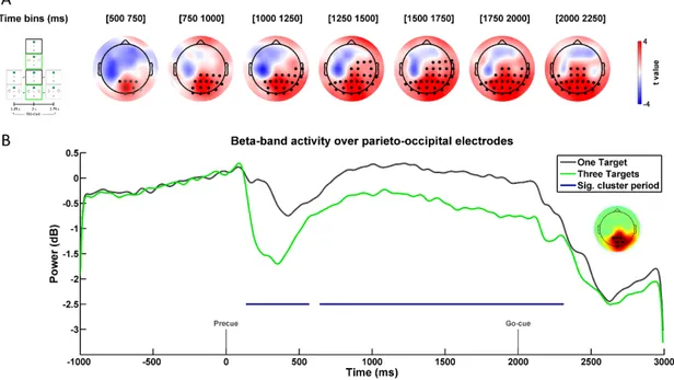
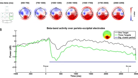
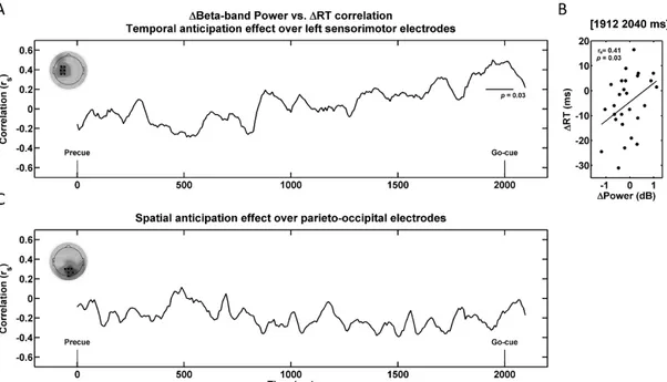
It is well documented that providing advanced information regarding the spatial location of a target stimulus (i.e., spatial anticipation) or its timing of occurrence (i.e. temporal anticipation) influences action preparation, reducing reaction times (RTs). Yet, it remains unknown whether the RT gains attributable to temporal and spatial anticipation are subtended by similar preparatory dynamics. Here this issue is addressed in humans by investigating EEG beta-band activity during movement preparation. Participants performed a reach RT task in which they initiated a movement as fast as possible toward visual targets following their appearance. Temporal anticipation was manipulated by having the target appear after a constant or variable foreperiod, whereas spatial anticipation was manipulated by precueing participants about the upcoming target location in advance or not. Results revealed that temporal and spatial anticipation both reduced reach RTs, with no interaction. Interestingly, temporal and spatial anticipation were associated with fundamentally different patterns of beta-band modulations. Temporal anticipation was associated with beta-band desynchronization over contralateral sensorimotor regions specifically around the expected moment of target onset, the magnitude of which was correlated with RT modulations across participants. In contrast, spatial anticipation did not influence sensorimotor activity, but rather led to increased beta-band power over bilateral parieto-occipital regions during the entire delay period. These results argue for distinct states of preparation incurred by temporal and spatial anticipation. In particular, sensorimotor beta-band desynchronization may reflect the timely disinhibition of movement-related neuronal ensembles at the expected time of movement initiation, without reflecting its spatial parameters per se.
28 Introduction
The time necessary to initiate a reaching movement toward an appearing stimulus, referred to as reaction time (RT), is known to be influenced by prior knowledge as to when and where it will appear. In support, studies manipulating the temporal predictability of an impending target have shown that RTs are faster as temporal anticipation increases (Niemi and Näätänen, 1981; Nobre et al., 2007). Similarly, studies manipulating the number of possible target locations have shown that RTs are faster as spatial anticipation increases (Hick, 1952; Schmidt and Lee, 2011). In spite of considerable work, it remains unclear whether the RT gains associated with spatial and temporal anticipation are subtended by similar preparatory dynamics at the neural level.
Of interest, recent behavioral work suggests that the mechanisms involved in the preparation of the spatial aspects of a movement (i.e. direction) are independent from those mediating its initiation (Haith et al., 2016). Specifically, these authors compared RTs in a task in which participants initiated reaching movements after target presentation to a task in which they were forced to initiate movements with lower-than-normal RTs using rhythmic cues. They showed that in the latter condition, participants reduced their RTs by ~80 ms, but strikingly were still able to produce spatially accurate movements. In further support, recent electrophysiological work in monkeys showed that movement initiation is accompanied by a large change in neural activity in primary motor (M1) and dorsal premotor (PMd) cortex that reflects when the movement will occur, but carries no essential information about reach direction (Kaufman et al., 2016). This indicates that separate "components" of the population response in these regions encode movement direction and timing. Overall, these studies suggest that RT gains associated with temporal and spatial anticipation may be subtended by distinct neural preparatory dynamics.
One method allowing to characterize preparatory activity with high temporal resolution is electroencephalography (EEG), with known modulations in many frequency bands. Most notable is the event-related desynchronization (ERD) in the beta-band (13-30 Hz) that is observed over contralateral sensorimotor regions before
29 and during movement (Pfurtscheller, 1981; Pfurtscheller and Lopes da Silva, 1999; Kilavik et al., 2013). While beta-band ERD has long been linked to "motor readiness," there remains ambiguity as to whether it relates more to the spatial specification of the movement or its initiation. For one, the status quo hypothesis, according to which reduced power reflects a release from inhibition necessary for a change in motor state (Gilbertson et al., 2005; Engel and Fries, 2010; Jenkinson and Brown, 2011; Perfetti et al., 2011), suggests that ERD is closely tied to movement initiation. As such, movements for which the go-cue is rhythmic are associated with greater pre-movement beta-band ERD than when the go-cue is unpredictable (Alegre et al., 2003). This possibility is further supported by a growing body of literature relating beta-band oscillations to predictive timing (Saleh et al., 2010; Arnal, 2012; Fujioka et al., 2012). However, there is also evidence that beta-band ERD is modulated by the degree of directional uncertainty of an upcoming movement, suggesting a possible role in encoding the spatial aspects of movements. For example, pre-movement beta-band ERD is greater when reducing the number of possible target directions (Tzagarakis et al., 2010) or the angle of separation between two alternative targets (Grent-’t-Jong et al., 2014).
Overall, these studies demonstrate that temporal and spatial precueing both influence the pattern of the beta-band ERD. However, direct comparison between the two types of precueing, as well as their possible interaction during reach preparation, has never been explicitly tested. Here this is addressed using a factorial design in a reach RT task. Temporal anticipation was manipulated by having separate blocks in which the foreperiod was either constant (i.e. 2 s) or variable (i.e. 1.25, 2 or 2.75 s). Spatial anticipation was manipulated by using spatial precues that were either informative (i.e. one location) or non-informative (i.e. three possible locations) as to the upcoming target location.
Materials and Methods
30 Twenty-seven young adults (23.1 ± 2.1 years old, 14 female and 13 male) without any known neurological or psychiatric condition took part in the experiment. They were all self-declared right-handed and had no visual impairment left uncorrected. All participants provided informed consent prior to the experiment by signing a consent form approved by the ethics committee of the Centre Hospitalier de l’Université de Sherbrooke and they all received a monetary compensation of 20 $ (CAD) for their participation.
Apparatus
Participants sat comfortably facing a CRT monitor and a digitizing tablet. The monitor (LG Studioworks 995E, Seoul, KR) was positioned ~77 cm in front of participants. The tablet (GTCO CalComp DB6 1218, Scottsdale, AZ, USA) was placed directly in front of participants and recorded the position of a hand-held stylus in real-time, which was presented as a cursor on the monitor (green circle; 0.5 cm in diameter). Participants were instructed to control the cursor by sliding the stylus across the tablet with the right hand. A custom-made box covered the digitizing tablet, such that participants could not see their arm while moving.
Stimuli and task
The experimental task consisted of center-out reaching movements toward one of three possible visual targets (see Figure 1). All movements were initiated from a starting circle (grey; 0.75 cm in diameter) located at the bottom of the screen. The targets (white circles; 1.65 cm in diameter) were situated 7 cm away from the starting circle, at 30°, 90° and 150° relative to the trigonometric circle (i.e. rightwards, straight-ahead and leftwards relative to midline, respectively). Participants were required to gaze at a fixation cross (red; 0.3 x 0.3 cm) situated 4 cm above the starting circle throughout the entire experiment to prevent eye movements.
31
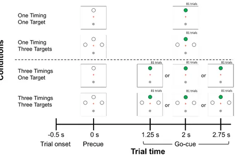
Figure 1. Trial sequence and experimental design. Temporal and Spatial anticipation were manipulated in four experimental conditions: 1) One Timing - One Target; 2) One Timing - Three Targets; 3) Three Timings - One Target; 4) Three Timings - Three Targets. Trials started with a 0.5 s baseline period after which a precue was provided. The precue consisted in the presentation of either a single target or three targets. The two levels of the factor Temporal anticipation were conducted in separate experimental blocks (One Timing; Three Timings). In the One Timing block (above dotted line), the timing of the go-cue (i.e., target turning green) was constant at 2 s, whereas in the Three Timings block (below dotted line), it varied between 1.25, 2 or 2.75 s. Only trials for which the go-cue occurred at 2 s were kept for primary experimental analysis (n = 81 per participant per condition).
Figure 1 illustrates the sequence of events for a given trial. Participants brought the cursor into the starting circle to begin the trial. After a 0.5 s baseline period, a precue specifying the possible locations of the targets was presented, marking the beginning of the foreperiod. At the end of the foreperiod, a target turned green (i.e. go-cue), prompting participants to perform their reach towards it. Participants were instructed to initiate and execute their movements as fast and accurately as possible. They were told not to stop on the target but to "strike" through it with a single uncorrected movement. After movement completion, participants were instructed to hold their final hand position for 250 ms, after which the cursor disappeared, prompting the return to the starting circle for the initiation of the next trial.
32
Experimental design
The temporal and spatial anticipation of target onset were independently manipulated using a 2 × 2 factorial design (see Figure 1). Temporal anticipation was manipulated by having participants take part in two separate experimental blocks in which the duration of the foreperiod was either constant at 2 s (i.e. One Timing), or could vary pseudo-randomly between three possibilities (i.e. 1.25, 2 or 2.75 s; Three Timings). The objective of using repeated exposure to either constant or variable foreperiods was to manipulate participants’ expectancy of the go-cue in order to build an internal representation of the moment of target onset (Niemi and Näätänen, 1981; Nobre et al., 2007). Although different approaches could have been used to influence temporal anticipation, such as rhythmic entrainment to a tone (Alegre et al., 2003), here the rationale for building a temporal prior of target onset was to keep the preparatory period exempt of additional sensory stimuli (i.e. rhythmic tones) which would have themselves influenced oscillatory activity and made it difficult to compare EEG modulations across conditions. Thus, in the present context, preparatory activity was identical from a sensory standpoint across the two levels of Temporal anticipation, therefore allowing to ascribe all spectral modulations to movement preparation only. Importantly, the ordering of the two blocks was counterbalanced across participants to rule out any ordering effect behavioral and EEG dependent variables.
Within each of the two experimental blocks, Spatial anticipation was manipulated by having the precue being fully informative as to the spatial location of the upcoming target (i.e. One Target; straight ahead) or not (i.e. Three Targets; leftwards, straight-ahead, and leftwards) (see Figure 1). The experiment thus consisted of four distinct conditions.
In a first block, participants could be submitted to either the One Timing - One Target condition or the One Timing - Three Targets condition. This block comprised a total of 178 trials: 81 for the former and 81 for the later (i.e. 27 trials per target), as well as 16 no-go trials. No-go trials were identical to the other trials with the exception that the go-cue was not presented. Participants were informed of these trials and were instructed not to move in this context. These trials served to prevent
33 participants from jumping the start, which was especially relevant for the One Timing - One Target condition since both spatial and temporal information were known in advance. In a second block, participants could be submitted to either the Three Timings - One Target condition or the Three Timings - Three Targets condition. This block comprised a total of 510 trials: 243 for the former (i.e. 81 trials per possible timing) and 243 for the later (i.e. 81 trials per possible timing, comprising 27 trials per target), as well as 24 no-go trials. Trials were pseudo-randomized throughout each experimental block. Overall, the experiment comprised 688 trials and lasted ~75 min.
By design, the One Timing conditions (One Timing - One Target and One Timing - Three Targets) only had data for the 2 s foreperiod. Hence, the primary experimental strategy was to compare preparatory activity across all four conditions using only trials in which the foreperiod was 2 s. Doing so ensured that the delay period was identical in every respect across the four conditions, such that any difference would be solely attributable to differential movement preparation incurred by Temporal or Spatial anticipation. In additional analyses, data from the 2.75 s foreperiods were used to provide further validation of the results obtained in the main 2 s foreperiod analysis. To ensure that an internal representation of the timing of target onset (i.e. prior) would be achieved through repeated exposure to either constant or variable foreperiods (Temporal anticipation factor), it was decided a priori that the first 12 trials of each of the two One Timing conditions (One Timing - One Target and One Timing - Three Targets) would be discarded, as well as the first 36 trials of each of the two Three Timings conditions (12 trials per possible timing in both the Three Timings - One Target and the Three Timings - Three Targets conditions). This corresponded to 96 trials out of the 688 per participant (14 % of the trials).
Behavioral data recording and analysis
Visual stimuli were presented using functions from the psychophysics toolbox [Psychtoolbox (Brainard, 1997; Pelli, 1997)], which were run with MATLAB (v2014a, MathWorks, Natick, MA, USA) using the Windows 7 operating system (Microsoft, Redmond, WA, USA) on a desktop computer (Dell Optiplex 7010, Round Rock, TX, USA). All hand position-related data, obtained from the digitizing tablet, were recorded
34 at 100 Hz and analyzed offline with custom MATLAB routines. Movement initiation was defined as the moment when the stylus left the starting circle. RT was calculated as the time difference between the go-cue and movement initiation. Time to target was calculated as the time difference between movement initiation and the moment the radial distance between the stylus and the starting circle exceeded 7 cm. Endpoint error was defined as the Euclidian distance in cm between the location of the cursor at the 7 cm radial distance and the aiming target.
Outlier trials were rejected based on several criteria. First, trials for which 1) RT was under 160 or over 600 ms, 2) time to target exceeded 500 ms or 3) endpoint error was greater than 5 cm were discarded. In addition, trials for which RT and time to target were beyond ± 2 SD from a participant’s mean were rejected. All these criteria led to the rejection of 21 ± 14 trials per participant (3.5% of the data).
EEG data acquisition, processing and time-frequency decomposition EEG recording and analysis
Scalp EEG data were recorded from a 64-electrode actiCAP (Brain Products, Gilching, Germany) and BrainAmp system (Brainproducts, Munich, Germany). Electrodes were positioned in accordance with the extended 10/20 system (Falk Minow Services, Herrsching-Breitbrunn, Germany) and it was ensured that the Cz electrode was at the participant’s vertex. The reference electrode was located at FCz and impedances were kept below 20 kΩ. The EEG signals were digitized online (sampling rate 500 Hz; BrainVision Recorder 2.0) using a Laptop (Dell Latitude E6530, Round Rock, TX, USA) running on Windows 7 (Microsoft, Redmond, WA, USA).
All EEG analyses were done offline using custom MATLAB routines, as well as functions derived from the EEGLAB toolbox (Delorme and Makeig, 2004). First, trials that had been rejected based on movement kinematics were discarded from the EEG datasets. Then data were digitally bandpass-filtered between 1 – 55 Hz and epoched from -1000 ms to +3000 ms around precue onset for all conditions. EEG data were then baseline-corrected to the average potential recorded during the 500 ms preceding the precue. This period was chosen as a baseline since participants were motionless and the precue had not been presented yet. Cortical activity was thus
35 considered neutral at that moment. Thereafter, EEG epochs showing voltage values exceeding ± 80 μV were discarded. Based on this criterion, 47 ± 55 trials per participant (6.8 % of data) were discarded from further behavioral and EEG analyses.
In sum, after considering both kinematic-based and EEG-based trial rejections, all analyses (both EEG and movement kinematics) were conducted on a total of 62 ± 8, 64 ± 7, 60 ± 9, and 63 ± 8 trials per participant for One Timing - One Target, One Timing - Three Targets, Three Timings - One Target, and Three Timings - Three Targets, respectively.
The data were further inspected for artifacts with a procedure based on independent component (IC) analysis, a blind separation technique that decomposes the EEG signal into maximally independent components in order to remove artifacts from EEG activity without having to discard entire epochs (Makeig et al., 2002; Delorme and Makeig, 2004; Hammon et al., 2008; Gwin et al., 2010; Gwin and Ferris, 2012a,b). The ‘runica’ function in EEGLAB was applied to decompose EEG signals into statistically maximal ICs. ICs were analyzed with respect to scalp topography and frequency characteristics, and were identified as being artifactual and removed if they met two of the following three criteria: 1) their time-course showed spurious bursts of activity, 2) their spectral power did not generally decrease as a function of frequency, as expected for EEG spectral power (Buzsáki, 2006) and 3) their scalp map showed activity concentrated at the far edges of the scalp, which are often indicative of muscle and/or ocular artifacts (Jung et al., 2000). Cleaned EEG data were generated by projecting back the time-course of activity of the remaining ICs to the electrode space.
To assess time-frequency power modulations across experimental conditions, the EEG time-series of each electrode and trial were convolved with a series of complex Morlet wavelets (1-50 Hz, 1 Hz steps). Spectral power estimates were obtained by multiplying the resulting complex signal by its complex conjugate. Wavelet cycles were linearly increased from 3 - 7.9 in 0.1 steps to improve frequency resolution at higher frequencies (Cohen, 2014). The obtained power time-series were then baseline-normalized, and changes in power were expressed in decibels as follows:
36 dB = 10log10(𝑅𝑃
𝐵𝑃)
where dB corresponds to the decibel-converted mean power, RP corresponds to the mean power value at a given time point, and BP corresponds to the average raw power during the baseline period, which was defined as the average power during the 500 ms preceding the precue. This measure was computed separately for each condition. Finally, the spectral power data were downsampled to 125 Hz.
Behavioral Statistical analysis
All behavioral dependant variables (i.e. RT, time to target and endpoint error) were submitted to separate 2 Temporal anticipation (One timing, Three timings) × 2 Spatial anticipation (One target, Three targets) repeated measures ANOVAs. All statistical tests were two-tailed and the threshold for significance was set to 0.05. Data normality was tested with the Shapiro-Wilk test prior to all analyses. The statistical analysis of the all behavioral dependent variables was done with IBM SPSS statistics (version 23, IBM Canada, ON, Canada). For all these analyses, the F statistic, statistical significance (p), effect size [partial eta squared, 𝜂𝑝2 (Field, 2009)] and descriptive statistics (Mean ± Standard error of the mean) are reported in the text. According to Fritz and colleagues (2012), the thresholds past which 𝜂𝑝2 denotes small, moderate and large effect sizes are 0.01, 0.06 and 0.14, respectively (Fritz et al., 2012).
EEG statistical analysis
Regarding EEG data, the goal was to assess whether beta-band power during the delay period was modulated by either Temporal or Spatial anticipation. To do so, non-parametric permutation tests were conducted to identify clusters of spatially and temporally adjacent electrode/time pairs showing statistically significant differences across conditions (Maris and Oostenveld, 2007). This method does not make assumptions about the distribution of the data and it provides an efficient solution to the multiple comparisons problem, making it particularly interesting for EEG analysis. Specifically, for each comparison, two-tailed dependent t-tests were computed for all electrode/time pairs in the true EEG data. Adjacent electrode/time pairs whose test statistic exceeded statistical significance threshold (t(26) = 2.056, α = 0.05, two-tailed),
37 were then identified. To be considered as a cluster, at least three adjacent electrodes had to show statistically significant t values. The size of a cluster was obtained by summing the t values across all adjacent electrode/time pairs constituting the cluster. Then, permutations (N = 1000) were undertaken, which consisted of randomly shuffling the experimental condition labels across participants. Following each permutation, the largest permuted cluster was identified. Ultimately, a Monte Carlo estimate (i.e. the proportion of permuted clusters whose size was larger than the clusters identified in the true data) was used to yield p values for each cluster.
All non-parametric permutation tests were conducted over the entire delay period starting from the precue (0 ms) until trial end (3000 ms). To probe for differences across the Temporal anticipation factor, data were pooled across Spatial anticipation levels and dependent t-tests were used to compare the One Timing to the Three Timings trials. Similarly, to probe for differences across the Spatial anticipation factor, data were pooled across Temporal anticipation levels and dependent t-tests were used to compare the One target and the Three Targets trials. To probe for an interaction between the two factors, dependent t-tests were used to compare the differences between the One Target and the Three Targets trials across the two Temporal anticipation levels. Clusters were deemed statistically significant if their p value was smaller or equal to the significance threshold (α = 0.05). All non-parametric permutation tests were done using the Fieldtrip toolbox (Oostenveld et al., 2011). For each identified cluster, the size, average statistic (t), statistical significance (p), and effect size (Cohen’s dz) are reported in the text. Cohen’s dz was calculated using the
average t-value of a cluster (Rosenthal, 1986; Lakens, 2013). According to Cohen (1988), dz is considered small, moderate, or large if it exceeds 0.2, 0.5 or 0.8,
respectively.
Results
Behavioral results
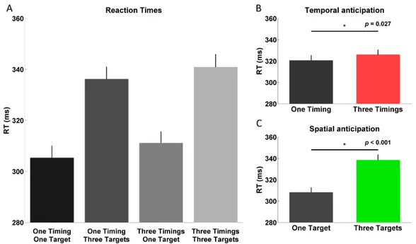
Mean RT data for all conditions can be seen in Figure 2A. The ANOVA conducted on the RT data revealed both a significant main effect of Temporal and
38 Spatial anticipation. Specifically, as can be seen in Figure 2B, RTs were significantly faster in One Timing (321 ± 5 ms) as compared to Three Timings (326 ± 5 ms; F(1,26) = 5.478, p = 0.027, 𝜂𝑝2 = 0.174). Similarly, as can be observed in Figure 2C, RTs were significantly faster in One Target (308 ± 5 ms) as compared to Three Targets (339 ± 5 ms; F(1,26) = 139.079, p < 0.001, 𝜂𝑝2 = 0.842). Importantly, there was no significant interaction between factors (F(1,26) = 0.279, p = 0.602, 𝜂𝑝2 = 0.011).
Figure 2. Reaction times A. Mean reaction times in each of the four conditions using only trials for which the go-cue occurred at 2 s. B. Main effect of Temporal anticipation. C. Main effect of Spatial anticipation. Error bars represent standard errors of the mean.
The ANOVA conducted on the time to target data revealed both a significant main effect of Temporal and Spatial anticipation. Specifically, time to target was slightly but significantly lower for One Timing (85 ± 2 ms) than for Three Timings (88 ± 3 ms; F(1,26) = 5.651, p = 0.025, 𝜂𝑝2 = 0.179). Similarly, time to target was slightly but significantly lower for One Target (85 ± 2 ms) than for Three Targets (88 ± 2 ms; F(1,26) = 9.048, p = 0.006, 𝜂𝑝2 = 0.258). There was no interaction between factors (F(1, 26) = 1.452, p = 0.239, 𝜂𝑝2 = 0.053).
The ANOVA conducted on the endpoint error data revealed a significant main effect of Spatial anticipation, with errors being slightly but significantly lower in One