HAL Id: hal-00180710
https://hal.archives-ouvertes.fr/hal-00180710
Submitted on 20 Nov 2019HAL is a multi-disciplinary open access archive for the deposit and dissemination of sci-entific research documents, whether they are pub-lished or not. The documents may come from teaching and research institutions in France or abroad, or from public or private research centers.
L’archive ouverte pluridisciplinaire HAL, est destinée au dépôt et à la diffusion de documents scientifiques de niveau recherche, publiés ou non, émanant des établissements d’enseignement et de recherche français ou étrangers, des laboratoires publics ou privés.
Nanoparticles loaded with ferrocenyl tamoxifen
derivatives for breast cancer treatment.
Anh Nguyen, Véronique Marsaud, Céline Bouclier, Siden Top, Anne Vessieres,
Pascal Pigeon, Ruxandra Gref, Philippe Legrand, Gérard Jaouen, Jack-Michel
Renoir
To cite this version:
Anh Nguyen, Véronique Marsaud, Céline Bouclier, Siden Top, Anne Vessieres, et al.. Nanoparticles loaded with ferrocenyl tamoxifen derivatives for breast cancer treatment.. International Journal of Pharmaceutics, Elsevier, 2008, 347 (1-2), pp.128-35. �10.1016/j.ijpharm.2007.06.033�. �hal-00180710�
Nanoparticles loaded with ferrocenyl tamoxifen derivatives for
breast cancer treatment
Anh Nguyen b, Véronique Marsaud a, Céline Bouclier a, Siden Top b, Anne Vessieres b, Pascal Pigeon b, Ruxandra Gref a, Philippe Legrand c, Gérard Jaouen b, Jack-Michel Renoir a
a
Laboratoire de physico-chimie, pharmacotechnie, biopharmacie, UMR CNRS 8612 and IFR 141, université Paris XI, 92296 Châtenay-Malabry, France
b
Laboratoire de chimie et biochimie des complexes moléculaires, UMR CNRS 7576, école nationale supérieure de chimie de Paris, 11, rue Pierre-et-Marie-Curie, 75231 Paris cedex 05, France
c
Institut Charles-Gerhardt UMR 5253 CNRS/UM2/ENSCM/UM1, équipe MACS, faculté de pharmacie, 15, avenue Charles-Flahault, B.P. 14491, 34093 Montpellier cedex 05, France
Abstract
For the first time, two organometallic triphenylethylene compounds (Fc-diOH and DFO), with strong antiproliferative activity in breast cancer cells, but insoluble in biological fluids, were incorporated in two types of stealth nanoparticles (NP): PEG/PLA nanospheres (NS) and nanocapsules (NC). Their physicochemical parameters were measured (size, zeta potential, encapsulation and loading efficiency), and their biological activity was assessed. In vitro drug release after high dilution of loaded NPs was measured by estradiol binding competition in MELN cells. The influence of the encapsulated drugs on the cell cycle and apoptosis was studied by flow cytometry analyses. Notwithstanding potential drug adsorption at the NP surface, Fc-diOH and DFO were incorporated efficiently in NC and NS, which slowly released both compounds. They arrested the cell cycle in the S-phase and induced apoptosis, whose activity is increased by loaded NS. A decrease in their antiproliferative activity by the antioxidant α-tocopherol indicated that reactive oxygen species (ROS) may be involved. Therefore, nanosystems, containing for the first time a high load of anticancer organometallic triphenylethylenes, have been developed. Their small size and delayed drug release, combined
with their enhanced apoptotic potential, are compatible with an increased persistence in the blood and a promising antitumour activity.
Keywords
Breast cancer; Antiestrogens; Estradiol receptors; Organometallic bioligands; SERMs
1. Introduction
The female hormone estradiol (E2 in Fig. 1) plays a critical role in controlling the female
reproductive system. One of its many functions is to regulate the proliferation and differentiation of the healthy breast epithelium. Therefore it is not surprising that this molecule is involved in two-thirds of breast tumours, the second most deadly cancer for women. These hormone-dependent cancer cells (ER+ cases) exhibit a higher accumulation of the estrogen receptor (ER), a transcription factor of the thyroid/steroid superfamily, which can be found in two different isoforms: ERα and ERβ ((Shiau et al., 1998), (Gustafsson, 1999)). However, a third of breast tumours are not hormone-dependent and are called ER negative (ER–). In the hormone induced pathway, when estradiol binds to its intranuclear receptor, it induces a typical conformational change in the protein structure, involving Helix 12 ((Shiau et al., 1998), (Jordan, 2003)). This enables the recruitment of coactivators and the dimerization of the ER. The homo/hetero-dimer then binds to small palindromic ERE sequences of DNA. This interaction allows the recruitment of transcriptional factors from the general transcription machinery around Polymerase II, and therefore initiates gene transcription and specific protein synthesis leading finally to cell proliferation. Thus, in ER+ tumours, the increased concentration of estrogen receptors favours cell multiplication.
Since estradiol binding to its receptor seems to be a key step required for the proliferation of breast cancer cells, many molecules targeting this protein have been synthesized to counteract the action of estradiol ((Magarian et al., 1994), (MacGregor and Jordan, 1998), (Meegan and Lloyd, 2003)). The most popular and widely prescribed antiestrogen for hormone-dependent breast cancer is tamoxifen. Its active metabolite, hydroxytamoxifen (OH-Tam, in Fig. 1), acts as an antagonist in breast tissue. The dimethylaminoethoxy side chain interaction with Asp351 of the ER is held responsible for the observed antiestrogenic effect of hydroxytamoxifen, because it prevents the recruitment of coactivators, and favours the
binding of corepressors instead. However, depending on the nature of the promoter to which hydroxytamoxifen binds, and the cellular context (e.g. the major type of ER (α or β) present in the tissue and the coactivator/corepressor ratio in the cell), this selective estrogen receptor modulator (SERM) can also act as an agonist. Thus, like estradiol, OH-Tam can induce beneficial effects such as maintaining bone density, but it also enhances endometrial tumour growth (MacGregor and Jordan, 1998).
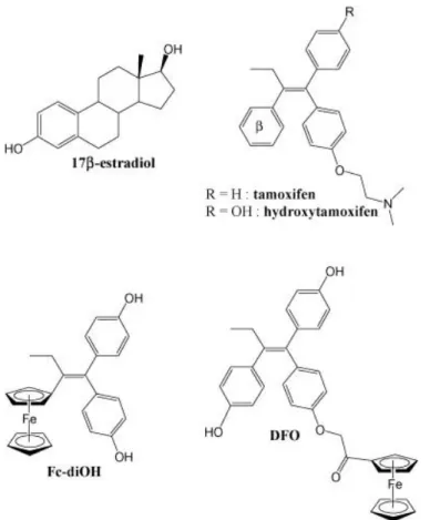
Fig. 1. Structures of Fc-diOH and DFO as compared to 4-hydroxy-tamoxifen (4-OH-Tam) and 17β-estradiol (E2).
Unfortunately, the successful treatment of breast cancer by hydroxytamoxifen is also overshadowed by the fact that some hormone-dependent breast tumours do not respond to endocrine therapy, and others respond initially to the antihormonal treatment, but acquire resistance in the long term (Lewis and Jordan, 2005, Osborne, 1998). To those cases of intrinsic and acquired resistance, the insensitive ER– tumour cells have to be added to point
out the limitation of hydroxytamoxifen. These cases highlight the dire need for new active molecules with broader therapeutic scopes.
After the discovery of the potent anticancer activity of cis-platin (Rosenberg et al., 1965, Rosenberg et al., 1969), many similar metal coordination complexes have been proposed (Jakupec et al., 2003). Though highly cytotoxic, most of them faced problems of instability. Compared to metal coordination complexes, organometallic compounds should be more stable, because their metal–ligand bond has a stronger covalent character (Crabtree, 2005). Our approach was to add a potentially cytotoxic moiety, i.e. the robust organometallic group ferrocene, to the competitive bioligand hydroxytamoxifen scaffold. We have shown that attaching such a neutral and stable moiety enhanced the cytotoxicity of hydroxytamoxifen, and increased the lipophilicity of the compound to facilitate its passage through the cellular membrane. (Jaouen et al., 2000, Jaouen et al., 1996, Top et al., 1996, Top et al., 2003, Vessières et al., 2005).
Among the various organometallic compounds we synthesised, two of them were singled out. Compound Fc-diOH (1,1-di(4′-hydroxyphenyl)-2-ferrocenylbut-1-ene, Fig. 1) is an analog of OH-Tam, where the tamoxifen β- aromatic benzyl ring has been replaced by the aromatic ferrocene moiety, and the amino side-chain has been replaced by a second hydroxyl group (Vessières et al., 2005). It is our most efficacious cytotoxic compound to date (IC50 ≈ 0.7 μM
on MCF-7, 0.44 μM on MDA-MB231). In compound DFO (1,2-di(4′-hydroxyphenyl)-1-[4″-(2″-ferrocenyl-2″-oxoethoxy)phenyl]but-1-ene, Fig. 1), the amino side-chain of OH-Tam has been changed into a carbonylferrocene moiety (Nguyen et al., 2007). DFO has shown a weaker antiproliferative activity towards cancer cells (IC50 ≈ 10 μM on MCF-7), but good
affinity for the estrogen receptors (RBA ≈ 14% on ERα and ERβ), probably thanks to the additional hydroxyl group on the β-benzyl ring. Interestingly, both compounds are active against ER+ and ER– cancer cells, suggesting that they do not target only the ERs in cancer cells.
Although Fc-diOH and DFO appear to be promising potential drugs, they still encounter the problem of bioavailibility, so common to active biomolecules discovered in laboratories, especially for phenols. Indeed, once injected in the bloodstream, the active molecule is often degraded or opsonized and removed from blood circulation by macrophages of the reticuloendothelial system. Hence, in order to increase the circulation time in the bloodstream and to enhance the probability of the molecule to extravasate in tumour tissues, we chose to protect the active molecule inside nanoparticles (NP). These drug carriers have hydrophilic
polyethyleneglycol (PEG) chains at their surface, to reduce the opsonization process (Soppimath et al., 2001). They could transport and concentrate the antiproliferative agents in the tumours, because their size enables them to cross the gaps of the discontinuous endothelium of cancer cells, which are richly vascularised (Gabizon and Papahadjopoulos, 1988). This method to improve the bioavailibility has already been used for many bioactive molecules, including tamoxifen (Hu et al., 2006, Shenoy and Amiji, 2005).
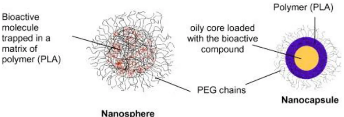
Therefore, for a more targeted and effective drug delivery, Fc-diOH and DFO were loaded inside nanospheres (NS) and nanocapsules (NC). A nanosphere is a matrix of polymer into which the active molecules are trapped. A nanocapsule is a vesicular system, whose oily core traps and retains the active molecules, and is surrounded by a protective polymeric membrane (Fig. 2). The polymer chosen is a biodegradable poly(D, L-lactic acid), or PLA. It is covalently bound to hydrophilic PEG chains, which are at the surface of the NPs. The ability of the nanospheres and nanocapsules to modulate the antiproliferative activity of Fc-diOH and DFO was then assessed by in vitro biological evaluation.
Fig. 2. Structures of the PEG-PLA nanosphere and nanocapsule.
2. Materials and methods
2.1. Chemicals
Soy phosphatidylcholine Lipoïd S75® (about 70% of phosphatidylcholine) was purchased from Lipoïd GmbH (Germany). Miglyol 810N was kindly provided by Hüls (Germany). Poly(D, L-lactide) PLA50 of MW 42 kDa was supplied by Phusis (France). PLA-PEG (D,
L-PLA50 MW 45 kDa and PEG MW 5 kDa) was synthesized and characterized as previously described (Gref et al., 1995).
Compounds Fc-diOH and DFO were prepared according to literature procedures (Top et al., 2003) and (Nguyen et al., 2007).
The solvents were analytical grade, and all other chemicals were commercially available at reagent grade. Water was purified by reverse osmosis (MilliQ, Millipore®).
2.2. Preparation of PEG-Coating Nanoparticles
The preparation of nanospheres (NS) according to Fessi et al. (1989) was based on interfacial deposition of preformed polymer, following solvent displacement.
Briefly, 20 mg of polymer or copolymer were dissolved in 1 mL of acetone containing 10−3 M of Fc-diOH or DFO, and rapidly dispersed into 2 mL of demineralised and sterilised water, followed by acetone evaporation under nitrogen flow for 2 h, and left overnight before use.
In the case of the nanocapsules (NC), only 5 mg of preformed polymer was dissolved in 1 mL of acetone. An amount of 2,1 mg of Fc-diOH or 5,9 mg of DFO was dissolved in a mixture of lipophilic surfactant (Lipoïd S75®, 66 mg/mL of acetone) and oil (Miglyol 810, 25 μL/mL of acetone). An amount of 25 μL of this preparation was added to the organic phase, before dispersion in water.
Control particles without Fc-diOH and DFO were prepared under the same conditions.
2.3. Physicochemical characterization of nanoparticles
The size of nanoparticles was measured by QuasiElastic Laser Light Scattering using a Nanosizer N4 Plus (Coulter Electronics, Florida, USA). The potential measurements (ς potentials) were determined with a Zetasizer 4 (Malvern Instruments, UK) after dilution of NS or NC suspensions in 1 mM KCl in a 1:2 volume ratio (NP suspension/1 mM KCl).
2.4. Measurement of encapsulation efficacy
Incorporation efficiency of the ferrocenyl derivatives was expressed both as encapsulation rate (w/w ratio) and percentage of encapsulation (%). The amount of Fc-diOH and DFO associated with the drug carriers was indirectly determined by UV–vis spectroscopy (Jenway 6405 UV–vis spectrometer). These values were calculated as the difference between the total
amount of Fc-diOH or DFO used to prepare the loaded nanoparticles and the amount of free molecules in the aqueous phase, after separation by centrifugation (60,000 g, 1 h, 4 °C) for nanospheres, or by ultrafiltration/centrifugation (450,000 g, 1 h, 4 °C) using Millicon eppendorfs (10,000 NMWL, Millipore®, France) for nanocapsules. The concentrations of
Fc-diOH and DFO in the aqueous phase after centrifugation were determined by the absorbance
at 304 nm for Fc-diOH and at 276 nm for DFO. This spectroscopic method was validated for drugs with concentrations between 1 and 100 μM.
2.5. Cell culture and transcription measurements
We used a method based on the capacity of free and encapsulated antiestrogens to inhibit estradiol-induced transcription in MELN cells, as previously described (Ameller et al., 2003b). MELN cells are MCF-7 cells stably transfected with a construct in which the luciferase (LUC) reporter gene is placed downstream of an estrogen responsive element (ERE) linked to the minimal β-globin promoter (ERE-β-globin-LUC). MELN and MCF-7 cells were cultured in DMEM (DMEM Biological Industries, Inc., Kibbutz Beit Haemek, Israel) supplemented with L-glutamine (2 mM), penicillin (50 UI/mL), streptomycin (50 UI/mL) and 10% FCS and maintained at 37 °C, with 5% CO2 in a humidified atmosphere.
Before steroid treatment, MELN cells were grown for 3 days in phenol red free DMEM containing 10% charcoal-stripped FCS. Under these experimental conditions, 0.1 nM of 17β-estradiol (E2) was found to induce the maximum LUC transcription. In order to evaluate the
release of Fc-diOH and DFO from the formulations, NCs and NSs containing Fc-diOH or
DFO were added to the culture medium containing 0.1 nM E2. After a total incubation time of
18 h at 37 °C, cells were collected, rinsed and lysed in 250 μL of LUC buffer (25 mM Tris/HPO4 at pH 7.8, 10 mM MgCl2, 1% Triton X100, 15% glycerol, 1 mM EDTA, 1 mM
DTT). Protein concentration was determined by the Biorad assay (Bio-Rad GmbH, Munich, Germany). Quantification of LUC activity was performed in triplicate in a luminometer (TD 20/20, Tuner Designs, Sunnyvale, CA), after injection of the LUC buffer (100 μL) supplemented with 100 mM ATP and 87 μg of luciferin/mL to 100 μL of cellular extract. Each experiment was repeated twice.
MCF-7 cells (2 × 10⁵) were exposed or not to the free or the encapsulated drugs (1 or 10 μM) during 48 h and 72 h. The cells were washed twice in cold phosphate-buffered saline (PBS), pelleted and suspended in ice-cold ethanol (80% in MilliQ water). In some experiments, 10 μM α-tocopherol was added for 1 h to the cell medium prior to further treatment. Fixed cells were then centrifuged and resuspended in PBS at 25 °C containing 100 μg/mL RNase A (Roche Molecular Biochemicals, Meylan, France) and 20 μg/mL propidium iodide (PI, Sigma–Aldrich) for 30 min at 37 °C. Cells were further analysed with a FACS Calibur (Becton–Dickinson, Le-Pont-de-Claix, France) equipped with an argon laser tuned at 488 nm and data were obtained with the CellQuest 1.2.2 and the ModFit LT 1.01 softwares (Becton– Dickinson).
3. Results
3.1. Characterization of NPs
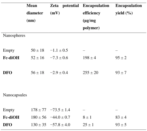
The size and the zeta (ς) potential of both empty NSs and NCs loaded with the organometallic compounds Fc-diOH and DFO are summarized in Table 1. The ς potential indicates the surface charge of the NP. While the size of the NCs was in the range of 150–200 nm, that of the NSs was three times smaller. The incorporation of the drug did not modify this parameter for either formulation. This was not the case for the ς potential: the decrease of the ς potential of Fc-diOH- and DFO-charged NSs, as well as the increase of NC ς potential indicate that some amount (though small) of the compounds were adsorbed at the NP surface. Indeed, the ς potential of the aqueous suspension of Fc-diOH and DFO at 1 mM was definitely negative, and measured as −35.5 ± 5.2 and −36.6 ± 0.5 mV, respectively. These values are lower than the ς potential of the unloaded NSs. Hence, if the organometallic compounds were adsorbed at the surface of the NSs, the ς potential of the loaded NSs should decrease. On the other hand, the ς potential of the free compounds are higher than those of the empty NCs. Therefore, we expected the ς potential of the loaded NCs to increase, if the organometallic compounds were adsorbed at the surface of the NCs.
Table 1. Characterization of the nanoparticles (mean values ± S.D., n = 6) Mean diameter (nm) Zeta potential (mV) Encapsulation efficiency (μg/mg polymer) Encapsulation yield (%) Nanospheres Empty 50 ± 18 −1.1 ± 0.5 – – Fc-diOH 52 ± 16 −7.3 ± 0.6 198 ± 4 95 ± 2 DFO 56 ± 18 −2.9 ± 0.4 255 ± 20 93 ± 7 Nanocapsules Empty 178 ± 77 −73.5 ± 1.4 – – Fc-diOH 180 ± 56 −44.0 ± 0.7 8 ± 1 83 ± 4 DFO 130 ± 35 −57.8 ± 4.0 25 ± 1 93 ± 5
The encapsulation yield of both molecules in the NSs was higher than that in the NCs. The maximum concentrations of encapsulated Fc-diOH and DFO were similar in the NSs (1 mM) and were estimated at 70 μM and 160 μM in the NCs respectively. The encapsulation efficiency and encapsulation yield of both types of NP showed that the organometallic compounds have a high affinity for the copolymer. Nevertheless, NC had a tendency to incorporate a lower concentration of both compounds, a feature characteristic of such nanoparticles, because of the reduced oil solubility (Ameller et al., 2003b).
3.2. Modulation of the antiestrogenic activity by ferrocifen-loaded NP
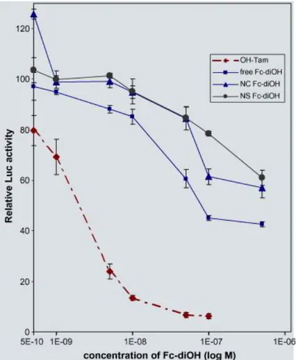
We wondered next about the biological activity of the NPs. In a previous work, we showed that unloaded PEG/PLA NPs have no inhibitory activity on the luciferase gene expression in MELN cells (Ameller et al., 2003b). As shown in Fig. 3, free Fc-diOH had a weak inhibition on estradiol-induced transcription in MELN cells at 16 h, with an IC50 value about 100-fold
that of OH-Tam. However, the trapped Fc-diOH has an even weaker ability to inhibit the expression of luciferase than the free compound at 16 h. This feature suggests that only a limited amount of drug was released from the NPs. Surprisingly, the type of NP does not seem to affect the transcription inhibition activity.
Fig. 3. Inhibitory capacity of free and PEG/PLA NS and NC loaded with Fc-diOH on E2
-mediated transcription. MELN cells (106) grown in phenol red free medium for 3 days were treated in triplicates during one night (16 h) with increasing concentrations of Fc-diOH, free and entrapped in either NS or NC as described in Materials and Methods. In the control experiment, E2-induced transcription (at 1 nM) was estimated as 100%. It corresponds to a 8–
9 fold increase as compared to the basal level of transcription (obtained without E2, but only
vehicle). After harvest and lysis, luciferase activity was measured. Data are expressed as mean% ± S.D. (n = 3) of inhibition of the E2-induced luciferase (LUC) activity.
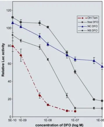
Data from Fig. 4 indicate that DFO has a better antiestrogenic activity than compound
Fc-diOH. Precisely, its luciferase inhibition activity is ten times stronger than that of Fc-Fc-diOH.
Again, both DFO-loaded NCs and NSs have a weaker transcription inhibition activity than free DFO. Interestingly, DFO released from the NC has an inhibitory activity somewhat similar to that of Fc-diOH trapped in the NPs (data from Fig. 3 compared to data from Fig. 4). However, in the case of DFO, the type of NP in which the molecule is encapsulated seems to influence its activity. Comparatively, the DFO-loaded NS inhibits the luciferase activity 100 times more than DFO trapped in a NC. This suggests that NCs release DFO more slowly than NSs, and this feature is even more striking at high concentrations (10−7–10−6 M).
Fig. 4. Inhibitory capacity of free and PEG/PLA NS and NC loaded with DFO on E2
-mediated transcription. MELN cells were treated as in Fig. 3 with free and NS, NC-trapped compound DFO. Results are expressed as in Fig. 3.
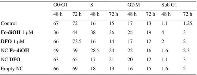
3.3. Activity of ferrocifen-NP on cell cycle and apoptosis
We then wanted to know if these nanosystems behave differently towards MCF-7 cell proliferation. Flow cytometry experiments were carried out at 48 h and 72 h with both free and encapsulated Fc-diOH and DFO at 1 μM and/or at 10 μM. Data (FACS analysis) are summarized in Table 2, Table 3. Both empty NSs (Table 2) and empty NCs (Table 3) had no effect on the cell distribution as already observed (Renoir et al., 2006). Interestingly, the number of cells in the S-phase of the cycle has increased more clearly with Fc-diOH than with DFO, whether the compounds were trapped or not in NS. At a similar concentration, once trapped in the NS, DFO had a strong capacity to arrest cell cycle in the G0/G1 phase at 48 h, unlike the free drug. This effect was no longer observed at 72 h, suggesting maybe some instability of DFO, which is protected only as long as it stays encapsulated.
Table 2. FACS analysis on MCF-7 cells exposed to free and encapsulated ferrocene derivatives in nanospheres (NS) of PEG/PLA
G0/G1 S G2/M Sub G1 48 h 72 h 48 h 72 h 48 h 72 h 48 h 72 h Control 69 79 14 12 14 8 3 2 Fc-diOH 10 μM 63 54 24 37 6 6 11 10 DFO 10 μM 66 62 13 18 19 16 5 10 NS Fc-diOH 64 52 25 38 8 7 8 17 NS DFO 80 66 10 20 9 9 5 15 Empty NS 70 73 13 11 14 9 2.5 3
Table 3. FACS analysis on MCF-7 cells exposed to free and encapsulated ferrocene derivatives in nanocapsules (NC) of PEG/PLA
G0/G1 S G2/M Sub G1 48 h 72 h 48 h 72 h 48 h 72 h 48 h 72 h Control 67 72 16 15 17 13 1.1 1.25 Fc-diOH 1 μM 36 44 38 36 25 19 4 3 DFO 1 μM 66 73.5 16 14 17 12 2 2 NC Fc-diOH 49 59 28.5 24 22 16 1.6 2.3 NC DFO 63 65 17 21 20 12 1.1 3 Empty NC 66 69 18 19 16 15 1.6 2
Interestingly, an increase of cells in apoptosis was noticed after cell exposure to both free
Fc-diOH and DFO. This fate was stronger in the case of cell incubation with the trapped
compounds. Altogether, data from Table 2 suggest that at 10 μM, Fc-diOH has a greater capacity to arrest the cell cycle in the S-phase than DFO. They have comparable ability to induce apoptosis in MCF-7 cells and this potency is enhanced and delayed by entrapment in PEG/PLA nanospheres.
We also compared the activity of Fc-diOH/DFO-loaded NCs on the cell cycle. Since the encapsulation loading was weaker in the NCs than in the NSs, MCF-7 cells were exposed to weaker drug concentrations in the case of the NC experiments, in order to avoid too high an amount of polymer in the cell culture. Results presented in Table 3 clearly indicate that
Fc-diOH at 1 μM, whether free or encapsulated in the NCs, arrests cell cycle in the S-phase, and
to a more limited extent in the G2/M phase. This feature is less clearly observed at 10 μM, a concentration which rather induces apoptosis (see Table 4). Additionally, a smaller number of cells in apoptosis were noticed after 48 h when the compounds were encapsulated, hence suggesting again the delay induced by the nanovectors. At this low concentration, free DFO at 1 μM had no significant effect on MCF-7 cells, contrary to the encapsulated drug which slightly increased the number of cells in the S-phase.
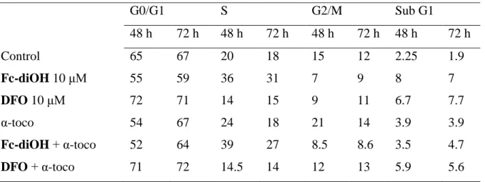
Table 4. Effect on free radical inhibition by α-tocopherol (α-toco) on MCF-7 cells exposed to free compounds Fc-diOH and DFO
G0/G1 S G2/M Sub G1 48 h 72 h 48 h 72 h 48 h 72 h 48 h 72 h Control 65 67 20 18 15 12 2.25 1.9 Fc-diOH 10 μM 55 59 36 31 7 9 8 7 DFO 10 μM 72 71 14 15 9 11 6.7 7.7 α-toco 54 67 24 18 21 14 3.9 3.9 Fc-diOH + α-toco 52 64 39 27 8.5 8.6 3.5 4.7 DFO + α-toco 71 72 14.5 14 12 13 5.9 5.6
3.4. Influence of α-tocopherol on cell cycle and apoptosis
It has been suggested that OH-Tam at high concentrations may induce the production of reactive oxygen species (ROS) in breast cancer cells (Obrero et al., 2002). Similarly, ferrocene derivatives have also been suspected of producing ROS ((Joy et al., 1989), (Osella et al., 2000), (Tabbi et al., 2002)). We next examined if the effects of the ferrocene derivatives observed in MCF-7 cells could imply ROS production. Similar experiments to those described in Table 3 were performed in the presence or absence of the antioxidant α-tocopherol. Data from Table 4 suggest that α-tocopherol alone has practically no effect on the MCF-7 cell cycle in the experimental conditions, although it slightly induced apoptosis. However, we predicted that α-tocopherol would decrease apoptosis of cells exposed to
Fc-diOH and DFO. Indeed, in the presence of α-tocopherol, the number of cells in subG1
dropped from 7 to 4.7, and from 7.7 to 5.6, respectively, after 72 h of treatment. These data also indicate that DFO could have a dual effect on MCF-7 cells, an antiproliferative effect through its inhibitory capacity towards ERα activation and downstream consequences, and the capacity to produce free radicals.
4. Discussion
The coupling of organometallic moieties tethered to SERMs has led to the synthesis of various types of compounds showing variable affinities for the two main ER isoforms ERα and ERβ (Jaouen et al., 2000, Jaouen et al., 1996, Top et al., 1996, Top et al., 2003, Vessières
et al., 2005). In such a program, a ferrocenyl substituent has been tethered to the 4-hydroxy-tamoxifen scaffold, the active metabolite of 4-hydroxy-tamoxifen (Top et al., 2001, Top et al., 2003). Ferrocene is lipophilic, compact, and stable in non-oxidizing media. Its derivatives have shown some antitumour potential, when oxidized to the ferrocenium salt (Osella et al., 2000, Tamura and Miwa, 1997), albeit at rather high concentration (0.1 mM). Amongst the large variety of ferrocenyl derivatives of 4-OH-Tam created in our laboratory, compounds
Fc-diOH and DFO displayed antiproliferative activity on both estradiol-dependent (MCF-7) and
independent (MDA-MB-231) breast cancer cell lines, probably due to ER-independent cytotoxicity of the compounds.
In the present work, we have designed two types of nanovectors, aiming at the in vivo delivery of insoluble anticancer molecules. We took advantage of previously well established formulations based on the copolymer PEG/PLA, which proved to be capable of incorporating high concentrations of a SERM like 4-OH-Tam (Renoir et al., 2006) and a pure antiestrogen like RU58668 (Ameller et al., 2003a, Ameller et al., 2003b). Both organometallic compounds
Fc-diOH and DFO were incorporated in these formulations at a high yield, although the NS
incorporated higher concentrations than the NC. The size of the resulting NPs were within the range of the gaps in the discontinuous cancerous endothelium, and thus they are possible vectors for the organometallic bioligands.
Both compounds showed efficiency in inhibiting estradiol-induced transcription in ER+ breast cancer cells, arresting cell cycle, and inducing apoptosis in the same cells. A great benefit of such devices is their endocytosis ability, leading to the delivery of their trapped drug inside the cell (Gabizon and Papahadjopoulos, 1988). They have, among others, the advantage of protecting the drugs against hydrolysis and oxidation. The synthesised nanovectors tend to promote the activity of Fc-diOH and DFO, maybe because encapsulation reduces their degradation. This observation is consistent with the results from the estradiol-induced transcription experiments on MELN cells and from the flow cytometry experiments.
It is worthwhile to differentiate the results from antiestrogenic experiments on MELN cells and those from the FACS Analysis. The distinction lies in the concentrations used for each method. In the antiestrogenic experiments, the concentrations reached very small values such as 0.5 nM, whereas in the FACS experiments the concentrations were 1 μM and 10 μM. Therefore, in the MELN cells where the NPs were very diluted, a greater percentage of
Fc-diOH or DFO was liberated. This is a function of the partition coefficient between
species through the polymeric matrix of the NSs was favoured for small concentrations. Thus, there was a greater percentage of organometallic compounds Fc-diOH or DFO liberated from the NPs in the estradiol-induced transcription experiments than in the flow cytometry experiments. So the relative reduced activity of the trapped compounds compared to the free ones was different according to the type of experiment.
The mechanism of action of both organometallic compounds has been investigated, in order to better understand their antiproliferative effects. Fc-diOH and DFO must each follow different mechanisms, due to their structural differences. Indeed, it has been proposed that the ferrocene moiety in Fc-diOH acts as an intramolecular oxidation assistant (Hillard et al., 2006b, Vessières et al., 2005). It is thought that the oxidation of iron (II) to iron (III) provokes the deprotonation of the phenol which is concerted with an intramolecular electron transfer to the ferrocenium moiety. The resulting organic radical species is oxidized again, followed by another proton abstraction from the ethyl group, resulting in a reactive quinone methide-type structure. This species can then form adducts with DNA, GSH, or proteins. It has been shown that the pattern ferrocene–C=C–phenol is required for this mechanism, being the electron carrier π-system. However, in the structure of DFO, the ferrocene moiety cannot act as an intramolecular oxidation “antenna”, having no electron carrier system to link the ferrocene moiety to a phenol group. So the antiproliferative effect observed must follow another mechanism. It might involve the oxidation of the ferrocene to a ferrocenium salt and production of hydroxyl radicals via the Fenton process (Hillard et al., 2006a).
Our experiments suggest that α-tocopherol does reverse the antiproliferative effect of both organometallic compounds, since the number of cells in subG1 dropped when the antioxidant agent was added. This result is consistent with our hypothesis that Fe plays an important role in inhibiting the proliferation of breast cancer cells. As a well-known antioxidant, α-tocopherol could prevent the oxidation of Fe(II) into Fe(III), and also help to reduce the downstream production of ROS, thus lowering the number of cells in subG1.
5. Conclusion
Interest in nanovectors of highly cytotoxic compounds has fostered many drug formulations, such as the lipid-coated aggregates of the metal coordination complex cis-platin (Burger et al., 2002). This article describes for the first time an efficient encapsulation of organometallic compounds, which should possess stronger metal–ligand bonds than metal coordination
complexes. The ferrocene derivatives Fc-diOH and DFO have shown interesting cytotoxic effects on MCF-7 cells, which very likely involve ROS production. But since their water-insolubility impedes their potential biological activity, both were successfully loaded at high yield in nanospheres and nanocapsules. Thanks to their appropriate physicochemical characteristics, the nanoparticles were able to deliver the water-insoluble compounds to cancer cells, and shield the active molecules from degradation by delaying their release.
Furthermore, according to the metal introduced, organometallic compounds could offer different possibilities. For instance, the Fe of DFO may be converted into radioactive Re or Tc, without greatly changing the binding affinity with ERα and β. Then it would be possible to follow the bioavailibility of the labelled bioligands in the body, once released from their protective nanovectors.
Acknowledgements
We thank the “ministère de la Recherche”, the “Centre national de la recherche scientifique” and the “Ligue nationale contre le cancer” for their financial support (grants from the Cher and Indre departments to J.-M. R.), and M. Pons and P. Ballaguer for the gift of MELN cells. We are grateful to E. Hillard for proofreading the manuscript.
References
Ameller et al., 2003a
T. Ameller, V. Marsaud, P. Legrand, R. Gref, B. Gillian, J.-M. Renoir. Polyester-poly(ethylene glycol) nanoparticles loaded with the pure antiestrogen RU 58668: physicochemical and opsonization properties. Pharm Res, 20 (2003), pp. 1063-1070
Ameller et al., 2003b
T. Ameller, V. Marsaud, P. Legrand, R. Gref, J.-M. Renoir. In vitro and in vivo biologic evaluation of long-circulating biodegradable drug carriers loaded with the pure antiestrogen RU 58668. Int J Cancer, 106 (2003), pp. 446-454
Burger et al., 2002
K.N.J. Burger, R.W.H.M. Staffhorst, H.C. de Vijlder, M.J. Velinova, P.H. Bomans, P.M. Frederik, B. de Kruijff. Nanocapsules: lipid-coated aggregates of cis-platin with high cytotoxicity. Nat Med, 8 (2002), pp. 81-84
Crabtree, 2005
R.H. Crabtree. The Organometallic Chemistry of the Transition Elements. (4th Edition), J. Wiley and Sons, Hoboken, New Jersey (2005)
Fessi et al., 1989
H. Fessi, F. Puisieux, J.P. Devissaguet, N. Ammoury, S. Benita. Nanocapsule formation by interfacial polymer deposition following solvent displacement. Int J Pharm, 55 (1989), pp. R1-R4
Gabizon and Papahadjopoulos, 1988
A. Gabizon, D. Papahadjopoulos. Liposome formulations with prolonged circulation time in blood and enhanced uptake by tumours. Proc Natl Acad Sci USA, 85 (1988), pp. 6949-6953
Gref et al., 1995
R. Gref, A. Domb, P. Quellec, T. Blunk, R.H. Muller, J.M. Verbavatz, R. Langer. The controlled intravenous delivery of drugs using PEG-coated sterically stabilized nanospheres. Adv Drug Deliv Rev, 16 (1995), pp. 215-233
Gustafsson, 1999
J.A. Gustafsson. Estrogen receptor ß: a new dimension in estrogen mechanism of action. J Endocrinol, 163 (1999), pp. 379-383
Hillard et al., 2006a
E.A. Hillard, A. Vessières, F. Le Bideau, D. Plazuk, D. Spera, M. Huché, G. Jaouen. A series of unconjugated ferrocenyl phenols: prospects as anticancer agents. Chem Med Chem, 1 (2006), pp. 551-559
Hillard et al., 2006b
E.A. Hillard, A. Vessières, L. Thouin, G. Jaouen, C. Amatore. Ferrocene-mediated proton-coupled electron transfer in a series of ferrocifen-type breast cancer drug candidates. Angew Chem Int Ed, 45 (2006), pp. 285-290
Hu et al., 2006
F.X. Hu, K.G. Neoh, E.T. Kang. Synthesis and in vitro anticancer evaluation of tamoxifen-loaded magnetite/PLLA composite nanoparticles. Biomaterials, 27 (2006), pp. 5725-5733
Jakupec et al., 2003
M.A. Jakupec, M. Galanski, B.K. Keppler. Tumour-inhibiting platinum complexes-state of the art and future perspectives. Rev Physiol Biochem Pharmacol, 146 (2003), pp. 1-53
Jaouen et al., 2000
G. Jaouen, S. Top, A. Vessières, R. Alberto. New paradigms for the synthetic pathways inspired by bioorganometallic chemistry. J Organomet Chem, 600 (2000), pp. 23-36
Jaouen et al., 1996
G. Jaouen, A. Vessières, S. Top, M. Salmain. La chimie bioorganométallique en réceptorologie et analyse. Première partie: étude du récepteur des estrogènes. Actual Chim, 15 (1996), pp. 6-9
Jordan, 2003
V.C. Jordan. Antiestrogens and selective estrogen receptor modulators as multifunctional medicines. 1. Receptor interactions. J Med Chem, 46 (2003), pp. 883-908
Joy et al., 1989
A.M. Joy, D.M.L. Goodgame, I.J. Stratford. Int J Radiat Oncol Biol Phys, 16 (1989), pp. 1053-1056
Lewis and Jordan, 2005
J.S. Lewis, V.C. Jordan. Selective estrogen receptor modulators (SERMs): Mechanisms of anticarcinogenesis and drug resistance. Mut Res/Fundam Mol Mech Mutagen, 591 (2005), pp. 247-263
MacGregor and Jordan, 1998
J.I. MacGregor, V.C. Jordan. Basic guide to the mechanisms of antiestrogen action. Pharmacol Rev, 50 (1998), pp. 151-196
Magarian et al., 1994
R.A. Magarian, L.B. Overacre, S. Singh, K.L. Meyer. The medicinal chemistry of nonsteroidal antiestrogens: a review. Curr Med Chem, 1 (1994), p. 61
Meegan and Lloyd, 2003
M.J. Meegan, D.G. Lloyd. Advances in the science of estrogen receptor modulation. Curr Med Chem, 10 (2003), pp. 181-210
Nguyen et al., 2007
A. Nguyen, S. Top, A. Vessieres, P. Pigeon, M. Huche, E.A. Hillard, G. Jaouen. Organometallic analogues of tamoxifen: Effect of the amino side-chain replacement by a carbonyl ferrocenyl moiety in hydroxytamoxifen. J Organometal Chem, 692 (2007), pp. 1219-1225
Obrero et al., 2002
M. Obrero, D.V. Yu, D.J. Shapiro. Estrogen dependent and estrogen receptor-independent pathways for tamoxifen and 4-hydroxytamoxifen-induced programmed cell death. J Biol Chem, 277 (2002), pp. 45695-45703
Osborne, 1998
C.K. Osborne. Tamoxifen in the treatment of breast cancer. N Eng J Med, 339 (1998), pp. 1609-1618
Osella et al., 2000
D. Osella, M. Ferrali, P. Zanello, F. Laschi, M. Fontani, C. Nervi, G. Cavigiolio. On the mechanism of the antitumor activity of ferrocenium derivatives. Inorg Chim Acta, 306 (2000), pp. 42-48
Renoir et al., 2006
J.-M. Renoir, B. Stella, T. Ameller, E. Connault, P. Opolon, V. Marsaud. Improved antitumoral capacity of mixed and pure antiestrogens in breast cancer cell xenografts after their administration by entrapment in colloidal nanosystems. J Steroid Biochem Mol Biol, 102 (2006), pp. 114-127
B. Rosenberg, L. Vancamp, T. Krigas. Inhibition of cell division in Escherichia coli by electrolysis products from a platinum electrode. Nature, 205 (1965), pp. 698-699
Rosenberg et al., 1969
B. Rosenberg, L. Vancamp, J.E. Trosko, V.H. Mansour. Platinum compounds: a new class of potent antitumour agents. Nature, 222 (1969), pp. 385-386
Shenoy and Amiji, 2005
D.B. Shenoy, M.M. Amiji. Poly(ethylene oxide)-modified poly(epsilon-caprolactone) nanoparticles for targeted delivery of tamoxifen in breast cancer. Int J Pharm, 293 (2005), pp. 261-270
Shiau et al., 1998
A.K. Shiau, D. Barstad, P.M. Loria, L. Cheng, P.J. Kushner, D.A. Agard, G.L. Greene. The structural basis of estrogen receptor/coactivator recognition and the antagonism of this interaction by tamoxifen. Cell, 95 (1998), pp. 927-937
Soppimath et al., 2001
K.S. Soppimath, T.M. Aminabhavi, A.R. Kulkarni, W.E. Rudzinski. Biodegradable polymeric nanoparticles as drug delivery devices. J Control Release, 70 (2001), pp. 1-20
Tabbi et al., 2002
G. Tabbi, C. Cassino, G. Cavigiolio, D. Colangelo, A. Ghiglia, I. Viano, D. Osella. Water stability and cytotoxic activity relationship of a series of ferricenium derivatives. ESR insights on the radical production during the degradation process. J Med Chem, 45 (2002), pp. 5786-5796
Tamura and Miwa, 1997
H. Tamura, M. Miwa. DNA cleaving activity and cytotoxic activity of ferricenium cations. Chem Lett (1997), pp. 1177-1178
Top et al., 1996
S. Top, J. Tang, A. Vessières, D. Carrez, C. Provot, G. Jaouen. Ferrocenyl hydroxytamoxifen: a prototype for a new range of estradiol receptor site-directed cytotoxics. Chem Commun (1996), pp. 955-956
Top et al., 2001
S. Top, A. Vessières, C. Cabestaing, I. Laios, G. Leclercq, C. Provot, G. Jaouen. Studies on organometallic selective receptor modulators (SERMs). Dual activity in the hydroxy ferrocifen series. J Organomet Chem, 637 (2001), pp. 500-506
Top et al., 2003
S. Top, A. Vessières, G. Leclercq, J. Quivy, J. Tang, J. Vaissermann, M. Huché, G. Jaouen. Synthesis, biochemical properties and molecular modelling studies of organometallic specific estrogen receptor modulators (SERMs), the ferrocifens and hydroxyferrocifens: evidence for an antiproliferative effect of hydroxyferrocifens on both dependent and hormone-independent breast cancer cell lines. Chem Eur J, 9 (2003), pp. 5223-5236
Vessières et al., 2005 A.
Vessières, S. Top, P. Pigeon, E.A. Hillard, L. Boubeker, D. Spera, G. Jaouen. Modification of the estrogenic properties of diphenols by the incorporation of ferrocene. Generation of antiproliferative effects in vitro. J Med Chem, 48 (2005), pp. 3937-3940