Role of the Cationic C-Terminal Segment of Melittin on Membrane
Fragmentation
Alexandre Therrien1, Alain Fournier2, Michel Lafleur1*
1Department of chemistry, Center for Self-Assembled Chemical Structures (CSACS), Université de Montréal, C.P. 6128, Succ. Centre Ville, Montréal (Québec) H3C 3J7, Canada.
2Centre INRS–Institut Armand-Frappier, Institut national de la recherche scientifique, 531 boul. des Prairies, Ville de Laval (Québec) H7V 1B7, Canada.
*Corresponding author: Michel Lafleur Fax: (514) 343-7586 e-mail: [email protected] 3 4 5 6 7 8 9 10 11 12 13 14 15 16 17 18 19 20 21 22 23 24 25 26 27 28 29 30 31 32 33 34 35 36 37 38 39 40 41 42 43 44 45 46 47 48 49 50 51 52 53 54 55 56 57
Abstract
The widespread distribution of cationic antimicrobial peptides capable of membrane fragmentation in nature underlines their importance to living organisms. In the present work, we determined the impact of the electrostatic interactions associated with the cationic C-terminal segment of melittin, a 26-amino acid peptide from bee venom (net charge +6), on its binding to model membranes and on the resulting fragmentation. In order to detail the role played by the C-terminal charges, we prepared a melittin analogue for which the 4 cationic amino acids in positions 21 to 24 were substituted with the polar residue citrulline, providing a peptide with the same length and amphiphilicity but with a lower net charge (+2). We compared the peptide bilayer affinity and the membrane fragmentation for bilayers prepared from dipalmitoyl-sn-glycero-3-phosphocholine (DPPC)/1,2-dipalmitoyl-sn-glycero-3-phospho-L-serine (DPPS) mixtures. It is shown that neutralization of the C-terminal considerably increased melittin affinity for zwitterionic membranes. The unfavorable contribution associated with transferring the cationic C-terminal in a less polar environment was reduced, leaving the hydrophobic interactions, which drive the peptide insertion in bilayers, with limited counterbalancing interactions. The presence of negatively charged lipids (DPPS) in bilayers increased melittin binding by introducing attractive electrostatic interactions, the augmentation being, as expected, greater for native melittin than for its citrullinated analogue. The membrane fragmentation power of the peptide was shown to be controlled by electrostatic interactions and could be modulated by the charge carried by both the membrane and the lytic peptide. The analysis of the lipid composition of the extracted fragments from DPPC/DPPS bilayers revealed no lipid specificity. It is proposed that extended phase separations are more susceptible to lead to the extraction of a lipid species in a specific manner than a specific lipid-peptide affinity. The present work on the lipid extraction by melittin and citrullinated melittin with model membranes emphasizes the complex relation between the affinity, the lipid extraction/membrane fragmentation, and the lipid specificity.
3 4 5 6 7 8 9 10 11 12 13 14 15 16 17 18 19 20 21 22 23 24 25 26 27 28 29 30 31 32 33 34 35 36 37 38 39 40 41 42 43 44 45 46 47 48 49 50 51 52 53 54 55 56 57
1. Introduction
The widespread distribution of cationic antimicrobial peptides (CAPs) in animal and plants underlines their importance to living organisms. To this day, more than 2500 naturally occurring CAPs have been catalogued. They present a wide range of activity: antibacterial, antiviral, antifungal, and anticancer, to name a few 1-2. Notably, the interest they generate in modern medicine comes from their straightforward mode of action: in general, CAPs directly target and disrupt vital membrane features, allowing CAPs to preserve their potency despite some microbial mutations.
A characteristic feature of CAP structures is the coexistence of an amphipathic character, and positively charged amino acids. Upon binding to membranes, they often organize as α-helices with hydrophobic and hydrophilic residues on opposite faces, a feature referred to as secondary amphipathic character. Designers of synthetic CAPs found that it was possible to modify CAP activity towards microbial and mammalian cells independently by varying the hydrophobicity, as well as the number and the position of the positive charges in the amphiphilic structure 3-9. Therefore, fine-tuning the primary structure of CAPs can optimize cell selectivity. It is believed that the cationic charge of CAPs plays a fundamental role in the targeting of bacterial cells 10-12 as these typically possess negatively charged membranes 13. For example, the increase of the net positive charge of synthetic CAPs was found to improve their antimicrobial activity while maintaining low toxicity towards mammalian cells 6, 8. It appeared, however, that there was a positive-charge threshold as further increase of the CAP net positive charge led to a higher activity toward erythrocyte membranes 7, a phenomenon that was associated with the putative attraction of these highly charged CAPs by the negative membrane potential inside the cell 14.
3 4 5 6 7 8 9 10 11 12 13 14 15 16 17 18 19 20 21 22 23 24 25 26 27 28 29 30 31 32 33 34 35 36 37 38 39 40 41 42 43 44 45 46 47 48 49 50 51 52 53 54 55 56 57
In the present work, we determined the impact of electrostatic interactions associated with the cationic C-terminal segment of melittin on bilayer affinity and on the resulting lipid extraction. Melittin, the main component of dry bee venom, is a 26-aminoacid CAP 15. It has been used as a model peptide for various purposes over the last decades (for a general review, see 16), and has shown therapeutic potential with antimicrobial, anti-inflammatory 17-20, antiparasitic 21, and anticancer 22-23 activity, as well as for treatment 24 and prevention 25 of HIV. Like many CAPs, melittin binds to membranes as an amphipathic α-helix 26-28. Its sequence, GIGAVLKVLTTGLPALISWIKRKRQQ-NH2 15, includes a highly hydrophilic C-terminal segment with 4 positive charges (K21-R22-K23-R24). The 1-20 segment folds as an amphipathic α-helix upon the binding of the peptide to bilayers. When interacting with membranes, melittin induces leakage 29-34 and it leads, at higher concentrations, to membrane fragmentation and the formation of small lipid/peptide bicelles, or nano-disks 35-37.
The cationic character of melittin was shown to be essential to its activity on membranes. It is well documented that the presence of negatively charged lipids in membranes, such as phosphatidylglycerol (PG), phosphatidylserine (PS), and phosphatidic acid (PA), significantly increases the melittin affinity due to attractive electrostatic interactions 38-42. However, it was observed that melittin-induced bicellization of membranes was strongly hindered by the presence of anionic lipids in membranes 26, 38, 40, 43-44. This observation was proposed to be due to an electrostatic anchoring of the peptide to the interface, preventing the relocation of the peptide deeper in the hydrophobic core, an essential step for the membrane fragmentation. This inhibition of the lipid extraction was found to be proportional to the interfacial negative charge density of the bilayer, and independent of the nature of the anionic lipid 38.
3 4 5 6 7 8 9 10 11 12 13 14 15 16 17 18 19 20 21 22 23 24 25 26 27 28 29 30 31 32 33 34 35 36 37 38 39 40 41 42 43 44 45 46 47 48 49 50 51 52 53 54 55 56 57
Melittin analogues with modified or omitted residues have been used to determine the role of the cationic C-terminal part. At this point, there is no coherent conclusion regarding the impact of the electrostatic interactions on bilayer binding. For instance, the properties of melittin-21Q, a truncated analogue of melittin in which residues 21 to 25 (K21-R22-K23-R24-Q25) were omitted, were investigated. Its binding to zwitterionic 1,2-dimyristoyl-sn-glycero-3-phosphocholine (DMPC) bilayers as well as to anionic 1,2-dimyristoyl-sn-glycero-3-phosphoglycerol (DMPG) bilayers was measured by Surface Plasmon Resonance (SPR) 45-46. Melittin-21Q exhibited a reduced or similar binding to DMPC and to DMPG bilayers compared to native melittin, suggesting a favorable or a limited contribution of the cationic C-terminal portion to membrane affinity. Another investigation, using tryptophan fluorescence, revealed that melittin and its C-terminal truncated versions 1-22 (Mel1-22), and 1-20 (Mel1-20), all showed an increased affinity for 1-palmitoyl-2-oleoyl-sn-glycero-3-phosphocholine (POPC)/1-palmitoyl-2-oleoyl-sn-glycero-3-phospho-(1'-rac-glycerol) (POPG) 70/30 large unilamellar vesicles (LUVs) compared POPC LUVs 47 (See Supporting Information). These findings highlighted the role of attractive electrostatic interactions. However, it was also reported that Mel1-22 and Mel1-20 showed a larger affinity for POPC bilayers than melittin, hinting for a detrimental role of the cationic C-terminal in the association with neutral membranes. Similarly, the disordering effect of membrane apolar core by melittin fragments, as assessed by Attenuated Total Reflectance - infrared (ATR-IR) spectroscopy, suggested that the hydrophobic 1-15 fragment of melittin inserted well into 1,2-dipalmitoyl-sn-glycero-3-phosphocholine (DPPC) and POPC membranes, while the binding of the hydrophilic 16-26 fragment was limited 48. The results relative to the impact of the C-terminal residues on cell lysis do not provide a consistent description either. Mel1-22 was shown to induce less leakage of encapsulated fluorescein from DPPC LUVs than melittin 49. This conclusion was in agreement with the findings resulting from the comparison of melittin cytolytic activity
3 4 5 6 7 8 9 10 11 12 13 14 15 16 17 18 19 20 21 22 23 24 25 26 27 28 29 30 31 32 33 34 35 36 37 38 39 40 41 42 43 44 45 46 47 48 49 50 51 52 53 54 55 56 57
with that of Mel1-22 or Mel1-20, evaluated by 51Cr release assays from human lymphoblast cells; this study also found that the truncated analogues were less active 50. However, it was shown that the removal of any of the 21 to 26 (KRKRQQ) residues to form 25-residue analogues of melittin (net charge +5 or +6) had no effect on its lytic power; the induced leakage of cell material from erythrocytes and bacterial cells was similar between analogues and native melittin 51.
In the present paper, we detail the role played by the electrostatic interactions involving the C-terminal charges of melittin in the lipid extraction induced upon interacting with membranes. We compared the bilayer affinity and the extent of lipid extraction of melittin bearing either its native cationic C-terminal segment or a neutral hydrophilic segment. The neutral segment was prepared by substituting the 4 basic residues of melittin (21K-22R-23K-24R) by citrullines, a polar but neutral amino acid 52. Citrullination of the peptide isolated the effect of the charges but preserved the solubility, the length and the number of peptide bonds of the native peptide. We characterized the interactions with membranes prepared from DPPC and 1,2-dipalmitoyl-sn-glycero-3-phospho-L-serine (DPPS) in various proportion in order to modulate the negative charge of the bilayer interface. In addition, we examined whether melittin-driven lipid extraction was specific for anionic lipids, considering the attractive interactions between the two species. It has been recently shown that melittin could preferentially extract DPPC molecules when interacting with DPPC/1,2-dipalmitoyl-sn-glycero-3-phosphoethanolamine (DPPE) membranes 53. It was proposed that the surrounding of bound melittin was depleted in DPPE, as the stronger inter-PE interactions were unfavorable to the peptide insertion in the bilayer. The resulting enrichment in DPPC of the peptide environment was proposed to be at the origin of the enhanced DPPC extraction relative to DPPE. The specificity of the lipid extraction was, in that case, based on lipid polymorphic propensities. In the present work, we examined whether
3 4 5 6 7 8 9 10 11 12 13 14 15 16 17 18 19 20 21 22 23 24 25 26 27 28 29 30 31 32 33 34 35 36 37 38 39 40 41 42 43 44 45 46 47 48 49 50 51 52 53 54 55 56 57
electrostatic interactions may also lead to the preferential extraction of a lipid species by melittin.
2. Experimental Methods
2.1 Chemicals
Melittin was purified from bee venom (Sigma, St. Louis, MO, USA) by ion exchange chromatography on SP-Sephadex C-25 54. Solid phase synthesis of citrullinated melittin (Cit-Mel) was carried out on methylbenzhydrylamine resin, using commercial Boc-amino acid residues (Chem-Impex, Wood Dale, IL, USA) and BOP as the coupling reagent (Matrix Innovation, Quebec City, QC, CAN). Acidolytic Boc removal was obtained by treating the protected peptide-resin with trifluoroacetic acid (TFA)/methylene chloride (45%). After a final Boc deprotection step, following a cleavage with hydrofluoric acid (Matheson, Edmonton, AB, CAN) containing m-cresol (10%) as a scavenger, a crude peptide preparation was isolated and washed with ethylether. The crude material was purified by reverse-phase HPLC using an acetonitrile (ACN) gradient in aqueous TFA (0.1%). Pure fractions corresponding to the expected mass of Cit-Mel, as established by MALDI-TOF mass spectrometry, were pooled, evaporated to remove ACN, and lyophilized. DPPC and DPPS were purchased from Avanti Polar Lipids (Alabaster, AL, USA). Ethylenediaminetetraacetic acid (EDTA), NaCl, and 3-[N-morpholino]propanesulfonic acid (MOPS) were obtained from Sigma (St. Louis, MO, USA). All chemicals were used as received. 2.2 Lipid membrane preparation
First dissolved in a benzene/methanol mixture (90/10 (v/v)), individual lipids were mixed to obtain the desired molar ratio and then lyophilized. The lipid powders were hydrated in a MOPS buffer (50 mM) containing 100 mM NaCl and 100 µM EDTA, and adjusted to pH 7.4. The samples
3 4 5 6 7 8 9 10 11 12 13 14 15 16 17 18 19 20 21 22 23 24 25 26 27 28 29 30 31 32 33 34 35 36 37 38 39 40 41 42 43 44 45 46 47 48 49 50 51 52 53 54 55 56 57
were submitted to 3 freeze-and-thaw cycles (from liquid nitrogen temperature to 65 °C) to form the multilamellar vesicle (MLV) suspensions used for the extraction experiments. For the fluorescence experiments, the MLVs were extruded at 75 °C using a manual extruder (Northern Lipids, Vancouver, BC, CAN) to obtain 100-nm LUVs.
2.3 Fluorescence measurements
Tryptophan fluorescence spectra were recorded to assess the binding of melittin to lipid bilayers. For these binding studies, LUV aliquots were added to a melittin solution (14 µM in the MOPS buffer; its concentration was determined from its absorbance at 280 nm, using a molar absorptivity coefficient of 5 570 M-1 cm-1 55). After each addition, the fluorescence spectrum of melittin was acquired at 65 °C; this temperature was selected to ensure that all the investigated lipid systems were in the fluid phase. The wavelengths at maximum for free (λfree) and bound (λbound) melittin were determined from the spectra recorded from samples with a lipid to peptide incubation ratio (L/P) of 0, and 400, respectively. The fraction of bound melittin, Xb, was then calculated from the ratio of the fluorescence intensity at λfree (Iλfree) and at λbound (Iλbound) using
𝑋𝑋𝑏𝑏 =𝑅𝑅400𝑅𝑅−𝑅𝑅−𝑅𝑅00 ,
where R is Iλbound/Iλfree, R0 is Iλbound/Iλfree at L/P=0, and R400 is Iλbound/Iλfree at L/P=400. The association constants of melittin to the membranes (Ka) were calculated as the slope of the fitted lines describing the variations of Xb/(1-Xb) as a function of [lipid]external, according to :
𝐾𝐾𝑎𝑎=[𝑝𝑝𝑝𝑝𝑝𝑝𝑝𝑝𝑝𝑝𝑝𝑝𝑝𝑝][𝑝𝑝𝑝𝑝𝑝𝑝𝑝𝑝𝑝𝑝𝑝𝑝𝑝𝑝]𝑓𝑓𝑓𝑓𝑓𝑓𝑓𝑓 × [𝑙𝑙𝑝𝑝𝑝𝑝𝑝𝑝𝑝𝑝𝑙𝑙]𝑏𝑏𝑏𝑏𝑏𝑏𝑏𝑏𝑏𝑏𝑓𝑓𝑒𝑒𝑒𝑒𝑓𝑓𝑓𝑓𝑏𝑏𝑒𝑒𝑒𝑒.
The data points for which most of melittin was bound (Xb≥0.8) deviated from the model and were discarded for the determination of Ka. The value [lipids]external took into account only the lipid external leaflet (i.e. the total lipid concentration/2) as the association was assumed to
3 4 5 6 7 8 9 10 11 12 13 14 15 16 17 18 19 20 21 22 23 24 25 26 27 28 29 30 31 32 33 34 35 36 37 38 39 40 41 42 43 44 45 46 47 48 49 50 51 52 53 54 55 56 57
occur essentially at the bilayer interface. The molar Gibbs free energy of binding (ΔGbind) was calculated using:
𝛥𝛥𝛥𝛥𝑏𝑏𝑝𝑝𝑏𝑏𝑝𝑝= RT ln �Kc°a�,
where R is the ideal gas constant, T is the temperature, and c°=1 M is the standard reference concentration. Fluorescence measurements were carried using a Photon Technology International fluorometer. The excitation wavelength was set at 270 nm and the bandwidths for the excitation and emission monochromators were set at 1.0 and 2.0 nm, respectively. In these conditions, no significant light diffusion caused by the liposomes was observed.
2.4 Circular dichroism
Circular dichroism spectra were acquired to determine the secondary structure of the bilayer-bound peptides. Spectra were acquired from a melittin solution (14 µM in the MOPS buffer), and after the addition of a quantity of LUV ensuring complete binding. The CD spectra were recorded, at 65 °C, on an Applied Photophysics ChirascanTM spectropolarimeter, using a cuvette with an optical-path length of 5 mm. The helical content, f, was estimated using :
𝑓𝑓 = 𝜃𝜃−𝜃𝜃𝑅𝑅𝑅𝑅
𝜃𝜃𝐻𝐻−𝜃𝜃𝑅𝑅𝑅𝑅 ,
where θ is the measured mean residue molar ellipticity at 222 nm, θRC is the predicted value for 100% random coil (-1 500 deg cm2 dmol-1) and θ
H is the predicted value for 100% α-helix (-33 400 deg cm2 dmol-1)56.
2.5 Lipid Extraction
Lipid extraction induced by melittin was determined using an approach previously described 53. Briefly, a melittin solution and a MLV suspension prepared in the MOPS buffer were mixed in a microcentrifuge tube to obtain the desired L/P and a fixed phospholipid concentration of 1
3 4 5 6 7 8 9 10 11 12 13 14 15 16 17 18 19 20 21 22 23 24 25 26 27 28 29 30 31 32 33 34 35 36 37 38 39 40 41 42 43 44 45 46 47 48 49 50 51 52 53 54 55 56 57
mg/mL. The suspensions were then incubated for at least 30 min at 65 °C, a temperature above the membrane gel-to-fluid phase transition temperature (Tm = 41 °C for DPPC and 54 °C for DPPS). After the incubation, the samples were centrifuged for 5 min at 20 800 g and 1 °C. It was assumed that the extracted lipids, existing as small melittin-lipid assemblies, stay in the supernatant while the remaining MLVs (possibly with bound melittin) pellet. Centrifugation of control samples (without melittin) showed that more than 85% of lipids were found in the pellets for all phospholipid mixtures. The supernatants were isolated and the pellets were resuspended in the MOPS buffer for their analysis. The phospholipid contents in the supernatants and in the pellets were determined by a Bartlett’s phosphorus assay 57. The extent of extraction was calculated using
Extraction % =𝑝𝑝ℎ𝑜𝑜𝑙𝑙𝑝𝑝ℎ𝑜𝑜𝑙𝑙𝑝𝑝𝑝𝑝𝑝𝑝𝑝𝑝𝑙𝑙 𝑝𝑝𝑏𝑏 𝑝𝑝ℎ𝑝𝑝 𝑙𝑙𝑠𝑠𝑝𝑝𝑝𝑝𝑠𝑠𝑏𝑏𝑎𝑎𝑝𝑝𝑎𝑎𝑏𝑏𝑝𝑝𝑝𝑝𝑜𝑜𝑝𝑝𝑎𝑎𝑙𝑙 𝑝𝑝ℎ𝑜𝑜𝑙𝑙𝑝𝑝ℎ𝑜𝑜𝑙𝑙𝑝𝑝𝑝𝑝𝑝𝑝𝑝𝑝𝑙𝑙 .
The lipid composition of the supernatants and of the pellets were determined by HPLC-MS analysis, using an Agilent Technologies 1100 series system equipped with a 1100 MSD mass spectrometer. Samples were eluted on a YMC diol column (4.6 x 150 mm, 5-µm particle size) (Agilent Technologies), maintained at 50 °C. Elution of the phospholipids was achieved in 7 min, using ACN/aqueous ammonium acetate solution (100 mM) (85/15) at 0.6 mL/min. The ESI source was used in the positive ionization mode. Nitrogen was used as drying gas at 250 °C and 12 L/min. Nebulizing gas was also nitrogen, held at 241 kPa. The extent of extraction was determined for each lipid species, using
𝐸𝐸𝐸𝐸𝐸𝐸𝐸𝐸𝐸𝐸𝐸𝐸𝐸𝐸𝐸𝐸𝐸𝐸𝐸𝐸 % = 𝐴𝐴𝑠𝑠
𝐴𝐴𝑠𝑠+ 𝐴𝐴𝑝𝑝,
where As and Ap are the lipid peak area from the supernatant and the pellet analysis, respectively. Experiments were carried out in triplicates unless stated otherwise.
3 4 5 6 7 8 9 10 11 12 13 14 15 16 17 18 19 20 21 22 23 24 25 26 27 28 29 30 31 32 33 34 35 36 37 38 39 40 41 42 43 44 45 46 47 48 49 50 51 52 53 54 55 56 57
3. Results
3.1 Association to LUVs
First, the impact of the C-terminal charges of melittin on bilayer affinity was determined. The hypsochromic shift of the tryptophan fluorescence band was used to characterize the transfer of this amino acid from water (in the case of free melittin) to a more hydrophobic environment (associated with membranes) 58. The binding of melittin and Cit-Mel to DPPC zwitterionic bilayers and to DPPC/DPPS anionic membranes was characterized (Figure 1). The binding experiments were carried out at 65 °C, a temperature at which membranes were in the fluid phase. The effect of citrullination on the melittin association to pure DPPC membranes is presented in Figure 1A. Free melittin displayed a fluorescence maximum at 348 nm; the band was progressively shifted upon the addition of DPPC LUVs towards 335 nm, a value characteristic of the bound state 41 (See Supporting Information). From these fluorescence data, the proportion of membrane-bound melittin was estimated as described in the Materials and Methods section. 3 4 5 6 7 8 9 10 11 12 13 14 15 16 17 18 19 20 21 22 23 24 25 26 27 28 29 30 31 32 33 34 35 36 37 38 39 40 41 42 43 44 45 46 47 48 49 50 51 52 53 54 55 56 57
Figure 1: Evolution of tryptophan fluorescence of melittin (full symbols) and of citrullinated melittin (empty symbols), characteristic of melittin association with DPPC (A) or DPPC/DPPS 70/30 (B) bilayers at 65 °C.
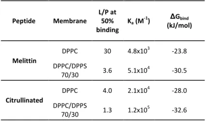
The L/P ratio at which 50% of melittin was bound (L/P50) was evaluated at 30 for DPPC. Ka and ΔGbind were also inferred from these data (Table 1). The ΔGbind value for the association of melittin to DPPC membranes was -23.8 kJ/mol, which is similar to the values that have been reported for melittin binding to fluid POPC LUVs (varying between -21.3 to -24.7 kJ/mol) 59-60. The titration of Cit-Mel with DPPC LUVs showed a similar hypsochromic shift (from 344 nm to 336 nm; see Supporting Information) but occurring at lower L/Ps, with L/P50=4. This result indicated that ∼7 times less lipids were required to bind half the peptides. In addition, ΔGbind was -28.0 kJ/mol, indicating a stronger association to DPPC than native melittin.
3 4 5 6 7 8 9 10 11 12 13 14 15 16 17 18 19 20 21 22 23 24 25 26 27 28 29 30 31 32 33 34 35 36 37 38 39 40 41 42 43 44 45 46 47 48 49 50 51 52 53 54 55 56 57
Table 1: Association parameters obtained from binding data. Peptide Membrane L/P at 50% binding Ka (M -1) ΔGbind (kJ/mol) Melittin DPPC 30 4.8x10³ -23.8 DPPC/DPPS 70/30 3.6 5.1x10⁴ -30.5 Citrullinated DPPC 4.0 2.1x10 4 -28.0 DPPC/DPPS 70/30 1.3 1.2x10⁵ -32.6
The peptide affinity was also determined for negatively charged membranes formed by DPPC/DPPS 70/30 (mol/mol) mixture (Figure 1B). The titration curves indicated that melittin had a stronger affinity for negatively charged bilayers than for pure DPPC membranes; in the case of DPPC/DPPS 70/30 bilayers, the L/P50 was shifted to 3.6 and ΔGbind was -30.5 kJ/mol. A consistent increased melittin affinity had already been reported for anionic membranes of PS, of phosphatidylglycerol (PG), or containing unprotonated palmitic acid (PA-) 41, 59, 61. This enhanced attraction was proposed to be due to electrostatic interactions. The binding experiments with Cit-Mel also revealed an increased affinity for DPPC/DPPS membranes compared to pure DPPC bilayers, with L/P50 reaching 1.3, and a ΔGbind of -32.6 kJ/mol. It should be noted that these results indicated that the affinity of Cit-Mel for DPPC/DPPS bilayers was stronger than that of melittin, even though 4 of the 6 positively charged amino acids of the latter have been substituted with citrullines.
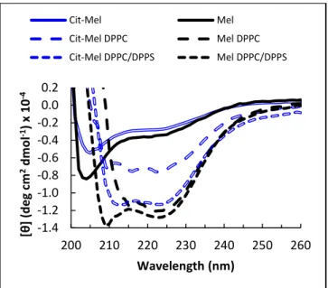
We have determined the variation of secondary structure of the peptides upon their binding to bilayers, using CD spectroscopy (Figure 2). The spectra of the free peptides were typical of a random coil structure. Upon binding, the peptides adopted an α-helix structure, characterized
3 4 5 6 7 8 9 10 11 12 13 14 15 16 17 18 19 20 21 22 23 24 25 26 27 28 29 30 31 32 33 34 35 36 37 38 39 40 41 42 43 44 45 46 47 48 49 50 51 52 53 54 55 56 57
by the minimum at 222 nm. The inferred α-helix content was between 31 and 36% except for Cit-Mel bound to DPPC that showed an estimated α-helix content of 18%. The formation of a helical structure by melittin upon its binding to bilayers is well established 9, 46, 62, and this structure appeared to be preserved upon heating the systems to 65 °C.
Figure 2 - CD spectra of melittin or Cit-Mel (14 µM) in solution, before and after the addition of DPPC or DPPC/DPPS 70/30 (mol/mol) LUVs. The lipid/peptide incubation ratio was 200 for the Mel/DPPC system, and 100 for the others, in order to obtain complete binding of the peptides. T = 65 °C.
3.2 Lipid extraction by melittin and citrullinated melittin
The impact of electrostatic interactions on melittin-induced lipid extraction was characterized by modulating both the bilayer charge, using DPPC/DPPS membranes in different proportions, and
-1.4 -1.2 -1.0 -0.8 -0.6 -0.4 -0.2 0.0 0.2 200 210 220 230 240 250 260 [θ ] ( deg c m 2 d mo l -1) x 1 0 -4 Wavelength (nm) Cit-Mel Mel Cit-Mel DPPC Mel DPPC Cit-Mel DPPC/DPPS Mel DPPC/DPPS 3 4 5 6 7 8 9 10 11 12 13 14 15 16 17 18 19 20 21 22 23 24 25 26 27 28 29 30 31 32 33 34 35 36 37 38 39 40 41 42 43 44 45 46 47 48 49 50 51 52 53 54 55 56 57
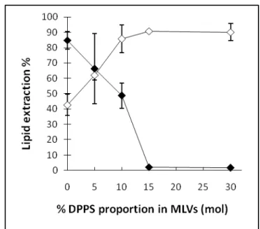
the peptide charge, by comparing melittin and Cit-Mel. Figure 3 indicates that the addition of anionic DPPS to neutral DPPC MLVs modulated lipid extraction induced by melittin and Cit-Mel in opposite ways.
Figure 3: Lipid extraction after incubation (L/P=20) of melittin (filled diamonds) or Cit-Mel (empty diamonds) with DPPC/DPPS MLVs in different proportions of neutral/anionic lipids.
At L/P=20, DPPC bilayers were almost completely destructed after an incubation with melittin as the lipid extraction reached 85%; this extent is in agreement with previous results 38, 53. The insertion of DPPS in membranes progressively inhibited the lipid extraction, a phenomenon previously demonstrated for other negatively charged phospholipids 38. Membranes with 15% DPPS or more were completely resistant to the lipid extraction by melittin at L/P=20; this inhibition is consistent with previous 2H-NMR results obtained with DPPC bilayers containing 10 (mol)% DPPG or DMPS 38. The lipid extraction from pure DPPC bilayers by Cit-Mel was ∼40 %, about half of the level observed for melittin. Conversely to the inhibiting effect on melittin-induced extraction, the addition of DPPS appeared to enhance the ability of Cit-Mel to extract lipids; the addition of 5 and 10 % DPPS increased the extent of the lipid extraction by about 20%
3 4 5 6 7 8 9 10 11 12 13 14 15 16 17 18 19 20 21 22 23 24 25 26 27 28 29 30 31 32 33 34 35 36 37 38 39 40 41 42 43 44 45 46 47 48 49 50 51 52 53 54 55 56 57
and 40% respectively, mirroring the pattern observed with melittin. The lipid extraction reached a plateau at about 90% for membranes with 10% DPPS or more.
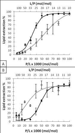
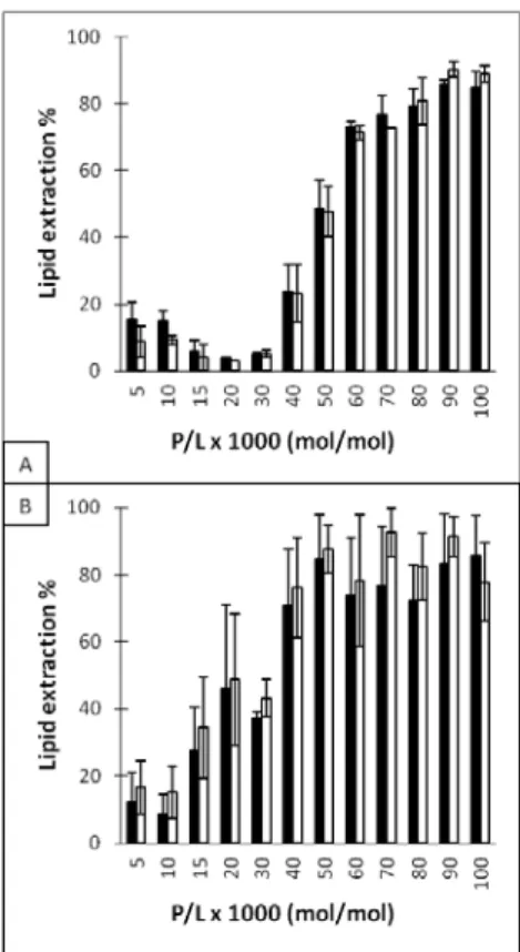
The lipid extraction as a function of the melittin concentration was detailed for DPPC and DPPC/DPPS 90/10 membranes (Figure 4) to better distinguish the activity of both peptides towards neutral and anionic liposomes. Melittin started to cause a lipid extraction from DPPC membranes when the peptide to lipid incubation ratio (P/L) reached 15×10-3 (L/P=67). The lipid extraction increased abruptly with an increased melittin concentration, from 6% at P/L=15×10-3 (L/P=67) to 91% at P/L=50×10-3 (L/P=20). These results are in agreement with previous studies using the same centrifugation approach 53 as well as those using 2H-NMR measurements to assess the formation of extracted small lipid/peptide particles 38. The curve obtained with DPPC/DPPS 90/10 bilayers illustrates the resistance of anionic bilayers to the lipid extraction by melittin. The lipid extraction was observed for P/L proportions greater than 30×10-3 (L/P=33); it increased from 5% at P/L=30×10-3 to 80% at P/L=70×10-3. 3 4 5 6 7 8 9 10 11 12 13 14 15 16 17 18 19 20 21 22 23 24 25 26 27 28 29 30 31 32 33 34 35 36 37 38 39 40 41 42 43 44 45 46 47 48 49 50 51 52 53 54 55 56 57
Figure 4: Quantification of lipid extraction after incubation of melittin (full symbols) or Cit-Mel (empty symbols) with MLVs of DPPC (A) or DPPC/DPPS 90/10 (B) at different ratios of incubation. The incubation ratio is displayed as peptide per 1000 lipids, P/L, or L/P.
The lipid extraction from pure DPPC bilayers by Cit-Mel reflected the reduced activity of this peptide compared to melittin. The lipid extraction was observed when P/L reached 20×10-3: 9% lipid extraction was observed. It increased with the peptide concentration, reaching >95% at P/L=90×10-3. The results obtained with DPPC/DPPS 90/10 bilayers showed the enabling effect of DPPS on the lipid extraction by Cit-Mel, as a P/L as low as 15×10-3 led to 28% lipid extraction. The maximal bilayer disruption was already obtained for a P/L of 50×10-3. It should be noted that DPPC/DPPS 90/10 bilayers were more difficult to pellet by centrifugation in the absence of peptide, likely because of the electrostatic repulsion between the anionic membranes (Figure 4B). This was reflected by a small extent of lipid extraction reported for very low peptide
3 4 5 6 7 8 9 10 11 12 13 14 15 16 17 18 19 20 21 22 23 24 25 26 27 28 29 30 31 32 33 34 35 36 37 38 39 40 41 42 43 44 45 46 47 48 49 50 51 52 53 54 55 56 57
contents (P/L≤10) that was, in fact, corresponding to the amount of lipid obtained for the blank. Upon the addition of more melittin (10≤P/L≤30), the MLVs were pelleted more efficiently, leading to an estimated lipid extraction closer to 0%. This observation is in agreement with the proposed bridging of adjacent anionic bilayers by melittin that would force the expulsion of some interlamellar water 26, 61.
3.3 Absence of lipid selectivity in melittin-induced extraction
The composition of the extracted lipid fraction after the incubation of DPPC/DPPS membranes with the peptides was determined in order to identify whether the extraction was specific for one of the two lipid species (Figure 5). The 90/10 DPPC/DPPS mixture was chosen for these experiments since the resulting membranes led to intermediate levels of lipid extraction; these were required to obtain sufficient DPPS for reliable quantitation and to get remaining MLVs to allow specific lipid extraction. The overall lipid extraction could be inferred from the weighted extraction average of the two lipids. As shown on Figure 3, the lipid extraction augmented as P/L increased. The higher lipid extraction extent observed for Cit-Mel at P/Ls between 15×10-3 and 60×10-3 demonstrated again its increased activity towards DPPS-containing membranes. The results show that both DPPC and DPPS were extracted by the peptides to a similar extent. Therefore, melittin and Cit-Mel extracted lipids from DPPC/DPPS 90/10 bilayers in a non-selective manner. No significant difference between the DPPS content in the initial membrane composition and in the extracted lipid fractions was observed for membranes with up to 30% of DPPS for P/L of 50×10-3 (L/P=20) (Supporting Information).
3 4 5 6 7 8 9 10 11 12 13 14 15 16 17 18 19 20 21 22 23 24 25 26 27 28 29 30 31 32 33 34 35 36 37 38 39 40 41 42 43 44 45 46 47 48 49 50 51 52 53 54 55 56 57
Figure 5: Melittin (A) and Cit-Mel (B) extraction selectivity after incubation with DPPC/DPPS 90/10 MLVs. Black bars represent PC extraction whereas white bars represent PS extraction. The incubation ratio is displayed as peptide per 1000 lipids, P/L.
4. Discussion
4.1 The electrostatic effect on the melittin association
It is generally accepted that the positive character of CAPs is pivotal for the peptide association with negatively charged membranes; the more negative surface potential of bacterial membranes compared to eukaryotic cell is assumed to be a key feature for providing cell
3 4 5 6 7 8 9 10 11 12 13 14 15 16 17 18 19 20 21 22 23 24 25 26 27 28 29 30 31 32 33 34 35 36 37 38 39 40 41 42 43 44 45 46 47 48 49 50 51 52 53 54 55 56 57
specificity 10-12, 63. The stronger affinity of melittin for anionic than for neutral membranes had already been demonstrated 40-41, 61 and is corroborated by the present results (Figure 1). This preference for anionic membranes is likely due to attractive electrostatic interactions associated with the cationic residues of the peptide. However, the present findings suggest that the cationic C-terminal grants cell binding specificity to melittin not only by providing attractive electrostatic interactions with anionic membranes, but also by reducing its association with zwitterionic bilayers. The binding experiments (Table 1) showed that the substitution with citrullines of the C-terminal cationic amino acids increased melittin affinity for zwitterionic DPPC membranes, rising ΔGbind from -23.8 to -28.0 kJ/mol. This result is consistent with a previous study showing that the affinity of truncated amidated Mel1-22 (net charge +4) or Mel1-20 (net charge +2) for POPC membranes was increased compared to native melittin (47, see Supporting Information). In that study, half-association was reached at L/P50=5 and L/P50=2 for melittin 1-22 and 1-20, compared to L/P50=19 for melittin. Considering these observations, it is concluded that the cationic C-terminal K21-R22-K23-R24 segment has an adverse effect on the association of melittin to neutral membranes. Melittin binding to membranes is a process involving substantial hydrophobic interactions. A continuum mean-field model describing the melittin transfer from the polar aqueous medium to the membrane apolar core isolated the contributions of the hydrophobic and of the electrostatic interactions to the ΔG of association 64. It was found that the favorable hydrophobic interactions were dominating, overcoming the energy required for peptide desolvation. The thermodynamics of the melittin association to POPC LUVs was also determined experimentally by Isothermal Titration Calorimetry (ITC), and concluded that ΔGbind was the combination of a favorable entropic contribution, driven by hydrophobic interactions, and an unfavorable enthalpic contribution 59. The increased affinity of Cit-Mel for zwitterionic membranes could originate from at least two consequences resulting from the substitution of
3 4 5 6 7 8 9 10 11 12 13 14 15 16 17 18 19 20 21 22 23 24 25 26 27 28 29 30 31 32 33 34 35 36 37 38 39 40 41 42 43 44 45 46 47 48 49 50 51 52 53 54 55 56 57
the charged residues with citrullines. First, it is possible that neutralization of the cationic C-terminal segment lowered the free energy of peptide desolvation mentioned above, and effectively decreased the free energy of the peptide association. ΔG associated with the water-to-interface transfer of citrulline is not reported but such a value is typically lower for neutral residues like glutamine (2.4 kJ/mol) or asparagine (1.8 kJ/mol) than for cationic arginine (3.4 kJ/mol) and lysine (4.2 kJ/mol) 65. Such contributions would be consistent with the more negative ΔG of the bilayer association found for Cit-Mel compared to that for melittin. It should be noted that the α-helical content of Cit-Mel bound to DPPC bilayers appeared to be lower than that of bound melittin (Figure 2) even though their Trp fluorescence maximum was roughly the same (Figure S-3), suggesting a similar polarity of the environment of this residue. The CD results would suggest a difference in the mode of binding. Second, the positive membrane surface potential associated with the presence of bound melittin has been proposed to create a melittin concentration gradient, from the bulk to the interface, as a response for intermelittin electrostatic repulsion 39, 60. The reduced melittin concentration close to the bilayer interface was proposed to reduce the apparent binding of melittin 60. Such an interpeptide repulsion was also proposed to affect the binding of cecropins and may be a general feature of the association of CAPs to membranes 66. In the case of Cit-Mel, the neutralization of the C-terminal charges would considerably decrease the impact of this phenomenon and would increase the apparent binding constant, as observed here. The present results therefore reveal that the C-terminal charges of melittin reduce the affinity of the peptide for neutral membranes. These conclusions are in contradiction with those inferred from the SPR binding studies concluding that truncated amidated melittin-21Q exhibits, relative to melittin, a lower or a similar affinity for DMPC membranes 45,46. These SPR experiments were conducted on supported bilayers and the nature
3 4 5 6 7 8 9 10 11 12 13 14 15 16 17 18 19 20 21 22 23 24 25 26 27 28 29 30 31 32 33 34 35 36 37 38 39 40 41 42 43 44 45 46 47 48 49 50 51 52 53 54 55 56 57
of the interactions with the peptides, particularly regarding the peptide insertion, is probably different.
As a matter of fact, the association of Mel and Cit-Mel with negatively charged bilayers included attractive electrostatic interactions. Melittin (charge +6) and Cit-Mel (charge +2) both displayed a greater association with DPPC/DPPS bilayers than with DPPC ones (Figure 1). The ΔGbind (Table 1) of melittin increased by 28% when 30 (mol)% DPPS were included in DPPC bilayers, whereas the augmentation was 16% for Cit-Mel; these differences highlight the fact that the extent of increase was dependent on the peptide charge. However, despite the fact that negatively charged membranes led to larger attractive electrostatic interactions, it appeared that hydrophobic interactions remained a prevailing contribution as Cit-Mel still displayed greater affinity for DPPC/DPPS bilayers than native melittin. It must be noted that truncated Mel1-22 and Mel1-20 (net charge +4 and +2 respectively) displayed also a greater association with POPC/POPG 70/30 membranes than melittin (47, see Supporting Information). The present findings suggest that the C-terminal charges of melittin actually play two roles in membrane association. They improve the association to anionic membranes by attractive electrostatic interactions. In addition, they reduce the peptide affinity for neutral membranes by counterbalancing the favorable hydrophobic interactions associated with the penetration of apolar segments of the peptide in the bilayer core with an unfavorable desolvation energy of the cationic C-terminal. These combined contributions are proposed to play a central role in the cell selectivity of melittin towards bacterial cell membranes compared to mammalian cell membranes 67-68. This fine modulation of affinity should be investigated for other CAPs in order to establish whether this phenomenon is a general feature.
3 4 5 6 7 8 9 10 11 12 13 14 15 16 17 18 19 20 21 22 23 24 25 26 27 28 29 30 31 32 33 34 35 36 37 38 39 40 41 42 43 44 45 46 47 48 49 50 51 52 53 54 55 56 57
4.2 Electrostatic effect on lipid extraction
Proposed mechanisms for the lipid extraction by melittin generally include two steps: first, the binding of the peptide to the bilayer interface, and second, its relocation in a transmembrane position that would cause the fragmentation of a part of the bilayer if the peptide amount is sufficient 38, 69. The details of the mechanism of membrane fragmentation occurring upon the relocation of the peptide are not well identified, but at least 3 phenomena have been suggested to trigger this relocation. First, the relocation is observed when the peptide reaches a critical/limit interfacial concentration. The insertion of melittin in bilayers causes their thinning 69-70, leading to a membrane tension. Hence, as more peptide molecules are inserted at the bilayer interface level, the tension is increased. Eventually, a critical peptide concentration is reached, and melittin then relocates from the interface to a transmembrane position. This change is proposed to lead to the creation of membrane defects and leaks and, at high peptide concentrations, it would cause the membrane fragmentation. The second trigger is the fluid-to-gel phase transition. Cooling the membrane to the more ordered fluid-to-gel phase reduces its capacity to accommodate melittin at the interface level and, as a consequence, induces the redistribution of the peptide towards the apolar core 36, 71-74. Third, melittin relocation from the interface can also be triggered by electrostatic repulsions at the interfacial level. For example, the presence of positively charged 1,2-dipalmitoyl-3-(trimethylammonium)propane (DPTAP) in DPPC membranes has been shown to promote melittin-induced lipid extraction 38. Accordingly, it was proposed that the enhanced activity of melittin was due to the electrostatic repulsion between melittin and DPTAP, thus causing a disfavored interfacial location of the peptide and a deeper insertion in the membrane, and thereby resulting in bilayer fragmentation. Similarly, intermelittin electrostatic repulsion was also proposed as a trigger for the change in peptide location 75. 3 4 5 6 7 8 9 10 11 12 13 14 15 16 17 18 19 20 21 22 23 24 25 26 27 28 29 30 31 32 33 34 35 36 37 38 39 40 41 42 43 44 45 46 47 48 49 50 51 52 53 54 55 56 57
The present results show that the substitution of the 4 positively charged C-terminal K21-R22-K23 -R24 residues with the neutral polar amino acid citrulline decreased the lipid extraction potential of melittin from neutral DPPC membranes. This inhibition highlights the fact that there is no direct relationship between the extent of lipid extraction by a peptide and its affinity for a particular membrane since Cit-Mel showed a greater association than melittin for DPPC membranes. Therefore, the reported reduction of lipid extraction potential upon citrullination of melittin is not due to a smaller number of bound (active) melittin but results from a reduced ability of the peptide to cause membrane fragmentation. It was mentioned above that the electrostatic repulsion between melittin bound at a bilayer interface could lead to the peptide relocation in a transmembrane conformation and, consequently, to the bilayer fragmentation. The fact that Cit-Mel possesses a decreased net charge should reduce the interpeptide repulsion. It is then possible that the critical number of bound peptide molecules leading to relocation is higher for Cit-Mel than for native melittin.
Negatively charged phospholipids are known to inhibit melittin-induced lipid extraction 26, 38, 40, 43-44 as the attractive electrostatic interactions between melittin and the lipid head groups prevent the relocation of the peptide by anchoring it to the interface. However, the present results indicate that Cit-Mel is more efficient for the fragmentation of anionic membranes compared to native melittin (Figures 3 and 4B). Like melittin, Cit-Mel affinity for bilayers was increased by the presence of DPPS (Figure 1B and Table 1). However, in the conditions used for the fragmentation (lipid/peptide ratios > 20), most Cit-Mel peptides were membrane-bound, even when the LUVs were prepared exclusively from DPPC. The observed increase in Cit-Mel-induced fragmentation caused by the presence of anionic lipids cannot be rationalized on the basis of the electrostatic interactions discussed above. In fact, the anchoring of the peptide at the bilayer interface via negatively charged lipids in membranes, and the resulting inhibition of
3 4 5 6 7 8 9 10 11 12 13 14 15 16 17 18 19 20 21 22 23 24 25 26 27 28 29 30 31 32 33 34 35 36 37 38 39 40 41 42 43 44 45 46 47 48 49 50 51 52 53 54 55 56 57
the membrane fragmentation, should be less significant for Cit-Mel. Hence, the observed increase of fragmentation must imply particular phenomena. It is possible that a positive intrinsic curvature of the bilayer, provided by electrostatic repulsion between anionic phospholipids at the head group level, lowers the energy of penetration of Cit-Mel, as proposed
for surfactins 76. Combined with the absence of anchoring effect, this would rationalize the
increased activity of Cit-Mel on anionic membranes compared to native melittin. Consequently, these results demonstrate that the extent of the membrane fragmentation can be regulated by the electrostatic interactions, which are modulated by the charges carried by both membranes and the lytic peptides. Interestingly, in our experimental conditions, no straightforward correlation between the peptide secondary structure and its ability to fragment bilayers was observed. Indeed, the -helical content of melittin and its citrullinated mutant bound to DPPC/DPPS bilayers is similar whereas their ability to induce bilayer fragmentation is considerably different. This shows that the physicochemical properties of the substituted residues are a prevailing parameter compared to the α-helix molecular arrangement.
4.3 Lipid extraction specificity
The present work reveals the absence of specificity in the lipid extraction from anionic membranes induced by melittin: melittin extracted the same proportions of DPPC and of DPPS from membranes made of binary mixtures of these lipids. The same absence of specificity was observed for Cit-Mel. This absence of specificity was somehow unexpected given the strong electrostatic attraction between melittin and DPPS. Recently, it was shown that melittin-induced lipid extraction from PC/PE bilayers was specific. For example, the PC/PE ratio increased from 1/1 in the original membranes to up to 6/1 in the small particles resulting from the membrane
3 4 5 6 7 8 9 10 11 12 13 14 15 16 17 18 19 20 21 22 23 24 25 26 27 28 29 30 31 32 33 34 35 36 37 38 39 40 41 42 43 44 45 46 47 48 49 50 51 52 53 54 55 56 57
fragmentation53. It was proposed that this specificity was due to a local DPPC enrichment near the membrane-inserted melittin molecules, a consequence of the stronger lipid-lipid interactions between PE molecules, as compared to those existing between PC molecules. Melittin would extract lipids from this PC-enriched environment, leading to PC-enriched fragments. Incubation of melittin with DPPC/cholesterol bilayers also led to PC-specific lipid extraction 77 and a similar mechanism was proposed. The absence of specificity in the lipid extraction by melittin with DPPC/DPPS bilayers suggests that there was no local enrichment of the anionic lipid species in the environment of the adsorbed peptide. Actually, two studies using either Raman spectroscopy 78 or 2H-NMR 44 have reported the absence of lipid phase separation upon melittin insertion in DPPC/DPPG or DMPC/DMPS bilayers. A putative homogeneous lipid distribution in melittin surroundings in anionic membranes would be consistent with the absence of lipid selectivity in the fragmentation process reported here.
Our current knowledge of the lipid selectivity in membrane fragmentation by CAPs is very limited. The present work on the bilayer activity of melittin and citrullinated melittin emphasizes the complex relation between affinity, lipid extraction/membrane fragmentation and lipid specificity. It is shown that an augmented affinity for membranes via neutralization of cationic C-terminal can lead either to an increased (on anionic membranes) or a decreased (on zwitterionic membranes) lipid extraction activity. It is also shown that lipids inducing a stronger binding via electrostatic interactions do not necessarily lead to lipid specificity in the extraction process. Other CAPs have been found to extract lipids from phospholipid membranes, including magainin 79, δ-lysin 80-83, and aurein 84, and the formation of bilayer fragments at high peptide concentrations was proposed as a general consequence of the carpet mechanism 80. The incubation of phospholipid vesicles with one of these peptides gave rise to a narrow signal in 31P-NMR, indicating that, like melittin, they extracted phospholipids from bilayers to form small
3 4 5 6 7 8 9 10 11 12 13 14 15 16 17 18 19 20 21 22 23 24 25 26 27 28 29 30 31 32 33 34 35 36 37 38 39 40 41 42 43 44 45 46 47 48 49 50 51 52 53 54 55 56 57
fast-tumbling particles. Furthermore, it was proposed that magainin 79 and δ-lysin 81 form bicelles out of PC bilayers, as does melittin. Interestingly, magainin was shown to be unable to fragment 1-palmitoyl-2-oleoyl-sn-glycero-3-phospho-L-serine (POPS) anionic bilayers 79. Also, following a study using three histidine-containing amphipathic helical CAPs, it was proposed that their penetration in anionic bilayers was reduced due to anchoring electrostatic interactions at the interface 85. These reports, including the present work, suggest that a partial neutralization of the cationic residues of CAPs would modulate their membrane fragmentation capacity. Therefore, the study of a wider selection of CAPs needs to be carried out in order to precisely establish the role played by electrostatics in the mechanism of CAP-induced bilayer fragmentation.
Acknowledgments
This work was supported by the Natural Sciences and Engineering Research Council of Canada, and by the Fonds de recherche du Québec – Nature et technologies through its Strategic Cluster program.
Supporting Information Available
Figure S-1 - Affinity of the peptides for POPC vesicles, as reported by the fluorescence shift of Trp-19. Melittin, Mel1-22, and Mel1-20 . (From ref. 44)
3 4 5 6 7 8 9 10 11 12 13 14 15 16 17 18 19 20 21 22 23 24 25 26 27 28 29 30 31 32 33 34 35 36 37 38 39 40 41 42 43 44 45 46 47 48 49 50 51 52 53 54 55 56 57
Figure S-2 - Affinity of the peptides for POPC:POPG (70:30) vesicles, as reported by the fluorescence shift of Trp-19. Melittin, Mel1-22, and Mel1-20 . (From ref. 44)
Figure S-3: Evolution of tryptophan fluorescence of melittin and of citrullinated melittin, characteristic of melittin association with DPPC or DPPC/DPPS 70/30 bilayers at 65 °C.
Figure S-4: DPPC content as % of total lipids in lipid extraction experiments before and after incubation and centrifugation with melittin or Cit-Mel at P/L=50x10-3. The initial DPPC/DPPS ratios of the liposomes are indicated under the x-axis.
This information is available free of charge via the Internet at http://pubs.acs.org
References
(1) Zasloff, M., Antimicrobial peptides of multicellular organisms. Nature 2002, 415, 389-395. (2) Wang, G. S.; Li, X.; Wang, Z., APD2: the updated antimicrobial peptide database and its application in peptide design. Nucleic Acids Res. 2009, 37, D933-D937.
(3) Chen, Y. X.; Mant, C. T.; Farmer, S. W.; Hancock, R. E. W.; Vasil, M. L.; Hodges, R. S., Rational design of alpha-helical antimicrobial peptides with enhanced activities and specificity/therapeutic index. J. Biol. Chem. 2005, 280, 12316-12329.
(4) Meng, H.; Kumar, K., Antimicrobial activity and protease stability of peptides containing fluorinated amino acids. J. Am. Chem. Soc. 2007, 129, 15615-15622.
(5) Sarig, H.; Rotem, S.; Ziserman, L.; Danino, D.; Mor, A., Impact of self-assembly properties on antibacterial activity of short acyl-lysine oligomers. Antimicrob. Agents Chemother. 2008, 52, 4308-4314. 3 4 5 6 7 8 9 10 11 12 13 14 15 16 17 18 19 20 21 22 23 24 25 26 27 28 29 30 31 32 33 34 35 36 37 38 39 40 41 42 43 44 45 46 47 48 49 50 51 52 53 54 55 56 57
(6) Mowery, B. P.; Lee, S. E.; Kissounko, D. A.; Epand, R. F.; Epand, R. M.; Weisblum, B.; Stahl, S. S.; Gellman, S. H., Mimicry of antimicrobial host-defense peptides by random copolymers. J. Am.
Chem. Soc. 2007, 129, 15474-15476.
(7) Jiang, Z. Q.; Vasil, A. I.; Hale, J. D.; Hancock, R. E. W.; Vasil, M. L.; Hodges, R. S., Effects of net charge and the number of positively charged residues on the biological activity of amphipathic alpha-helical cationic antimicrobial peptides. Biopolymers 2008, 90, 369-383.
(8) Zhu, W. L.; Lan, H. L.; Park, I. S.; Kim, J. I.; Jin, H. Z.; Hahm, K. S.; Shin, S. Y., Design and mechanism of action of a novel bacteria-selective antimicrobial peptide from the cell-penetrating peptide Pep-1. Biochem. Biophys. Res. Commun. 2006, 349, 769-774.
(9) Krauson, A. J.; He, J.; Wimley, W. C., Gain-of-function analogues of the pore-forming peptide melittin selected by orthogonal high-throughput screening. J. Am. Chem. Soc. 2012, 134, 12732-12741.
(10) Teixeira, V.; Feio, M. J.; Bastos, M., Role of lipids in the interaction of antimicrobial peptides with membranes. Prog. Lipid Res. 2012, 51, 149-177.
(11) Stromstedt, A. A.; Ringstad, L.; Schmidtchen, A.; Malmsten, M., Interaction between amphiphilic peptides and phospholipid membranes. Curr. Opin. Colloid Interface Sci. 2010, 15, 467-478.
(12) Lee, T.-H.; Hall, K. N.; Aguilar, M.-I., Antimicrobial peptide structure and mechanism of action: a focus on the role of membrane structure. Curr. Top. Med. Chem. 2016, 16, 25-39. (13) Cevc, G., Phospholipids Handbook. Marcel Dekker, inc.: New York, 1993; p 1004.
(14) Stewart, K. M.; Horton, K. L.; Kelley, S. O., Cell-penetrating peptides as delivery vehicles for biology and medicine. Org. Biomol. Chem. 2008, 6, 2242-2255.
(15) Habermann, E., Bee and wasp venoms. Science 1972, 177, 314-322.
(16) Raghuraman, H.; Chattopadhyay, A., Melittin: A membrane-active peptide with diverse functions. Biosci. Rep. 2007, 27, 189-223.
(17) Vila-Farres, X.; Giralt, E.; Vila, J., Update of peptides with antibacterial activity. Curr. Med.
Chem. 2012, 19, 6188-6198.
(18) Marr, A. K.; McGwire, B. S.; McMaster, W. R., Modes of action of Leishmanicidal antimicrobial peptides. Future Microbiol. 2012, 7, 1047-1059.
(19) Srivastava, R. M.; Srivastava, S.; Singh, M.; Bajpai, V. K.; Ghosh, J. K., Consequences of alteration in leucine zipper sequence of melittin in its neutralization of lipopolysaccharide-induced proinflammatory response in macrophage cells and interaction with lipopolysaccharide.
J. Biol. Chem. 2012, 287, 1980-1995.
(20) Bhunia, A.; Domadia, P. N.; Bhattacharjya, S., Structural and thermodynamic analyses of the interaction between melittin and lipopolysaccharide. Biochim. Biophys. Acta 2007, 1768, 3282-3291.
(21) Torrent, M.; Pulido, D.; Rivas, L.; Andreu, D., Antimicrobial peptide action on parasites.
Curr. Drug Targets 2012, 13, 1138-1147. 3 4 5 6 7 8 9 10 11 12 13 14 15 16 17 18 19 20 21 22 23 24 25 26 27 28 29 30 31 32 33 34 35 36 37 38 39 40 41 42 43 44 45 46 47 48 49 50 51 52 53 54 55 56 57
(22) Gajski, G.; Garaj-Vrhovac, V., Melittin: A lytic peptide with anticancer properties. Environ.
Toxicol. Pharmacol. 2013, 36, 697-705.
(23) Orsolic, N., Bee venom in cancer therapy. Cancer Metastasis Rev. 2012, 31, 173-194.
(24) De Clercq, E., Current lead natural products for the chemotherapy of human immunodefiency virus (HIV) infection. Med. Res. Rev. 2000, 20, 323-349.
(25) Castaneda-Delgado, J. E.; Cervantes-Villagrana, A. R.; Rivas-Santiago, B., Antimicrobial peptides: a potential arsenal against HIV infection. Invest. Clin. 2012, 53, 71-83.
(26) Lafleur, M.; Samson, I.; Pézolet, M., Investigation of the interaction between melittin and dipalmitoylphosphatidylglycerol bilayers by vibrational spectroscopy. Chem. Phys. Lipids 1991,
59, 233-244.
(27) Lauterwein, J.; Bosch, C.; Brown, L. R.; Wuthrich, K., Physicochemical studies of the protein-lipid interactions in melittin-containing micelles. Biochim. Biophys. Acta 1979, 556, 244-264. (28) Vogel, H.; Jähnig, F., The structure of melittin in membranes. Biophys. J. 1986, 50, 573-582. (29) van den Bogaart, G.; Guzman, J. V.; Mika, J. T.; Poolman, B., On the mechanism of pore formation by melittin. J. Biol. Chem. 2008, 283, 33854-33857.
(30) Santo, K. P.; Irudayam, S. J.; Berkowitz, M. L., Melittin creates transient pores in a lipid bilayer: results from computer simulations. J. Phys. Chem. B 2013, 117, 5031-5042.
(31) Benachir, T.; Lafleur, M., Study of vesicle leakage induced by melittin. Biochim. Biophys.
Acta 1995, 1235, 452-460.
(32) Rex, S.; Schwarz, G., Quantitative studies on the melittin-induced leakage mechanism of lipid vesicles. Biochemistry 1998, 37, 2336-2345.
(33) Hanke, W.; Methfessel, C.; Wilmsen, H. U.; Katz, E.; Jung, G.; Boheim, G., Melittin and a chemically modified trichotoxin form alamethicin-type multi-state pores. Biochim. Biophys. Acta 1983, 727, 108-14.
(34) Yianni, Y. P.; Fitton, J. E.; Morgan, C. G., Lytic effects of melittin and delta-hemolysin from staphylococcus-aureus on vesicles of dipalmitoylphosphatidylcholine. Biochim. Biophys. Acta 1986, 856, 91-100.
(35) Dufourc, E. J.; Smith, I. C. P.; Dufourcq, J., Molecular details of melittin-induced lysis of phospholipid membranes as revealed by deuterium and phosphorus NMR. Biochemistry 1986,
25, 6448-6455.
(36) Dufourcq, J.; Faucon, J.-F.; Fourche, G.; Dasseux, J.-L.; Le Maire, M.; Gulik-Krzywicki, T., Morphological changes of phosphatidylcholine bilayers induced by melittin: vesicularization, fusion, discoidal particles. Biochim. Biophys. Acta 1986, 859, 33-48.
(37) Lafleur, M.; Dasseux, J.-L.; Pigeon, M.; Dufourcq, J.; Pézolet, M., Study of the effect of melittin on the thermotropism of dipalmitoylphosphatidylcholine by Raman spectroscopy.
Biochemistry 1987, 26, 1173-1179.
(38) Monette, M.; Lafleur, M., Modulation of melittin-induced lysis by surface charge density of membranes. Biophys. J. 1995, 68, 187-195. 3 4 5 6 7 8 9 10 11 12 13 14 15 16 17 18 19 20 21 22 23 24 25 26 27 28 29 30 31 32 33 34 35 36 37 38 39 40 41 42 43 44 45 46 47 48 49 50 51 52 53 54 55 56 57
(39) Beschiaschvili, G.; Seelig, J., Melittin binding to mixed phosphatidylglycerol/ phosphatidylcholine membranes. Biochemistry 1990, 29, 52-58.
(40) Batenburg, A. M.; van Esch, J. H.; Leunissen-Bijvelt, J.; Verkleij, A. J.; de Kruijff, B., Interaction of melittin with negatively charged phospholipids: consequences for lipid organisation. FEBS Lett. 1987, 223, 148-154.
(41) Dufourcq, J.; Faucon, J. F., Intrinsic fluorescence study of lipid-protein interactions in membrane models - binding of melittin, an amphipathic peptide, to phospholipid vesicles.
Biochim. Biophys. Acta 1977, 467, 1-11.
(42) Klocek, G.; Schulthess, T.; Shai, Y.; Seelig, J., Thermodynamics of melittin binding to lipid bilayers. aggregation and pore formation. Biochemistry 2009, 48, 2586-2596.
(43) Pott, T.; Maillet, J.-C.; Abad, C.; Campos, A.; Dufourcq, J.; Dufourc, E. J., The lipid charge density at the bilayer surface modulates the effects of melittin on membranes. Chem. Phys.
Lipids 2001, 109, 209-223.
(44) Dempsey, C.; Bitbol, M.; Watts, A., Interaction of melittin with mixed phospholipid membranes composed of dimyristoylphosphatidylcholine and dimyristoylphosphatidylserine studied by deuterium NMR. Biochemistry 1989, 28, 6590-6596.
(45) Mozsolits, H.; Wirth, H. J.; Werkmeister, J.; Aguilar, M. I., Analysis of antimicrobial peptide interactions with hybrid bilayer membrane systems using surface plasmon resonance. Biochim.
Biophys. Acta 2001, 1512, 64-76.
(46) Hall, K.; Lee, T. H.; Aguilar, M. I., The role of electrostatic interactions in the membrane binding of melittin. J. Mol. Recognit. 2011, 24, 108-118.
(47) Grenier, J. Importance des charges de la mélittine dans ses interactions avec les membranes modèles. M. Sc. thesis, Université de Montréal, Montréal, 1996.
(48) Brauner, J. W.; Mendelsohn, R.; Prendergast, F. G., Attenuated total reflectance Fourier transform infrared studies of the interaction of melittin, two fragments of melittin, and delta-hemolysin with phosphatidylcholines. Biochemistry 1987, 26, 8151-8.
(49) Otoda, K.; Kimura, S.; Imanishi, Y., Interaction of melittin derivatives with lipid bilayer-membrane - role of basic residues at the c-terminal and their replacement with lactose. Biochim.
Biophys. Acta 1992, 1112, 1-6.
(50) Werkmeister, J. A.; Kirkpatrick, A.; McKenzie, J. A.; Rivett, D. E., The effect of sequence variations and structure on the cytolytic activity of melittin peptides. Biochim. Biophys. Acta 1993, 1157, 50-54.
(51) Blondelle, S. E.; Houghten, R. A., Hemolytic and antimicrobial activities of the 24 individual omission analogs of melittin. Biochemistry 1991, 30, 4671-4678.
(52) Wada, M., Über citrullin, eine neue aminosäure im preβsaft der wassermelone, citrullus vulgaris schrad. Biochemische Zeitschrift 1930, 224, 420.
(53) Therrien, A.; Lafleur, M., Melittin-induced lipid extraction modulated by the methylation level of phosphatidylcholine headgroups. Biophys. J. 2015, 110, 400-410.
3 4 5 6 7 8 9 10 11 12 13 14 15 16 17 18 19 20 21 22 23 24 25 26 27 28 29 30 31 32 33 34 35 36 37 38 39 40 41 42 43 44 45 46 47 48 49 50 51 52 53 54 55 56 57
(54) Monette, M.; Lafleur, M., Influence of lipid chain unsaturation on melittin-induced micellization. Biophys. J. 1996, 70, 2195-2202.
(55) Quay, S. C.; Condie, C. C., Conformational studies of aqueous melittin: thermodynamic parameters of the monomer-tetramer self-association reaction. Biochemistry 1983, 22, 695-700. (56) Ladokhin, A. S.; White, S. H., Folding of amphipathic α−helices on membranes: Energetics of helix formation by melittin. J. Mol. Biol. 1999, 285, 1363-1369.
(57) Bartlett, G. R., Phosphorous assay in column chromatography. J. Biol. Chem. 1958, 234, 466-468.
(58) Georghiou, S.; Thompson, M.; Mukhopadhyay, A. K., Melittin-phospholipid interaction studied by employing the single tryptophan residue as an intrinsic fluorescent probe. Biochim.
Biophys. Acta 1982, 688, 441-52.
(59) Fernandez-Vidal, M.; White, S. H.; Ladokhin, A. S., Membrane partitioning: "classical" and "nonclassical" hydrophobic effects. J. Membr. Biol. 2011, 239, 5-14.
(60) Kuchinka, E.; Seelig, J., Interaction of melittin with phosphatidylcholine membranes. Binding isotherm and lipid head-group conformation. Biochemistry 1989, 28, 4216-4221.
(61) Batenburg, A. M.; Vanesch, J. H.; Leunissenbijvelt, J.; Verkleij, A. J.; Dekruijff, B., Interaction of melittin with negatively charged phospholipids - consequences for lipid organization. FEBS
Lett. 1987, 223, 148-154.
(62) Appadu, A.; Jelokhani-Niaraki, M.; DeBruin, L., Conformational changes and association of membrane-interacting peptides in myelin membrane models: a case of the C-terminal peptide of proteolipid protein and the antimicrobial peptide melittin. J. Phys. Chem. B 2015, 119, 14821-14830.
(63) Sato, H.; Felix, J. B., Peptide-membrane interactions and mechanisms of membrane destruction by amphipathic alpha-helical antimicrobial peptides. Biochim. Biophys. Acta 2006,
1758, 1245-1256.
(64) Bernèche, S.; Nina, M.; Roux, B., Molecular dynamics simulation of melittin in a dimyristoylphosphtidylcholine bilayer membrane. Biophys. J. 1998, 75, 1603-1618.
(65) Wimley, W. C.; White, S. H., Experimentally determined hydrophobicity scale for proteins at membrane interfaces. Nature Struct. Biol. 1996, 3, 842-848.
(66) McHaourab, H. S.; Hyde, J. S.; Feix, J. B., Binding and state of aggregation of spin-labeled cecropin AD in phospholipid-bilayers - effects of surface-charge and fatty acyl-chain length.
Biochemistry 1994, 33, 6691-6699.
(67) Bechinger, B.; Salnikov, E. S., The membrane interactions of antimicrobial peptides revealed by solid-state NMR spectroscopy. Chem. Phys. Lipids 2012, 165, 282-301.
(68) Brogden, K. A., Antimicrobial peptides: Pore formers or metabolic inhibitors in bacteria?
Nat. Rev. Microbiol. 2005, 3, 238-250.
(69) Huang, H. W.; Chen, F. Y.; Lee, M. T., Molecular mechanism of peptide-induced pores in membranes. Phys. Rev. Lett. 2004, 92.
3 4 5 6 7 8 9 10 11 12 13 14 15 16 17 18 19 20 21 22 23 24 25 26 27 28 29 30 31 32 33 34 35 36 37 38 39 40 41 42 43 44 45 46 47 48 49 50 51 52 53 54 55 56 57
(70) Lee, M.-T.; Chen, F.-Y.; Huang, H. W., Energetics of pore formation induced by membrane active peptides. Biochemistry 2004, 43, 3590-3599.
(71) Dasseux, J.-L.; Faucon, J.-F.; Lafleur, M.; Pézolet, M.; Dufourcq, J., A restatement of melittin-induced effects on the thermotropism of zwitterionic membranes. Biochim. Biophys.
Acta 1984, 775, 37-50.
(72) Dufourc, E. J.; Faucon, J. F.; Fourche, G.; Dufourcq, J.; Gulik-Krzywicki, T.; Lemaire, M., Reversible disk-to-vesicle transition of melittin-DPPC complexes triggered by the phospholipid acyl chain melting. FEBS Lett. 1986, 201, 205-209.
(73) Faucon, J.-F.; Bonmatin, J.-M.; Dufourcq, J.; Dufourc, E. J., Acyl chain length dependence in the stability of melittin-phosphatidylcholine complexes. A light scattering and 31P-NMR study.
Biochim. Biophys. Acta 1995, 1234, 235-243.
(74) Dempsey, C. E.; Watts, A., A deuterium and 31-P nuclear-magnetic-resonance study of the interaction of melittin with dimyristoylphosphatidylcholine bilayers and the effects of contaminating phospholipase-A2. Biochemistry 1987, 26, 5803-5811.
(75) Sun, D.; Forsman, J.; Woodward, C. E., Multi-step molecular dynamics simulations identify the highly cooperative activity of melittin in recognizing and stabilizing membrane pores.
Langmuir 2015, 31, 9388-9401.
(76) Fiedler, S.; Heerklotz, H., Vesicle leakage reflects the target selectivity of antimicrobial lipopeptides from bacillus subtilis. Biophys. J. 2015, 109, 2079-2089.
(77) Monette, M.; Van Calsteren, M.-R.; Lafleur, M., Effect of cholesterol on the polymorphism of dipalmitoylphosphatidylcholine/melittin complexes: an NMR study. Biochim. Biophys. Acta 1993, 1149, 319-328.
(78) Lafleur, M.; Faucon, J.-F.; Dufourcq, J.; Pézolet, M., Perturbation of binary phospholipid mixtures by melittin - a fluorescence and Raman spectroscopy study. Biochim. Biophys. Acta 1989, 980, 85-92.
(79) Bechinger, B., Detergent-like properties of magainin antibiotic peptides: A 31P solid-state NMR spectroscopy study. Biochim. Biophys. Acta 2005, 1712, 101-108.
(80) Bechinger, B.; Lohner, K., Detergent-like actions of linear amphipathic cationic antimicrobial peptides. Biochim. Biophys. Acta 2006, 1758, 1529-1539.
(81) Lohner, K.; Staudegger, E.; Prenner, E. J.; Lewis, R.; Kriechbaum, M.; Degovics, G.; McElhaney, R. N., Effect of staphylococcal delta-lysin on the thermotropic phase behavior and vesicle morphology of dimyristoylphosphatidylcholine lipid bilayer model membranes. Differential scanning calorimetric, 31P nuclear magnetic resonance and Fourier transform infrared spectroscopic, and X-ray diffraction studies. Biochemistry 1999, 38, 16514-16528.
(82) Dufourc, E. J.; Dufourcq, J.; Birkbeck, T. H.; Freer, J. H., Delta-hemolysin from staphylococcus-aureus and model membranes - a solid-state 2H-NMR and 31P-NMR study. Eur.
J. Biochem. 1990, 187, 581-587.
(83) Rydall, J. R.; Macdonald, P. M., Influence of staphylococcal delta-toxin on the phosphatidylcholine headgroup as observed using 2H-NMR. Biochim. Biophys. Acta 1992, 1111, 211-220. 3 4 5 6 7 8 9 10 11 12 13 14 15 16 17 18 19 20 21 22 23 24 25 26 27 28 29 30 31 32 33 34 35 36 37 38 39 40 41 42 43 44 45 46 47 48 49 50 51 52 53 54 55 56 57
(84) Fernandez, D. I.; Le Brun, A. P.; Whitwell, T. C.; Sani, M. A.; James, M.; Separovic, F., The antimicrobial peptide aurein 1.2 disrupts model membranes via the carpet mechanism. Phys.
Chem. Chem. Phys. 2012, 14, 15739-15751.
(85) Vogt, T. C. B.; Bechinger, B., The interactions of histidine-containing amphipathic helical peptide antibiotics with lipid bilayers - The effects of charges and pH. J. Biol. Chem. 1999, 274, 29115-29121. 3 4 5 6 7 8 9 10 11 12 13 14 15 16 17 18 19 20 21 22 23 24 25 26 27 28 29 30 31 32 33 34 35 36 37 38 39 40 41 42 43 44 45 46 47 48 49 50 51 52 53 54 55 56 57
Table of Contents - Graphic 3 4 5 6 7 8 9 10 11 12 13 14 15 16 17 18 19 20 21 22 23 24 25 26 27 28 29 30 31 32 33 34 35 36 37 38 39 40 41 42 43 44 45 46 47 48 49 50 51 52 53 54 55 56 57