Receptor-binding loops in alphacoronavirus
adaptation and evolution
Alan H.M. Wong
1
, Aidan C.A. Tomlinson
1
, Dongxia Zhou
2
, Malathy Satkunarajah
2
, Kevin Chen
1
, Chetna Sharon
2
,
Marc Desforges
3
, Pierre J. Talbot
3
& James M. Rini
1,2
RNA viruses are characterized by a high mutation rate, a buffer against environmental
change. Nevertheless, the means by which random mutation improves viral
fitness is not well
characterized. Here we report the X-ray crystal structure of the receptor-binding domain
(RBD) of the human coronavirus, HCoV-229E, in complex with the ectodomain of its
receptor, aminopeptidase N (APN). Three extended loops are solely responsible for receptor
binding and the evolution of HCoV-229E and its close relatives is accompanied by changing
loop
–receptor interactions. Phylogenetic analysis shows that the natural HCoV-229E
receptor-binding loop variation observed de
fines six RBD classes whose viruses have
suc-cessively replaced each other in the human population over the past 50 years. These RBD
classes differ in their af
finity for APN and their ability to bind an HCoV-229E neutralizing
antibody. Together, our results provide a model for alphacoronavirus adaptation and
evolu-tion based on the use of extended loops for receptor binding.
DOI: 10.1038/s41467-017-01706-x
OPEN
1Department of Biochemistry, University of Toronto, 1 King’s College Circle, Toronto, Ontario, Canada M5S 1A8.2Department of Molecular Genetics,
University of Toronto, 1 King’s College Circle, Toronto, Ontario, Canada M5S 1A8.3Laboratory of Neuroimmunovirology, INRS-Institut Armand-Frappier,
Institut National de la Recherche Scientifique, Université du Québec, 531 Boulevard des Prairies, Laval, Québec, Canada H7V 1B7. Correspondence and requests for materials should be addressed to J.M.R. (email:james.rini@utoronto.ca)
123456789
C
oronaviruses are enveloped, positive-stranded RNA
viru-ses that cause a number of respiratory, gastrointestinal,
and neurological diseases in birds and mammals
1,2. The
coronaviruses all possess a common ancestor and four different
genera (alpha, beta, gamma, and delta) that collectively use at
least four different glycoproteins and acetylated sialic acids as
host receptors or attachment factors have evolved
3–5. Four
cor-onaviruses,
HCoV-229E,
HCoV-NL63,
HCoV-OC43,
and
HCoV-HKU1 circulate in the human population and collectively
they are responsible for a significant percentage of the common
cold as well as more severe respiratory disease in vulnerable
populations
6, 7. HCoV-229E and HCoV-NL63 are both
alpha-coronaviruses and although closely related, they have evolved to
use two different receptors, aminopeptidase N (APN) and
angiotensin converting enzyme 2 (ACE2), respectively
8, 9. The
more distantly related betacoronaviruses, HCoV-OC43 and
HKU1, are less well characterized and although
HCoV-OC43 uses 9-O-acetylsialic acid as its receptor
10, the receptor for
HCoV-HKU1 has not yet been determined
11–13. Recent zoonotic
transmission of betacoronaviruses from bats is responsible for
SARS and MERS, and in these cases infection is associated with
much more serious disease and high rates of mortality
14–16. Like
HCoV-NL63, SARS-CoV uses ACE2
17as its receptor and the
observation that MERS-CoV uses dipeptidyl peptidase 4
18high-lights the fact that coronaviruses with new receptor specificities
continue to arise.
The coronavirus spike protein (S-protein) is a trimeric
single-pass membrane protein that mediates receptor binding and
fusion of the viral and host cell membranes
19. It is a type-1 viral
fusion protein possessing two regions, the S1 region that contains
the receptor-binding domain (RBD) and the S2 region that
contains the fusion peptide and heptad repeats involved in
membrane fusion
20–25. The coronavirus S-protein is also a major
target of neutralizing antibodies and one outcome of
host-induced neutralizing antibodies is the selection of viral variants
capable of evading them, a process known to drive variation
26–28.
As shown by both in vivo and in vitro studies, changes in host,
host cell type, cross-species transmission, receptor expression
levels, serial passage, and tissue culture conditions can also drive
viral variation
29–33.
RNA viruses are characterized by a high mutation rate, a
property serving as a buffer against environmental change
34. A
host-elicited immune response, the introduction of antiviral
drugs, and the transmission to a new species provide important
examples of environmental change
35. Nevertheless, the means by
which random mutations lead to viral variants with increased
fitness and enhanced survival in the new environment are not
well characterized. Given their wide host range, diverse receptor
usage and ongoing zoonotic transmission to humans, the
cor-onaviruses provide an important system for studying RNA virus
adaptation and evolution. The alphacoronavirus, HCoV-229E, is
particularly valuable as it circulates in the human population and
a sequence database of natural variants isolated over the past
fifty
years is available. Moreover, changes in sequence and serology
have suggested that HCoV-229E is changing over time in the
human population
36–38.
Reported here is the X-ray structure of the HCoV-229E RBD in
complex with human APN (hAPN). The structure shows that
receptor binding is mediated solely by three extended loops, a
feature shared by HCoV-NL63 and the closely related porcine
respiratory coronavirus, PRCoV. It also shows that the
HCoV-229E RBD binds at a site on hAPN that differs from the site
where the PRCoV RBD binds on porcine APN (pAPN), evidence
of an ability of the RBD to acquire novel receptor interactions.
Remarkably, we
find that the natural HCoV-229E sequence
var-iation observed over the past
fifty years is highly skewed to the
receptor-binding loops. Moreover, we
find that the loop variation
defines six RBD classes (Classes I–VI) whose viruses have
suc-cessively replaced each other in the human population. These
RBD classes differ in their affinity for hAPN and their ability to
be bound by a neutralizing antibody elicited by the HCoV-229E
reference strain (Class I). Taken together, our results provide a
model for alphacoronavirus adaptation and evolution stemming
from the use of extended loops for receptor binding.
Results
Characterization of the HCoV-229E RBD interaction with
hAPN. To define the limits of the HCoV-229E RBD, we
expressed a series of soluble S-protein fragments and measured
their affinity to a soluble fragment (residues 66–967)
39of hAPN,
the HCoV-229E receptor. The smallest S-protein fragment made
(residues 293–435) bound hAPN with an affinity (K
dof 0.43
±
0.1
µM) similar to that of the entire S1 region (residues 17–560)
(Table
1
, Supplementary Fig.
1
A, B) and this fragment was used
in the structure determination. To confirm the importance of the
Table 1 Analysis of the hAPN ectodomain (residues 66
–967, WT and mutants) interaction with fragments of the HCoV-229E
S-protein (WT and mutants) using surface plasmon resonance
HCoV-229E kon(×105M−1s−1) koff(s−1) Kd(μM) 17-560 (S1) WT 0.39± 0.03 0.06± 0.02 1.63± 0.17 293-435 (RBD) WT 3.6± 0.53 0.16± 0.02 0.43± 0.06 293-435 (RBD) F318A 1.4± 0.15 0.84± 0.06 5.8± 0.05 293-435 (RBD) N319A — — n.b. at 25μM 293-435 (RBD) W404A — — n.b. at 2.2μM 293-435 (RBD) C317S/C320S (double mutant) — — n.b. at 15μM hAPN kon(×105M−1s−1) koff(s−1) Kd(μM) WT hAPN 3.6± 0.53 0.16± 0.02 0.43± 0.06 hAPN D288A 1.4± 0.32 0.67± 0.20 4.6± 0.35 hAPN Y289A 1.3± 0.27 1.0 ± 0.1 7.8± 0.71 hAPN V290G 6.0± 1.27 0.74± 0.01 12.8± 2.8 hAPN I309A 1.4± 0.4 1.45± 0.22 10.7± 1.8 hAPN L318A 2.8± 0.3 1.43± 0.30 5.2± 0.92
hAPN E291N/K292E/Q293T (triple mutant) — — n.b. at 8μM
n.b. no binding
HCoV-229E RBD–hAPN interaction for viral infection, we
showed that both the RBD and the hAPN ectodomain inhibited
viral infection in a cell-based assay (Fig.
1
a, b, c).
Crystals of the HCoV-229E RBD–hAPN complex were
obtained by co-crystallization of the complex after size exclusion
chromatography. The crystallographic data collection and
refine-ment statistics are shown in Table
2
. The asymmetric unit
contains one hAPN dimer (and associated RBDs) and one hAPN
monomer (and associated RBD) that is related to its dimeric mate
by a crystallographic two-fold rotation axis. Both dimers
(non-crystallographic and (non-crystallographic) are found in the closed
conformation and are essentially identical to that which we
previously reported
39for hAPN in its apo form (RMSD over all
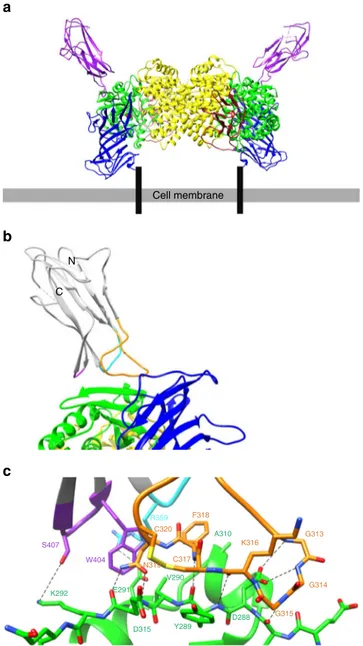
Cα atoms of 0.34 Å). Each APN monomer is bound to one RBD
as shown in Fig.
2
a. The HCoV-229E RBD–hAPN interaction
buries 510 Å
2of surface area on the RBD and 490 Å
2on hAPN.
The HCoV-229E RBD is an elongated six-stranded
β-structural
domain with three extended loops (loop 1: residues 308–325, loop
2: residues 352–359, loop 3: residues 404–408) at one end that
exclusively mediate the interaction with hAPN (Fig.
2
b). Loop 1 is
the longest and it contributes ~70% of the RBD surface buried on
complex formation (Figs.
2
c and
3
g). Within loop 1, residues
Cys
317and Cys
320form a disulfide bond that makes a stacking
interaction with the side chains of hAPN residues Tyr
289and
Glu
291(Fig.
2
c). The C317S/C320S RBD double mutant showed
no binding to hAPN at concentrations up to 15
μM (Table
1
,
Supplementary Fig.
1
D, and Supplementary Table
1
), evidence of
the importance of the stacking interaction and a likely role for the
disulfide bond in defining the conformation of loop 1. Notably,
loop 1 contains three tandemly repeated glycine residues
(residues 313–315) whose NH groups donate hydrogen bonds
to the side chain of Asp
288and the carbonyl oxygen of Phe
287of
hAPN (Fig.
2
c); mutation of hAPN residue Asp
288to alanine
leads to a ~10-fold reduction in affinity (Table
1
, Supplementary
Fig.
2
A, and Supplementary Table
1
). Apolar interactions
between RBD residues Cys
317and Phe
318and hAPN residues
Tyr
289, Val
290, Ile
309, Ala
310, and Leu
318are also observed
(Fig.
2
c); mutation of RBD residue Phe
318leads to a 13-fold
reduction in affinity while mutation of hAPN residues Tyr
289,
Val
290, Ile
309, and Leu
318lead to a 10- to 30-fold reduction in
affinity (Table
1
, Supplementary Fig.
1
C, Supplementary
Fig.
2
B–E, and Supplementary Table
1
). Centered in the contact
a
b
PBS
Percent of cells infected
Percent of cells infected
0 µM 0.094 µM 0.375 µM 0.75 µM 1.5 µM 3.0 µM 0 µ M 0.094 µ M 0.375 µ M 0.75 µ M 1.5 µ M 3.0 µ M
Soluble hAPN concentration
c
***
10 8 6 4 2 0 100***
80 60 40 20 0 S1 17–560 RBD 293–435Fig. 1 Characterization of soluble fragments of the HCoV-229E S-protein and hAPN.a HCoV-229E infection of L-132 cells in the presence of: PBS, the HCoV-229E S1 domain (residues 17–560 at 10 µM), and the HCoV-229E RBD (residues 293–435 at 30 µM). Statistics were obtained from three independent experiments. Statistical significance (ANOVA): ***p < 0.001; error bars correspond to the standard deviation.b Representative images of HCoV-229E infection of L-132 cells in the presence of the hAPN ectodomain at various concentrations. Greenfluorescence measures the expression of the viral S-protein. Magnification (100×) and scale bar = 20 µm. c Quantitation of the hAPN inhibition experiment. Statistics were obtained from three independent experiments. Statistical
significance (ANOVA): ***p < 0.001
Table 2 X-ray crystallographic data collection and
re
finement statistics
HCoV-229E RBD–hAPN Data collection Space group P3121 Cell dimensions a, b, c (Å) 153.8, 153.8, 322.1 α, β, γ (°) 90, 90, 120 Wavelength(Å) 0.9795 Resolution (Å) 50–3.5 (3.6–3.5)No. of total reflections 229,646 (22,754)
No. of unique reflections 55,987 (5490)
CC1/2 99.1 (68.1) CC* 99.8 (90) Rsym 0.16 (0.70) Rpim 0.08 (0.33) I/σI 10.9 (2.7) Completeness (%) 99.6 (99.8) Redundancy 4.1 (4.2) Refinement Resolution (Å) 50–3.5 No. of reflections 55,969 Rwork/Rfree 0.24 (0.31) /0.27 (0.32) No. of atoms Protein 23306 N-glycans 353 Water 0 B-factors (Å2) Protein 102 N-glycans 110 Wilson B-value (Å2) 95 R.m.s. deviations Bond lengths (Å) 0.004 Bond angles (°) 0.71 Ramachandran stats. (%) Favored 97 Outlier 0
area between the RBD and hAPN is a hydrogen bond between the
side chain of RBD residue Asn
319and the carbonyl oxygen of
hAPN residue Glu
291(Fig.
2
c); mutation of RBD residue Asn
319to alanine also ablates binding at the highest concentrations
achievable (Table
1
, Supplementary Fig.
1
E, and Supplementary
Table
1
). The remaining loop 1 residues serve to satisfy most of
the hydrogen bond donor/acceptor pairs of the edge
β-strand on
subdomain 2 of the hAPN molecule. Most prominent of the
remaining RBD–hAPN interactions is the salt bridge between
loop 2 residue Arg
359and hAPN residue Asp
315and the
interactions made by loop 3 residues Trp
404and Ser
407with
hAPN residues Asp
315and Lys
292(Fig.
2
c); the importance of
Trp
404of loop 3 is evidenced by the fact that mutating it also
ablates binding (Table
1
, Supplementary Fig.
1
F, and
Supple-mentary Table
1
).
HCoV-229E and PRCoV bind at different sites on APN. As
with HCoV-229E, the porcine respiratory alphacoronavirus,
PRCoV, also uses APN as its receptor
40. As our complex shows,
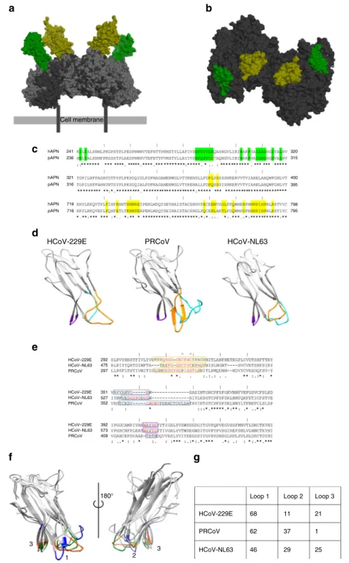
HCoV-229E binds at a site on hAPN (H-site) that differs from
the site on pAPN (P-site) used by PRCoV (Fig.
3
a, b). Glu
291in
hAPN, a residue in the hAPN–RBD interface, is an
N-glycosy-lated asparagine (Asn
286) in pAPN and attempts to dock the
HCoV-229E RBD at the H-site on pAPN leads to a steric clash
with the N-glycan (Supplementary Fig.
3
A). Consistent with this
observation, the HCoV-229E RBD cannot bind to a mutant form
of hAPN (E291N/K292E/Q293T) that possesses an N-glycan at
position 291, as we have shown (Table
1
, Supplementary
Fig.
4
A–C). Attempts to dock the PRCoV RBD at the P-site on
hAPN also leads to a steric clash, in this case with hAPN residue
Arg
741(Supplementary Fig.
3
B). Notably, porcine transmissible
gastroenteritis virus (TGEV) can bind hAPN, and HCoV-229E
can bind mouse APN, once the Arg side chain (on hAPN) and
the N-glycan (on mouse APN) on the respective APNs have been
mutated
41. Across species, the sequence identity at the H- and
P-sites is only ~60% (Fig.
3
c and Supplementary Fig.
3
C) and the
receptor-binding loops of these viruses must be accommodating
the remaining APN structural differences on receptors from
species that they do not infect. Together these results provide
evidence that the extended receptor-binding loops of these
alphacoronaviruses possess conformational plasticity.
The observation that HCoV-229E and PRCoV bind to different
sites on APN has important consequences. Among species, APN
is found in open/intermediate and closed conformations and
conversion between them is thought to be important for the
catalysis of its substrates
39, 42. The HCoV-229E RBD binds to
hAPN in its closed conformation and structural comparison
shows that the H-site does not differ between the open and closed
conformations. This is to be contrasted with the P-site of pAPN
that differs in the open and closed conformations. Indeed, the
PRCoV RBD has recently been shown to bind to pAPN in the
open conformation as a result of P-site interactions made possible
in the open form
42. These differences in binding and receptor
conformation are reflected in the fact that enzyme inhibitors that
promote the closed conformation of APN block TGEV
infection
42, but not HCoV-229E infection
8, and the fact that
the PRCoV S-protein
42, but not HCoV-229E
43, inhibits APN
catalytic activity.
The receptor-binding loops of HCoV-229E vary extensively.
Sequence data from viruses isolated over the past 50 years
pro-vides a wealth of data on the natural variation shown by
HCoV-229E (Supplementary Fig.
5
). With reference to the HCoV-229E
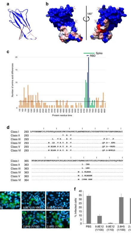
RBD–hAPN complex reported here, we now show that 73% of
the amino acids in the receptor-binding loops and supporting
residues vary among the sequences analyzed (52 sequences in
total), while only 11% of the RBD surface residues outside of the
receptor-binding loops show variation (Fig.
4
a, b). Moreover, for
the eight variants where full genome sequences were reported, the
receptor-binding loops represent the location at which the
greatest variation in the entire genome is observed (Fig.
4
c).
Analysis of the HCoV-229E RBD–hAPN interface further shows
that of the 16 RBD surface residues that are fully or partially
buried on complex formation, 10 of them vary in at least one of
the 52 sequences analyzed and a pairwise comparison of the
C N Cell membrane R359 C320 C317 N319 W404 S407 F318 K316 G313 G314 G315 D288 Y289 D315 E291 K292 V290 A310a
b
c
Fig. 2 HCoV-229E RBD in complex with the ectodomain of hAPN. a The complex between dimeric hAPN (domain I: blue, domain II: green, domain III: brown, and domain IV: yellow) and the HCoV-229E RBD (purple) is depicted in its likely orientation relative to the plasma membrane. The hAPN peptide and zinc ion (red spheres) binding sites are located inside a cavity distant from the virus binding site. Black bars represent the hAPN N-terminal transmembrane region.b Ribbon representation of the HCoV-229E RBD (gray) in complex with hAPN (same coloring as ina). The three receptor-binding loops are colored, orange (loop 1), cyan (loop 2), and purple (loop 3). N and C label the N- and C-termini of the RBD.c Atomic details of the interaction at the binding interface. Hydrogen bonds and salt bridges are indicated by dashed lines. Red and blue correspond to oxygen and nitrogen atoms, respectively. Loop and hAPN coloring as inb
a
b
c
e
d
g
f
HCoV-229E PRCoV HCoV-NL63
Cell membrane hAPN 241 320 315 400 395 798 795 236 321 316 719 716 292 475 297 351 527 352 392 573 409 hAPN hAPN pAPN pAPN HCoV–229E HCoV–NL63 PRCoV HCoV–229E HCoV–NL63 PRCoV HCoV–229E HCoV–NL63 PRCoV pAPN ** * * * 180° 1 3 3 Loop 1 HCoV-229E PRCoV HCoV-NL63 Loop 2 Loop 3 68 11 21 1 37 29 25 62 46 2 * *** * * *** * * ** *** ***** * ** * * * ** * * ** * * * ** *** ** ** * ** * ********** ********** * * * *** * *********** ** *** ***** ***** * *** *** *** *********** ** * * * * * ** *** * * ** ***** * * ** ***** * * ** ***** * * ** ***** * ** ** * * * **** * *** * * * ** ***** * * ** ***** * * *** ******** *****
Fig. 3 Alphacoronavirus receptor-binding domains. a Surface representation of an APN-based overlay of the HCoV-229E RBD–hAPN and PRCoV RBD–pAPN complexes. Human APN (dark gray), porcine APN (light gray), HCoV-229E RBD (green), and PRCoV RBD (yellow). APNs are aligned on domain IV.b Top view of the APN surface buried by HCoV-229E RBD binding (H-site, green) and PRCoV RBD binding (P-site; yellow) mapped onto hAPN. c Sequence alignment of human and porcine APN. Residues in the H-site are highlighted in green and residues in the P-site are highlighted in yellow. The“|“ symbol demarcates every 10 residues in the alignment. The N-glycosylation sequon (Asn residue 286) in porcine APN is shown in red (Glu residue 291 in human).d Ribbon representation of the HCoV-229E RBD (receptor: hAPN), the PRCoV RBD (receptor: pAPN), and the HCoV-NL63 RBD (receptor: hACE2). Loops 1, 2, and 3 are colored in orange, cyan, and purple, respectively.e Sequence alignment of the HCoV-229E, PRCoV, and HCoV-NL63 RBDs. Residues in loops 1, 2, and 3 are enclosed by orange, cyan, and purple boxes, respectively. The cysteine residues involved in the loop 1 disulfide bond are indicated by“^“. The “|“ symbol demarcates every 10 residues in the alignment. Residues directly interacting with the receptor are colored red. f Structural alignment of the HCoV-229E, HCoV-NL63, and PRCoV RBDs with receptor interacting residues colored orange, green, and blue, respectively. Numbers indicate the loop numbers. The structures are shown in two views rotated by 180orelative to each other.g The percentage contribution made by each loop to the total surface area buried on the RBD in the receptor complexes
sequences suggests that many of these positions can vary
simul-taneously (Supplementary Fig.
5
). Finally, we show that the six
invariant interface residues on the RBD (Gly
313, Gly
315, Cys
317,
Cys
320, Asn
319, and Arg
359) constitute only 45% of the viral
surface area buried, the very region expected to be the most
highly conserved from a receptor-binding standpoint. The
% Infected cells PBS PBS 9.8E12 (1/100) 9.8E12 (1/10) 2.8H5 (1/100) 2.8H5 (1/10)
f
e
d
RBD Spikea
b
c
Number of amino acid differences
Protein residue bins
180° 20 15 10 5 0 Class I 293 293 293 293 293 293 365 363 363 363 363 364 40 35 30 25 20 15 10 5 0 Class II Class III Class IV Class V Class VI Class I Class II Class III Class IV Class V Class VI 100 400 700 1000 1300 1600 1900 2200 2500 2800 3100 3400 3700 4000 4300 4600 4900 5200 5500 5800 6100 6400 6700 7000 7300 7600 7900 8200 8500 8800 9.8E12 (1/100) 9.8E12 (1/10) 2.8H5 (1/100) 2.8H5 (1/10)
Fig. 4 Naturally occurring HCoV-229E sequence variation. a Color-coded amino-acid sequence conservation index (Chimera) mapped onto a ribbon representation of the HCoV-229E RBD. Blue represents a high percentage sequence identity and red represents a low percentage sequence identity among the 52 viral isolates analyzed.b Surface representation in the same orientation as in (a, left), and rotated 180° (right). The Asn-GlcNAc moiety of the N-glycans are shown in stick representation. Color coding as ina. c Amino-acid sequence variation shown by the eight viral isolates whose entire genome sequences have been reported. The entire protein coding region of the viral genome was treated as a continuous amino acid string (8850 residues in total). Amino acid differences among the eight sequences were analyzed in 100 residue bins and for each bin the sum was plotted. Green-colored bins correspond to residues in the S-protein and purple-colored bins correspond to residues in the RBD. The horizontal dotted line denotes the average number of amino-acid differences per bin across the protein-coding region of the whole viral genome.d Alignment of the sequences selected for each of the six classes. The “|“ symbol demarcates every 10 residues in the alignment. e Representative images showing HCoV-229E infection of L-132 cells in the presence of: PBS, monoclonal antibody 9.8.E12 at two different concentrations, and monoclonal antibody 2.8H5 at two different concentrations (anti-HCoV-OC43 antibody). The nucleus is stained blue and green staining indicates viral infection. Magnification (×200) and scale bar = 10 µm. f Statistical quantification of the monoclonal antibody inhibition experiment. Error bars correspond to standard deviations obtained from three independent experiments
remaining 55% (i.e., 279 Å
2) of the viral surface area buried is
made up of 10 residues that differ in their variability and the role
they play in complex formation (Supplementary Table
2
).
Loop variation leads to phylogenetic classes. Phylogenetic
analysis of the HCoV-229E RBD sequences found in the database
showed that they segregate into six classes (Supplementary Fig.
6
).
Class I contains the ATCC-740 reference strain (originally
iso-lated in 1967 and deposited in 1973) and reiso-lated lab strains, while
Classes II–VI, represent clinical isolates that have successively
replaced each other in the human population over time since the
1970s. To characterize these classes, a representative sequence
from each was selected; for Class I, the RBD of the reference
strain, also used in our structural analysis, was selected. To
simplify characterization, the RBDs of the other
five classes were
synthesized with the Class I sequence in all but the loop regions
(Fig.
4
d). As observed for Class I, the other RBDs do not bind to
the hAPN mutant that introduces an N-glycan at Glu
291(Sup-plementary Fig.
4
D), an observation suggesting that they all bind
at the same site on hAPN. The RBDs bound hAPN with an
~16-fold range in affinity (K
dfrom ~30 to ~440 nM). These
differences in affinity are largely a result of differences in k
offwith
little difference in k
on(Table
3
and Supplementary Fig.
7
).
Notably, the Class I RBD binds with the lowest affinity, while the
RBDs from viral classes that have emerged most recently (Class
V: viruses isolated in 2001–2004 and Class VI: viruses isolated in
2007–2015) bind with the highest affinity. For each of the six
classes, Supplementary Table
2
shows the identity of the loop
residues that have shown variation. Of those buried in the
RBD–hAPN interface, residues 314, 404, and 407 are particularly
noteworthy as they undergo considerable variation in amino-acid
character. Residue 314, for example, accounts for 9% of the total
buried surface area on complex formation and changes from Gly
to Val to Pro in the transition from Classes I to VI. Variation of
this sort provides insight into how changes in receptor-binding
affinity might be mediated during the process of viral adaptation.
Each of the six RBD classes were also characterized using a
neutralizing mouse monoclonal antibody (9.8E12) that we
generated against the HCoV-229E reference strain (Class I). As
shown in Fig.
4
e, f, 9.8E12 inhibits HCoV-229E infection of the
L132 cell-line. This antibody binds to the Class I RBD with a K
dof 66 nM (k
on= 6.3 × 10
5M
−1s
−1, k
off= 0.041 s
−1) and as shown
by a competition binding experiment, it blocks the RBD–hAPN
interaction (Supplementary Fig.
8
A, B). In contrast, 9.8E12 shows
no binding to the other
five RBD classes at a concentration of 1
μM (Supplementary Fig.
8
C), strong evidence that the
receptor-binding loops of the Class I RBD are important for antibody
binding and that loop variation can abrogate antibody binding.
Consistent with this observation, non-conserved amino-acid
changes both within and outside of the RBD–hAPN interface
are observed across all classes (Supplementary Table
2
).
Discussion
Correlating structure and function with natural sequence data is a
powerful means of studying viral adaptation and evolution. To
this end, we have delimited the HCoV-229E RBD and determined
its X-ray structure in complex with the ectodomain of its
recep-tor, hAPN. We found that three extended loops on the RBD are
solely responsible for receptor binding, and that these loops are
highly variable among viruses isolated over the past 50 years. A
phylogenetic analysis also showed that the RBDs of these viruses
define six RBD classes whose viruses have successively replaced
each other in the human population. The six RBDs differ in their
receptor-binding affinity and their ability to be bound by a
neutralizing antibody (9.8E12) and taken together, our
findings
suggest that the HCoV-229E sequence variation observed arose
through adaptation and selection.
Antibodies that block receptor binding are a common route to
viral neutralization and exposed loops are known to be
particu-larly immunogenic
44. Loop-binding neutralizing antibodies are
elicited by the alphacoronavirus TGEV
40, and the
receptor-binding loops of HCoV-229E mediate the receptor-binding of the
neu-tralizing antibody, 9.8E12. As shown by the sequences of the viral
isolates analyzed, the RBDs differ almost exclusively in their
receptor-binding loops. 9.8E12 blocks the hAPN–RBD
interac-tion and it can only bind to the RBD (Class I) found in the virus
that elicited it. This observation shows that loop variability can
abrogate neutralizing antibody binding. Indeed, the successive
replacement or ladder-like phylogeny observed, when the
sequence of the HCoV-229E RBD is analyzed, is characteristic of
immune escape as shown by the influenza virus
45, 46. Taken
together, our results suggest that immune evasion contributes to
if not explains the extensive receptor-binding loop variation
shown by HCoV-229E over the past 50 years. HCoV-229E
infection in humans does not provide protection against different
isolates
37, and viruses that contain a new RBD class that cannot
be bound by the existing repertoire of loop-binding neutralizing
antibodies provide an explanation for this observation.
Neu-tralizing antibodies that block receptor binding can also be
thwarted by an increase in the affinity/avidity between the virus
and its host receptor. Increased receptor-binding affinity/avidity
allows the virus to more effectively compete with receptor
blocking neutralizing antibodies, a mechanism thought to be
important for evading a polyclonal antibody response
47. In
addition, an optimal receptor binding affinity is thought to exist
in a given environment. As such, adaptation in a new species,
changes in tissue tropism, and differences in receptor expression
levels can all lead to changes in receptor binding affinity
29,31,48.
Taken together, the observation that the most recent RBD classes
(Class V: viruses isolated in 2001–2004 and Class VI: viruses
isolated in 2007–2015) show a ~16-fold increase in affinity for
hAPN over that of Class I (viruses isolated in 1967) merits further
study.
Recent cryoEM analysis has shown that the receptor-binding
sites of HCoV-NL63, SARS-CoV, MERS-CoV, and by inference
HCoV-229E, are inaccessible in some conformations of the
pre-fusion S-protein trimer
21–25. Although the ramifications of this
structural arrangement are not yet clear, restricting access to the
binding site has been proposed to provide a means of limiting
B-cell receptor interactions against the receptor-binding site
23. How
this might work in mechanistic terms is also not clear given the
need to bind receptor. However, in a simple model, the
inac-cessible S-protein conformation(s) would be in equilibrium with a
less stable (higher energy) but accessible S-protein conformation
(s). The energy difference between these conformations is a
barrier to binding that decreases equally the intrinsic free energy
of binding of both the viral receptor and the B-cell receptor and
relative binding energies may be the key. Both soluble hAPN and
Table 3 Surface plasmon resonance-binding data for the
interaction between the six HCoV-229E RBDs and hAPN
Class kon(×105M−1s−1) koff(s−1) Kd(nM) I 3.6± 0.5 0.16± 0.02 434± 63 II 3.3± 0.5 0.08± 0.02 246± 19 III 7.3± 1.4 0.08± 0.02 113± 2.3 IV 3.6± 0.5 0.10± 0.02 261± 24 V 4.8± 1.1 0.01± 0.01 27.0± 1.7 VI 8.5± 0.6 0.03± 0.01 37.4± 3.5
Values after± correspond to the residual standard deviation reported by Scrubber 2. Two experiments were performed
antibody 9.8E12 can inhibit HCoV–229E infection in a cell-based
assay, an indication that their binding energies (K
dof 430 and 66
nM, respectively) are sufficient to efficiently overcome the barrier
to binding. However, B-cell receptors bind their antigens
rela-tively weakly prior to affinity maturation
49and they would be
much less able to do so. The dynamics of the interconversion
between accessible and inaccessible conformations may also be a
factor in the recognition of inaccessible antibody epitopes
50,51,
and further work will be required to establish if and how
restricting access to the receptor binding site enhances
cor-onavirus
fitness. The cryoEM structures also show that the
receptor-binding loops make intra- and inter-subunit contacts in
the inaccessible prefusion trimer. This suggests the intriguing
possibility that the magnitude of the energy barrier, or the
dynamics of the interconversion between accessible and
inac-cessible conformations, might be modulated by loop variation
during viral adaption.
Immune evasion and cross-species transmission involve viral
adaptation and we posit that the use of extended loops for
receptor binding represents a strategy employed by HCoV-229E
and the alphacoronaviruses to mediate the process. Such loops
can tolerate insertions, deletions, and amino acid substitutions
relatively free of the energetic penalties associated with the
mutation of other protein structural elements. Indeed, our
ana-lysis of the six RBD classes shows that the receptor-binding loops
possess a remarkable ability to both accommodate and
accumu-late mutational change while maintaining receptor binding.
Among the six classes, 73% of the loop residues show change and
only 45% of the receptor interface buried on receptor binding has
been conserved. As we have shown, variation in the
receptor-binding loops can abrogate neutralizing antibody receptor-binding and it
will also increase the likelihood of acquiring new receptor
inter-actions by chance. In this way, the selection of viral variants
capable of immune evasion and/or cross-species transmission will
be facilitated
27,28,52–54.
Cross-species transmission involves the acquisition of either a
conserved (i.e., a similar interaction with a homologous receptor)
or a non-conserved receptor interaction (i.e., an interaction with a
non-homologous receptor, or an interaction at a new site on a
homologous receptor) in the new host. HCoV-229E binds to a
site on hAPN that differs from the site where PRCoV
40binds
to pAPN (Fig.
3
a, b), and HCoV-NL63 is known to bind the
non-homologous receptor, ACE2
55. Clearly, conserved receptor
interactions have not accompanied the evolution of these
alpha-coronaviruses (Fig.
3
d–g). In mechanistic terms, receptor-binding
loop variability and plasticity would facilitate the acquisition of
both conserved and non-conserved receptor interactions.
How-ever, compared to conserved receptor interactions, the successful
acquisition of non-conserved interactions would be expected to
be relatively infrequent and more likely to require viral replication
and mutation in the new host to optimize receptor-binding
affinity.
Many coronaviruses have originated in bats
3, 4and it is
tempting to speculate that viral transmission between bats has
facilitated the emergence of non-conserved receptor interactions.
Bats account for ~20% of all mammalian species and they possess
a unique ecology/biology that facilitates viral spread between
them
56,57. Moreover, the barriers to viral replication in a new
host are lower among closely related species
58,59. It follows that
the viral replication required to optimize non-conserved receptor
interactions in the new host would be facilitated by transmission
between closely related bat species. By a similar reasoning, the use
of conserved receptor interactions requiring little optimization
would facilitate large species jumps. Several bat coronaviruses
showing a high degree of sequence similarity with HCoV-229E
have recently been identified
60,61and an analysis of how they
interact with bat APN will inform this discussion.
Predicting the emergence of new viral threats is an important
aspect of public health planning
62and our work suggests that
RNA viruses that use loops to bind their receptors should be
viewed as a particular risk. RNA viruses are best described as
populations
34, and extended loops—inherently capable of
accommodating and accumulating mutational change—will
enable populations with loop diversity. Such populations will
provide routes to escaping receptor loop-binding neutralizing
antibodies, optimizing receptor-binding affinity, and acquiring
new receptor interactions, interrelated processes that drive viral
evolution and the emergence of new viral threats.
Methods
Protein expression and purification. The soluble ectodomain of hAPN (residues 66–967) was expressed and purified from stably transfected HEK293S GnT1- cells (ATCC CRL-3022) as described previously39. The various soluble forms of the HCoV-229E S-protein were expressed and purified from stably transfected HEK293S GnT1-cells for X-ray crystallography, and from HEK293T (ATCC CRL-3216) and/or HEK293F (Invitrogen 51-0029) cells for cell-based and biochemical characterization, as described previously63. Point mutations were generated using the InFusion HD Site-Directed Mutagenesis protocol (Clontech). In all cases, the target proteins were secreted as N-terminal protein-A fusion proteins with a Tobacco Etch Virus (TEV) protease cleavage site following the protein-A tag. Harvested media was concentrated 10-fold and purified by IgG affinity chroma-tography (IgG Sepharose, GE). The bound proteins were liberated by on-column TEV protease cleavage and further purified by anion exchange chromatography (HiTrap Q HP, GE).
Protein crystallization. The RBD of the S-protein of HCoV-229E (residues 293–435) and the soluble ectodomain of hAPN (residues 66–967) were mixed in a ratio of 1.2:1 (RBD:hAPN) and the complex was purified by Superdex 200 (GE) gel filtration chromatography in 10 mM HEPES, 50 mM NaCl, pH 7.4. The complex was concentrated in gelfiltration buffer to 10 mg/ml for crystallization trials. Crystals were obtained by the hanging drop method using a 1:1 mixture of stock protein and well solution containing 8% PEG 8000, 1 mM GSSG, 1 mM GSH, 5% glycerol, 1µg/ml endo-β-N-acetylglucosaminidase A64and 100 mM MES, pH 6.5 at 298 K. Crystals were typically harvested after 3 days andflash-frozen with well solution supplemented with 22.5% glycerol as cryoprotectant.
Data collection and structure determination. Diffraction data were collected at the Canadian Light Source, Saskatoon, Saskatchewan (Beamline CMCF-08ID-1) at a wavelength of 0.9795 Å. Data were merged, processed, and scaled using HKL200065; 5% of the data set was used for the calculation of Rfree. Phases were
obtained by molecular replacement using the human APN structure as a search model (PDB ID: 4FYQ) using Phaser in Phenix66. Manual building of the HCoV-229E RBD was performed using COOT67. Alternate rounds of manual rebuilding and automated refinement using Phenix were performed. Secondary structural restraints and torsion-angle non-crystallographic symmetry restraints between the three monomers in the asymmetric unit were employed. Ramachandran analysis showed that 96% of the residues are in the most favored region, with 4% in the additionally allowed region. Data collection and refinement statistics are found in Table2. A stereo image of a portion of the electron density map in the HCoV–229E–hAPN interface is showed in Supplementary Fig.9. Figures were generated using the program chimera68. Buried surface calculations were per-formed using the PISA server.
Surface plasmon resonance binding assays. Surface plasmon resonance (Bia-core) assays were performed on CM-5 dextran chips (GE) covalently coupled to the ligand via amine coupling. The running and injection buffers were matched and consisted of 150 mM NaCl, 0.01% Tween-20, 0.1 mg/ml BSA, and 10 mM HEPES at pH 7.5. Response unit (RU) values were measured as a function of analyte concentration at 298 K. Kinetic analysis was performed using the globalfitting feature of Scrubber 2 (BioLogic Software) assuming a 1:1 binding model. For experiments using hAPN as a ligand, between 300 and 400 RU were coupled to the CM-5 dextran chips. For experiments using 9.8E12, 1900 RU was immobilized. Viral inhibition assay. HCoV-229E was originally obtained from the American Type Culture Collection (ATCC VR-740) and was produced in the human L132 cell line (ATCC CCL5) which was grown in minimum essential medium alpha (MEM-α) supplemented with 10% (v/v) FBS (PAA).
The L132 (1 × 10⁵) cells were seeded on coverslips and grown overnight in MEM-α supplemented with 10% (v/v) FBS. For inhibition assays in the presence of soluble hAPN, wild-type HCoV-229E (105.5TCID
50) was pre-incubated with the
added to cells for 2 h at 33 °C. For inhibition assays in the presence of the soluble S-protein fragments, the different fragments, diluted in PBS, were added to cells and kept at 4 °C on ice for 1 h. Medium was then removed and cells were inoculated with wild-type HCoV-229E (105TCID
50) for 2 h at 33 °C. For both inhibition
assays, after the 2-h incubation period, medium was replaced and cells were incubated at 33 °C with fresh MEM-α supplemented with 1% (v/v) FBS for 24 h before being analyzed by an immunofluorescence assay (IFA).
Cells on the coverslips were directlyfixed with 4% paraformaldehyde (PFA 4%) in PBS for 30 min at room temperature and then transferred to PBS. Cells were permeabilized in cold methanol (−20 °C) for 5 min and then washed with PBS for viral antigen detection. The S-protein-specific monoclonal antibody, 5-11H.6, raised against HCoV-229E (IgG1, produced in our laboratory by standard hybridoma technology), was used in conjunction with an AlexaFluor-488-labeled mouse-specific goat antibody (Life Technologies A-21202), for viral antigen detection69. After three washes with PBS, cells were incubated for 5 min with DAPI (Sigma-Aldrich) at 1µg/ml to stain the nuclear DNA. To determine the percentage of L-132 cells positive for the viral S-protein, 15fields containing a total of 150–200 cells were counted, at a magnification of ×200 using a Nikon Eclipse E800 microscope, for each condition tested in three independent experiments. Green fluorescent cells were counted as S-protein positive and expressed as a percentage of the total number of cells. Statistical significance was estimated by the analysis of variance (ANOVA) test and Tukey’s test post hoc.
Monoclonal antibodies (IgG1, produced in our laboratory by standard hybridoma technology) raised against HCoV-229E (9.8E12) or HCoV-OC43 (2.8H5, negative control) that were found to be S-protein specific were tested in an infectivity neutralization assay. Wild-type HCoV-229E (105.5TCID50) was
pre-incubated with the antibodies (1/100 of hybridoma supernatant) for 1 h at 37 °C before being added to L-132 cells for 2 h at 33 °C. Cells were washed with PBS and incubated at 33 °C with fresh MEM-α supplemented with 1% FBS (v/v) for 18 h before being analyzed by an immunofluorescence assay (IFA). Statistical significance was estimated by an ANOVA test, followed by post hoc Dunnett (two-sided) analysis.
Comparative sequence analysis of HCoV-229E viral isolates. The protein sequence of the HCoV-229E P100E isolate RBD (residues 293–435) was used to perform a search of the non-redundant protein sequence database using Blastp. Sequences were curated as of December 1, 2016. A total of 52 sequences were obtained with the GenBank Identifier numbers: NP_073551.1, AAK32188.1, AAK32189.1, AAK32190.1, AAK32191.1, CAA71056.1, CAA71146.1,
CAA71147.1, ADK37701.1, ADK37702.1, ADK37704.1, BAL45637.1, BAL45638.1, BAL45639.1, BAL45640.1, BAL45641.1, AAQ89995.1, AAQ89999.1, AAQ90002.1, AAQ90004.1, AAQ90005.1, AAQ90006.1, AAQ90008.1, AFI49431.1, AFR45554.1, AFR79250.1, AFR79257.1, AGT21338.1, AGT21345.1, AGT21353.1, AGT21367.1, AGW80932.1, AIG96686.1 ABB90506.1, ABB90507.1, ABB90508.1, ABB90509.1, ABB90510.1, ABB90513.1. ABB90514.1, ABB90515.1, ABB90516.1, ABB90519.1, ABB90520.1, ABB90522.1, ABB90523.1, ABB90526.1, ABB90527.1, ABB90528.1, ABB90529.1, ABB90530.1, AOG74783.1. The 52 sequences were then aligned using Muscle70and the residue-specific sequence conservation index was mapped onto the surface of the RBD using the“render by conservation” tool in Chimera68. Percentage identity is mapped using a color scale with blue indicating 100% identity and red indicating 30% identity. The protein-coding regions of the eight sequences for which the entire genome were reported (GenBank Identifier num-bers: NC_002645.1, JX503060.1, JX503061.1, KF514433.1, KF514430.1, KF514432.1, AF304460.1, and KU291448.1) were aligned using Muscle. The entire protein-coding region of the viral genome was treated as a continuous amino-acid string (8850 residues in total). Protein residues that were not identical among the eight sequences were counted as a difference and plotted in 100 residue bins. The sequence AAK32191.1 was chosen as the representative of Class I and the loop sequences of ABB90507.1, ABB90514.1, ABB90519.1, ABB90523.1, and AFR45554.1 were combined with the non-loop sequences of AAK32191.1 to generate the RBDs of Classes (II–VI), respectively.
Data availability. Coordinates and structure factors for the HCoV-229E RBD in complex with human APN were deposited in the protein data bank with PDB ID: 6ATK. The authors declare that all other data supporting thefindings of this study are available within the article and its Supplementary Informationfiles, or are available from the authors upon request.
Received: 29 May 2017 Accepted: 6 October 2017
References
1. Graham, R. L., Donaldson, E. F. & Baric, R. S. A decade after SARS: strategies for controlling emerging coronaviruses. Nat. Rev. Microbiol. 11, 836–848 (2013).
2. Su, S. et al. Epidemiology, genetic recombination, and pathogenesis of coronaviruses. Trends Microbiol. 24, 490–502 (2016).
3. Woo, P. C. et al. Discovery of seven novel Mammalian and avian coronaviruses in the genus deltacoronavirus supports bat coronaviruses as the gene source of alphacoronavirus and betacoronavirus and avian coronaviruses as the gene source of gammacoronavirus and deltacoronavirus. J. Virol. 86, 3995–4008 (2012).
4. Forni, D. et al. Molecular evolution of human coronavirus genomes. Trends Microbiol. 25, 35–48 (2017).
5. Peck, K. M. et al. Coronavirus host range expansion and middle east respiratory syndrome coronavirus emergence: biochemical mechanisms and evolutionary perspectives. Annu. Rev. Virol. 2, 95–117 (2015).
6. Desforges, M. et al. Neuroinvasive and neurotropic human respiratory coronaviruses: potential neurovirulent agents in humans. Adv. Exp. Med. Biol. 807, 75–96 (2014).
7. Gaunt, E. R. et al. Epidemiology and clinical presentations of the four human coronaviruses 229E, HKU1, NL63, and OC43 detected over 3 years using a novel multiplex real-time PCR method. J. Clin. Microbiol. 48, 2940–2947 (2010). 8. Yeager, C. L. et al. Human aminopeptidase N is a receptor for human
coronavirus 229E. Nature 357, 420–422 (1992).
9. Hofmann, H. et al. Human coronavirus NL63 employs the severe acute respiratory syndrome coronavirus receptor for cellular entry. Proc. Natl Acad. Sci. USA 102, 7988–7993 (2005).
10. Vlasak, R. et al. Human and bovine coronaviruses recognize sialic acid-containing receptors similar to those of influenza C viruses. Proc. Natl Acad. Sci. USA 85, 4526–4529 (1988).
11. Qian, Z. et al. Identification of the receptor-binding domain of the spike glycoprotein of human betacoronavirus HKU1. J. Virol. 89, 8816–8827 (2015). 12. Huang, X. et al. Human coronavirus HKU1 spike protein uses O-acetylated
sialic acid as an attachment receptor determinant and employs hemagglutinin-esterase protein as a receptor-destroying enzyme. J. Virol. 89, 7202–7213 (2015).
13. Ou, X. et al. Crystal structure of the receptor binding domain of the spike glycoprotein of human betacoronavirus HKU1. Nat. Commun. 8, 15216 (2017). 14. Lau, S. K. et al. Severe acute respiratory syndrome coronavirus-like virus in
Chinese horseshoe bats. Proc. Natl Acad. Sci. USA 102, 14040–14045 (2005). 15. Ge, X. Y. et al. Isolation and characterization of a bat SARS-like coronavirus
that uses the ACE2 receptor. Nature 503, 535–538 (2013).
16. Anthony, S. J. et al. Further Evidence for bats as the evolutionary source of Middle East respiratory syndrome coronavirus. MBio 8, e00373–17 (2017). 17. Li, W. et al. Angiotensin-converting enzyme 2 is a functional receptor for the
SARS coronavirus. Nature 426, 450–454 (2003).
18. Raj, V. S. et al. Dipeptidyl peptidase 4 is a functional receptor for the emerging human coronavirus-EMC. Nature 495, 251–254 (2013).
19. Li, F. Structure, function, and evolution of coronavirus spike proteins. Annu. Rev. Virol. 3, 237–261 (2016).
20. Bosch, B. J. et al. The coronavirus spike protein is a class I virus fusion protein: structural and functional characterization of the fusion core complex. J. Virol. 77, 8801–8811 (2003).
21. Gui, M. et al. Cryo-electron microscopy structures of the SARS-CoV spike glycoprotein reveal a prerequisite conformational state for receptor binding. Cell. Res. 27, 119–129 (2017).
22. Kirchdoerfer, R. N. et al. Pre-fusion structure of a human coronavirus spike protein. Nature 531, 118–121 (2016).
23. Walls, A. C. et al. Glycan shield and epitope masking of a coronavirus spike protein observed by cryo-electron microscopy. Nat. Struct. Mol. Biol. 23, 899–905 (2016).
24. Walls, A. C. et al. Cryo-electron microscopy structure of a coronavirus spike glycoprotein trimer. Nature 531, 114–117 (2016).
25. Yuan, Y. et al. Cryo-EM structures of MERS-CoV and SARS-CoV spike glycoproteins reveal the dynamic receptor binding domains. Nat. Commun. 8, 15092 (2017).
26. Buchholz, U. J. et al. Contributions of the structural proteins of severe acute respiratory syndrome coronavirus to protective immunity. Proc. Natl Acad. Sci. USA 101, 9804–9809 (2004).
27. Tang, X. C. et al. Identification of human neutralizing antibodies against MERS-CoV and their role in virus adaptive evolution. Proc. Natl Acad. Sci. USA 111, E2018–E2026 (2014).
28. Sui, J. et al. Effects of human anti-spike protein receptor binding domain antibodies on severe acute respiratory syndrome coronavirus neutralization escape andfitness. J. Virol. 88, 13769–13780 (2014).
29. Holmes, E. C. (ed.) in The Evolution and Emergence of RNA Viruses 254 (Oxford University Press, Oxford; New York, 2009).
30. Allison, A. B. et al. Host-specific parvovirus evolution in nature is recapitulated by in vitro adaptation to different carnivore species. PLoS Pathog. 10, e1004475 (2014).
31. Graham, R. L. & Baric, R. S. Recombination, reservoirs, and the modular spike: mechanisms of coronavirus cross-species transmission. J. Virol. 84, 3134–3146 (2010).
32. Dijkman, R. et al. Human coronavirus 229E encodes a single ORF4 protein between the spike and the envelope genes. Virol. J. 3, 106 (2006).
33. Shirato, K. et al. Clinical isolates of human coronavirus 229E bypass the endosome for cell entry. J. Virol. 91, 1 (2017).
34. Lauring, A. S., Frydman, J. & Andino, R. The role of mutational robustness in RNA virus evolution. Nat. Rev. Microbiol. 11, 327–336 (2013).
35. Coffin, J. & Swanstrom, R. HIV pathogenesis: dynamics and genetics of viral populations and infected cells. Cold Spring Harb. Perspect. Med. 3, a012526 (2013).
36. Chibo, D. & Birch, C. Analysis of human coronavirus 229E spike and nucleoprotein genes demonstrates genetic drift between chronologically distinct strains. J. Gen. Virol. 87(Pt 5), 1203–1208 (2006).
37. Reed, S. E. The behaviour of recent isolates of human respiratory coronavirus in vitro and in volunteers: evidence of heterogeneity among 229E-related strains. J. Med. Virol. 13, 179–192 (1984).
38. Shirato, K. et al. Differences in neutralizing antigenicity between laboratory and clinical isolates of HCoV-229E isolated in Japan in 2004-2008 depend on the S1 region sequence of the spike protein. J. Gen. Virol. 93(Pt 9), 1908–1917 (2012). 39. Wong, A. H., Zhou, D. & Rini, J. M. The X-ray crystal structure of human
aminopeptidase N reveals a novel dimer and the basis for peptide processing. J. Biol. Chem. 287, 36804–36813 (2012).
40. Reguera, J. et al. Structural bases of coronavirus attachment to host aminopeptidase N and its inhibition by neutralizing antibodies. PLoS Pathog. 8, e1002859 (2012).
41. Tusell, S. M., Schittone, S. A. & Holmes, K. V. Mutational analysis of aminopeptidase N, a receptor for several group 1 coronaviruses, identifies key determinants of viral host range. J. Virol. 81, 1261–1273 (2007).
42. Santiago, C. et al. Allosteric inhibition of aminopeptidase N functions related to tumor growth and virus infection. Sci. Rep. 7, 46045 (2017).
43. Breslin, J. J. et al. Human coronavirus 229E: receptor binding domain and neutralization by soluble receptor at 37 degrees C. J. Virol. 77, 4435–4438 (2003).
44. Corti, D. & Lanzavecchia, A. Broadly neutralizing antiviral antibodies. Annu. Rev. Immunol. 31, 705–742 (2013).
45. Grenfell, B. T. et al. Unifying the epidemiological and evolutionary dynamics of pathogens. Science 303, 327–332 (2004).
46. Volz, E. M., Koelle, K. & Bedford, T. Viral phylodynamics. PLoS Comput. Biol. 9, e1002947 (2013).
47. Hensley, S. E. et al. Hemagglutinin receptor binding avidity drives influenza A virus antigenic drift. Science 326, 734–736 (2009).
48. de Vries, R. P. et al. Evolution of the hemagglutinin protein of the new pandemic H1N1 influenza virus: maintaining optimal receptor binding by compensatory substitutions. J. Virol. 87, 13868–13877 (2013).
49. Schmidt, A. G. et al. Preconfiguration of the antigen-binding site during affinity maturation of a broadly neutralizing influenza virus antibody. Proc. Natl Acad. Sci. USA 110, 264–269 (2013).
50. Goo, L. et al. A single mutation in the envelope protein modulatesflavivirus antigenicity, stability, and pathogenesis. PLoS Pathog. 13, e1006178 (2017). 51. Munro, J. B. et al. Conformational dynamics of single HIV-1 envelope trimers
on the surface of native virions. Science 346, 759–763 (2014).
52. Lynch, R. M. et al. HIV-1fitness cost associated with escape from the VRC01 class of CD4 binding site neutralizing antibodies. J. Virol. 89, 4201–4213 (2015).
53. Kim, Y. et al. Spread of mutant Middle East respiratory syndrome coronavirus with reduced affinity to human CD26 during the South Korean outbreak. MBio 7, e00019 (2016).
54. Rockx, B. et al. Escape from human monoclonal antibody neutralization affects in vitro and in vivofitness of severe acute respiratory syndrome coronavirus. J. Infect. Dis. 201, 946–955 (2010).
55. Wu, K. et al. Crystal structure of NL63 respiratory coronavirus receptor-binding domain complexed with its human receptor. Proc. Natl Acad. Sci. USA 106, 19970–19974 (2009).
56. Brook, C. E. & Dobson, A. P. Bats as“special” reservoirs for emerging zoonotic pathogens. Trends Microbiol. 23, 172–180 (2015).
57. Hayman, D. T. Bats as viral reservoirs. Annu. Rev. Virol. 3, 77–99 (2016). 58. Streicker, D. G. et al. Host phylogeny constrains cross-species emergence and
establishment of rabies virus in bats. Science 329, 676–679 (2010).
59. Menachery, V. D., Graham, R. L. & Baric, R. S. Jumping species-a mechanism for coronavirus persistence and survival. Curr. Opin. Virol. 23, 1–7 (2017).
60. Corman, V. M. et al. Evidence for an ancestral association of human coronavirus 229E with bats. J. Virol. 89, 11858–11870 (2015).
61. Tao, Y. et al. Surveillance of bat coronaviruses in Kenya identifies relatives of human coronaviruses NL63 and 229E and their recombination history. J. Virol. 91, e01953–16 (2017).
62. Holmes, E. C. What can we predict about viral evolution and emergence? Curr. Opin. Virol. 3, 180–184 (2013).
63. Li, Z. et al. Simple piggyBac transposon-based mammalian cell expression system for inducible protein production. Proc. Natl Acad. Sci. USA 110, 5004–5009 (2013).
64. Fujita, K. et al. Synthesis of neoglycoenzymes with homogeneous N-linked oligosaccharides using immobilized endo-beta-N-acetylglucosaminidase A. Biochem. Biophys. Res. Commun. 267, 134–138 (2000).
65. Otwinowski, Z. & Minor, W. in Processing of X-ray diffraction data collected in oscillation mode. Methods Enzymol. Vol. 276 (eds Carter, C. W. & Sweet, R. M.) 307–326 (Academic Press, New York, 1997).
66. Adams, P. D. et al. PHENIX: a comprehensive Python-based system for macromolecular structure solution. Acta Crystallogr. D Biol. Crystallogr. 66, 213–221 (2010).
67. Emsley, P. & Cowtan, K. Coot: model-building tools for molecular graphics. Acta Crystallogr. D Biol. Crystallogr. 60, 2126–2132 (2004).
68. Pettersen, E. F. et al. UCSF Chimera--a visualization system for exploratory research and analysis. J. Comput. Chem. 25, 1605–1612 (2004).
69. Arbour, N. et al. Persistent infection of human oligodendrocytic and neuroglial cell lines by human coronavirus 229E. J. Virol. 73, 3326–3337 (1999). 70. Edgar, R. C. MUSCLE: multiple sequence alignment with high accuracy and
high throughput. Nucleic Acids Res. 32, 1792–1797 (2004).
Acknowledgements
The work was supported by CIHR operating grants to J.M.R. and P.J.T. and a Canada Research Chair to P.J.T. The Canadian Light Source is acknowledged for synchrotron data collection.
Author contributions
A.H.M.W. and J.M.R. designed the research. A.H.M.W., A.C.A.T., M.D., K.C. and C.S. performed the experiments. D.Z. and M.S. provided technical assistance. A.H.M.W. and J.M.R. wrote the manuscript with input from M.D. and P.J.T.
Additional information
Supplementary Informationaccompanies this paper at doi:10.1038/s41467-017-01706-x. Competing interests:The authors declare no competingfinancial interests.
Reprints and permissioninformation is available online athttp://npg.nature.com/ reprintsandpermissions/
Publisher's note:Springer Nature remains neutral with regard to jurisdictional claims in published maps and institutional affiliations.
Open Access This article is licensed under a Creative Commons Attribution 4.0 International License, which permits use, sharing, adaptation, distribution and reproduction in any medium or format, as long as you give appropriate credit to the original author(s) and the source, provide a link to the Creative Commons license, and indicate if changes were made. The images or other third party material in this article are included in the article’s Creative Commons license, unless indicated otherwise in a credit line to the material. If material is not included in the article’s Creative Commons license and your intended use is not permitted by statutory regulation or exceeds the permitted use, you will need to obtain permission directly from the copyright holder. To view a copy of this license, visithttp://creativecommons.org/ licenses/by/4.0/.


