Mémoire de Maitrise, Hanauer Nicolas 2
Université de Montréal
Fabrication et caractérisation de matrices polymériques
structurées pour le génie des tissus articulaires
par Hanauer Nicolas
Axe Formulation et Analyse du Médicament (AFAM),
Faculté de Pharmacie
Mémoire présenté en vue de l’obtention du grade de M.Sc
en sciences pharmaceutiques option technologie
pharmaceutique
Mai 2017
Mémoire de Maitrise, Hanauer Nicolas 3 Résumé et mots clés en français
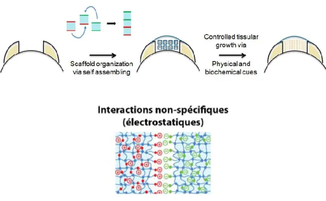
Hypothèse : La fonctionnalisation de blocs hydrogels neutres à l’aide de polyélectrolytes ou de microgels chargés permet leur assemblage contrôlé en milieux aqueux via des interactions électrostatiques
Méthode : Des blocs hydrogels de taille contrôlée ont été photosynthétisés à l’aide d’un support d’injection fabriqué à partir de lames de verre et de photomasques imprimés sur papier transparent. Les formulations se basent sur une structure (hydroxyéthyl)méthacrylate - poly(éthylène glycol diméthacrylate) (HEMA-PEGDMA) interpénétrée soit par du polyéthylèneimine(PEI) chargé positivement soit par de l’acide hyaluronique (HA) chargé négativement. Un autre type de blocs négatifs a été obtenu par le traitement des blocs PEI à l’aide de microgels N-isopropylacrylamide - acide méthacrylique (NIPAM-MAA) chargés négativement. Les propriétés d’assemblage dirigé de deux couples de blocs (PEI-HA et PEI-MG) ont été testées par la mise en contact aléatoire de population de blocs en milieu aqueux. L’effet de la salinité et du pH sur les propriétés d’assemblage ont été étudiés par des tests d’assemblage en milieux salin (NaCl) ou acide/basique.
Résultats : Le support d’injection développé a permis l’obtention de blocs hydrogels de différentes formes et tailles. Différentes fonctionnalisations à base de polyélectrolytes et de microgels ont été testées. Les tests d’assemblage ont résulté en l’obtention d’agrégats de blocs hydrogels liés par des contacts adhésifs spécifiques entre blocs chargés positivement et négativement. Les deux systèmes étudiés présentent cependant des caractéristiques d’assemblage différentes puisque les agrégats PEI-MG sont plus compacts et rigides que les agrégats PEI-HA. En milieu acide (pH=3) et basique (pH=10,5), aucun assemblage n’a pu être
Mémoire de Maitrise, Hanauer Nicolas 4 observé. L’augmentation de la salinité s’est accompagnée d’une perte croissante des propriétés d’assemblage. Cet effet délétère du sel est plus marqué pour les systèmes PEI-MG.
Mémoire de Maitrise, Hanauer Nicolas 5 Résumé et mots clés en anglais
Hypothesis: Functionalization of neutral hydrogel blocks with polyelectrolytes and charged microgels allow their directed assembly in aqueous medium via electrostatic interactions
Methods: Hydrogel blocks of controlled size were photosynthesized thank to an injection support composed of glass slides and photomasks imprinted on transparent sheet. Formulations are based on an HEMA-PEGDMA structure interpenetrated either with positively charged PEI or with negatively charged HA. Another type of negative block was obtained by treating PEI blocks with NIPAM-MAA negatively charged microgels. The assembling properties of two block couples (PEI-HA and PEI-MG) were tested by randomly putting in contact block population in water. Effects of salinity and pH on assembling properties were studied with assembling tests in saline (NaCl) or acidic/basic media.
Results: The injection support developed allowed obtaining hydrogel blocks with different shapes and sizes. Various formulations based on polyélectrolytes and microgels were tested. The assembling tests resulted in aggregates formation of hydrogel blocks linked together by specific adhesive contacts between positively and negatively charged blocks. The two systems studied nevertheless present differences in their assembling characteristics: PEI-MG aggregates are indeed more compact and rigid than the PEI-HA aggregates. In acidic (pH=3) and basic (pH=10,5) media, no assembling was observed. The augmentation in salinity resulted in an increasing loss of assembling properties. This deleterious effect was more important for PEI -MG systems.
Mémoire de Maitrise, Hanauer Nicolas 6 Remerciements
Je tiens à remercier l’ensemble de l’équipe de l’axe Formulation et Analyse du Médicament, dite du 4ème étage, et plus particulièrement aux membres du Labo Banquy.
Mémoire de Maitrise, Hanauer Nicolas 7 Table des matières
Résumé et mots clés en français ... 3
Résumé et mots clés en anglais ... 5
Remerciements ... 6
Liste des tableaux ... 9
Liste des figures ... 9
Liste des abréviations ... 10
Chapitre 1 : Introduction ... 11
1-1: Médecine régénérative et ingénierie tissulaire... 11
1-2 : Échafaudages hydrogel ... 11
Article 1: 2D, 3D and 4D Active Compound Delivery in Tissue Engineering and Regenerative Medicine... 13
Rôle des auteurs ... 13
Résumé en français ... 13
Abstract... 14
Introduction ... 15
Part I: 2D Tissue Engineering ... 16
A. Distribution control of molecular cues in 2D ... 16
B. Cell patterning and co-culture ... 20
C. 3D constructs based on 2D assemblies ... 21
Part II: Hydrogel scaffolds ... 22
A. Effect of active compounds loading and release ... 23
B. Effect of cells loading and culture ... 25
C. Active carriers for tissue regeneration... 27
Part III: Fibrous scaffolds ... 31
A. Controlled release of AC from fibrous scaffolds ... 31
B. Encapsulation of cells into fibers ... 33
C. Promoting regeneration with cells and growth factor delivery in fibrous scaffold ... 35
Conclusion ... 37
Mémoire de Maitrise, Hanauer Nicolas 8
1-3 : Échafaudages structurés injectables ... 44
1-4 : Formation d’agrégats à partir de blocs hydrogels ... 44
Chapitre 2 : Objectifs et méthodologie... 45
2-1 : Objectifs ... 45
2-2 : Méthodologie ... 47
2-2-1 : Fabrication de blocs hydrogels... 47
2-2-2 : Fonctionnalisation des blocs hydrogels ... 49
2-2-3 : Tests d’assemblage ... 50
Chapitre 3: Résultats... 50
Article 2: Assembly of hydrogel blocks mediated by polyelectrolytes or microgels: effect of salinity, pH and microgel surface charge ... 51
Rôle des auteurs ... 51
Résumé en français ... 51
Abstract... 52
Introduction ... 53
Materials and methods ... 55
Results ... 59 Discussion ... 67 Conclusion ... 71 Acknowledgements ... 72 References ... 73 Chapitre 4: Conclusion ... 76
Chapitre 5 : Perspectives et travaux futurs ... 77
Mémoire de Maitrise, Hanauer Nicolas 9 Liste des tableaux
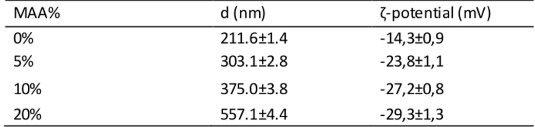
Table 1: Particle size and ζ-potential of the NIPAM-MAA microgels
Liste des figures
Figure 1-1: AC and cells deposition processes in 2D.
Figure 1-2: Two-cell patterned co-culture, adapted from [39]. Figure 1-3: BAEP fabrication, adapted from [42].
Figure 1-4: Strategies for tissue growth based on AC delivery.
Figure 1-5: Expected cumulative release of different cells/ACs distributions in hydrogel scaffolds. Figure 1-6: Release curve of FGF (a), FGF loaded (b) and variation of GAG concentration (c) in different chitosan hydrogel treated by freeze drying and/or by gas foaming prior to layer-by-layer assembling of chondroitin sulfate and chitosan. Adapted from [56].
Figure 1-7: Representation of possible uses of fibrous scaffolds in drug encapsulation and in cell encapsulation.
Figure 2-1 : Principe de la croissance contrôlée de néo-tissus à l’aide d’un échafaudage hydrogel formé par l’assemblage dirigé de blocs hydrogels injectables fonctionnalisés électrostatiquement
Figure 2-2 : Principe de l’utilisation de moules A) Pour obtenir des blocs B) Pour obtenir un pain d’hydrogel pouvant être découpé
Figure 2-3 : Principe de l’utilisation de photomasques pour la synthèse de blocs de forme et taille contrôlées
Figure 2-4 : Différentes formes de blocs obtenues à l’aide du support d’injection et des photomasques
Figure 3-1: Stereomicroscopies (A, B, C, E, F, G) and camera pictures (D, H) of PEI/HA and PEI/MG aggregates of different sizes (scale bar: 1mm)
Figure 3-2: A) to C): Cumulative % of blocks aggregation as a function of the aggregation number (number of blocks per aggregates. D) to F): Average aggregation number as a function of the experiment iteration. Lines are guides for the eye.
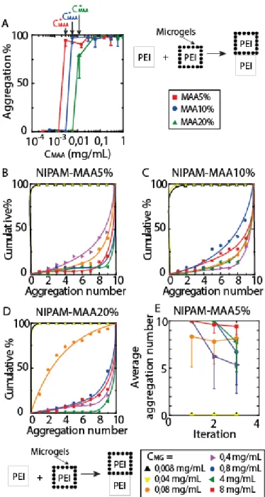
Figure 3-3: Effect of the MAA content in microgels on the directed assembly of PEI containing hydrogel blocks. A) to D) Cumulative % of blocks aggregation as a function of the aggregation number (number of blocks per aggregates). E) Average aggregation number as a function of the experiment iteration. Curves are guides for the eye.
Figure 3-4: Effect of the microgel concentration on the directed assembly of PEI blocks. (A) Blocks aggregation % as a function of MAA content in the microgels during the pretreatment. B-D) Cumulative % of aggregated blocks as a function of the average aggregation number (represented values are averages of three assembly iterations). E) Average blocks aggregation number in presence of NIPAM-MAA5% microgels only. Error bars represent the standard deviation of 5 separate experiments. Lines are guides for the eye.
Mémoire de Maitrise, Hanauer Nicolas 10 Figure 3-5: Effect of salt concentration on the directed assembly of A) PEI/HA and PEI/MG systems B) PEI/MG system in presence of microgels NIPAM-MAA20% at different concentrations. Lines are guides for the eye.
Figure 3-6: Effect of pH on the directed assembly of PEI/HA and PEI/MG systems
Figure 3-7: Models of supposed interactions at hydrogel blocks surfaces during adhesive contacts, A: steric entanglement guided by electrostatic cues between HA and PEI polyelectrolytes chains, B: bridging between PEI chains and NIPAM-MAA microgels without any entanglements involved.
Liste des abréviations
HA: Acide hyaluronique
HEMA: (Hydroxyéthyl)méthacrylate MAA: Acide méthacrylique
MG: Microgels
NIPAM: N-isopropylacrylamide PDMS: Polydiméthylsiloxane
PEGDMA: Poly(éthylène glycol diméthacrylate) PEI: Polyéthylèneimine
Mémoire de Maitrise, Hanauer Nicolas 11
Chapitre 1 : Introduction
1-1: Médecine régénérative et ingénierie tissulaire
La médecine régénérative est un domaine émergent des sciences pharmaceutiques et médicales s’intéressant à l’élaboration de traitements basés sur la création de tissus vivants fonctionnels dans le but de soigner ou revitaliser des tissus ou organes endommagés [1,2]. La croissance de tels tissus réparateurs peut être menée ex-vivo suivie d’une implantation [3] ou directement
in-vivo[4-6]. Le développement de telles solutions médicales se base ainsi sur la compréhension,
apportée par l’ingénierie tissulaire, de l’influence des facteurs biochimiques (composition du milieu, facteurs de croissance, enzymes…) et physiques (contraintes mécaniques, porosité) sur la différenciation et la prolifération des cellules composant un tissu sain [7-9]. Sans contrôle sur leur croissance, les néo-tissus obtenus ne présentent pas la même organisation structurelle qu’un tissu sain et ne possèdent pas des propriétés biologiques et mécaniques équivalentes. La reproduction spatio-temporelle de contraintes s’approchant du microenvironnement naturel sur des populations cellulaires est donc au cœur des traitements régénératifs [10,11].
1-2 : Échafaudages hydrogels
En réponse à ce problème, l’utilisation de matrices biocompatibles permettant un meilleur contrôle de la croissance cellulaire s’est développée
[12]
. Ces matrices prennent la forme d’échafaudages polymériques composés d’hydrogels et/ou de fibres polymériques. Le contrôle sur leur structure et leur composition permet de définir l’environnement physique et biochimique des cellules transportées afin d’obtenir un certain contrôle sur leur croissance. Les hydrogels sont de parfaits matériaux pour la réalisation de telles matrices puisqu’ils sont composés d’une part d’une structure polymérique aux propriétés hautement modulables tantMémoire de Maitrise, Hanauer Nicolas 12 au niveau physique (gonflement, rigidité, porosité…) que chimiques (fonctionnalisations, sensibilité aux conditions environnantes…) et d’autre part d’une phase liquide «immobilisée » permettant l’embarquement de différents composés actifs (espèces chimiques, facteurs de croissance, cellules) [13,14].
Si l’utilisation de tels dispositifs a permis le développement de solutions d’ingénierie tissulaire 2Det 3D pour de nombreux tissus (peau [15,16], cartilage [17,18], os [19]..) , une emphase est mise de nos jours sur l’incorporation d’un facteur temporel (relargage s rapides ou progressifs, simultanées ou consécutifs de composés actifs, modification des propriétés physiques avec la croissance cellulaire) pour le développement de solutions dites 4D approchant plus l’évolution des conditions environnant la croissance d’un tissu sain. L’article présenté ci-après est une revue de la littérature scientifique sur les dernières avancées dans la livraison 2D, 3D et 4D de composés actifs en médecine régénérative et en ingénierie tissulaire. La 4D correspondant aux 3 dimensions spatiales avec un contrôle temporel. Il a été écrit en collaboration avec Pierre-Luc Latreille, Shaker Alsharif et Xavier Banquy et a été publié en 2015 dans le Volume 21 Numéro 12 de la revue Current Pharmaceutical Design.
Mémoire de Maitrise, Hanauer Nicolas 13 Article 1:
2D, 3D and 4D Active Compound Delivery in Tissue Engineering and
Regenerative Medicine
Nicolas Hanauer
1, Pierre Luc Latreille
1, Shaker Alsharif
1, 2and Xavier Banquy
1 1Canada Research Chair in Bio-inspired Materials and Interfaces, Faculty of Pharmacy, Université de Montréal C.P. 6128, Succursale Centre Ville, Montréal, QC H3C 3J7, Canada
2
Pharmaceutical Chemistry Department, Faculty of Pharmacy, Umm Al-Qura University Al Taif Road, Makkah 24382, Saudi Arabia
Rôle des auteurs
Shaker Alsharif a effectué les recherches et rédigé la partie sur l’ingénierie tissulaire 2D, Pierre -Luc Latreille sur les échafaudages fibreux et moi-même sur les échafaudages hydrogel. Les recherches et la rédaction ont été effectuées sous la supervision du Pr. Banquy.
Résumé en français
Les avancées récentes dans les domaines de l’ingénierie tissulaire et de la médecine régénérative ont montré que le contrôle du microenvironnement des cellules durant leur croissance est un facteur clé dans le développement de systèmes thérapeutiques efficaces. Pour atteindre un tel contrôle, les chercheurs ont d’abord proposé l’utilisation d’échafaudages polymériques capables de supporter la croissance cellulaire et, dans une certaine mesure, favoriser l’organisation cellulaire et la structure tissulaire. De nos jours, avec la disponibilité de nombreuses lignées de cellules souches, cette approche semble plutôt limitée puisqu’elle n’offre pas un contrôle précis du microenvironnement cellulaire dans le temps et l’espace (4D). Les chercheurs concentrent ainsi leurs efforts sur le développement de stratégies incluant un système de livraison de composés actifs dans le but d’ajouter une dimension aux échafaudages 3D. Cette revue se concentrera sur des concepts et applications récents de techniques 2D et 3D qui ont été utilisés pour contrôler le chargement et le relargage de composés actifs pour
Mémoire de Maitrise, Hanauer Nicolas 14 promouvoir la différentiation et la prolifération cellulaire à l’intérieur ou à l’extérieur d’un échafaudage. Nous présenterons les avancées récentes dans le design d’échafaudages polymériques 2D et les différentes techniques utilisées pour le dépôt contrôlé de signaux moléculaires et de cellules. Nous continuerons en présentant les avancées récentes effectuées dans le design d’échafaudages 3D basés sur des hydrogels ou de s fibres polymères. Nous finirons en présentant certaines des pistes de recherche encore à explorer.
Abstract
Recent advances in tissue engineering and regenerative medicine have shown that controlling cells micro-environment during growth is a key element to the development of successful therapeutic system. To achieve such control, researchers have first proposed the use of polymeric scaffolds that were able to support cellular growth and, to a certain extent, favor cell organization and tissue structure. With nowadays availability of a large pool of stem cell lines, such approach has appeared to be rather limited since it does not offer the fine control of the cell micro-environment in space and time (4D). Therefore, researchers are currently focusing their efforts in developing strategies that include active compound delivery systems in order to add a fourth dimension to the design of 3D scaffolds. This review will focus on recent concepts and applications of 2D and 3D techniques that have been used to control the load and release of active compounds used to promote cell differentiation and proliferation in or out of a scaffold. We will first present recent advances in the design of 2D polymeric scaffolds and the different techniques that have been used to deposit molecular cues and cells in a controlled fashion. We will continue by presenting the recent advances made in the design of 3D scaffolds based on hydrogels as well as polymeric fibers and we will finish by presenting some of the research avenues that are still to be explored.
Mémoire de Maitrise, Hanauer Nicolas 15 Keywords: niche engineering, controlled release, scaffolding, hydrogel, fiber
Introduction
Driven by the increasing demand of organ transplantation, tissue engineering and more recently regenerative medicine have developed numerous strategies to grow such organs in vivo or ex
vivo. After more than two decades of intense research, it is clear that organ engineering requires
the use of a scaffold that serves as a synthetic extracellular matrix (ECM) to support and organize cell growth [1-4]. With the increasing number of available biomaterials that possess all the desirable properties required for tissue engineering as well as the constantly widening spectrum of manufacturing techniques to generate complex and finely tuned structures, researchers have been able to develop a tremendous variety of materials and scaffolds designed for specific tissues and applications [5-8]. Within the last few years, the use of stem cells in regenerative medicine and tissue engineering has become predominant [9-13]. Building a suitable micro-environment for their differentiation and proliferation is a challenging task. The rational design of such micro environment must involve a combination of many different expertises such as material micro-engineering, biological engineering and more recently pharmaceutical technology. The requirement of such diverse set of expertise has been driven by the intrinsic behavior of stem cells in their environment [14, 15]. Stem cells are present in many different places in any mammalian organism. They are inherently sensitive to many biophysical as well as biochemical stimuli generated from their direct surroundings [16]. Their differentiation and proliferation is not only dictated by very specific molecules such as growth factors but also by the concentration of such factors and their spatiotemporal distribution in the surroundings [17, 18]. It is believed that these spatiotemporal distributions (also called niche) of key factors are paramount elements determining cell recruitment, migration, proliferation,
Mémoire de Maitrise, Hanauer Nicolas 16 protein production and finally organ architecture [19, 20]. Artificially reproducing such complex dynamic environment is the main goal of nowadays tissue engineering research and the main focus of this review article.
Early studies in tissue engineering predominantly used 2D polymeric scaffolds functionalized with adhesives molecules in order to mimic the interactions between cells and the ECM [21, 22]. In parallel, 2D devices such as patches, micro-electro-mechanical-systems or microchips were already reported for the controlled delivery of actives compounds (AC) [23-25]. It is only recently that these two worlds have collided and nurtured each other beneficially. To better mimic biological tissues, the transition from 2D to 3D scaffolds has become a necessary step. Interestingly, the tremendous large body of AC delivery systems using 3D devices such as particles or macromolecules have not been fully explored in tissue engineering. This provides an excellent opportunity for development and promising future discoveries.
Part I: 2D Tissue Engineering
A. Distribution control of molecular cues in 2D
It is well known that most of human body organs and tissues have a 3D structure while some other important body tissues such as blood and lymphatic vessels have a 2D structure. Therefore, engineering of tissues in 2D has proven to be of importance. For this purpose, different technologies in the realm of AC release and cell delivery have emerged in the past few years which we discuss here the most relevant ones (see figure 1).
Mémoire de Maitrise, Hanauer Nicolas 17 Figure 1-1: AC and cells deposition processes in 2D.
1. Gradient technology
Tissue engineering and regenerative medicine deal directly with ECM, which carries various macromolecules or proteins such as growth factors and chemokines. Their physiological functions such as wound healing and morphogenesis are majorly regulated by mole cular concentration gradient phenomena. Many studies related to cellular processes such as in vitro migration, signal transmission, cellular proliferation, viability had shown the significance of using gradient materials in tissue engineering [26, 27].
Developing molecular gradients in a material can be extremely challenging especially when it comes to fine controlling. Ostrovidovet al.[28] have developed a microfluidic device acting as concentration gradient generator. The device made from micro-engineered poly(ethylene glycol) diacrylate (PEGDA) hydrogel contains concentration gradient of okadaic acid as a model drug released by diffusion. The authors showed that the drug gradient was able to modulate the viability of MC3T3 cells.
Mémoire de Maitrise, Hanauer Nicolas 18 Controlling the distribution of AC is not the only benefit of using gradient technology. The mechanical properties of a cell substrate can be controlled as well. In vitro techniques based on photolithography [29] or on polymerization of adjoining solutions with variable concentrations [30] in order to obtain crosslinking density gradients have shown that it is possible to achieve good control over the elastic properties of a substrate in 2D.
In a recent study, Tse et al.[31] have discussed whether undifferentiated mesenchymal stem cells (MSCs) can experience durotaxis in the absence of any pathological stimulation under exposure to a physiological stiffness gradient. The authors created crosslinking gradient in polyacrylamide hydrogels using radial greyscale pattern with a photomask. In addition, type -I collagen was added to the gradient hydrogel to allow MSCs attachment. Results evidenced that MSCs were subjected to durotaxis on substrates with stiffness gradient values within physiological range and initiated differentiation at the stiffest regions instead of remaining in stationary position as had been hypothesised.
2. Patterning technology
The ability to spatially deposit and control the release of AC of variable size, including drugs and growth factors from patterned biomaterials is crucial to the development of bioactive surfaces for regenerative medicine. One of the scalable methods in patterning such surfaces is lithography. Stern et al.[32] have used patterned electropolymerized polypyrroles surfaces to attach and release AC such as ovalbumin and interleukin-2 respectively. These proteins act as vaccine components for binding to dendritic cells that process the antigen and present it to T-cell surface. The patterning was obtained by deposing photoresistant masks on the conductive substrate where electropolymerization of the dissolved monomers containing the AC took place.
Mémoire de Maitrise, Hanauer Nicolas 19 The authors showed that surface patterning offered a very high control of the spatial distribution of the AC while their release rate was electrically controlled.
Recent advancement in nanopaterning opens the opportunity to combine colloidal lithography and surface-initiated atom-transfer radical polymerization to finely control molecular cues distribution such as cell adhesive proteins. Li et al.[33] used hierarchical polymer brush nanopatterns to graft fibronectin on a planar substrate. As a result, fibronectin was covalently immobilized and showed biological activity without denaturation. Furthermore, MC3T3-E1 mice osteoblasts had cohered to fibronectin patterns immediately and displayed uniformity along the stripes, which suggest that these protein patterns are excellent candidates for cell patterning. Thissenet al.[34] have recently described a method based on surface patterning to control the growth of bovine corneal epithelial tissue on surfaces by creating protein adsorbing and non-adsorbing sites via cell-collagen-I interactions. This manipulation was accomplished by applying a thin layer of acetaldehyde polymer coating (adhesive site for subsequent collagen I deposition) and poly(ethylene oxide) PEO (non-adhesive site) on the substrate.
Common lithographic patterning techniques require either UV exposure or jarring solvents, which are not suitable for most biomolecules. A new patterning technique that does not damage biomolecules was recently reported [35]. The process uses hydrofluoroether solvents which solubilise fluorinated UV resistant materials used to pattern AC through imprint lithography. Such process has been applied to protein and DNA patterning without damaging the AC.
Alternatively, inkjet printing has been used to create spatial patterns of fibroblast growth factor-2 (FGF-factor-2) on fibrin films for studying preosteoblastic cells response in vitro [36]. The authors showed that under cell culture conditions for over one week, printed patterns as well as FGF-2 remained persistent and active.
Mémoire de Maitrise, Hanauer Nicolas 20 Most of the previously described reports carry great potentials and opportunities for future development regarding tissue engineering. However, it has not been found yet advanced studies involving such methods upon major in vivo applications in regenerative medicine.
B. Cell patterning and co-culture
Spatial control of living cells distribution has attracted great attention due to its broad potential applications in regenerative medicine. The development of microfabrication technology in the past decade has largely enriched cell patterning methods by introducing precise surface engineering, in which spatial patterning of cells is confined by regulating surface chemistry. Cells are often patterned on a planar surface, which can be further controlled to prepare a 3D bioactive structure or scaffold.
Inkjet printing method was reported as an advantageous technique for human fibroblast cells patterning. Using this method Saunders et al. [37] were able to create cells patterns on agarose gel without damaging the cells.
In order to modify surface chemistry and to improve cell patterning, Chienet al.[38] have combined microcontact-printing method with mussel inspired surface chemistry. Controlled imprints of polydopamine (PDA)/poly ethylene imine (PEI) were fabricated using poly(dimethylsiloxane) (PDMS) stamps. These imprints were used to control cell adhesion using the high binding affinity of PDA enhanced by deposition of PEI. In vitro tests conducted with co-cultured hepatocytes and neural cells lead to spatially controlled distribution of cells. This technique could be used to favor cells adhesion at specific sites by recover them of cell adhesion promoting imprints.
In another study Tanaka et al.[39] discussed how to manage the PDMS stamping force and the importance of stamp stiffness to improve cell patterning. The authors reported a method to
Mémoire de Maitrise, Hanauer Nicolas 21 improve printing precision by controlling the stamp stiffness via microscope observation of stamp deformation due to the applied force. The proposed micro printing method gave a high printing quality with 2.5% error of micro stamping area and was tested by patterning GFP-HUVEC (GFP Expressing Human Umbilical Vein Endothelial Cells) and NIH/3T3 co culture on fibronectin covered substrates (see figure 2)
Figure 1-2: Two-cell patterned co-culture, adapted from [39].
C. 3D constructs based on 2D assemblies
In the area of 3D microfabrication, a recent novel strategy based on 2D scaffold folding, which enables production of 3D microstructure simply by folding 2D sheets was recently reported [40]. Origami folding and polyhedral capsule rolls are two examples using such strategy [41].
Bioartificial endocrine pancreas (BAEP) was created by encapsulating pancreatic B-cells for diabetes treatment purposes [42]. This BAEP was found more advantageous over gel encapsulation method in terms of mass transfer efficiency of AC due to its unique architectural design and geometry. The BAEP fabrication was based on folded polyhedral capsules wrapped up within an alginate sheet (see figure 3). Consequently, insulin release was confirmed suggesting that this approach could be convenient for regenerative medicine.
Mémoire de Maitrise, Hanauer Nicolas 22 Figure 1-3: BAEP fabrication, adapted from [42].
This emerging technology is extremely promising due to its potential scalability, its versatility in terms of structures and materials that can be used. Such approach though, requires very specific expertises and equipment which limits its exploration and use at the present time. Instead, other approaches based on readily available materials such as hydrogels have attracted much more attention and will be described in the next section.
Part II: Hydrogel scaffolds
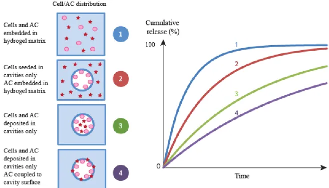
Techniques of 3D hydrogels scaffolding have been developed for two major regenerative medicine related purposes: cell viability, proliferation and differentiation as well as AC delivery [43]. This was commonly achieved by the incorporation of AC and/or cells inside the hydrogel matrix via different techniques leading to various architectures that can be used for diverse applications (see figure 4).
Mémoire de Maitrise, Hanauer Nicolas 23 Figure 1-4: Strategies for tissue growth based on AC delivery.
A. Effect of active compounds loading and release
Incorporating an AC such as a growth factor, a drug or genetic material into a polymeric scaffold can be achieved by embedding this compound inside the scaffold using chemical or physical bounding [44]. Control over such bindings and loading mechanics is a key parameter to achieve simultaneous or sequential controlled release of multiple AC [45].
Incorporation of an AC into a hydrogel matrix usually results in a fast release of the AC, at least during the initial period of the release (see figure 5). Such effect, known as initial burst effect is problematic for tissue engineering applications where long lasting delivery is often desirable. Tang et al.[46] have controlled the burst effect by embedding N-(2-hydroxyl) propyl-3-trimethyl ammonium chitosan chloride (HTCC) – carboxymethyl chitosan (CM) nanoparticles into
Mémoire de Maitrise, Hanauer Nicolas 24 chitosan/poly(vinyl alcohol) hydrogel by adding them prior to gelation. Propranolol, as positively charged model drug, diclofenac sodium, as negatively charged model drug, and nanoparticles were added prior to gelation. The authors obtained nanoparticles with different charges by varying the ratio between HTCC and CM. The interaction between the drug and the nanoparticles was shown to have a direct effect on the release. The release of the positively charged drug was found to be much slower in negatively charged hydrogel than in neutral hydrogel and vice versa.
The controlled release of growth factors is crucial in regenerative medicine due to their roles as biological cues for cell fates. Pakulska et al.[47] have prepared chondroitinase ABC (ChABC), a promising therapeutic agent for spinal cord injury to a methylcellulose (MC) hydrogel by grafting a small protein domain (Src homology 3: SH3) on the AC and a binding peptide (weak or strong) on the hydrogel. The release rate of the AC was then tuned either by varying the SH3-protein/SH3-peptide pair binding strength or ratio. Even if the release process was disturbed by the thermal instability of ChABC at 37°C, the authors were able to observe a tunable release: 90% of release was obtained after 3 days for an unmodified MC hydrogel, while it decreased to 20% in 7 days with the strong ChABC-SH3 binder and to 50% and 10% in 7 days with a weaker binder at respectively 100 and 300-fold molar excess of SH3 peptide to ChABC.
AC release can be triggered by cell activity as well. Song et al.[48] have studied the effect of combining two AC (stromal derived factor 1: SDF-1 and angiogenic peptides: Ac-SDKP) in an acrylated hyaluronic acid hydrogel on a chronic myocardial infarction rat model. The authors loaded SDF-1 directly within the hydrogel and Ac-SDKP was bound to the polymer scaffold via thiol-acrylate reaction. The release of SDF-1 and Ac-SDKP was triggered by the action of matrix metalloproteinase (MMP) secreted by the surrounding cells or via hydrogel degradation. By providing an injectable 3D micro-environment to attract mesenchymal stem cells followed by
Mémoire de Maitrise, Hanauer Nicolas 25 growth factor release, this approach was found to promote stable vessels growth, and decreased fibrosis, which in turn leads to the recovery of heart function. Even if the mechanism of regeneration by SDF-1 and Ac-SDKP is still unclear, this study showed the strong positive synergistic effect of the two compounds.
These recent examples show that AC controlled loading and release in hydrogel scaffold play a crucial role for the development of therapeutic implants. By tuning the hydrogel scaffold properties and especially the AC-matrix bounding, multiple and sequential releases of ACs can be envisaged.
Figure 1-5: Expected cumulative release of different cells/ACs distributions in hydrogel scaffolds.
B. Effect of cells loading and culture
Due to their internal structure that can be tuned to mimic the ECM, hydrogels were firstly used for cell immobilization [49]. With the development of tissue engineering and progress in hydrogel scaffolding, these materials are now able to promote cells growth, differentiation and
Mémoire de Maitrise, Hanauer Nicolas 26 organization [50, 51]. Such properties can be achieved via incorporation of cells into structured 3D hydrogel scaffolds in multiple ways depending on what the final goal or application is. Cell embedment in the hydrogel can be achieved by directly inserting the cells during the gelation process. Wright et al.[52] studied human corneal epithelial cells viability in a calcium alginate-hydroxyethyl cellulose hydrogel. After mixing the cells with the hydrogel solution, cells were found to survive the gelation process, and were viable up to 7 days in ambient and chilled conditions, which makes this hydrogel potentially useful for cells transport and storage purposes.
Such technique can also be used to highlight the role of the hydrogel composition on loaded cells fate. Li et al[53] used fluorinated methacrylamide chitosan hydrogels for neural stem cell differentiation. Neural cells were added with scaffold components prior to photopolymerization. The authors studied the proliferation and differentiation of neural cells in fluorinated methacrylamide chitosan hydrogels which had the ability to uptake oxygen from the environment or from supplemental oxygen. Fluorine moieties in the hydrogel were found to modulate oxygen uptake and release which resulted in improved cell proliferation and differentiation.
Introducing the receptor sites in the hydrogel is an easy way to increase the amount of introduced cells and to achieve a better control over their near environment. Halstenberg et
al.[54] created an artificial protein with matrix degradation capacity containing two cell binding
sites (RGD integrin-binding and heparin binding site), matrix degradation sites (two plasmin degradation sites) and an acrylate moiety. The authors used this protein in conjunction with poly(ethylene glycol) diacrylate to form a hydrogel. Human fibroblasts-fibrin clusters were embedded via cell solution deposition on the hybrid hydrogel. These clusters were used to assess cell attachment on 3D binding sites, proliferation for at least 7-9 days in vitro and cell
Mémoire de Maitrise, Hanauer Nicolas 27 induced matrix degradation. Using the artificial protein resulte d in an improved cellular penetration in the hydrogel due to the combination of cellular outgrowth and triggered matrix degradation.
Incorporation of niches inside the hydrogel matrix prior to cell embedment was found to have positive effect on cells development. Hwang et al.[55] used gelatin beads (150-300µm) included in a cell laden alginate hydrogel, which after dissolution and washing left occlusions of controlled size. Use of such scaffolds in tissue engineering was tested using hepatocarcinoma cells (HepG2). The cells were positioned inside the cavities and significantly enhanced cell proliferation was observed compared to non porous scaffold, due to better mass transfer of nutrients, oxygen and waste removal through the hydrogel.
C. Active carriers for tissue regeneration
Cell fate in a tissue depends on two main factors: mechanical stress and cell -ECM biochemistry. Chemical interactions between cells and AC are based on 3D-signaling which is a result of AC spatiotemporal availability and cells motility. Active carriers for tissue regeneration combine cells encapsulation and triggered AC release to promote timed-control cell growth.
Du et al.[56] obtained chitosan hydrogel exhibiting an interconnected network of cavities using 10 µm CaCO3microparticles encapsulation followed by gas foaming. After freeze-drying treatment of the hydrogel, the authors obtained a hierarchical porous structure later treated by layer-by-layer molecular deposition of oppositely charged chondroitin sulfate (CS) and chitosan to mimic the ECM. The hydrogel cavities were then loaded on their surface with fibroblast growth factor (FGF) via CS binding and then with human lung fibroblast cells. The authors showed that the architecture of the interconnected network of cavities did not have any significant effect on FGF cumulative release but improved FGF loading resulting in a higher
Mémoire de Maitrise, Hanauer Nicolas 28 amount of FGF reaching encapsulated cells (see figure 6). The authors showed that combining the hierarchical porous structure of the chitosan hydrogel with the controlled loading and release of the growth factor via CS binding of FGF had a positive effect on human lung fibroblast growth.
Figure 1-6: Release curve of FGF (a), FGF loaded (b) and variation of GAG concentration (c) in different chitosan hydrogel treated by freeze drying and/or by gas foaming prior to
layer-by-layer assembling of chondroitin sulfate and chitosan. Adapted from [56].
In another study, the same authors [57] used two growth factors that could be released successively. They incorporated two growth factors, native TGF-β and bFGF modified to specifically bind to collagen in a CS/collagen hydrogel. Such modification allowed the growth factor to be loaded in the scaffold to a much higher content and to be released much slower than TGF-β. These successive releases were used by the authors to induce differentiation of hMSCs into chondrocytes (see figure 7).
Mémoire de Maitrise, Hanauer Nicolas 29 Figure 1-7: Representation of possible uses of fibrous scaffolds in drug encapsulation and in cell
encapsulation.
Hydrogel scaffolds have been tested invivo as well. Using direct loading of two growth factors (VEGF: vascular endothelial growth factor and IGF-1) in an alginate hydrogel precursors mix prior to gelation and followed by freeze drying to create a niche for myoblast encapsulation lead to a scaffold able to promote muscle regeneration. Borselliet al.[58] tested this hydrogel in the context of a severe injury to mice skeletal muscle tissue. A synergistic effect between VEGF and SDF-1was demonstrated on muscle growth in comparison to implantation of blank alginate scaffold or single growth factor loaded hydrogel. It was also shown that cell seeding in the hydrogel allows even better muscle regeneration. Even if the impact of such scaffold on the muscle size and weight was not always significant, it allowed an improve d fiber growth and higher blood vessel density leading to normal tissue perfusion levels.
It is possible also to pattern the hydrogel containing growth factors and cells before implantation in order to better mimic in vivo tissue organization. Chen et al. [59] have combined two genes (TGF-B1 and BMP-2) activated chitosan/gelatin scaffolds (freeze dried for
Mémoire de Maitrise, Hanauer Nicolas 30 mesenchymal stem cell loading) to create a bilayered hydrogel for articular cartilage and subchondral bone simultaneous regeneration. Once both scaffold were loaded with mesenchymal stem cells and have supported cells differentiation (chondrogenic and osteogenic respectively), they were glued together via fibrin glue. The bilayered materiel was tested on a rabbit knee defect and was found to perfectly support articular cartilage and subchondral regenerations, leading to complete repair.
The strategies described so far involve controlled but passive release of the AC. This is, in principle, not efficient to maximize contact of the AC with the surrounding cells. To overcome this problem Yang et al.[59] developed an active PEG scaffold able to release locally synthetic glucocorticoid Dexamethasone (DEX) only in presence of a neighboring cell. The authors conjugated DEX to the scaffold using a peptidic linker. The linker was degraded by production of matrix metalloproteinase from the proliferating hMSCs. This resulted in a localized stimulation of alkaline phosphatase (ALP) and calcium deposition for over 21 days whereas no elevated cellular responses were observed in co-cultured hMSCs surrounding the gel, suggesting possible applications in bone regenerative medicine.
With their highly tunable internal structure, hydrogels are the main material used for scaffolding. Such scaffolds have various applications for regenerative medicine going from storage to multiple controlled releases of AC and/or cells. Even if some hydrogel scaffolding techniques are already commercially available, the innovations discussed above present promising future as therapeutic treatments. However hydrogel scaffolding is not the only solution for 4D AC delivery, other materials are gaining interest, such as fibrous scaffolding.
Mémoire de Maitrise, Hanauer Nicolas 31
Part III: Fibrous scaffolds
Fibrous scaffolds represent a popular substrate for tissue engineering. Their fibrous nature mimics biological tissues matrices at a microscopic scale compared to plain materials such as hydrogels. Many strategies were developed and characterized in an objective of delivering AC and cells to animals (see figure 7). As mentioned, those concepts focused on the control of AC release from promising fibrous scaffolds have emerged and are summarized in the next section.
A. Controlled release of AC from fibrous scaffolds
Many different strategies exist to incorporate an AC into a fibrous material [60-62]. Most of them face a common problem of short release time which is problematic from a tissue engineering perspective. To tackle this problem, a first approach consists in using structured fibers. Novajra et al. [63] have recently developed a biodegradable scaffold based on hollow fibers of biodegradable glass for the long term release of neurotropic factors. Fibers were filled with genipin crosslinked agar/gelatin hydrogel in presence of fluorescein isothiocyanate -dextran (FD-20). The authors did not report any correlation of the release rate and the fibers diameter since all of them achieved 100% release of FD-20 after 24h. Also, no significant cytotoxicity of fiber dissolution products was reported on neonatal rat olfactory bulb ensheathing cell line. Another efficient strategy to develop structured fibers is to use mixture polymers during the electrospinning process. Bonani et al.[64] have developed a fibrous scaffold by making use of electrospunnanofibers of poly ε-caprolactone (PCL) and poly(D,L-lactide-co-glycolide acid) (PLGA). The authors designed different patterns of fibers of PCL and PLGA by spatially controlling their distribution on both side of the scaffold. Therefore, PLGA (with ending carboxylic group, PLGAac or with ester end group, PLGAes) polymers were loaded with either rhodamine B (RhB), fluorescein or tetra-methyl-rhodamine conjugated bovine serum albumin (AlbF and AlbT) to
Mémoire de Maitrise, Hanauer Nicolas 32 perform double-sided release. PLGAac-PCL and PLGAes-PCL releasing RhB (from a uniform gradient of PLGA-PCL polymers) both showed a majorly one-sided release with a burst effect. PLGAes has demonstrated lower side release selectivity. Release of AlbT of PLGAac-PCL with the same gradient showed that 24% of protein was released in 24 hours and 80% was released in 9 days on mainly one side only of the scaffold. After this period, the authors had estimated a constant release rate of AlbT of 1% per week. Moreover, the authors were able to sequentially release both proteins (AlbT and ALbF) by altering the materials distribution in the scaffold (see figure 8). Highest release rate of AlbT occurred at the first day, while AlbF release was delayed until day 5. A similar approach was used to release AlbT and AlbF each one from a differe nt side of the scaffold.
Alternatively, Lee et al. [65] successfully immobilized bone-forming peptide-1 BFP-1 on the surface of PLGA fibers coated with PDA and then characterized the differentiation of hMSCs into osteocytes and bone volume increase in mice calvarial defect models. hMSCs culture has shown for BFP-1 immobilized fibers higher cellular differentiation, calcium production and ALP activity than controls (PLGA and PLGA-PDA coated scaffolds). Similar correlations were also observed in
vivo in the mice calvarial defect models. Immobilization of the growth factor at the surface of
the fibrous scaffold significantly increased bone regeneration in animal model by potentially increasing cell differentiation and ALP activities, supported by in vitro results.
Fibrous scaffold based on polyester polymers such as PLA, PLGA or PCL are most commonly used without any further modifications [66]. An improvement to this methodology is to build hybrid scaffolds incorporating different components within the fibers designed to perform very specific tasks. In that line of research, Lee et al.[67] have incorporated a self-assembled nanofiber gel of heparin-binding peptide amphiphiles (HBPA) and heparin-sulfate (HS) into a porous collagen scaffold. The objective was to increase bone regeneration by mimicking biological BMP-2
Mémoire de Maitrise, Hanauer Nicolas 33 signaling. The authors found that the natural affinity of BMP-2 to HBPA/HS complex made it able to modulate its release from the nanofibers gel. Implantation of the hybrid collagen scaffold loaded with low dose of BMP-2 significantly increased bone regeneration compared to controls in a rat model of femoral defect. These results clearly demonstrated that the architecture of the scaffold or its capacity to release AC are not the only crucial factors determining tissue regeneration. Incorporation of key signaling mechanism of the ECM can certainly amplify the regenerative capacity of growth factors as well.
Silk fibers are increasingly used as scaffolds for their biodegradability, biocompatibility and mechanical properties and were found to be very suitable for bone tissue bioengineering [68-70]. Li et al.[71] designed a scaffold using electrospun silk fibers, poly(ethylene oxide) and incorporated nanoparticles of hydroxyapatite (silk/PEO/nHAP). BMP-2 was incorporated in the scaffold without any specific link and hMSCs were seeded on the surface of the scaffolds. Higher deposition of calcium and presence of bone-specific markers of differentiated hMSCs was observed and presence of nHAP further improved the results compared to controls. A similar scaffold developed by Bimanet al. [72] utilized silk fibers embedded in polyacrylamide hydrogel. The authors showed, by using different fibers/hydrogel ratios, that the release rate of a model peptide (FITC linked insulin) increased significantly at higher concentration of fibroins in the scaffold.
B. Encapsulation of cells into fibers
Scaffold designed for cell delivery aim to recreate microenvironments with 3D cell-cell interaction of tissues. Promoting such 3D interaction might constitute a major leap for the treatment of a broad spectrum of degenerative diseases. Onoe et al.[73] developed a calcium alginate micro-fibrous hydrogel (Ca-alginate) embedding cells and ECM proteins. These fibers
Mémoire de Maitrise, Hanauer Nicolas 34 were generated from a double-coaxial microfluidic device with a flow of hydrogel and ECM with cells. Cells were then cultured until a cell fiber had being formed and the Ca-alginate was removed to obtain a cell-ECM fiber. Using a weaving and reeling technique, the authors were able to produce a scaffold with multiple cell fibers. This technology was then tested in diabetic mice model in the attempt to treat mellitus diabetes. The authors injected pepsin-solubilized type I collagen as ECM and fibers made of rat dissociated pancreatic cells and mouse pancreatic beta cells into the subrenal capsular space. After implantation, a significant decrease in blood glucose concentration was found while upon removal, blood glucose levels were readjusted to their initial concentrations, demonstrating efficacy for diabetes treatment. Potentially, this technique is very promising for fibrous scaffolding in regenerative medicine because of its versatility of application along with its customizability of ECM patterning, cell type or cell line. Similarly, Wan et al.[74] fabricated an interesting multi-component hydrogel fibers made from water soluble chitin (WSC) and sodium alginate in a matrix of WSC, galactose and collagen to spatially co-culture differentiated human hepatocytes and endothelial cells. The resulting 3D fibrous scaffold was utilized for encapsulation and culture of differentiated cells and then implanted in mice where 70% of the original liver was removed. Human albumin in mice serum was detected at 2 and 4 weeks after implantation. The addition of structured endothelial cells to human hepatocytes on fibers increased albumin secretion in vivo.
Electrospun fibers recently received increased attention as a potential AC and cell encapsulating and delivering scaffold [75-78]. The capacity to guide cell adhesion and simulating native ECM makes this material attractive for various cellular applications. Mirahmadi et al.[79] prepared different hybrid scaffolds of chitosan/glycerophosphate (CS/GP) hydrogels by incorporating electrospun silk fibroins. No cytotoxicity was reported on seeded chondrocytes. Silk fibers incorporated in the scaffold increased glycosaminoglycan producti on after 21 days. This
Mémoire de Maitrise, Hanauer Nicolas 35 production was further increased by homogenous dispersions of silk fibers. Scaffolds comprising homogenously dispersed fibers in the hydrogels produced lower collagen II amount compared to a multi layered construct. The authors concluded that the multi-layer construct was the most suitable for collagen and proteoglycan deposit and therefore the most viable option for cartilage tissue engineering.
Using a comparable method, Xiao et al.[80] incorporated different concentration of silk fibroins into a gelatin methacrylate (Gel-MA) hydrogel scaffold. NIH-3T3 cells were seeded on the surface of the scaffold. The authors showed that cell spreading was similar for all fiber concentrations tested, including blank Gel-MA hydrogels. Cell number was consequently higher among the lowest fiber concentrations and blank Gel-MA hydrogels. Interestingly, similar results for metabolic activities after 5 days were reported. Scaffolds presenting the lower fibroin concentration (5 mg.mL-1) in Gel-Ma hydrogels were displaying the best properties for regenerative medicine application.
C. Promoting regeneration with cells and growth factor delivery in fibrous scaffold
As mentioned previously fiber electrospinning is a process suitable for creating fibers of various materials. The convenience of growth factors loading as well as simultaneous cells incorporation lead Du et al.[81] in making use of CS/PCL electrospun nano fibers. By controlling the distribution of CS in the scaffold, either highly concentrated at the surface or homogeneously distributed, the authors were able to tune the distribution and release of VEGF from the scaffold. The release of VEGF was measured and the burst effect from the gradient scaffold was found approximately 42.5% reduced in comparison to uniformly loaded scaffold (see figure 9). After approximately 72 hours, nearly 80% of the total loading was released scaffolds for both. Cytotoxicity assay on human umbilical vein endothelial cells (HUVECs) cultured on fibers was
Mémoire de Maitrise, Hanauer Nicolas 36 performed testing 4 preparations: both uniformed and gradient scaffolds with or wi thout immobilized VEGFt. Gradient scaffold with VEGF presented significant increased cell growth compared to the three other scaffolds after 24h, 48h and 72h incubation time, but not after 4h and 12h (see figure 9). Co-culture of HUVECs and vascular smooth muscle cells (vSMCs) on the CS/PCL-VEGF gradient scaffold demonstrated that HUVEC were proliferating on the surface of the scaffold to form a monolayer, while vSMCs were growing at the bottom surface, forming a vascular-like structure.
Alternatively, Lee et al.[82] combined photolithography and electrospun fibers of PCL/gelatin in a poly(ethylene glycol) (PEG) micropatterned hydrogel. According to the authors, the technique has the potential to release multiple growth factors in a controlled fashion to help stem cell s to differentiate. The authors first synthesized PCL-gelatin fibers and then PEG hydrogel was micropatterned on the fibers by photopolymerisation in presence of bone morphogenetic protein-2 (BMP-2) in solution. The resulting composite gel was swollen into basic fibroblast growth factor (bFGF) solution in order to load bFGF on the surface of the exposed fibers. Both loaded growth factors (BMP-2 and bFGF) were released in PBS. The release of bFGF deposited on the fibers was faster with a significant burst during the first days, while BMP-2 entrapped in the hydrogel scaffold exhibited a slower burst extended over 5 days. Both factors showed a slow release rate for 30 days after burst release. The authors demonstrated that hMSCs proliferated only on PCL/gelatin fibers and not on PEG micropatterns. 2 and combination of BMP-2/bFGF significantly increased hMSCs differentiation compared to bFGF and control, suggesting that bFGF had no effect on both parameters. Faster differentiation into osteocytes was also correlated to stronger mineralisation.
Alternating layer of fibers may also be an interesting avenue to achieve a structured material capable of releasing AC and delivering stem cells. Manning et al.[83] created a scaffold
Mémoire de Maitrise, Hanauer Nicolas 37 alternating 11 layers of electrospun PLGA nanofiber and heparin/fibrin-based delivery system (HBDS). The release profile of platelet-derived growth factor (PDGF) was measured for fibrin and HDBS with and without fibers. PDGF was released faster from fibrin and slower for HDBS and fiber addition was found to have a versatile effect, decreasing the release rate with fibrin and increasing it for HDBS. In vivo cell viability tests using adipose stem cells were performed on adult mongrel dogs and shown successful cell delivery and viability after 9 days.
Lee et al.[84] have demonstrated that hydroxyapatite mineralized polycaprolactone-gelatin fibers (PCL-gelatin), combined with a fibronectin fusion protein and osteocalcin (OCN), were able to stimulate hMSC functions. A release study of FN-OCN on non-mineralized fibers have shown that release was completed after 3 days, while for mineralized fibers only, 10-15% was released in 10 days, showing a more sustainable release. In vitro models, using hMSCs, comparing fibers with and without FN-OCN protein showed that cells were adhering and spreading faster in presence of FN-OCN protein. Further in vivo testing in a rat calvarium model showed that mineralized PCL-gelatin fibers with FN-OCN were increasing bone volume and density compared to PCL-gelatin without FN-OCN protein. Moreover, the addition of hMSCs and OCN in the scaffold further increased bone volume, but not density.
Conclusion
As we just described, fine control of AC release from a scaffold can be achieved in many different ways. The most commonly used strategies to date involve either the physical conjugation of AC to the scaffold, the encapsulation of the AC into a drug delivery system embedded into the scaffold or the direct incorporation of the AC into the scaffold. These approaches have shown to be able to modulate, to a certain extent, the release profile of the AC from a few hours to several weeks. Correlation between internal structure and AC release is the
Mémoire de Maitrise, Hanauer Nicolas 38 key parameter to use such scaffolds for tissue engineering and regenerative medicine applications. Mimicking internal architecture and AC regulation of native tissue allows controlling cells fate and organization which dictate neo-tissue properties. As we have seen, 2D gradient and patterning technologies have been developed for many years but their transition to 3D and 4D AC release is not yet achieved. Even if 3D printing, currently an important field of experimentations, and folding technique, a more recent explored phenomenon, show interesting properties for scaffold design, their capacity for AC embedment still needs to be improved. Due to their tunable internal structure, hydrogels and fibers have been majorly used for regenerative medicine scaffold systems development. These systems complexity is increasing, resulting in a better control over AC release, but it could also be a drawback over their transition to clinical use. Future development of such systems will have to put emphasis on cells environment via controlled organization and multiple triggered AC release. Nevertheless, besides the large body of work that have been reported, it is quite surprising to notice that only a few systematical studies have tried to quantitatively correlate AC release profile to cell differentiation and proliferation. In fact, the few existing studies, as we showed in this review, are performed in vitro and do not focus on such correlations yet. It is also interesting to notice that besides the extremely rich population of drug delivery systems that have been designed and tested in vivo, only a handful have been incorporated into an engineered scaffold. Such observation confirms that the control of AC release, in space and time (4D) can still be improved and explored in order to improve existing regenerative therapies.
Mémoire de Maitrise, Hanauer Nicolas 39
References
[1] Yang SF, Leong KF, Du ZH, Chua CK. The design of scaffolds for use in tissue engineering. Part 1. Traditional factors. Tissue Eng., 2001; 7: 679-689.
[2] Yang SF, Leong KF, Du ZH, Chua CK. The design of scaffolds for use in tissue engineering. Part II. Rapid prototyping techniques. Tissue Eng., 2002; 8: 1-11.
[3] Langer R, Vacanti JP. Tissue engineering. Science (New York, N.Y.), 1993; 260: 920-6. [4] Rabanel JM, Banquy X, Zouaoui H, Mokhtar M, Hildgen P. Progress technology in
microencapsulation methods for cell therapy. Biotechnol. Prog., 2009; 25: 946-963. [5] Hutmacher DW, Goh JC, Teoh SH. An introduction to biodegradable materials for tissue
engineering applications. Ann. Acad. Med. Singapore, 2001; 30: 183-91.
[6] Kytai Truong N, Jennifer LW. Photopolymerizable hydrogels for tissue engineering applications. Biomaterials, 2002; 23.
[7] Pham QP, Sharma U, Mikos AG. Electrospinning of polymeric nanofibers for tissue engineering applications: a review. Tissue Eng., 2006; 12: 1197-211.
[8] Chen W, Tabata Y, Wah Tong Y. Fabricating tissue engineering scaffolds for simultaneous cell growth and drug delivery. Curr. Pharm. Des., 2010; 16: 2388-2394.
[9] X. W. Spatial Effects of Stem Cell Engagement in 3 D Printing Constructs. Journal of Stem Cells Research, Reviews & Reports, 2014; 1.
[10] Eberli D, Atala A. Tissue engineering using adult stem cells. Methods Enzymol., 2006; 420: 287-302.
[11] Ciapetti G, Granchi D, Baldini N. The combined use of mesenchymal stromal cells and scaffolds for bone repair. Curr. Pharm. Des., 2011; 18: 1796-1820.
[12] Thorrez L, Sampaolesi M. The future of induced pluripotent stem cells for cardiac therapy and drug development. Curr. Pharm. Des., 2011; 17: 3258-70.
[13] Pelled G, Turgeman G, Aslan H, Gazit Z, Gazit D. Mesenchymal stem cells for bone gene therapy and tissue engineering. Curr. Pharm. Des., 2002; 8: 1917-1928.
[14] Bianco P, Robey PG. Stem cells in tissue engineering. Nature, 2001; 414: 118-121. [15] Caplan AI. Adult mesenchymal stem cells for tissue engineering versus regenerative
medicine. J. Cell. Physiol., 2007; 213: 341-347.
[16] Tuan RS, Boland G, Tuli R. Adult mesenchymal stem cells and cell-based tissue engineering. Arthrit. Res. Ther., 2003; 5: 32-45.
[17] Lutolf MP, Hubbell JA. Synthetic biomaterials as instructive extracellular microenvironments for morphogenesis in tissue engineering. Nat. Biotechnol., 2005; 23: 47-55.
[18] Adam C, Mathis R. Tissue engineering: the biophysical background. Phys. Med. Biol., 2001; 46: R47.
[19] Place ES, Evans ND, Stevens MM. Complexity in biomaterials for tissue engineering. Nat. Mater., 2009; 8: 457-470.
[20] Rezwan K, Chen QZ, Blaker JJ, Boccaccini AR. Biodegradable and bioactive porous polymer/inorganic composite scaffolds for bone tissue engineering. Biomaterials, 2006; 27: 3413-3431.
[21] Nikolovski J, Mooney DJ. Smooth muscle cell adhesion to tissue engineering scaffolds. Biomaterials, 2000; 21: 2025-32.
[22] Mann BK, Tsai AT, Scott-Burden T, West JL. Modification of surfaces with cell adhesion peptides alters extracellular matrix deposition. Biomaterials, 1999; 20: 2281-6.
Mémoire de Maitrise, Hanauer Nicolas 40 [23] Staples M, Daniel K, Cima MJ, Langer R. Application of micro- and
nano-electromechanical devices to drug delivery. Pharm. Res., 2006; 23: 847-63.
[24] Reed ML, Wu C, Kneller J, Watkins S, Vorp DA, Nadeem A, Weiss LE, Rebello K, Mescher M, Smith AJ, Rosenblum W, Feldman MD. Micromechanical devices for intravascular drug delivery. J. Pharm. Sci., 1998; 87: 1387-94.
[25] Simon DT, Kurup S, Larsson KC, Hori R, Tybrandt K, Goiny M, Jager EW, Berggren M, Canlon B, Richter-Dahlfors A. Organic electronics for precise delivery of neurotransmitters to modulate mammalian sensory function. Nat. Mater., 2009; 8: 742-6.
[26] Sant S, Hancock MJ, Donnelly JP, Iyer D, Khademhosseini A. Biomimetic gradient hydrogels for tissue engineering. Can. J. Chem. Eng., 2010; 88: 899-911.
[27] Singh M, Berkland C, Detamore MS. Strategies and applications for incorporating physical and chemical signal gradients in tissue engineering. Tissue Eng. Pt B-Rev, 2008; 14: 341-366.
[28] Ostrovidov S, Annabi N, Seidi A, Ramalingam M, Dehghani F, Kaji H, Khademhosseini A. Controlled Release of Drugs from Gradient Hydrogels for High-Throughput Analysis of Cell–Drug Interactions. Anal. Chem., 2011; 84: 1302-1309.
[29] Zaari N, Rajagopalan P, Kim SK, Engler AJ, Wong JY. Photopolymerization in Microfluidic Gradient Generators: Microscale Control of Substrate Compliance to Manipulate Cell Response. Adv. Mater., 2004; 16: 2133-2137.
[30] Lo C-M, Wang H-B, Dembo M, Wang Y-l. Cell Movement Is Guided by the Rigidity of the Substrate. Biophys. J., 2000; 79: 144-152.
[31] Justin RT, Engler AJ. Stiffness gradients mimicking in vivo tissue variation regulate mesenchymal stem cell fate. PLOS ONE, 2011; 6: e15978.
[32] Stern E, Jay SM, Demento SL, Murelli RP, Reed MA, Malinski T, Spiegel DA, Mooney DJ, Fahmy TM. Spatiotemporal Control over Molecular Delivery and Cellular Encapsulation from Electropolymerized Micro- and Nanopatterned Surfaces. Adv. Funct. Mater., 2009; 19: 2888-2895.
[33] Li Y, Zhang J, Fang L, Jiang L, Liu W, Wang T, Cui L, Sun H, Yang B. Polymer brush nanopatterns with controllable features for protein pattern applications. J. Mater. Chem., 2012; 22: 25116-25122.
[34] Thissen H, Johnson G, Hartley PG, Kingshott P, Griesser HJ. Two-dimensional patterning of thin coatings for the control of tissue outgrowth. Biomaterials, 2006; 27: 35-43. [35] Midthun KM, Taylor PG, Newby C, Chatzichristidi M, Petrou PS, Lee J-K, Kakabakos SE,
Baird BA, Ober CK. Orthogonal Patterning of Multiple Biomolecules Using an Organic Fluorinated Resist and Imprint Lithography. Biomacromolecules, 2013; 14: 993-1002. [36] Campbell PG, Miller ED, Fisher GW, Walker LM, Weiss LE. Engineered spatial patterns of
FGF-2 immobilized on fibrin direct cell organization. Biomaterials, 2005; 26: 6762-6770. [37] Saunders RE, Gough JE, Derby B. Delivery of human fibroblast cells by piezoelectric
drop-on-demand inkjet printing. Biomaterials, 2008; 29: 193-203.
[38] Chien H-W, Tsai W-B. Fabrication of tunable micropatterned substrates for cell patterning via microcontact printing of polydopamine with poly(ethylene imine)-grafted copolymers. Acta Biomater., 2012; 8: 3678-3686.
[39] Tanaka N, Ota H, Fukumori K, Yamato M, Okano T. Stamp-stiffness calibrated micro contact printing. In: ed.^eds., Robotics and Automation (ICRA), 2013 IEEE International Conference on, 2013; pp. 2582-2587.
[40] DeForest CA, Polizzotti BD, Anseth KS. Sequential click reactions for synthesizing and patterning three-dimensional cell microenvironments. Nat. Mater., 2009; 8: 659-664.
![Figure 1-2: Two-cell patterned co-culture, adapted from [39].](https://thumb-eu.123doks.com/thumbv2/123doknet/8049967.269854/21.918.140.788.349.488/figure-cell-patterned-culture-adapted.webp)