Introduction
The electroencephalography (EEG) has a long his-tory of use in the intensive care unit, and there is a
well documented literature on EEG abnormalities in comatose patients and patients in vegetative state (VS) or newly coined unresponsive wakefulness syndrome (UWS) (Laureys et al., 2010). The use of
Electrophysiological investigations of brain
function in coma, vegetative and minimally
conscious patients
R. LEHEMBRE
1*, O. GOSSERIES
1*, Z. LUGO
1, Z. JEDIDI
2, C. CHATELLE
1,
B. SADZOT
2, S. LAUREYS
1,2, Q. NOIRHOMME
11 Coma Science Group, Cyclotron Research Centre and Neurology Department, University of
Liège, Belgium; 2 Neurology Department, CHU Sart Tilman Hospital, University of Liège, Belgium
A B S T R A C T
Electroencephalographic activity in the context of disorders of consciousness is a Swiss knife like tool that can evalu-ate different aspects of cognitive residual function, detect consciousness and provide a mean to communicevalu-ate with the outside world without using muscular channels. Standard recordings in the neurological department offer a first global view of the electrogenesis of a patient and can spot abnormal epileptiform activity and therefore guide treatment. Although visual patterns have a prognosis value, they are not sufficient to provide a diagnosis between vegetative state/ unresponsive wakefulness syndrome (VS/UWS) and minimally conscious state (MCS) patients. Quantitative electroen-cephalography (qEEG) processes the data and retrieves features, not visible on the raw traces, which can then be clas-sified. Current results using qEEG show that MCS can be differentiated from VS/UWS patients at the group level. Event Related Potentials (ERP) are triggered by varying stimuli and reflect the time course of information processing related to the stimuli from low-level peripheral receptive structures to high-order associative cortices. It is hence possible to assess auditory, visual, or emotive pathways. Different stimuli elicit positive or negative components with different time signatures. The presence of these components when observed in passive paradigms is usually a sign of good prognosis but it cannot differentiate VS/UWS and MCS patients. Recently, researchers have developed active paradigms show-ing that the amplitude of the component is modulated when the subject’s attention is focused on a task durshow-ing stimulus presentation. Hence significant differences between ERPs of a patient in a passive compared to an active paradigm can be a proof of consciousness. An EEG-based brain-computer interface (BCI) can then be tested to provide the patient with a communication tool. BCIs have considerably improved the past two decades. However they are not easily adapt-able to comatose patients as they can have visual or auditory impairments or different lesions affecting their EEG signal. Future progress will require large databases of resting state-EEG and ERPs experiment of patients of different etiologies. This will allow the identification of specific patterns related to the diagnosis of consciousness. Standardized procedures in the use of BCIs will also be needed to find the most suited technique for each individual patient.
Key words
Electroencephalography • Evoked related potential • Brain computer interface • Coma • Vegetative state • Unresponsive wakefulness syndrome • Minimally conscious state • Locked in syndrome
Corresponding Author: Dr Quentin Noirhomme, Cyclotron Research Centre, Allée du 6 août B30, 4000 Liège, Belgium -
Email: quentin.noirhomme@ulg.ac.be
EEG as a diagnostic tool is subject to controversies (Guérit, 2007), but the prognostic value of EEG patterns when combined with the etiology is well acknowledged (Zandbergen et al., 1998). However, the study of patients with disorders of conscious-ness (DOC) has experienced an important turn with the definition in 2002 of the minimally conscious state (MCS) as a state “characterized by inconsistent but clearly discernable behavioral evidence of con-sciousness and can be distinguished from coma and VS/UWS by documenting the presence of specific behavioral features not found in either of these con-ditions” (Giacino et al., 2002). This new definition has triggered the challenge to untangle VS/UWS and MCS patients. At bedside, behavioral scales such as the Coma Recovery Scale-Revised (CRS-R) (Giacino et al., 2004) are used to assess patients. This evaluation can give hints on the prognosis as MCS patients have better recovery chances than VS/UWS patients. Still, behavioral assessments are subjective and not always accurate (Schnakers et al., 2009a) hence the need of objective multimodal assessments covering all aspects of brain function-ing, ranging from the metabolism assessed with positron emission tomography (PET), the structural images captured with the magnetic resonance imag-ing (MRI), the hemodynamic response obtained with functional MRI, the structural changes in the white matter observed with diffusion tensor imag-ing (DTI) and the dynamics of cortical activations measured with EEG. In the following only EEG aspects of coma, VS/UWS and MCS patients will be covered as the other brain imaging methods are treated in detail in other papers of this issue.
Aside of standard clinical measurements, different applications have originated from EEG. For exam-ple, quantitative EEG (qEEG) consists in numerical computations of parameters from the EEG such as power spectra, connectivity values or entropy. These parameters offer better validity than visual scoring (Thatcher, 2010) and subsequently they can be used to train classifiers in order to separate groups of patients in different states. Next, evoked potentials, allows the assessment of specific corti-cal functions by measuring responses to repeated given stimuli. Furthermore, the past twenty years have seen advances in the field of brain computer interfaces (BCI) using the EEG. Direct communica-tion from the brain to a computer has been
demon-strated (Wolpaw et al., 2002; Birbaumer and Cohen, 2007). Such devices can be of great help to assess consciousness in severe brain injured patients with DOC. In the following, standard clinical EEG pat-terns in coma and related states will be reviewed and illustrated. Second, the investigation of qEEG meth-ods will be presented followed by a description of oscillatory brain activity in response to stimuli, i.e. event related potentials (ERPs). Finally, advances in brain computer interfaces and their applications at bedside will also be discussed.
Standard clinical EEG
The EEG is often mandatory in the neurological management. A routine clinical EEG recording lasts approximately seven to thirty minutes. Electrode failure and artifacts are common issues impeding the quality of the recording; hence its duration must be long enough to allow the practitioner to deter-mine the background activity or main rhythm of the patient. Moreover, some physiological or even pathological events could be missed if the recording duration is too short. To unravel some of these fea-tures, clinical EEG often involve provocative tests such as photic, auditory or painful stimulation. Visual inspection of the EEG traces can give insights on the origin and severity of the encephalopathy. First of all the clinician must identify the predomi-nant rhythm. Healthy awake adults display at rest a posterior and symmetric alpha rhythm which oscil-lates between 8 and 12 Hz depending on the subject. The amplitude of the alpha rhythm increases when the eyes are closed and decreases in case of stress. The distribution and amplitude of the alpha rhythm can differ between individuals, some do not exhibit any alpha rhythm (approximately 2% of the world population (Schomer, 2007)), others might have an alpha rhythm of low amplitude. While most indi-viduals have an alpha rhythm mainly distributed over posterior regions, in some subjects it can be concentrated on occipital leads and in others it might be more widely distributed (Niedermeyer and Da Silva, 2005). An irregular beta rhythm (13-30 Hz) of smaller amplitude can be superimposed on the alpha rhythm, especially when the eyes are opened or when the subject is attentive to the environment. Those beta rhythms are also often present in case of
benzodiazepine intake. When the subject is tired and drowsy, slower waves appear, and theta (4-7 Hz) becomes the predominant rhythm. In deep sleep states, the predominant rhythm is delta (0.5-4 Hz) (see Fig. 1.I-III for alpha, theta and delta waves). Following a brain injury, whether it is of traumatic or anoxic origin, the EEG can be altered and display abnormalities. A visible main effect is a slowing of the brain activity proportional to the severity of the injury. If cerebral suffering is diffuse, the predomi-nant rhythm is no longer posterior alpha but diffuse theta or delta. In some cases of severe brain lesions alpha or theta activity can be observed but it does not resemble a normal adult alpha activity as it is frontally distributed and not reactive to stimuli such as eye opening or closing. These rhythms are coined alpha-coma or theta-coma (Kaplan et al., 1999) (see Figure 1.VII for an illustration of alpha coma). Along with the diffuse slowing commonly observed in DOC patients, several additional patterns have been reported. In case of supratentorial lesions, polymorphic focal delta rhythm can be visible over the damaged regions (Brenner, 2005). If there is asymmetric brain damage, the EEG will likely also be asymmetric; the electrogenesis of one hemisphere can appear almost normal whereas that of the other is severely impaired. Still a precise location of a lesion cannot be achieved with the EEG as its spatial resolution is low. When the lesions are infratento-rial, the EEG can display a close to normal activity as it is sometimes but not always the case in locked-in patients (LIS) (Markand, 1976; Patterson and Grabois, 1986; Jacome and Morilla-Pastor, 1990; Bassetti et al., 1994; Gutling et al., 1996; Gosseries et al., 2009).
Another pattern found in DOC patients is termed burst suppression, where bursts of slow waves min-gled with high frequency transients are followed by periods of flat EEG (see Fig. 1.VIII). When the entire recording is flat or isoelectric, i.e. there is no cerebral activity of more than two micro volts; the patient can be in a state of electrocerebral inactivity (Husain, 2006) (Fig. 1.IX). In this case, repeated recordings are necessary because an inactive EEG can be reversible as it is the case after drug intoxication for example. Furthermore, some VS/UWS patients can have an inactive EEG since they no longer have cortical activ-ity while brainstem electrical activactiv-ity is not detected by standard EEG recording (Brenner, 2005).
Along with these abnormal patterns, the EEG recording can be very useful to detect epileptiform activity which can take different forms such as continuous generalized paroxysmal activity (Fig. 1. VI), paroxystic bursts, periodic lateralized epileptic discharges (Fig. 1.V), or focal sharp waves which can indicate epileptic discharges due to a lesion. In the case of therapeutic hypothermia after cardiac arrest, the brain activity can be followed in real time and helps guide the treatment (Rossetti et al., 2010). The scoring of an EEG recording is subjective and therefore dependent on the investigator. To assess the severity of the state of DOC patient, Synek sug-gested the use of a scale consisting of five grades and including sub-grades (Synek, 1988). The num-ber of the grade increases with the severity of the injury, grade one being associated with regular alpha and some theta, while grade two has a pre-dominant theta that may be either reactive or non-reactive. Grade three includes predominant delta and/or spindles reactive or non reactive. Grade four is reached when there is either burst suppression activity or alpha/theta coma or low amplitude activ-ity (< 20 µV). Finally grade five corresponds to electro-cerebral silence (<2 µV). Another scale was introduced by Young and colleagues to improve inter-observer agreement (Young et al., 1997). Furthermore, nomenclatures have been proposed to formalize the reading of an EEG trace (Hirsch et al., 2005).
Establishing a diagnosis solely based on a single standard EEG is difficult since the patterns are not specific of the etiology and the same subject can have varying patterns in short intervals. A study based on patients in persistent VS/UWS concluded that there was no possible diagnostic use of EEG (Kulkarni et al., 2007) due to its heterogeneous and varying aspect. It can still be used to confirm the diagnosis of brain death and can still be of diag-nostic importance in some cases of complete LIS patients.
Despite the limited diagnostic role of standard EEG recording, a prognosis is possible but challenging as one specific pattern can be found in encephalopathy of different origins. Furthermore, the outcome does not depend uniquely on the brain affection itself but on the overall condition of the patient. EEG information needs therefore to be backed up by etiology in order to have insights on the prognosis.
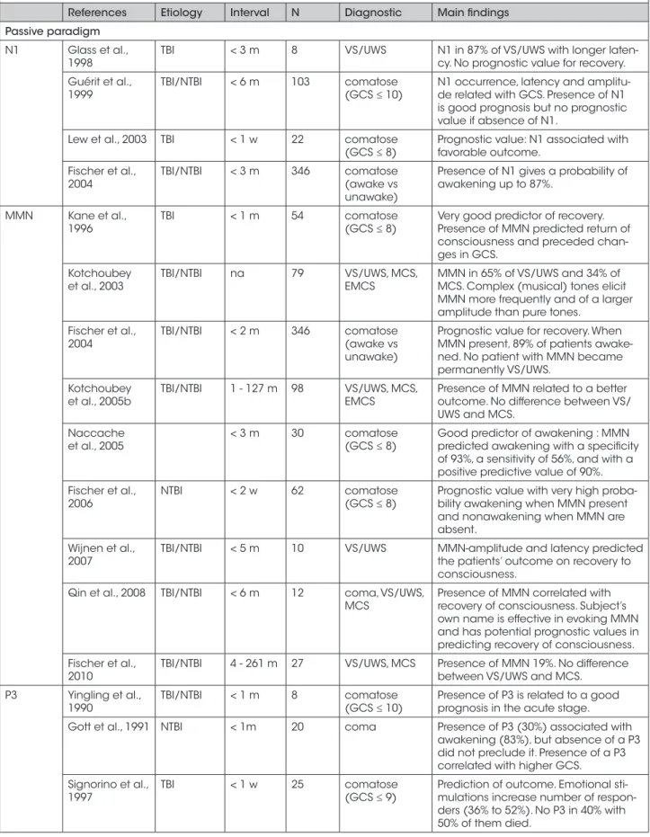
Fig. 1. - Caption: Nine different EEG patterns of five second duration in a “standard zero” bipolar montage with three electrodes on the right hemisphere (Fp2-C4, C4-T4 and T4-02) and three electrodes on the left hemisphere (Fp1-C3, C3-T3 and T3-01).
I. Alpha waves in a control patient, the eeg is symmetric and there is a posterior distribution of the alpha waves (higher amplitude at T4-02, T3-01). II. Theta waves of 4 cycles per second showing a general slowing of the brain electrical activity. III. Delta waves oscillating at one cycle per second. It can be seen in deep sleep or coma. IV. Triphasic waves, a distinctive but non-specific pattern linked to a moderate alteration of consciousness. V. Focal epi-leptiform activity is a sign of underlying brain lesions; here it is well seen in the fronto-central right hemisphere (Fp2-C4, C4-T4). VI. Generalized epileptiform activity. In the figure rhythmic spike waves are well visible on all electrodes. VII. Alpha coma, a pattern of bad prognosis is visible within 72 hours following the injury. It consists in diffuse irregular waves of 8-12 Hz. VIII. Burst-suppression, a pattern associated with a poor prognosis corresponds to brief periods of bursts followed by periods of electrical silence (< 10 uV). It often precedes cerebral death. IX. Inactivity is described as no cerebral electrical activity (< 2 uV). Except in cases of severe intoxication or hypothermia, it is irreversible.
For example, in the case of cardiac arrest coma, reactivity is an important feature for good progno-sis; indeed a recent study showed that in the case of cardiac arrest, 10 out of 11 patients with reactivity evolved positively while only one out of 18 patients that presented a non reactive EEG had a good out-come (Thenayan et al., 2010). General scales such as the Synek scale also gave a significant correlation with the variation of the level of cognitive function between admission and after 3 months in traumatic and non-traumatic patients (Bagnato et al., 2010). In the case of coma due to cardiac arrest, burst suppres-sion patterns are of bad prognosis (Niedermeyer and Da Silva, 2005).
There has been much debate on the prognosis value of alpha or theta coma. It was found that unreactive alpha or theta coma is of bad prognosis but a reactive alpha or theta coma can be associated with a better outcome (Berkhoff et al., 2000). EEG variation car-ries a better prognosis than an unvaried EEG, and the presence of sleep patterns in night recordings of patients are of better prognosis than if absent (Valente et al., 2002). Furthermore, a recent study (Landness et al., 2011) including 5 VS/UWS and 6 MCS show that only MCS patients have sleep patterns, more precisely rapid eye movement sleep and slow waves cycles while the VS/UWS patients did not. Sleep pat-terns could therefore be used as a diagnostic tool.
Quantitative EEG
Standard EEG scores can be different from one reader to another. Furthermore not all the informa-tion contained in the EEG can be unveiled by visual inspection of the EEG traces and only qualitative information can be retrieved.
With the advent of digital EEG technology, the computation of complex parameters has been made possible hence providing objective measures lead-ing to a quantitative analysis of the EEG (qEEG). For instance, one can compute the power spectral density (i.e. the distribution of the power of a signal in the frequency domain) at each electrodes thus giving local power at each frequency. The level of consciousness of a patient can be monitored with measures of signal complexity such as entropy. Levels of connectivity between electrodes can be assessed with measures of synchronization,
coher-ence or mutual information (Nunez et al., 1997; 1999). Furthermore, using an inverse model, one can project the information at the sensor level to a source level and hence localize specific activity in the brain such as epileptic focus.
To quantitatively analyze an EEG trace, a prepro-cessing step is required. The EEG must be band passed to remove slow direct current drifts due to gel drying and to remove high frequencies such as mus-cle artifacts and line noise. Then, segments contain-ing artifacts such as eye blinks or other movements are marked for removal. Artifacts removal can be automated using Independent Component Analysis (ICA) which decomposes the signal into independent components (Comon, 1994). A component contain-ing eye blinks or muscle tone can be removed and the signal rebuilt from the remaining components. The advantage of using ICA is that segments that would have otherwise been discarded can be used for fur-ther analysis, but the drawback is that it is unknown whether if neural information is lost in the process. Once artifact free segments of one or more seconds have been extracted, power or connectivity is com-puted on each segment and mean as well as standard deviation values are obtained. Statistical analysis can then be performed allowing the diagnosis and prognosis of groups of patients. For an overview of qEEG, see Tong and Takhor (2009).
The power spectral density can be computed with the fast Fourier transform or with a wavelet trans-form computed on the extracted segments (Tong and Thakor, 2009). It can be expressed in absolute power (uV2) or in relative power (%) for each individual electrode or on a selected number of electrodes of a region of interest. In patients with DOC, power in higher bands such as alpha and beta decreases while power in lower bands such as delta and theta increas-es in relationship with the severity of the disorder (León-Carrión et al., 2008; Lehembre et al., 2012). Measures of signal complexity such as the bispectral index (BIS, Aspect Medical Systems, Newton, USA) were initially developed for anesthesia monitoring (Johansen and Sebel, 2000; Rosow and Manberg, 2001) but can be used to evaluate consciousness levels in DOC patients. The BIS, a unit less measure ranging from 0 (isoelectric EEG) to 100 (normal activity) derives from a combination of temporal, frequency parameters and bispectral measures. It can be seen as a black box which parameters have been
empirically set and optimized on a large database of EEG recordings during anesthesia (Rampil, 1998). In sleep, BIS values gradually decrease (Noirhomme et al., 2009). In DOC patients, one study found correlations between BIS values and scores of the Glasgow Coma Scale (Gill et al., 2003). However, although statistically significant, these correlations were highly variable and thus difficult to use as a diagnostic tool. Another study showed that at the group level, VS/UWS patients have significantly lower BIS than MCS patients although BIS values do not allow the diagnosis of patients at the indi-vidual level (Schnakers et al., 2008a), still measures of BIS have a prognosis value as patients with a higher BIS recovered better. Other flexible methods based on entropy have been designed in order to synthesize the EEG information into one number. The Datex-Ohmeda S/5 entropy monitoring (GE Healthcare, Helsinki, Finland) for instance imple-ments a time-frequency balanced spectral entropy which gives an information on the complexity of the EEG. A recent study showed a 48% reduction of the entropy in VS/UWS patients compared to controls while in MCS patients it was only reduced by 18% (Gosseries et al., 2011). More importantly, entropy could discriminate VS/UWS and MCS patients with a specificity and sensitivity of 90% in the acute setting, it is therefore a candidate tool for diagno-sis. Furthermore this study corroborates the idea that VS/UWS have a decrease of neural network complexity, as suggested by results presented with yet another entropy measurement, the approximate entropy (ApEn) (Sarà and Pistoia, 2010). ApEn also showed prognostic capabilities in a group of VS/ UWS patients, low values were associated with bad outcome while higher values led to MCS, partial or total recovery. Moreover, testing of ApEn under three conditions, eyes closed, auditory and painful stimuli, yielded similar results, and could quantify the degree of complexity suppression as a function of loss of consciousness (Wu et al., 2010).
The EEG can also quantify connectivity between brain regions (Pereda et al., 2005). Connectivity is disrupt-ed in patients suffering from DOC (Laureys, 2005), it provides complementary information of diagnostic and prognostic importance (Thatcher et al., 1991). Coupling parameters between electrodes provide a connectivity measure between underlying regions. A broad array of methods can be used to assess this
coupling, correlation in the time domain, coherence which is a linear correlation between two signals as a measure of frequency, or non linear methods such as mutual information. Such measures require care-ful interpretation as they are entailed to two inherent problems of the EEG, the reference problem and the volume conduction problem. Indeed, a single refer-ence adds a common component to all the signals of all electrodes which will subsequently appear coupled. To address this problem, bipolar montages are used which provide reference free electrodes with the drawback of reducing the number of available electrodes. Volume conduction also adds artificial coupling between electrodes. Indeed, electrical cur-rents produced by neural assemblies diffuse through the cerebrospinal fluid and the bone and are mea-sured by different electrodes simultaneously, giving rise to correlated electric signals. Hence, correlation and coherence measures are affected by these biases which will typically result in high values of con-nectivity in neighboring electrodes. Still, coherence is well-known, and easy to use, it is therefore the most used method to assess connectivity in clinical settings. A case study on a VS/UWS patient with severe asymmetric brain damage revealed a drop of coherence in the damaged hemisphere (Davey et al., 2000). A decrease in coherence has also been observed in MCS patients (Schiff et al., 2007). Advanced connectivity methods aiming to answer the volume conduction problem and offering more information such as the directionality of the connec-tion have been proposed such as the imaginary part of coherency (Nolte et al., 2004), the phase lag index (Stam et al., 2007) or granger causality (Pereda et al., 2005). A preliminary study using the phase lag index and the imaginary part of coherency showed that MCS patients had stronger connections in theta and alpha band than VS/UWS patients at the group level (Lehembre et al., 2012). Likewise, granger causality showed that patients in severe neurocognitive disor-ders (SND), an upper boundary of MCS significantly displayed more connections than MCS patients. Furthermore, a classifier based on the number of con-nections computed with granger causality achieved a 100% accurate classification into MCS and SND class of the 16 (7 MCS, 9 SND) patients included in the study (Pollonini et al., 2010).
Finally, qEEG can also include approaches that allow the reconstitution of cerebral activity using
source reconstruction methods (Michel et al., 2004). These methods are based on models of ranging complexity, from spherical brain models to realistic models based on magnetic resonance images. All parameters computed at the electrode level can also be computed at the source level, resulting in a better spatial resolution and providing a 3D visualization of the activity of the brain. Source reconstruction techniques require however a minimum of 32 elec-trodes, and 64 or more being recommended (Michel et al., 2004). A prospective study including 50 VS/ UWS patients showed correlations between the level of recovery at three months and the power of occipi-tal sources in the alpha range (Babiloni et al., 2009). Recovery level, either MCS or complete recovery of consciousness could also be differentiated.
Event related potentials
Since the electroencephalography (EEG) cannot quantify small changes induced by sensory, motor or cognitive activities, more subtle functional varia-tions can be investigated by averaging the EEG activity, according to the onset of a repeated stimu-lus. Averaging increases the signal to noise ratio therefore revealing activity that is time-locked to the stimulus while other non-stimulus related activ-ity is averaged out. Event Related Potentials (ERP) reflects therefore the time course of information pro-cessing from low-level peripheral receptive struc-tures to high-order associative cortices. ERPs are frequently used in clinical routine and can be clas-sified in two categories: short latency or exogenous components, and cognitive or endogenous compo-nents (Luck, 2005).
Short-Latency ERPs
Short-latency ERPs or exogenous ERPs are elicited within a time range between 0 and 100 ms after the presentation of a stimulus. They correspond to the activation of the ascending pathways to the primary cortex and are affected by the physical properties of the stimulus. The absence of exogenous components is a marker of poor outcome (Laureys et al., 2005b), most patients who do not present these components bilaterally die or end their life in a vegetative state. The presence of short-latency ERP is however not sufficient to be a good predictor of recovery since
well-preserved potentials are recorded in patient who never recover.
Brainstem auditory evoked potentials (BAEP) are most often used with sounds (“clicks”) using head-phone. They are evoked in the first 10 ms revealing the activity from the auditory nerve to the infe-rior colliculus (Picton et al., 1974). The absence of BAEPs is also a reliable marker of bad outcome when there is no evidence of peripheral auditory injury. However, the presence of normal BAEPs does not indicate a good outcome (Fischer et al., 1988; Garcia-Larrea et al., 1992; Guerit et al., 1993; Fischer et al., 2001). Somatosensory evoked poten-tials (SEP) are elicited by sensory electrical stimu-lation of the median nerve at the wrists. Bilateral absence of SEPs in coma patients is strongly related with unfavourable outcome (i.e. death or VS/UWS) especially in patients with hypoxia–ischemia etiol-ogy (Cant et al., 1986; Lew et al., 2003; Logi et al., 2003; Robinson et al., 2003). A large scale review including 2891 patients can be consulted (Robinson and Micklesen, 2004). All patients with good out-come have developed normal SEPs but they are also present in patients with unfavorable outcome (Cant et al., 1986; De Giorgio, et al., 1993; Carter and Butt, 2001; Amantini et al., 2005). Likewise, mid-dle-latency auditory-evoked potentials (MLAEPs) appearing between 10 and 50 ms and possibly related to the activation of primary auditory cortex and thalamus is strongly associated, when absent, with poor outcome in post-anoxic coma (Fischer et al., 2006). Finally, visual evoked potentials, elicited using flashing LEDs, are less used at the intensive care because these are not systematically present even in healthy subjects (Vanhaudenhuyse et al., 2008).
Cognitive ERPs
Cognitive ERPs or endogenous ERPs are obtained after 100 ms of the presentation of a stimulus and reflect the activity of both cortical and sub-cortical structures, including associative areas (Vanhaudenhuyse et al., 2008). They depend on the psychological significance of the stimulus and are linked to the level of arousal or attention but also to the experimental condition. Using passive paradigms, cognitive ERPs assess residual cognitive functions and with active paradigms, they can be used to detect sign of consciousness in severe brain
injured patients (Table I). Moreover, ERPs generally appear to be good predictors of favorable outcome (Daltrozzo et al., 2007). Five cognitive compo-nents have been measured in DOC patients: the N1 component in response to a stimulus, the mismatch negativity and the P3 in response to novelty, and the N400 and P600 components in response to a seman-tic change.
Passive paradigms
The N1 component is a negative inflection that appears around 100 ms in response to any auditory stimulus (Hillyard et al., 1973), showing an activa-tion of the auditory cortex (Naatanen and Picton, 1987). The presence of a N1 in comatose patients indicates that the primary auditory cortex is func-tionally preserved. Some studies suggest that the presence of the N1 component is a good marker of recovery (Fischer et al., 2001; Lew et al., 2003; Fischer et al., 2004) whereas others suggest that its absence does not appear to be a predictor of bad out-come (Glass et al., 1998; Guerit et al., 1999). The mismatch negativity (MMN) is another nega-tive component that appears around 100-250 ms after any auditory change in a monotonous sequence of sounds (i.e., an oddball paradigm) (Naatanen et al., 1997), involving the primary auditory and prefrontal cortices (Alho, 1995). Its low amplitude implies that many repetitions are needed to observe a response. As subjects do not need to be attentive to the sound to detect a MMN, it indicates an auto-matic response generated by a comparison process between the afferent input and a memory trace developed by the repetitive stimulation. MMN is however not present in every healthy subjects. As most of healthy subjects, a few patients with DOC are likely to process sound deviance, mainly when their state is not due to anoxia. Many stud-ies showed a high prognostic value for recovery, all etiology confounded (Kan et al., 1996; Fischer et al., 2001; 2004; 2006; Kotchoubey, 2005; Naccache et al., 2005). Using the subject’s own name, another study also showed a prognostic val-ues of the MMN in recovery of consciousness from coma and VS/UWS patients (i.e., 4 of the 5 patients showing a MMN response recovered to MCS 3 months later, the other 5 patients without MMN response failed to show any clinical improvement) (Qin et al., 2008). Comparing VS/UWS to MCS
patients, Kotchoubey et al. reported a MMN in 65% and 34% of them respectively, with complex tones eliciting a MMN more frequently than pure tones (Kotchoubey et al., 2005b). In 10 VS/UWS patients, MMN amplitude has been shown to be increased with recovery of consciousness, when patients switched to MCS (Wijnen et al., 2007). However, MMN cannot be used to differentiate between VS/UWS and MCS patients (Kotchoubey et al., 2005b; Fischer et al., 2010).
The P3 is a positive component generated when subjects detect a rare and unexpected stimulus in a regular train of standard stimuli (i.e., oddball paradigm) (Sutton et al., 1965). MMN and P3 are two different brain responses generated by similar stimuli (deviant or novel) but they differ by their latency after a stimulus. For an auditory potential, P3 appears around 300 ms after the presentation of the stimulus and for a visual potential it may appear after 500 or 600 ms. The P3 corresponds to the activation of a fronto-parietal network (Pegado et al., 2010). Note again that as the MMN, not all healthy subjects present a P3. Even if simple sounds can generate a P3 or a MMN, they can also be produced by more complex stimuli with emotional valence that influence the amplitude of the evoked potential (Kotchoubey and Lang, 2001; Mazzini et al., 2001; Lew et al., 2003) leading to a larger number of post comatose patients exhibiting P3 response (Marosi et al., 1993; Signorino et al., 1997). For example, the patient’s own name, which is a salient attention-grabbing stimulus, is more likely to generate a P3 than a simple sound (“click”) (Perrin et al., 2006; Schnakers et al., 2008b). As the MMN, in passive paradigms, P3 component does not allow to distinguish between VS/UWS and MCS patients (Kotchoubey, 2005b; Perrin et al., 2006; Vanhaudenhuyse et al., 2008; Fischer et al., 2010). Indeed, delayed P3 responses have been observed following an own name stimulus in both VS/UWS and MCS patients, suggesting partially preserved semantic processing in non-communicative brain-damaged patients (Perrin et al., 2006). The presence of P3 in patients with impaired consciousness in the acute stage seems also related to a good prognosis (Yingling et al., 1990; Gott et al., 1991; Glass et al., 1998; Guerit et al., 1999; Lew et al., 2003; Fischer et al., 2008). Similarly to the MMN component, the presence of a P3 component has also been found to
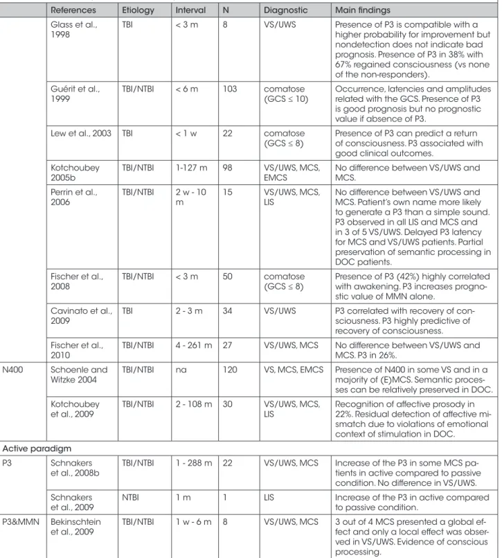
Table I. - Event related potentials studies in patients with disorders of consciousness.
References Etiology Interval N Diagnostic Main findings Passive paradigm
N1 Glass et al.,
1998 TBI < 3 m 8 VS/UWS N1 in 87% of VS/UWS with longer laten-cy. No prognostic value for recovery. Guérit et al.,
1999 TBI/NTBI < 6 m 103 comatose (GCS ≤ 10) N1 occurrence, latency and amplitu-de related with GCS. Presence of N1 is good prognosis but no prognostic value if absence of N1.
Lew et al., 2003 TBI < 1 w 22 comatose
(GCS ≤ 8) Prognostic value: N1 associated with favorable outcome. Fischer et al.,
2004 TBI/NTBI < 3 m 346 comatose (awake vs unawake)
Presence of N1 gives a probability of awakening up to 87%.
MMN Kane et al.,
1996 TBI < 1 m 54 comatose (GCS ≤ 8) Very good predictor of recovery. Presence of MMN predicted return of consciousness and preceded chan-ges in GCS.
Kotchoubey
et al., 2003 TBI/NTBI na 79 VS/UWS, MCS, EMCS MMN in 65% of VS/UWS and 34% of MCS. Complex (musical) tones elicit MMN more frequently and of a larger amplitude than pure tones.
Fischer et al.,
2004 TBI/NTBI < 2 m 346 comatose (awake vs unawake)
Prognostic value for recovery. When MMN present, 89% of patients awake-ned. No patient with MMN became permanently VS/UWS.
Kotchoubey
et al., 2005b TBI/NTBI 1 - 127 m 98 VS/UWS, MCS, EMCS Presence of MMN related to a better outcome. No difference between VS/ UWS and MCS.
Naccache
et al., 2005 < 3 m 30 comatose (GCS ≤ 8) Good predictor of awakening : MMN predicted awakening with a specificity of 93%, a sensitivity of 56%, and with a positive predictive value of 90%. Fischer et al.,
2006 NTBI < 2 w 62 comatose (GCS ≤ 8) Prognostic value with very high proba-bility awakening when MMN present and nonawakening when MMN are absent.
Wijnen et al.,
2007 TBI/NTBI < 5 m 10 VS/UWS MMN-amplitude and latency predicted the patients’ outcome on recovery to consciousness.
Qin et al., 2008 TBI/NTBI < 6 m 12 coma, VS/UWS,
MCS Presence of MMN correlated with recovery of consciousness. Subject’s own name is effective in evoking MMN and has potential prognostic values in predicting recovery of consciousness. Fischer et al.,
2010 TBI/NTBI 4 - 261 m 27 VS/UWS, MCS Presence of MMN 19%. No difference between VS/UWS and MCS. P3 Yingling et al.,
1990 TBI/NTBI < 1 m 8 comatose (GCS ≤ 10) Presence of P3 is related to a good prognosis in the acute stage. Gott et al., 1991 NTBI < 1m 20 coma Presence of P3 (30%) associated with
awakening (83%), but absence of a P3 did not preclude it. Presence of a P3 correlated with higher GCS.
Signorino et al.,
1997 TBI < 1 w 25 comatose (GCS ≤ 9) Prediction of outcome. Emotional sti-mulations increase number of respon-ders (36% to 52%). No P3 in 40% with 50% of them died.
Table I. - Event related potentials studies in patients with disorders of consciousness.
References Etiology Interval N Diagnostic Main findings Glass et al.,
1998 TBI < 3 m 8 VS/UWS Presence of P3 is compatible with a higher probability for improvement but nondetection does not indicate bad prognosis. Presence of P3 in 38% with 67% regained consciousness (vs none of the non-responders).
Guérit et al.,
1999 TBI/NTBI < 6 m 103 comatose (GCS ≤ 10) Occurrence, latencies and amplitudes related with the GCS. Presence of P3 is good prognosis but no prognostic value if absence of P3.
Lew et al., 2003 TBI < 1 w 22 comatose
(GCS ≤ 8) Presence of P3 can predict a return of consciousness. P3 associated with good clinical outcomes.
Kotchoubey
2005b TBI/NTBI 1-127 m 98 VS/UWS, MCS, EMCS No difference between VS/UWS and MCS. Perrin et al.,
2006 TBI/NTBI 2 w - 10 m 15 VS/UWS, MCS, LIS No difference between VS/UWS and MCS. Patient’s own name more likely to generate a P3 than a simple sound. P3 observed in all LIS and MCS and in 3 of 5 VS/UWS. Delayed P3 latency for MCS and VS/UWS patients. Partial preservation of semantic processing in DOC patients.
Fischer et al.,
2008 TBI/NTBI < 3 m 50 comatose (GCS ≤ 8) Presence of P3 (42%) highly correlated with awakening. P3 increases progno-stic value of MMN alone.
Cavinato et al.,
2009 TBI 2 - 3 m 34 VS/UWS P3 correlated with recovery of con-sciousness. P3 highly predictive of recovery of consciousness. Fischer et al.,
2010 TBI/NTBI 4 - 261 m 27 VS/UWS, MCS No difference between VS/UWS and MCS. P3 in 26%. N400 Schoenle and
Witzke 2004 TBI/NTBI na 120 VS, MCS, EMCS Presence of N400 in some VS and in a majority of (E)MCS. Semantic proces-ses can be relatively preserved in DOC. Kotchoubey
et al., 2009 TBI/NTBI 2 - 108 m 30 VS/UWS, MCS, LIS Recognition of affective prosody in 22%. Residual detection of affective mi-smatch due to violations of emotional context of stimulation in DOC.
Active paradigm P3 Schnakers
et al., 2008b TBI/NTBI 1 - 288 m 22 VS/UWS, MCS Increase of the P3 in some MCS pa-tients in active compared to passive condition. No difference in VS/UWS. Schnakers
et al., 2009 NTBI 1 m 1 LIS Increase of the P3 in active compared to passive condition. P3&MMN Bekinschtein
et al., 2009 TBI/NTBI 1 w - 6 m 8 VS/UWS, MCS 3 out of 4 MCS presented a global ef-fect and only a local effect was obser-ved in VS/UWS. Evidence of conscious processing.
MMN = mismatch negativity; N = Number of patients; VS/UWS = vegetative state/unresponsive wakefulness syndrome; MCS = minimally con-scious state; EMCS = emergence of MCS; LIS = locked-in syndrome; TBI = traumatic brain injury; NTBI = non traumatic brain injury; w = weeks; m: months; GCS = Glasgow Coma Scale.
correlate with recovery of consciousness in 34 post-traumatic VS/UWS patients (Cavinato et al., 2009). The last component is the N400 which is a negative potential appearing about 400 ms after the presen-tation of a word. Its amplitude is increased if the stimulus is discordant (phonological or semantic mismatch) with respect to the context (word or sen-tence) (Kutas and Hillyard, 1980). Semantic incon-gruence can also lead to a P600 which is a positive inflection 600 ms after the stimulus. Any positive or negative change can therefore be considered in the treatment of incongruence. Medial and lateral tem-poral cortex, as well as the left frontal and parietal cortex seem to contribute to N400 response (Smith et al., 1986; Hagoort et al., 1996). These inflections were observed in inattentive healthy subjects sug-gesting that like the MMN and the P3, N400 and P600 are also the result of an automatic process (Perrin and Garcia-Larrea, 2003; Vanhaudenhuyse et al., 2008). A N400 response to incongruous words has been reported in some VS/UWS and in a major-ity of MCS patients suggesting that semantic pro-cesses can be relatively preserved in DOC patients (Schoenle and Witzke, 2004). Similarly, recognition of affective prosody has been observed in 6 of 27 VS/UWS and MCS patients as well as in three LIS patients (Kotchoubey et al., 2009), suggesting resid-ual detection of affective mismatch due to violations of emotional context of stimulation in DOC patients. Active paradigms
While passive paradigms help to detect residual brain activity in severely brain injured patients, the use of new active paradigms (i.e. tasks requiring patient’s participation instead of passive listening) can help to determine if the patient is conscious or not (Table I). If the patient is conscious, EEG signal can be used to assess the possibility of a functional communication through brain computer interfaces (BCI) (Kübler, 2009; Millan et al., 2010). The case of a 21-year old comatose woman who failed to show any motor sign of consciousness after 49 days following a basilar artery thrombosis (Schnakers et al., 2009) illustrates the practical utility of dem-onstrating voluntary brain activity in non-clinical means. Only EEG evoked-potential based on com-mand following (i.e., counting her own name in a list of names) permitted to make the diagnosis of
complete locked-in syndrome (characterized by
tetraplegia, anarthria and paralysis of eye motility). Indeed, in the active condition, when asking to count her own name, the P3 response observed was larger than while passively listening. This active own name paradigm using P3 was also employed on a larger number of DOC patients. Schnakers et al. studied 22 severely brain injured patients which were instructed to count the number of times they heard their own name. Some MCS patients, including patients only showing visual tracking without behavioral com-mand following, showed an increase of the P3 responses when counting their own name compared to when listening to the names passively, docu-menting again command following based on brain activity only. VS/UWS patients did not demonstrate such responses (Schnakers et al., 2008b). Similarly, EEG-based evidence of conscious processing (i.e., count a deviant sound sequence in a series of beeps) was demonstrated in 3 out of 4 MCS patients while no voluntary modulation of evoked potentials was observed in 4 VS/UWS patients (Bekinschtein et al., 2009).
Brain-computer interface
Brain-computer interfaces (BCI) are systems allow-ing the brain to communicate with the outside world without going through the peripheral nerves and muscles. They convert directly the brain activity into control signals for electronic devices (Wolpaw et al., 2002; Wolpaw, 2010). BCI based on EEG brain activity can use different components such as the P3, the sensorimotor rhythms (SMRs) or the slow cortical potentials (SCPs).
The most widely component used in BCI is the P3. The advantage of using the P3 is that it allows up to 36 different commands with visual stimuli. A visual BCI has been developed using a matrix composed of letters (Donchin et al., 2000) where the rows and columns are illuminated one by one. Subjects have to focus their attention on the letter they want to spell, generating a P3 response after each illumination of the letter. With this device, users can spell up to 8 letters per minute with an accuracy of 80%. Communication could be established in 5 out of 6 patients with amyotrophic lateral sclerosis (ALS). Four of them continued to use this system to communicate with the external world in a daily routine (Nijboer et al., 2008a). This paradigm
has also been adapted to the auditory modality (e.g., for blind people), although the performances achieved are lower and higher concentration is required (Furdea et al., 2009; Kübler et al., 2009b). Another auditory BCI has been developed for a binary communica-tion (yes/no answer) (Sellers and Donchin, 2006) where users hear a sequence of four stimuli (i.e., yes, no, stop, go) presented in a random order. In order to answer a question, participants have to focus on either “yes” or “no” when hearing the sequence. A stable P3 response corresponding to the answer (yes/ no) has been observed in healthy volunteers but also in ALS patients, with a lower reliability (Sellers and Donchin, 2006). The first BCI study used with DOC patients was also carried out with this last paradigm in order to test its reliability as a diagnostic tool (“is the patient conscious?”). Results showed the benefits of the system to improve detection of consciousness in DOC patients. Two LIS patients were also evaluated, one showed reliable ability to use the BCI but not the other patient, suggesting that the system does not confirm the absence of consciousness in case of nega-tive results (Lulé et al., 2012). Other auditory BCI for communication have also been proposed for ALS and LIS patients (Kübler et al., 2009; Halder et al., 2010). A recent paradigm, the P3-Brain Painting, has been developed to paint pictures using P3 brain activity only enabling ALS and LIS patients to express them-selves creatively (Münßinger et al., 2010).
Changes in sensorimotor rhythms (SMRs) or mu rhythms have also been employed in BCIs. SMRs refer to an EEG activity of 8-15 Hz recorded in the primary sensorimotor cortex (Wolpaw et al., 2002), usually accompanied by a beta activity of 18-26 Hz. This activity can be reduced or desynchronized using preparation, execution, or imagination of movement, particularly in the contralateral motor cortex. An increase in the rate (or synchronization) occurs after the execution of a movement and dur-ing relaxation (Pfurtscheller and Lopes da Silva, 1999). The advantage is that these components do not require the actual execution of the movement but kinesthetic imagination is enough to make them appear (Pfurtscheller et al., 1997). It is however only possible to integrate two commands, more com-mands leading to a decrease in classification accura-cy. In healthy subjects, many BCIs based on SMRs have shown good results for writing words in both visual (Scherer et al., 2004) and auditory (Nijboer
et al., 2008b) modality. A completely paralyzed patient was also able to use a BCI based on SMRs to communicate through a virtual keyboard with a set of letters (Birbaumer et al., 1999; Neuper et al., 2003). To select a letter, the patient had to perform a mental task (i.e. movement imagery) and after several months, he was able to control the keyboard with an accuracy of 70%. The SMRs were also used recently with DOC patients showing the ability of three patients clinically diagnosed as VS/UWS out of 16 to respond to the command (imagine squeez-ing the right hand versus imagine movsqueez-ing all toes) (Cruse et al., 2011).
Finally, slow cortical potentials (SCPs) are the low-est frequencies generated by the cortex that can be used for BCI. They consist in slow potential shifts lasting 300 ms to several seconds. Negative SCPs are usually associated with movement involving cortical activation, while positive SCPs are associ-ated with a reduction in cortical activity (Birbaumer, 1997). Again, this system involves only 2 com-mands. Subjects learn to control their brain activity through operant conditioning and are subsequently able to move an object on a screen (Elbert et al., 1980). People with ALS in advanced stages have been able, after training, to use this device to write words by increasing or decreasing their brain activ-ity to select a target letter (Kübler et al., 1999).
Summary
Electroencephalography encompasses a palette of different techniques helping in the assessment of consciousness in brain injured patients. We reviewed herein results obtained with four main EEG-based approaches in the prognosis and diagnosis of disor-ders of consciousness.
First, standard clinical EEG, recorded in patients at rest is a routine examination in neurological units. In the context of comatose patients it is limited by a low spatial resolution and has little diagnostic value. It has however a prognosis value, in the cases of alpha/theta coma, or burst suppression patterns, it is of unfavorable prognosis. When the EEG is reactive to stimulations, or has varying patterns, it is a sign of better prognosis.
A step further can be taken with qEEG which gives access to a wide spectrum of objective measurements
of cerebral activity providing information on the state of consciousness of a patient. Encouraging results have shown differences between groups of patients at the diagnostic and prognostic level. It is however not possible at this date to identify a parameter provid-ing a correct diagnosis at the individual level. Future challenges will consist in linking the unmet temporal resolution of the EEG to the spatial resolution of the fMRI in order to have a better understanding of the state of consciousness of patients.
A third approach coined event related potentials is based on the averaging of stimulus time-locked events, hence improving the signal to noise ratio of the EEG. There are two categories of ERPs, firstly, short latency ERPs or exogenous components are elicited by external stimulations. When absent, these ERPs are associated with bad outcome, although if present they are not necessarily related to recovery. Second, endog-enous components or cognitive ERPs aim to assess cognitive residual functions (in passive paradigms) and consciousness (in active paradigms). The different components reviewed here, the N1, MMN, P3, N400 and P600 are all markers of good prognosis but can be present in both VS/UWS and MCS patients. They therefore cannot be used to establish a diagnosis in a passive paradigm. This can be achieved with active paradigms, where the patient is asked to focus on one task (for example counting the number of occurrences of own names). Indeed the amplitude of the com-ponents is different in passive and active paradigms hence providing a marker of consciousness.
Once that signs of consciousness have been observed in a patient, EEG based brain computer interface could prove useful in providing a mean of commu-nication for patients in minimally conscious states. Brain computer interfaces are in constant evolution and transfer bit rates as high as 50 bit/min have been reported. Preliminary studies in DOC underlined the challenges of these patients as one LIS patient could use the BCI while another could not. Furthermore, visual BCIs have to be adapted as comatose patients often have impaired eye movements or reduced visual acuity. The same goes for auditory BCIs as some patients have damaged auditory pathways. Providing a direct communication pathway from the brain in comatose patients requires therefore a case by case adaptation and future research should aim to identify for each individual the BCI design that will allow the best performances.
Acknowledgments
This work was supported by the Belgian Fonds National de la Recherche Scientifique (FNRS), European Commission, Mind Science Foundation, James McDonnell Foundation, French Speaking Community Concerted Research Action, Fondation Léon Fredericq, Public Utility Foundation “Université Européenne du Travail” and “Fondazione Europea di Ricerca Biomedica”. This work is supported by the European ICT Programme Projects FP7-247919 DECODER. The text reflects solely the views of its authors. The European Commission is not liable for any use that may be made of the information con-tained therein.
References
Alho K. Cerebral generators of mismatch negativity (MMN) and its magnetic counterpart (MMNm) elicited by sound changes. Ear Hear., 16: 38-51, 1995.
Amantini A., Grippo A., Fossi S., Cesaretti C., Piccioli A., Peris A., Ragazzoni A., Pinto F. Prediction of ‘awakening’ and outcome in pro-longed acute coma from severe traumatic brain injury: evidence for validity of short latency SEPs.
Clin. Neurophysiol., 116: 229-235, 2005.
Babiloni C., Sarà M., Vecchio F., Pistoia F., Sebastiano F., Onorati P., Albertini G., Pasqualetti P., Cibelli G., Buffo P., Rossini P.M. Cortical sources of resting-state alpha rhythms are abnor-mal in persistent vegetative state patients. Clin.
Neurophysiol., 120: 719-729, 2009.
Bagnato S., Boccagni C., Prestandrea C., Sant’Angelo A., Castiglione A., Galardi G. Prognostic value of standard EEG in traumatic and non-traumatic disorders of consciousness following coma. Clin.
Neurophysiol., 121: 274-280, 2010.
Bassetti C., Mathis J., Hess C.W. Multimodal elec-trophysiological studies including motor evoked potentials in patients with locked-in syndrome: report of six patients. Br. Med. J., 57: 1403, 1994. Bekinschtein T.A., Dehaene S., Rohaut B., Tadel F.,
Cohen L., Naccache L. Neural signature of the conscious processing of auditory regularities. Proc.
Natl. Acad. Sci. U S A, 106: 1672-1677, 2009. Berkhoff M., Donati F., Bassetti C. Postanoxic alpha
(theta) coma: a reappraisal of its prognostic signifi-cance. Clin. Neurophysiol., 111: 297-304, 2000.
Birbaumer N. Slow cortical potentials: their ori-gin, meaning, and clinical use. pp. 25-39. In: Van Boxtel G.J.M., Bocker K.B.E. (Eds.). Brain
and behavior past, present, and future. Tilburg, Tilburg University Press, 1997.
Birbaumer N., Ghanayim N., Hinterberger T., Iversen I., Kotchoubey B., Kübler A., Perelmouter J., Taub E., Flor H. A spelling device for the paralysed.
Nature 398: 297-298, 1999.
Birbaumer N. and Cohen L.G. Brain–computer inter-faces: communication and restoration of move-ment in paralysis. J. Physiol., 579: 621-636, 2007. Brenner R.P. The interpretation of the EEG in stupor
and coma. Neurologist, 11: 271-284, 2005.
Cant B.R., Hume A.L., Judson J.A., Shaw N.A. The assessment of severe head injury by short-latency somatosensory and brain-stem audito-ry evoked potentials. Electroencephalogr. Clin.
Neurophysiol., 65: 188-195, 1986.
Carter B.G. and Butt W. Review of the use of somatosensory evoked potentials in the prediction of outcome after severe brain injury. Crit. Care
Med., 29: 178-186, 2001.
Cavinato M., Freo U., Ori C., Zorzi M., Tonin P., Piccione F., Merico A. Post-acute P300 predicts recovery of consciousness from traumatic vegeta-tive state. Brain Inj., 23: 973-980, 2009.
Comon P. Independent component analysis, a new concept. Sign. Proc., 36: 287-314, 1994.
Cruse D., Chennu S., Chatelle C., Bekinschtein T.A., Fernández-Espejo D., Pickard J.D., Laureys S., Owen A.M. Bedside detection of awareness in the vegetative state: a cohort study. Lancet, 378: 2088-2094, 2011.
Daltrozzo J., Wioland N., Mutschler V., Kotchoubey B. Predicting coma and other low responsive patients outcome using event-related brain poten-tials: a meta-analysis. Clin. Neurophysiol., 118(3): 606-614, 2007.
Davey M.P., Victor J.D., Schiff N.D. Power spec-tra and coherence in the EEG of a vegetative patient with severe asymmetric brain damage.
Clin. Neurophysiol., 111: 1949-1954, 2000. De Giorgio C.M., Rabinowicz A.L., Gott P.S.
Predictive value of P300 event-related potentials compared with EEG and somatosensory evoked potentials in non-traumatic coma. Acta Neurol.
Scand., 87: 423-427, 1993.
Donchin E., Spencer K.M., Wijesinghe R. The men-tal prosthesis: assessing the speed of a P300-based brain-computer interface. IEEE Trans. Rehabil.
Eng., 8: 174-179, 2000.
Elbert T., Rockstroh B., Lutzenberger W., Birbaumer N. Biofeedback of slow cortical potentials.
Electroencephalogr. Clin. Neurophysiol., 48: 293-301, 1980.
Fischer C., Ibanez V., Jourdan C., Grau A., Mauguiere F., Artru F. Early and middle latency auditory evoked potentials and somatosensory evoked potentials in the vital and functional prog-nosis of severe brain injuries in intensive care.
Agressologie, 29: 359-363, 1988.
Fischer C., Luaute J., alord F., Jourdan C., Morlet D. Valeur pronostique des PEA au stade aigu du coma. PE auditifs precoces,de latence moyenne, tardifs et negativite de discordance (MMN). In: Guerit J. (Ed.). L’evaluation neurophysiologique
des comas, de la mort encephalique et des etats
vegetatifs. Marseille, Solal: 169-181, 2001. Fischer C., Luauté J., Adeleine P., Morlet D.
Predictive value of sensory and cognitive evoked potentials for awakening from coma. Neurology,
63: 669-673, 2004.
Fischer C., Luauté J., Némoz C., Morlet D., Kirkorian G., Mauguière F. Improved prediction of awaken-ing or nonawakenawaken-ing from severe anoxic coma using tree-based classification analysis. Crit. Care
Med., 34: 1520-1524, 2006.
Fischer C., Dailler F., Morlet D. Novelty P3 elicited by the subject’s own name in comatose patients.
Clin. Neurophysiol., 119: 2224-2230, 2008. Fischer C., Luaute J., Morlet D. Event-related
potentials (MMN and novelty P3) in permanent vegetative or minimally conscious states. Clin.
Neurophysiol., 121: 1032-1042, 2010.
Furdea A., Halder S., Krusienski D.J., Bross D., Nijboer F., Birbaumer N., Kübler A. An auditory oddball (P300) spelling system for brain-com-puter interfaces. Psychophysiology 46: 617-625, 2009.
García-Larrea L., Artru F., Bertrand O., Pernier J., Mauguière F. The combined monitoring of brain stem auditory evoked potentials and intracranial pressure in coma. A study of 57 patients. J. Neurol.
Neurosurg. Psychiatry, 55: 792-798, 1992.
Giacino J.T., Ashwal S., Childs N., Cranford R., Jennett B., Katz D.I., Kelly J.P., Rosenberg J.H., Whyte J., Zafonte R.D., Zasler N.D. The mini-mally conscious state: Definition and diagnostic criteria. Neurology, 58: 349-353, 2002.
Giacino J.T., Kalmar K., Whyte J. The JFK Coma Recovery Scale-Revised: measurement charac-teristics and diagnostic utility. Arch. Phys. Med.
Gill M., Green S.M., Krauss B. Can the bispectral index monitor quantify altered level of conscious-ness in emergency department patients? Acad.
Emerg. Med., 10: 175-179, 2003.
Glass I., Sazbon L., Groswasser Z. Mapping cogni-tive event-related potentials in prolonged postco-ma unawareness state. Clin. Electroencephalogr.,
29: 19-30, 1998.
Gosseries O., Bruno M., Vanhaudenhuyse A., Laureys S., Schnakers C. Consciousness in the Locked-In-Syndrom. In: Laureys S. and Tononi G. (Eds.). The Neurology of Consciousness:
Cognitive Neuroscience and Neuropathology, Oxford, Elsevier: 191-203, 2009.
Gosseries O., Schnakers C., Ledoux D., Vanhaudenhuyse A., Bruno M.A., Demertzi A., Noirhomme Q., Lehembre R., Damas P., Goldman S., Peeters E., Moonen G., Laureys S. Automated EEG entropy measurements in coma, vegeta-tive state/unresponsive wakefulness syndrome and minimally conscious state. Funct. Neurol., 26: 25-30, 2011.
Gott P.S., Rabinowicz A.L., DeGiorgio C.M. P300 auditory event-related potentials in nontraumatic coma: association with Glasgow Coma Score and awakening. Arch. Neurol., 48: 1267-1270, 1991. Guérit J.M., de Tourtchaninoff M., Soveges L.,
Mahieu P. The prognostic value of three-modality evoked potentials (TMEPs) in anoxic and traumat-ic comas. Neurophysiol. Clin., 23): 209-226, 1993. Guérit J.M., Verougstraete D., de Tourtchaninoff
M., Debatisse D., Witdoeckt C. ERPs obtained with the auditory oddball paradigm in coma and altered states of consciousness: clinical relation-ships, prognostic value, and origin of components.
Clin. Neurophysiol., 110: 1260-1269, 1999. Guérit J.M. Electroencephalography: the worst
tradi-tionally recommended tool for brain death confir-mation. Intensive Care Med., 33: 9-10, 2007. Gütling E., Isenmann S., Wichmann W.
Electrophysiology in the locked-in-syndrome.
Neurology, 46: 1092-1101, 1996.
Hagoort P., Brown C.M., Swaab T.Y. Lexical-semantic event-related potential effects in patients with left hemisphere lesions and aphasia, and patients with right hemisphere lesions without aphasia. Brain, 119 (Pt 2): 627-649, 1996.
Halder S., Rea M., Andreoni R., Nijboer F., Hammer E.M., Kleih S.C., Birbaumer N., Kübler A. An auditory oddball brain-computer interface for binary choices. Clin. Neurophysiol., 121: 516-523, 2010.
Hillyard S.A., Hink R.F., Schwent V.L., Picton T.W. Electrical signs of selective attention in the human brain. Science, 182: 177-180, 1973.
Hirsch L.J., Brenner R.P., Drislane F.W., So E., Kaplan P.W., Jordan K.G., Herman S.T., LaRoche S.M., Young B., Bleck T.P., Scheuer M.L., Emerson R.G. The ACNS subcommittee on research terminology for continuous EEG monitoring: proposed standard-ized terminology for rhythmic and periodic EEG patterns encountered in critically ill patients. J. Clin.
Neurophysiol., 22: 128-135 2005.
Husain A.M. Electroencephalographic Assessment of Coma. J. Clin. Neurophysiol., 23: 208-220, 2006. Jacome D. and Morilla-Pastor D. Unreactive
EEG: pattern in locked-in syndrome. Clin.
Electroencephalogr., 21: 31-36, 1990.
Johansen J.W. and Sebel P.S. Development and clinical application of electroencephalographic bispectrum monitoring. Anesthesiology, 93: 1336-1344, 2000.
Kane N.M., Curry S.H., Rowlands C.A., Manara A.R., Lewis T., Moss T., Cummins B.H., Butler S.R. Event-related potentials--neurophysiological tools for predicting emergence and early outcome from trau-matic coma. Intensive Care Med., 22: 39-46, 1996. Kaplan P.W., Genoud D., Ho T.W., Jallon P. Etiology,
neurologic correlations, and prognosis in alpha coma. Clin. Neurophysiol., 110: 205-213, 1999. Kotchoubey B. and Lang S. Event-related potentials
in an auditory semantic oddball task in humans.
Neurosci. Lett 310: 93-96, 2001.
Kotchoubey B. Event-related potential measures of consciousness: two equations with three unknowns.
Prog. Brain Res., 150: 427-444, 2005a.
Kotchoubey B., Lang S., Mezger G., Schmalohr D., Schneck M., Semmler A., Bostanov V., Birbaumer N. Information processing in severe disorders of consciousness: Vegetative state and minimally conscious state. Clin. Neurophysiol., 116: 2441-2453, 2005b.
Kotchoubey B., Kaiser J., Bostanov V., Lutzenberger W., Birbaumer N. Recognition of affective proso-dy in brain-damaged patients and healthy controls: a neurophysiological study using EEG and whole-head MEG. Cogn. Affect. Behav. Neurosci., 9: 153-167, 2009.
Kübler A., Kotchoubey B., Hinterberger T., Ghanayim N., Perelmouter J., Schauer M., Fritsch C., Taub E., Birbaumer N. The thought translation device: a neurophysiological approach to communication in total motor paralysis. Exp. Brain Res., 124: 223-232, 1999.
Kübler A. Brain-Computer Interfaces for Comm-unication in Paralysed Patients and Implications for Disorders of Consciousness. In: Laureys S. and Tononi G. The Neurology of Consciousness:
Cognitive Neuroscience and Neuropathology. Oxford, Elsevier: 217-233, 2009a.
Kübler A., Furdea A., Halder S., Hammer EM., Nijboer F., Kotchoubey B. A brain-computer inter-face controlled auditory event-related potential (p300) spelling system for locked-in patients. Ann.
N Y Acad. Sci., 1157: 90-100, 2009b.
Kulkarni V.P., Lin K., Benbadis S.R. EEG find-ings in the persistent vegetative state. J. Clin.
Neurophysiol., 24: 433, 2007.
Kutas M. and Hillyard S.A. Reading senseless sen-tences: brain potentials reflect semantic incongru-ity. Science, 207: 203-205, 1980.
Landsness E., Bruno M.A., Noirhomme Q., Riedner B., Gosseries O., Schnakers C., Massimini M., Laureys S., Tononi G., Boly M. Electrophysiological cor-relates of behavioural changes in vigilance in veg-etative state and minimally conscious state. Brain,
134: 2222-2232, 2011.
Laureys S. The neural correlate of (un) awareness: lessons from the vegetative state. Trends Cogn.
Sci., 9: 556-559, 2005a.
Laureys S., Perrin F., Schnakers C., Boly M., Majerus S. Residual cognitive function in comatose, vege-tative and minimally conscious states. Curr. Opin.
Neurol., 18: 726-733, 2005b.
Laureys S., Celesia G.G., Cohadon F., Lavrijsen J., León-Carrión J., Sannita W.G., Sazbon L., Schmutzhard E., von Wild K.R., Zeman A., Dolce G.; European Task Force on Disorders of Consciousness. Unresponsive wakefulness syn-drome: a new name for the vegetative state or apal-lic syndrome. BMC Med., 8: 68, 2010.
Lehembre R., Bruno M.A., Vanhaudenhuyse A., Chatelle C., Cologan V., Leclercq Y., Soddu A., Macq B., Laureys S., Noirhomme Q. Resting state EEG study of comatose patients: a connec-tivity and frequency analysis to find differences between Vegetative and Minimally Conscious States. Funct. Neurol., 27: 41-47, 2012.
Leon-Carrion J., Martin-Rodriguez J.F., Damas-Lopez J., Barroso y Martin J.M., Dominguez-Morales M.R. Brain function in the minimally conscious state: A quantitative neurophysiological study. Clin. Neurophysiol., 119: 1506-1514, 2008. Lew H.L., Dikmen S., Slimp J., Temkin N., Lee E.H.,
Newell D., Robinson L.R. Use of somatosenso-ry-evoked potentials and cognitive event-related
potentials in predicting outcomes of patients with severe traumatic brain injury. Am. J. Phys. Med.
Rehabil., 82: 53-61, 2003.
Logi F., Fischer C., Murri L., Mauguière F. The prognostic value of evoked responses from pri-mary somatosensory and auditory cortex in coma-tose patients. Clin. Neurophysiol. 114: 1615-1627, 2003.
Luck S. An introduction to the event-related poten-tials and their neural origins. In: An
introduc-tion to the Event-Related Potential Technique. Cambridge, MA, The MIT Press, 2005.
Lulé D., Noirhomme Q., Kleih S.C., Chatelle C., Halder S., Demertzi A., Bruno M.A., Gosseries O., Vanhaudenhuyse A., Schnakers C., Thonnard M., Soddu A., Kübler A., Laureys S. Probing com-mand following in patients with disorders of con-sciousness using a brain-computer interface. Clin.
Neurophysiol., 2012 [Epub ahead of print]
Markand, O.N. Electroencephalogram in locked-in syndrome. Electroencephalogr. Clin. Neurophysiol., 40: 529-534, 1976.
Marosi M., Prevec T., Masala C., Bramanti P., Giorganni R., Luef G., Berek K., Saltuari L., Bramanti M. Event-related potentials in vegetative state. Lancet 341: 1473, 1993.
Mazzini L., Zaccala M., Gareri F., Giordano A., Angelino E. Long-latency auditory-evoked poten-tials in severe traumatic brain injury. Arch. Phys.
Med. Rehabil., 82: 57-65, 2001.
Michel C.M., Murray M.M., Lantz G., Gonzalez S., Spinelli L., Grave de Peralta R. EEG source imag-ing. Clin. Neurophysio., 115: 2195-2222, 2004. Millán J.D., Rupp R., Müller-Putz G.R., Murray-Smith
R., Giugliemma C., Tangermann M., Vidaurre C., Cincotti F., Kübler A., Leeb R., Neuper C., Müller K.R., Mattia D. Combining Brain-Computer Interfaces and Assistive Technologies: State-of-the-Art and Challenges. Front. Neurosci.,
4: 161, 2010.
Münßinger J.I., Halder S., Kleih S.C., Furdea A., Raco V., Hösle A., Kübler A. Brain Painting: First Evaluation of a New Brain-Computer Interface Application with ALS-Patients and Healthy Volunteers. Front. Neurosci., 4: 182, 2010.
Näätänen R. and Picton T. The N1 wave of the human electric and magnetic response to sound: a review and an analysis of the component structure.
Psychophysiol., 24: 375-425, 1987.
Näätänen R., Lehtokoski A., Lennes M., Cheour M., Huotilainen M., Iivonen A., Vainio M., Alku P., Ilmoniemi R.J., Luuk A., Allik J., Sinkkonen J.,