Gammarus Roeseli
Exposed to Cadmium
Eric Gismondi*, Jean-Nicolas Beisel, Carole Cossu-LeguilleLaboratoire des Interactions Ecotoxicologie Biodiversite´ Ecosyste`mes (LIEBE), CNRS UMR 7146, Universite´ de Lorraine, Metz, France
Abstract
The acanthocephalan parasite Polymorphus minutus is a manipulator of its intermediate host Gammarus roeseli, which favours its transmission to the final host, a water bird. In contaminated environments, G. roeseli have to cope with two stresses, i.e. P. minutus infection and pollutants. As P. minutus survival relies on its host’s survival, we investigated the influence of P. minutus on the antitoxic defence capacities and the energy reserves of G. roeseli females after cadmium exposure. In parallel, malondialdehyde, a toxic effect biomarker, was measured in G. roeseli females and in P. minutus. The results revealed that infected females displayed higher cell damage than uninfected ones, despite an apparent increase in reduced glutathione and metallothionein production. In fact, the increase of these antitoxic systems could be counterbalanced by carotenoid intake by the parasite, so that the overall defence system seemed less efficient in infected females than in uninfected ones. In addition, we demonstrated that cadmium induced cell damage in P. minutus, probably linked with cadmium accumulation in the parasite. Altogether, we observed a paradoxical pattern of responses suggesting that P. minutus increases cadmium toxicity in G. roeseli females although (i) it tends to increase several host antitoxic defence capacities and (ii) it bears part of the pollutant, as reflected by cell damage in the parasite.
Citation: Gismondi E, Beisel J-N, Cossu-Leguille C (2012) Polymorphus Minutus Affects Antitoxic Responses of Gammarus Roeseli Exposed to Cadmium. PLoS ONE 7(7): e41475. doi:10.1371/journal.pone.0041475
Editor: Judith Korb, University of Osnabrueck, Germany
Received January 9, 2012; Accepted June 27, 2012; Published July 20, 2012
Copyright: ß 2012 Gismondi et al. This is an open-access article distributed under the terms of the Creative Commons Attribution License, which permits unrestricted use, distribution, and reproduction in any medium, provided the original author and source are credited.
Funding: This study was supported by the French Ministry of Education and Research (Ministe`re de 458 l’Enseignement Supe´rieur et de la Recherche), which the authors sincerely thank here. The present work is part of the research program EC2CO. The funders had no role in study design, data collection and analysis, decision to publish, or preparation of the manuscript.
Competing Interests: The authors have declared that no competing interests exist. * E-mail: eric.gismondi@hotmail.fr
Introduction
Over the last decade the study of parasitism in ecotoxicology has arisen increasing interest, and numerous studies have already underlined the influence of parasites on infected host physiology in contaminated environments (bivalves [1,2]; arthropods [3–5]; fish [6]). Parasites need energy for their own development; thus they affect the host energy allocation [7], which could weaken host detoxification processes. Parasites can disrupt the antitoxic defences of their hosts [5,8] as well as their immune system [9]. Conversely, some studies have described that parasites can help their host to cope with pollutants, for example by accumulating heavy metals in their own tissues [10,11] or by increasing antioxidant enzyme activities [12]. Most of these studies were conducted in freshwater environments.
Gammarid amphipods are increasingly used in ecotoxicological studies in both freshwater and marine environments, especially due to their important role in the trophic chain [13]. They can be infected by several parasites such as nematodes, trematodes [14,15], microsporidia [5,16–18] or acanthocephalans [19–22].
Acanthocephalans are the most widely studied parasites in gammarids. Their complex life cycle includes an intermediate host (an arthropod) to grow and a final host (a vertebrate) to mature and reproduce. Acanthocephalan parasites at the cystacanth stage are known to alter the phenotype of their intermediate host by a behavioural manipulation, in a way that makes it more prone to predation and thus favours their transmission to the final vertebrate host [23,24]. Although acanthocephalan parasites need
their gammarid hosts in order to develop before being transmitted to the final host, in polluted environments, they may represent an additional burden for gammarids. Indeed, in contaminated ecosystems, gammarids are faced with two different stresses: the presence of the parasite and the toxicity of pollutants. In a previous study, we demonstrated that the acanthocephalan Polymorphus minutus influenced the cadmium resistance of its intermediate host Gammarus roeseli, by increasing or decreasing the cadmium LC50
(lethal concentration that caused the death of 50% of individuals) after 96 hrs exposure in males and females, respectively [11]. In that study, cadmium accumulation in P. minutus as well as a lower cadmium bioaccumulation in infected gammarids than in uninfected ones were observed. However, up to now, only few studies have investigated the effect of an acanthocephalan parasite on the host antitoxic defence capacities, which could explain the difference in resistance between uninfected and infected gammar-ids [8,25,26]. Hence, in a previous study, we described a reduction of the reduced glutathione concentration, a scavenger of metallic and organic xenobiotics, as well as the activity of glutathione synthesis in P. minutus-infected G. roeseli in the absence of environmental stress [25]. It was also highlighted that the cystacanth stage of P. minutus prevents the synthesis of heat shock protein 70 in G. roeseli, subjected to thermal disturbance or palladium exposure [8]. In addition, a weakening of two major parameters of crustacean immunity, i.e. the prophenoloxidase system and haemocyte concentration, have been shown in Gammarus pulex infected by one of the three following
acantho-cephalan parasites: Pomphorhynchus laevis, P. tereticollis and P. minutus [26].
Accordingly, in this study we hypothesized that in a contam-ination context, the parasite could influence host antitoxic defences, to provide host with a better defence capacity and therefore lead to increased host survival. Host survival is believed to favour parasite survival and consequently its transmission. We tested our hypothesis by determining, in controlled laboratory experiments, the effect of a cadmium stress on the antitoxic defence capacities and energy reserves in uninfected and P. minutus-infected G. roeseli. Antitoxic defences were assayed by measuring concentrations of reduced glutathione (GSH), a tripeptide that plays an essential role in the detoxification system by scavenging organic or metallic xenobiotics, responsible of oxidative stress (i.e. production of reactive oxygen species), thanks to its thiol group [27]. In parallel, the activity of c-glutamylcysteine ligase (GCL, EC 6.3.2.2), the limiting enzyme of the de novo glutathione synthesis, was assayed. Concentrations of metallothio-neins (MT), which are involved in binding metal compounds thanks to the thiol groups of cysteine residues and contribute to protecting tissues against oxidative damage [28,29], were mea-sured. Their induction was related to metal exposure in many monitoring studies [30,31]. Carotenoids, which are involved in reproduction [32] and in antioxidant defences [33], were also measured; as well as levels of malondialdehyde (MDA), product of the lipid membrane degradation (i.e. lipoperoxidation) which reflects cell damage, and is thus considered as a biomarker of toxic effect. Moreover, energy reserves were assessed by measuring total lipid and glycogen contents. The levels of glycogen are represen-tative of the energy available for current activities [34] whereas lipids are used during starvation or reproduction periods [35]. Finally, as acanthocephalan parasites contained carotenoı¨ds [36], carotenoı¨d concentrations were measured in P. minutus, in parallel to lipid contents and malondialdehyde levels.
Materials and Methods
Sampling collection, maintenance and cadmium exposure
Due to the lack of infected males, this study was carried out only in G. roeseli females. Uninfected and P. minutus-infected non-ovigerous G. roeseli females were collected in April 2011 with a pond net in the French Nied River (Re´milly, North-eastern France, 49u009N and 6u239E), where cadmium concentrations were less than 0.2mg.L21(LADROME laboratory, Valence, France). Probably due to the fact that P. minutus castrate G. roeseli females, no infected females were found in precopulatory state (personal observation); thus, only females which were not mated to males were collected. Females were sorted out on the spot by observing gnathopods, which are smaller in females than in males. In addition, P. minutus cystacanths were easily identified in living individuals: they appeared as intense orange dots through the cuticle. The animals were transferred to the laboratory in large containers filled with river water, acclimated 5 days at 15uC in an Elendt M4 modified solution [37], and fed ad libitum with alder leaves. The Elendt M4 solution was modified due to the fact that no EDTA was added to avoid cadmium chelation during exposure.
Test solutions were prepared using Elendt M4 modified solution with CdCl2 added to obtain two cadmium exposure
concentra-tions: 2 and 8mg Cd.L21. Controls consisted of Elendt M4-modified solution only. Cadmium concentrations were defined (i) from the LC50(lethal concentration for 50% of individuals) within
96 hrs, which were 107 and 35mg Cd.L21for uninfected and P.
minutus-infected females, respectively [5]; and (ii) from the maximum admissible cadmium concentration in drinking water, which is 5mg.L21(CD 98/83/EC, 1998). For each condition, two replicates of 50 females were exposed, to the different conditions at 15uC for 96 hrs, in a 1.5 L aquarium previously saturated with the corresponding cadmium solutions for 5 days. During exposure, animals were not fed. At the end of the exposure, cadmium concentration were measured and averaged 1.9760.04mg Cd/L and 7.9660.05mg Cd/L.
As G. roeseli females can be infected with microsporidia parasites, and to avoid co-infected females, the presence of microsporidia was investigated by PCR-RFLP [5], which allowed us to keep females infected by P. minutus for biomarker analyses. In P. minutus infected individuals, cystacanths were removed and stored individually at 280uC awaiting marker analyses. Similarly, gammarid bodies were individually frozen in liquid nitrogen and stored at 280uC awaiting biomarker analyses.
G. roeseli biomarker assays
Assaying antitoxic defences is impossible to perform on individual gammarid, so a minimum number of six gammarids was necessary to get enough tissues to analyse all the parameters. Therefore, for each exposure condition, 6 replicates of 6 individual females with the same parasitic status (i.e. absence/presence of P. minutus) were made to measure energy reserves and antitoxic defences as described below. Two different conditions were established: (i) uninfected females and (ii) P. minutus-infected females.
Sample preparation. Each pool was homogenized with a Potter Elvejhem manual tissue grinder in 50 mM phosphate buffer KH2PO4/K2HPO4 (pH 7.6) supplemented with 1 mM
phenyl-methylsulphonylfluoride (PMSF) and 1 mM L-serine-borate mix-ture as proteases inhibitors, and 5 mM phenylglyoxal as a c-glutamyl transpeptidase inhibitor. The homogenization buffer was adjusted to a volume two-fold the wet weight of the sample pool (e.g. 200mL of homogenization buffer for 100 mg of wet weight tissue). The total homogenate was divided into seven parts to measure the different parameters. For each replicate, two independent measures were made for each biomarker.
Energy reserves. The measurement of total lipid and glycogen contents was adapted from Plaistow et al. [7]. A volume of 20mL of 2% sodium sulphate (w/v) and 540mL of chloroform/ methanol 1:2 (v/v) were added to 40mL of the total homogenate. After 1 hr on ice, the samples were centrifuged at 3,0006 g for 5 min at 4uC. The resulting supernatant and the pellet were used to determine the lipid and glycogen contents, respectively.
A volume of 100mL of supernatant was transferred into culture tubes and placed in a dry bath at 95uC to evaporate the solvent. Then, 200mL of 95% sulphuric acid were added in each tube and left for 10 min. The different tubes were removed and cooled on ice, and then 4.8 mL of phosphovanillin reagent were added. After a 10-min reaction, the optical density was measured at 535 nm. Commercial cholesterol was used as a standard and total lipid contents were expressed in mg.mL21.
The pellets were dissolved in 400mL of deionised water, 100mL of sample were placed into culture tubes and 4.9 mL of Anthrone reagent were added. The mixture was placed in a dry bath at 95uC for 17 min and then cooled on ice. Optical density was measured at 625 nm. Glucose was used as a standard and concentrations were expressed inmg.mg21tissue.
The total protein content of each sample was quantified according to Bradford [38] with bovine serum albumin (BSA) as a standard. Results were expressed in mg.mL21.
Antitoxic defence capacities
Reduced glutathione concentration and c-glutamylcysteine ligase activity. Reduced glutathione (GSH) concentrations were assessed by High-Pressure Liquid Chromatography (HPLC) separation adapted from Leroy et al. [39]. The proteins contained in 40mL of the total homogenate were precipitated with 10% perchloric acid (v/v). After a 10-min centrifugation at 20,0006 g and 4uC, the resulting supernatant was diluted 40-fold in 0.1 M HCl. Commercial reduced glutathi-one diluted in 0.1 M HCl was used as a standard and reduced glutathione concentrations were expressed in nmol GSH.mg21 protein.
The c-glutamylcysteine ligase (GCL) activity was assayed using an HPLC method adapted from Parmentier et al. [40]. Measurements were carried out on the S12000 fraction obtained after centrifuging 40mL of the total homogenate for 15 min at 5006g and then centrifuging the resulting supernatant at 12,0006 g and 4uC for 30 min. The resulting S12000 supernatant was diluted 20-fold in homogenization buffer and 40mL of this diluted solution were added to 112mL of incubation cocktail (0.5 M Tris HCl, 200 mM MgCl2 6H2O, 500 mM KCl, 45 mM glutamic
acid, 90 mM cystein, 1 mM DTT, 90 mM ATP, 0.5 mM phenylglyoxal, pH 8.25) to initiate the reaction. The mixture was incubated for 20 min at 25uC in a water bath and the reaction was stopped by a four-fold dilution with 0.1 M HCl. Commercial glutamylcysteine (GC) solution prepared in 0.1 M HCl was used as a standard and GCL activity was expressed in nmol GC.min21.mg21protein.
Metallothionein assay
Metallothionein (MT) concentrations were determined with an HPLC method adapted from Alhama et al. [41]. A volume of 40mL of the total homogenate was centrifuged at 3,5006 g for 10 min. Then, the resulting supernatant was centrifuged at 22,0006 g for 30 min and 4uC to obtain the S22000 fraction. A ten-fold dilution of the S22000 fraction was prepared in 100 mM Tris buffer (pH 9.5) supplemented with 1 mM DTT and 100 mM PMSF as a protease inhibitor. To reduce and denature the protein, 125mL of diluted sample were added to 108mL of incubation cocktail (230 mM Tris pH 9.5, 300 mM DTT, 100 mM EDTA and 10% sodium dodecyl sulfate) and placed in a water bath at 70uC for 20 min. Then, the incubation mixture was supplemented with 17mL of 180 mM monobromobimane (mBBr) and incubated in the dark at room temperature for 15 min, to mark metallothioneins. Commercial rabbit-liver metallothionein I solution prepared in 230 mM Tris, pH 9.5, was used as a standard and metallothionein concentrations were expressed in nmol MT.mg21protein.
Carotenoı¨d concentration
Carotenoid concentrations were measured by a spectropho-tometry method adapted from Rauque and Semenas [42]. A volume of 40mL of the total homogenate was diluted in 450mL of 96% ethanol and kept 6 hrs in the dark at 4uC, before being centrifuged 10 min at 3,5006 g. The optical density of the resulting supernatant was measured at 422, 448 and 476 nm, corresponding to the three major absorbance peaks observed in the absorption spectrum of G. roeseli (data not shown). A commercial carotene mixture (Sigma-Aldrich, France) was used as a standard. Carotenoid concentrations were expressed in ng carotenoids.mg21lipid.
Toxic effect biomarker
Malondialdehyde (MDA) levels were determined using an HPLC method adapted from Behrens and Made`re with UV detection at 267 nm [43]. A volume of 70mL of the total homogenate was diluted four-fold in 95% ethanol (HPLC grade) and cooled on ice for 1.5 hrs to deproteinize it. The mixture was then centrifuged at 18,0006 g for 30 min at 4uC. A volume of 100mL of the resulting supernatant was injected into the HPLC separation system. Malondialdehyde levels were expressed in ng MDA.mg21lipid.
P. minutus biomarker assays
In P. minutus, biomarkers were measured on 4 replicates of 9 P. minutus cystacanths each for each exposure condition. Each replicate was crushed in 350mL of 96% ethanol and kept 1.5 hrs in ice. The mixture was then centrifuged at 3,0006 g for 5 min at 4uC, and the resulting supernatant was divided into three parts to measure lipid contents, carotenoı¨d concentrations and malondialdehyde levels.
Lipid contents
A volume of 60mL of the resulting supernatant was two-fold diluted in chloroform-methanol 1:2 (v/v). Then, 100mL of this diluted solution were used to measure lipid contents as described above.
Carotenoı¨d assays
A volume of 150mL of the resulting supernatant was added to 300mL of 95% ethanol, mixed and kept 6 hrs in the dark at 4uC.
Carotenoı¨ds were then estimated as described above. Malondialdehyde levels
A volume of 100mL of the resulting supernatant was directly injected into the HPLC system, to measure malondialdehyde levels as described above.
Statistical analyses
All data met normality and homogeneity of variance assump-tions (Shapiro and Bartlett tests, p.0.05). Our data were analysed by using a multivariate analysis of variance (MANOVA, Pillai’s trace) with respect to ‘‘infection status’’ and ‘‘cadmium exposure’’ as fixed factors, to test global effect of parasites and cadmium on all biomarkers. Since the MANOVA test was significant, each biomarker was then analysed using ANOVA tests, to test the effect of the infection status, cadmium exposure and their interaction. Then, TukeyHSD post-hoc tests were used to describe significant differences. All tests were performed with a 5% type-I error risk, using R 2.9.0 Software.
Results
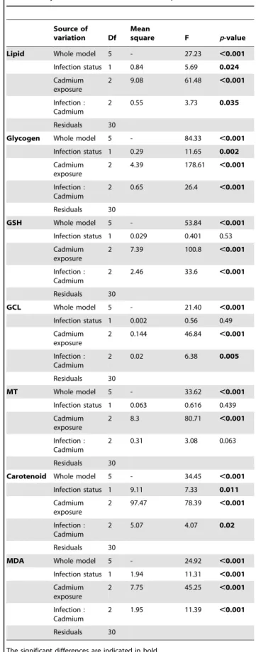
Global MANOVA analysis (Table 1) and ANOVAs tests (Table 2) revealed an effect of infection status, cadmium exposure, and their interactions on biomarker levels. Indeed, infection status had an effect on lipids and glycogen contents as well as on carotenoid and malondialdehyde levels. Cadmium exposure influenced all measured biomarkers. Finally, the interaction between infection status and cadmium exposure had influence all biomarkers, except metallothioneins concentration. As interac-tion between infecinterac-tion status and cadmium were significant for all biomarkers (almost significant for metallothionein), we described below biomarker results according to this interaction.
Effect of P. minutus without cadmium exposure
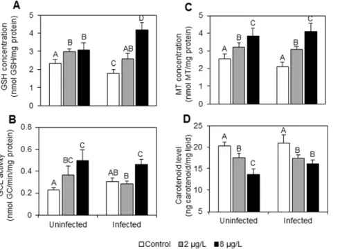
As shown in the figures and the post-hoc tests, infection by the acanthocephalan parasite P. minutus have influenced G. roeseli energy reserves, antitoxic defences and toxicity biomarkers (see white bars in Figure 1, Figure 2 and Figure 3). Indeed, malondialdehyde (MDA) level, lipid content and metallothionein (MT) concentration were unchanged but have a tendency to be lower in infected females than in uninfected ones (Figure 1, Figure 2C, Figure 3A). Reduced glutathione (GSH) concentrations were lower (Figure 2A) whereas c-glutamylcysteine ligase (GCL) activity has a tendency to be slightly higher in infected females as compared to uninfected ones (Figure 2B). Conversely, carotenoı¨d concentrations and lipid contents remained unchanged in the presence of P. minutus (Figure 2D, Figure 3A), while glycogen contents were higher in P. minutus-infected females than in uninfected ones (Figure 3B). Effects of P. minutus on G. roeseli exposed to cadmium
Toxicity biomarker. Malondialdehyde (MDA) levels in-creased in uninfected and in P. minutus-infected females exposed to cadmium as compared to the respective controls (Figure 1). In uninfected females, MDA levels were 1.5-fold higher at 8mg Cd.L21, while only a limited increase was observed at 2mg Cd.L21. In P. minutus-infected females, MDA levels increased according to a dose-response relationship. Indeed, they were 1.8-and 2.6-fold higher in infected females exposed to 2 1.8-and 8mg Cd.L21, respectively, as compared to infected controls. Infection by P. minutus was therefore linked to higher MDA levels in the case of cadmium exposure, especially at 8mg Cd.L21.
Defence capacities
Reduced glutathione (GSH) concentrations increased in both infection statuses (Figure 2A). In uninfected females, GSH concentrations significantly increased with cadmium exposure, but no significant difference was observed between the two cadmium concentrations. However, a dose-response relationship was observed in infected females. GSH concentrations were 1.3 and 2-fold higher when infected gammarids were exposed to 2 and 8mg Cd.L21, respectively, as compared to unexposed ones.
The c-glutamylcysteine ligase (GCL) responses were also influenced by the presence of P. minutus (Figure 2B). Indeed, in uninfected females, GCL activity increased according to a dose-response relationship. The activity was 1.7- and 2.3-fold higher with the lowest and the highest cadmium concentrations, respectively. In P. minutus-infected females, a significant increase was only observed at 8mg Cd.L21, when GCL activity increased 1.6-fold.
Table 1. Multivariate analyses of variance (Pillai’s trace) investigating variations in energy reserves (lipid, glycogen) and defence capacity (GSH, GCL, MT, Carotenoid, MDA) of Gammarus roeseli, as a function of infection by
acanthocephalan parasites and cadmium exposure.
Source of variation num d.f.a
, den d.f.b F p-value Infection status 7, 24 7.14 ,0.001 Cadmium exposure 14, 50 19.38 ,0.001 Infection : Cadmium 14, 50 15.73 ,0.001 a
Numerator degrees of freedom.
b
Denominator degrees of freedom doi:10.1371/journal.pone.0041475.t001
Table 2. Univariate analyses of variance (ANOVA) investigating variations in energy reserves (lipid, and glycogen), defence capacity (GSH, GCL, MT, carotenoid), and toxicity biomarker (MDA), in Gammarus roeseli, according to infection by P. minutus and cadmium exposure.
Source of variation Df
Mean
square F p-value Lipid Whole model 5 - 27.23 ,0.001 Infection status 1 0.84 5.69 0.024 Cadmium exposure 2 9.08 61.48 ,0.001 Infection : Cadmium 2 0.55 3.73 0.035 Residuals 30
Glycogen Whole model 5 - 84.33 ,0.001 Infection status 1 0.29 11.65 0.002 Cadmium exposure 2 4.39 178.61 ,0.001 Infection : Cadmium 2 0.65 26.4 ,0.001 Residuals 30 GSH Whole model 5 - 53.84 ,0.001 Infection status 1 0.029 0.401 0.53 Cadmium exposure 2 7.39 100.8 ,0.001 Infection : Cadmium 2 2.46 33.6 ,0.001 Residuals 30 GCL Whole model 5 - 21.40 ,0.001 Infection status 1 0.002 0.56 0.49 Cadmium exposure 2 0.144 46.84 ,0.001 Infection : Cadmium 2 0.02 6.38 0.005 Residuals 30 MT Whole model 5 - 33.62 ,0.001 Infection status 1 0.063 0.616 0.439 Cadmium exposure 2 8.3 80.71 ,0.001 Infection : Cadmium 2 0.31 3.08 0.063 Residuals 30
Carotenoid Whole model 5 - 34.45 ,0.001 Infection status 1 9.11 7.33 0.011 Cadmium exposure 2 97.47 78.39 ,0.001 Infection : Cadmium 2 5.07 4.07 0.02 Residuals 30
MDA Whole model 5 - 24.92 ,0.001 Infection status 1 1.94 11.31 ,0.001 Cadmium exposure 2 7.75 45.25 ,0.001 Infection : Cadmium 2 1.95 11.39 ,0.001 Residuals 30
The significant differences are indicated in bold. doi:10.1371/journal.pone.0041475.t002
Metallothionein (MT) concentrations in exposed gammarids did not significantly differ between uninfected and infected females (Figure 2C). Whatever the infection status, a dose-response relationship was observed. In uninfected females, MT concentra-tions were 1.2- and 1.6-fold higher with 2 and 8mg Cd.L21, respectively; while in infected females, they were 1.5- and 2-fold higher in the same conditions.
Carotenoı¨d levels decreased in uninfected females according to a dose-response relationship (Figure 2D). In fact, at 2mg Cd.L21, carotenoı¨d concentration was 1.2-fold lower, while at 8mg Cd.L21, it was 1.5-fold lower. In infected females, whatever the
cadmium exposure, carotenoı¨d concentrations were 1.2-fold lower.
Energy reserves
Energy reserves were influenced by the presence of P. minutus (Figure 3). Uninfected females showed a significant decrease in total lipid contents whatever the cadmium exposure (Figure 3A).
Figure 1. Malondialdehyde levels (ng.mg21lipids) in
uninfect-ed andP. minutus-infectedG. roeselifemales exposed at two cadmium concentrations (2 and 8 mg Cd.L21) for 96 hrs. Error
bar represent mean 6 SE. Different letters above the bars indicate significantly different values (Tukey’s HSD test, p-values,0.05). doi:10.1371/journal.pone.0041475.g001
Figure 2. Antitoxic defense responses in uninfected and P. minutus-infected G. roeseli females exposed at two cadmium concentrations (2 and 8 mg Cd.L21) for 96 hrs. A: GSH concentrations (nmol.mg21protein). B: GCL activity (nmol GC.min21.mg21protein). C: MT
concentrations (nmol.mg21protein). D: carotenoı¨d levels (ng.mg21lipids). Error bar represent mean 6 SE. Different letters above the bars indicate significantly different values (Tukey’s HSD test, p-values,0.05).
doi:10.1371/journal.pone.0041475.g002
Figure 3. Energy reserve levels in uninfected andP. minutus -infectedG. roeselifemales exposed at two cadmium concen-trations (2 and 8 mg Cd.L21) for 96 hrs. A: total lipid content
(mg.mL21). B: glycogen content (mg.mg21tissue). Error bar represent
mean 6 SE. Different letters above the bars indicate significantly different values (Tukey’s HSD test, p-values,0.05).
In fact, lipid contents were 1.3- and 1.9-fold lower at 2 and 8mg Cd.L21respectively. In infected females, lipid contents were also lower but no significant difference was observed between the two cadmium concentrations, as total lipid contents were 1.8-fold lower at 2 and 8mg Cd.L21.
The glycogen contents of uninfected and P. minutus-infected females were decreased following the same pattern, whatever the cadmium exposure (Figure 3B). Indeed, at the two cadmium concentrations, glycogen contents were 1.3-fold lower in uninfect-ed females, while they were 1.7-fold lower in infectuninfect-ed ones. Effect of cadmium on P. minutus biomarkers
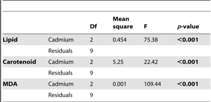
Global MANOVA analysis revealed an effect of cadmium exposure on biomarker levels (p-value,0.001). The ANOVAs tests were presented in table 3.
Lipid contents, carotenoı¨d and MDA levels were also measured directly in P. minutus. The results are presented in figure 4.
Lipid contents in the parasite also decreased according to a dose-response relationship (Figure 4A). In fact, lipid contents were 1.3- and 1.6-fold lower at 2 and 8mg Cd.L21, respectively. In parallel, carotenoı¨d concentrations were measured and the results revealed a significant decrease only at 2mg Cd.L21but not at 8mg Cd.L21(Figure 4B).
MDA levels were 1.9- and 1.4-fold higher in P. minutus exposed at 2 and 8mg Cd.L21, respectively (Figure 4C).
Discussion
This present work was carried out to investigate the influence of acanthocephalan cystacanth on the antitoxic defences and energy reserves of their intermediate host G. roeseli in an environmentally relevant cadmium exposure. Indeed, although macroparasites have already been shown to decrease significantly heavy metal accumulation in their hosts [8,44,45], the influence of these parasites on antitoxic responses had remained quite overlooked so far.
P. minutus effects in the absence of cadmium stress In the absence of cadmium stress (white bars in figures 1 to 3), P. minutus influenced G. roeseli biomarkers following different patterns. Malondialdehyde (MDA) levels were lower in infected-females than uninfected ones. This result is in accordance with one of our previous studies which showed a tendency towards lower MDA levels in P. minutus-infected gammarids [25]. This reflects lower cell damage, probably due to host protection by the parasite whose survival depends on its host’s survival. However, this result is
contradictory to the variations observed in defence capacities, especially reduced glutathione (GSH) and metallothionein (MT) concentrations which were lower in infected females than in uninfected ones, when gammarids were not exposed to stress. Indeed, weaker antitoxic defence capacities could allow us to predict that gammarid sensitivity should increase in contaminated environments. Other investigations have also shown lower MT and GSH concentrations in infected individuals [25,44,45], as well as lower immunity parameters (i.e. prophenoloxidase activity– [26]). Carotenoı¨d concentrations were not impacted by the presence of P. minutus although they are used by P. minutus for its own development [36,46]. This result was not expected as (i) carotenoı¨ds are synthesized neither by the parasite nor by the host, and (ii) the characteristic colour of the cystacanth stage is based on an accumulation of carotenoı¨ds coming from its host.
P. minutus cystacanths influenced the energy reserves of their hosts by increasing glycogen contents and decreasing (or tending to decrease in this study) lipid contents. These results are in accordance with those obtained in one of our previous studies [25] and also with those of Plaistow et al. [7] or Cornet et al. [47] who have observed an increase of glycogen content and a tendency of decrease in lipids content, respectively. Lipid depletion could be explained by the fact that parasites draw out energy for their own development from their host [48]. However, glycogen increases could be linked to gammarid immobility as a consequence of parasite infection that makes the host-parasite system more vulnerable to final host predation [20,49].
Table 3. Univariate analyses of variance (ANOVA) investigating variations in lipid content, carotenoid concentration and MDA levels in P. minutus according to cadmium exposure. Df Mean square F p-value Lipid Cadmium 2 0.454 75.38 ,0.001 Residuals 9 Carotenoid Cadmium 2 5.25 22.42 ,0.001 Residuals 9 MDA Cadmium 2 0.001 109.44 ,0.001 Residuals 9 doi:10.1371/journal.pone.0041475.t003
Figure 4. Lipid, carotenoid and malondialdehyde levels inP. minutusdissected fromG. roeselifemales exposed at 2 and 8 mg Cd.L21for 96 hrs. A: Total lipid contents (mg.mL21). B: carotenoid (ng.mg21lipids). C: MDA (ng.mg21lipids). Error bar represent mean 6
SE. Different letters above the bars indicate significantly different values (Tukey’s HSD test, p-values,0.05).
Influence of P. minutus on G. roeseli antitoxic responses under cadmium stress
Although a possible protective effect of P. minutus on G. roeseli was observed in the absence of stress, the results obtained under cadmium stress highlighted higher cell damage in infected females as compared to uninfected ones. Indeed, whatever the infection status, cadmium exposure induced lipoperoxidation (i.e. increased malondialdehyde levels), but cell damage was higher in the presence of the parasite. This result is in agreement with those obtained when G. roeseli females were infected by microsporidia parasites [5] and could be explained by a disruption of the antitoxic defence responses.
Thus, the antitoxic responses of infected females were different from those of uninfected ones. Metallothionein concentrations, whose synthesis is known to be induced by several metals including cadmium [50], tended to increase faster according to a dose-response relationship in P. minutus-infected females as compared to uninfected ones. We can hypothesize that P. minutus could influence the transcription of the metallothionein gene to provide higher concentrations in its host than in uninfected individuals and cope with cadmium stress by sequestering the metal. However, our result is in contradiction with those of Baudrimont et al. [51] who showed lower metallothionein concentrations in digenean-infected cockles exposed to cadmium as compared to uninfected ones, and of Paul-Pont et al. [52] who highlighted lower metallothionein concentrations in the bivalve Ruditapes philippinarum infected by the trematode Himasthla elongate and/or the bacterium Vibrio tapetis and exposed to cadmium. In these two studies, the results were explained by the fact that other efficient intracellular metal ligands such as glutathione could interfere with cadmium sequestration process [53]. Our results showed that glutathione concentrations also increased according to a dose-response relationship in infected females as compared to uninfected ones. The highest reduced glutathione concentration we measured was obtained with the highest Cd exposure in infected females. This increase observed in infected females could be linked to the increase in GCL activity at the highest cadmium concentration. In uninfected females exposed to the highest cadmium concentration, the relatively low GSH concentration combined with increased GCL activity could result from the fact that glutathione could be directly used as a metal scavenger as well as a substrate for antioxidant enzymes [54]. We can thus hypothesize that P. minutus could influence on the activity of these antioxidant enzymes, which could lead to less glutathione being used and thus a weaker decrease in GSH concentration.
The important increase in GSH and MT concentrations could also be linked to the fact that carotenoid concentration decreased less in infected females as compared to the uninfected ones, according to a dose-response relationship. Indeed, no difference was observed in carotenoı¨d concentrations between the lowest and the highest cadmium exposures in infected females. This could be explained by the fact that P. minutus needs carotenoı¨ds for its own metabolism [46]; therefore, it could reduce carotenoı¨d use of G. roeseli by drawing them from its host, and hence maintain a sufficient stock for itself. Indeed, in gammarids, this carotenoı¨d stock cannot be completed because gammarids cannot synthesize carotenoı¨ds and must get them from food [55]; however, in our study, gammarids were not fed during cadmium exposure.
All antitoxic defence capacities involve an energy cost for the organism; however, in infected females, energy could be reallo-cated by the parasite to its own metabolism [7]. Our results
highlighted a strong decreased in lipid and glycogen contents starting from the lowest cadmium exposure in infected females, whereas in uninfected females, these parameters decrease accord-ing to a dose-response relationship. On the one hand, these decreases could be explained by an energy mobilization by G. roeseli to cope with cadmium stress as already observed in several ecotoxicological investigations [5,54,56,57]. However, on the other hand, the strong decreases observed in infected females could also be linked to the fact that the parasite diverts energy from its host not only for its own development, but also for its own defence system. In a previous study, we observed that P. minutus located within the G. roeseli hemocoel accumulated cadmium [11], which could explain the higher cell damage measured in cadmium-exposed P. minutus than in controls. If the parasite accumulates cadmium, we hypothesize that it can also detoxify it. The carotenoı¨d depletions observed in this work in P. minutus at the lowest cadmium exposure could support this hypothesis as they are antioxidant compounds [33], possibly linked to a detoxification process. As in G. roeseli females, total lipid contents in P. minutus decreased in a dose-response relationship; this observation is consistent with a potential lipid mobilization to provide energy to detoxification processes. This hypothesis is supported by the fact that Sures and Radszuweit [8] described an induction of the Hsp70 defence protein in P. minutus exposed to thermal disturbance.
Conclusion
This study highlights the influence of the P. minutus cystacanth on the antitoxic defence capacities and the energy reserves of its intermediate host G. roeseli females in cadmium stress conditions. The results revealed that P. minutus could increase the use of some antitoxic defences (reduced glutathione and metallothionein), whereas others (carotenoı¨ds) necessary for its own metabolism were maintained. However, the fact that G. roeseli females did not use all these antitoxic defences could explain the toxicity increase reflected by higher MDA levels. To our knowledge, this is the first study that shows a toxic effect of cadmium in P. minutus cystacanths; probably linked to cadmium absorption by the parasite [11]. Since our study was conducted only in females, it could be interesting to compare these results to the influence of P. minutus on biomarkers of G. roeseli males. Indeed, in our previous study [11], we demonstrated that P. minutus-infected males were more resistant to cadmium as compared to uninfected ones. Thus, it would not be surprising to observe differences in the parasite influence according to the host gender, since different antitoxic defences capacities were observed between uninfected males and females [53].
Acknowledgments
The present work is part of the research program EC2CO (ECosphe`re COntinentale et COˆ tie`re). We are grateful to Annie Buchwalter for improving the English text and we wish to thank the three anonymous reviewers for their helpful comments on a previous draft of this paper.
Author Contributions
Conceived and designed the experiments: EG. Performed the experiments: EG. Analyzed the data: EG. Contributed reagents/materials/analysis tools: EG. Wrote the paper: EG JNB CCL.
References
1. Minguez L, Meyer A, Molloy DP, Giambe´rini L (2009) Interactions between parasitism and biological responses in zebra mussels (Dreissena polymorpha): Importance in ecotoxicological studies. Environ Res 109: 843–850. DOI: 10.1016/j.envres.2009.07.012.
2. Minguez L, Boiche´ A, Sroda S, Mastitsky S, Brule´ N, et al. (2011) Cross-effects of nickel contamination and parasitism on zebra mussel physiology. Ecotoxicol-ogy. Available:http://www.springerlink.com.gate1.inist.fr/content/ 13q6447701874j23/. Accessed 2011 Nov 22.
3. Sures B, Scheef G, Klar B, Kloas W, Taraschewski H (2002) Interaction between cadmium exposure and infection with the intestinal parasite Moniliformis moniliformis (Acanthocephala) on the stress hormone levels in rats. Environ Pollut 119: 333–340. DOI: 10.1016/S0269-7491(01)00340-2.
4. Sures B (2004) Environmental parasitology: relevancy of parasites in monitoring environmental pollution. Trends in Parasitology 20: 170–177. DOI:10.1016/ j.pt.2004.01.014.
5. Gismondi E, Rigaud T, Beisel J-N, Cossu-Leguille C (2012) Microsporidia parasites disrupt the responses to cadmium exposure in a gammarid. Environ Pollut 160: 17–23. DOI:10.1016/j.envpol.2011.09.021.
6. Sures B, Thielen F, Baska F, Messerschmidt J, von Bohlen A (2005) The intestinal parasite Pomphorhynchus laevis as a sensitive accumulation indicator for the platinum group metals Pt, Pd, and Rh. Environ Res 98: 83–88. DOI: 10.1016/j.envres.2004.05.010.
7. Plaistow SJ, Troussard J-P, Ce´zilly F (2001) The effect of the acanthocephalan parasite Pomphorhynchus laevis on the lipid and glycogen content of its intermediate host Gammarus pulex. Int J Parasitol 31: 346–351. DOI: 10.1016/S0020-7519(01)00115-1.
8. Sures B, Radszuweit H (2007) Pollution-induced heat shock protein expression in the amphipod Gammarus roeseli is affected by larvae of Polymorphus minutus (Acanthocephala). J Helminthol 81: 191–197. DOI:10.1017/S0022149X 07751465.
9. Rigaud T, Moret Y (2003) Differential phenoloxidase activity between native and invasive Gammarids infected by local acanthocephalans: Differential immunosuppression? Parasitology 127: 571–577. DOI:10.1017/S0031182003 004050.
10. Sures B, Siddall R, Taraschewski H (1999) Parasites as accumulation indicators of heavy metal pollution. Parasitology Today 15: 16–21. DOI: 10.1016/S0169-4758(98)01358-1.
11. Gismondi E, Cossu-Leguille C, Beisel J-N (2012) Acanthocephalan parasites: help or burden in Gammarid amphipods exposed to cadmium? Ecotoxicology 21:1188–1193. DOI 10.1007/s10646-012-0873-8.
12. Marcogliese DJ, Brambilla LG, Gagn F, Gendron AD (2005) Joint effects of parasitism and pollution on oxidative stress biomarkers in yellow perch Perca flavescens. Dis Aquat Org 63: 77–84. DOI:10.3354/dao063077.
13. Forrow DM, Maltby L (2000) Toward a mechanistic understanding of contaminant-induced changes in detritus processing in streams: Direct and indirect effects on detritivore feeding. Environ Toxicol Chem 19: 2100–2106. DOI:10.1002/etc.5620190820.
14. Thomas F, Guldner E, Renaud F (2000) Differential parasite (Trematoda) encapsulation in Gammarus aequicauda (Amphipoda). J Parasitol 86: 650–654. DOI: 10.1645/0022-3395(2000)086[0650:DPTEIG]2.0.CO;2.
15. Thomas F, Renaud F, Derothe JM, Lambert A, Meeu¨s T, et al. (2003) Assortative pairing in Gammarus insensibilis (Amphipoda) infected by a trematode parasite. Oecologia 104: 259–264–264. DOI:10.1007/BF00328591. 16. Haine ER, Brondani E, Hume KD, Perrot-Minnot M-J, Gaillard M, et al.
(2004) Coexistence of three microsporidia parasites in populations of the freshwater amphipod Gammarus roeseli: evidence for vertical transmission and positive effect on reproduction. Int J Parasitol 34: 1137–1146. DOI: 10.1016/ j.ijpara.2004.06.006.
17. Haine ER, Motreuil S, Rigaud T (2007) Infection by a vertically-transmitted microsporidian parasite is associated with a female-biased sex ratio and survival advantage in the amphipod Gammarus roeseli. Parasitology 134: 1363–1367. DOI:10.1017/S0031182007002715.
18. Terry RS, Smith JE, Sharpe RG, Rigaud T, Littlewood DTJ, et al. (2004) Widespread vertical transmission and associated host sex–ratio distortion within the eukaryotic phylum Microspora. P Roy Soc Lond B Bio 271: 1783–1789. DOI:10.1098/rspb.2004.2793.
19. Bollache L, Rigaud T, Ce´zilly F (2002) Effects of two acanthocephalan parasites on the fecundity and pairing status of female Gammarus pulex (Crustacea: Amphipoda). J Invertebr Pathol 79: 102–110. DOI: 10.1016/S0022-2011(02)00027-7.
20. Me´doc V, Bollache L, Beisel J-N (2006) Host manipulation of a freshwater crustacean (Gammarus roeseli) by an acanthocephalan parasite (Polymorphus minutus) in a biological invasion context. Int J Parasitol 36: 1351–1358. DOI: 10.1016/ j.ijpara.2006.07.001.
21. Me´doc V, Beisel J-N (2009) Field evidence for non-host predator avoidance in a manipulated amphipod. Naturwissenschaften 96: 513–523. DOI:10.1007/ s00114-008-0503-8.
22. Me´doc V, Beisel J-N (2011) When trophically-transmitted parasites combine predation enhancement with predation suppression to optimize their transmis-sion. Oikos 120: 1452–1458. DOI:10.1111/j.1600-0706.2011.19585.x.
23. Kennedy CR (2006) Ecology of the Acanthocephala. Cambridge Univ Press. p. 249.
24. Lagrue C, Kaldonski N, Perrot-Minnot MJ, Motreuil S, Bollache L (2007) Modification of hosts’ behavior by a parasite: Field evidence for adaptive manipulation. Ecology 88: 2839–2847.
25. Gismondi E, Cossu-Leguille C, Beisel J-N (2012) Does the acanthocephalan parasite Polymorphus minutus modify the energy reserves and antitoxic defences of its intermediate host Gammarus roeseli? Parasitology: 1–8. DOI:10.1017/ S0031182012000315.
26. Cornet S, Franceschi N, Bauer A, Rigaud T, Moret Y (2009) Immune depression induced by acanthocephalan parasites in their intermediate crustacean host: Consequences for the risk of super-infection and links with host behavioural manipulation. Int J Parasitol 39: 221–229. DOI: 10.1016/ j.ijpara.2008.06.007.
27. Vasseur P, Leguille C (2004) Defense systems of benthic invertebrates in response to environmental stressors. Environ Toxicol 19: 433–436. DOI:10.1002/tox.20024.
28. Roesijadi G (1992) Metallothioneins in metal regulation and toxicity in aquatic animals. Aquat Toxicol 22: 81–113. DOI:16/0166-445X(92)90026-J. 29. Bigot A, Minguez L, Giambe´rini L, Rodius F (2011) Early defense responses in
the freshwater bivalve Corbicula fluminea exposed to copper and cadmium: Transcriptional and histochemical studies. Environ Toxicol 26: 623–632. DOI:10.1002/tox.20599.
30. Amiard J-C, Amiard-Triquet C, Barka S, Pellerin J, Rainbow PS (2006) Metallothioneins in aquatic invertebrates: Their role in metal detoxification and their use as biomarkers. Aquat Toxicol 76: 160–202. DOI: 10.1016/ j.aquatox.2005.08.015.
31. Babin A, Biard C, Moret Y (2010) Dietary supplementation with carotenoids improves immunity without increasing its cost in a crustacean. The American Naturalist 176: 234–241. DOI:10.1086/653670.
32. Gilchrist BM, Lee WL (1972) Carotenoid pigments and their possible role in reproduction in the sand crab, Emerita analoga (Stimpson, 1857). Comp Biochem Phys B: 42: 263–294. DOI: 10.1016/0305-0491(72)90273-8.
33. Palozza P, Krinsky NI (1992) Antioxidant effects of carotenoids in Vivo and in Vitro: An overview. Carotenoids Part A: Chemistry, Separation, Quantitation, and Antioxidation. Academic Press, Vol. Volume 213. pp. 403–420. Available:http://www.sciencedirect.com/science/article/pii/007668799213142 K.
34. Sparkes TC, Keogh DP, Pary RA (1996) Energetic costs of mate guarding behavior in male stream-dwelling isopods. Oecologia 106: 166–171. DOI:10.1007/BF00328595.
35. Cargill AS, Cummins KW, Hanson BJ, Lowry RR (1985) The role of lipids as feeding stimulants for shredding aquatic insects. Freshwater Biol 15: 455–464. DOI:10.1111/j.1365-2427.1985.tb00215.x.
36. Gaillard M, Juillet C, Ce´zilly F, Perrot-Minnot M-J (2004) Carotenoids of two freshwater amphipod species (Gammarus pulex and G. roeseli) and their common acanthocephalan parasite Polymorphus minutus. Comp Biochem Phys B: 139: 129– 136. DOI: 10.1016/j.cbpc.2004.07.001.
37. Elendt B-P, Bias W-R (1990) Trace nutrient deficiency in Daphnia magna cultured in standard medium for toxicity testing. Effects of the optimization of culture conditions on life history parameters of D. magna. Water Res 24: 1157–1167. DOI:10.1016/0043-1354(90)90180-E.
38. Bradford MM (1976) A rapid and sensitive method for the quantitation of microgram quantities of protein utilizing the principle of protein-dye binding. Anal Biochem 72: 248–254. DOI: 10.1016/0003-2697(76)90527-3.
39. Leroy P, Nicolas A, Wellmann M, Michelet F, Oster T, et al. (1993) Evaluation of o-phthalaldehyde as bifunctional fluorogenic post-column reagent for glutathione in LC. Chromatographia 36: 130–134. DOI:10.1007/BF02263849. 40. Parmentier C, Leroy P, Wellman M, Nicolas A (1998) Determination of cellular thiols and glutathione-related enzyme activities: versatility of high-performance liquid chromatography-spectrofluorimetric detection. J Chromatogr B 719: 37– 46. DOI: 10.1016/S0378-4347(98)00414-9.
41. Alhama J, Romero-Ruiz A, Lopez-Barea J (2006) Metallothionein quantification in clams by reversed-phase high-performance liquid chromatography coupled to fluorescence detection after monobromobimane derivatization. J Chromatogr A 1107: 52–58. DOI: 10.1016/j.chroma.2005.11.057.
42. Rauque CA, Semenas L (2009) Effects of two acanthocephalan species on the reproduction of Hyalella patagonica (Amphipoda, Hyalellidae) in an Andean Patagonian Lake (Argentina). J Invertebr Pathol 100: 35–39. DOI: 10.1016/ j.jip.2008.10.001.
43. Behrens W, Made`re R (1991) Malonaldehyde determination in tissues and biological fluids by ion-pairing high-performance liquid chromatography. Lipids 26: 232–236. DOI:10.1007/BF02543977.
44. Siddall R, Sures B (1998) Uptake of lead by Pomphorhynchus laevis cystacanths in Gammarus pulex and immature worms in chub (Leuciscus cephalus). Parasitol Res 84: 573–577. DOI:10.1007/s004360050451.
45. Sures B, Taraschewski H (1995) Cadmium concentrations in two adult acanthocephalans, Pomphorhynchus laevis and Acanthocephalus lucii, as compared with their fish hosts and cadmium and lead levels in larvae of A. lucii as compared with their crustacean host. Parasitol Res 81: 494–497. DOI:10.1007/ BF00931792.
46. Barrett J, Butterworth PE (1968) The carotenoids of Polymorphus minutus (Acanthocephala) and its intermediate host, Gammarus pulex. Comp Biochem Phys 27: 575–581. DOI:10.1016/0010-406X(68)90254-5.
47. Cornet S, Sorci G, Moret Y (2010) Biological invasion and parasitism: invaders do not suffer from physiological alterations of the acanthocephalan Pomphor-hynchus laevis. Parasitology 137: 137–147.
48. Taraschewski H (2000) Host-parasite interactions in Acanthocephala: A morphological approach. Adv Parasit 46: 1–179. DOI:10.1016/S0065-308X(00)46008-2.
49. Ce´zilly F, Perrot-Minnot M-J (2005) Studying adaptive changes in the behaviour of infected hosts: a long and winding road. Behav Process 68: 223–228. DOI: 10.1016/j.beproc.2004.08.013.
50. Stillman MJ (1995) Metallothioneins. Coordination chemistry reviews 144: 461– 511. DOI:10.1016/0010-8545(95)01173-M.
51. Baudrimont M, de Montaudouin X, Palvadeau A (2006) Impact of digenean parasite infection on metallothionein synthesis by the cockle (Cerastoderma edule): A multivariate field monitoring. Mar Pollut Bull 52: 494–502. DOI: 10.1016/ j.marpolbul.2005.09.035.
52. Paul-Pont I, de Montaudouin X, Gonzalez P, Jude F, Raymond N, et al. (2010) Interactive effects of metal contamination and pathogenic organisms on the introduced marine bivalve Ruditapes philippinarum in European populations. Environ Pollut 158: 3401–3410. DOI:10.1016/j.envpol.2010.07.028. 53. Langston WJ (1998) Metal metabolism in aquatic environments. CRC Press. 54. Sroda S, Cossu-Leguille C (2011) Effects of sublethal copper exposure on two
gammarid species: which is the best competitor? Ecotoxicology 20: 264–273. DOI:10.1007/s10646-010-0578-9.
55. Ne`gre-Sadargues G, Castillo R, Segonzac M (2000) Carotenoid pigments and trophic behaviour of deep-sea shrimps (Crustacea, Decapoda, Alvinocarididae) from a hydrothermal area of the Mid-Atlantic Ridge. Comp Biochem Phys A 127: 293–300. DOI: 10.1016/S1095-6433(00)00258-0.
56. Lee WY, Macko SA, Nicol JAC (1981) Changes in nesting behavior and lipid content of a marine amphipod (Amphithoe valida) to the toxicity of a no. 2 fuel oil. Water, Air, & Soil Pollution 15: 185–195. DOI:10.1007/BF00161252. 57. Barata C, Varo I, Navarro JC, Arun S, Porte C (2005) Antioxidant enzyme
activities and lipid peroxidation in the freshwater cladoceran Daphnia magna exposed to redox cycling compounds. Comp Biochem Phys C 140: 175–186. DOI: 10.1016/j.cca.2005.01.013.