Abstract
RIPCR technology is becoming available for diagnosis of pome fruit virus detection in their nutur^t hosts,
thé'amplified
ploducts being usually visualizedafter
electrophoresis inàtfrlOlurn bromide stainèd gels. Although higlrly sensitive, this technology is not amenable to
ioutine testing
of
large numberof
samples, i-e.for
certification purposes. The aimof
our work wasto
develop a colorimetric RT-PCR (or RT-PCR ELISA) test that combines the pCR sensitiviry and ihe convenience of ELISA. The RT-PCR amplified products w9i9 eitherdirectiy labeled
with
digosigenin(DIG)
or,for
better specificity. unlabeled amplification-
oroduôts were hybridizedtù
an internal DlG-labeled detection probe.In
both .systems, aoiotin-labeled prôbe was used for trapping the amplification products on streptavidin-coated microplate strips. Revelation r,vas peifonned',.,'ith-an alkaline phosphatase labeled anti-DIG
conjugate (DIG Detection
Kit.
Boèhringer lvlannheim). The _results shorved hig.h sensitiv'it1. for itrà detection of ASGV in apple tissues. The impor-tance of specific primers allor,ving high cyciing temperatnres. in relation r,'v'ith the origin and pr"rrification status.of the Pl_uqt extracts usedfir
RT-PCR. and the rolesof
capture ànd detettion probes on the specificityof
the colorimetric RT-PC R are discussed.1. Introduction
nT,VELOPMENT
OF
COLORIMETRIC
RT.PCR
TESTSé-nOOVII..IG VIRUS DETECTION
IN
APPLE TREESI
Daniels, V.L.A. Marinho, J. Kummert and P. Lepoivre -iacultéUniversitaire des Sciences Agronomiques Unité de PhYtoPathologie
B-5030, Belgium
Proc. I 7th Int. Symp. on Fruit Tree Vinrs Diseases Ed. A. Hadidi
Àcta Hort. 17:. ISHS 1998
Apple stem grooving capillor.'irr.rs (ASGV) occurs
in
apple trees probably worldwide. utuufl!' r,vithoutiausin{obrious s1'mptoms (Lister.l9l0).
The pnmary control strateg,v for ASGV relies on the usJ of r ims-testeâ planting material and thus on the ability to detect and eliminate infected plants tiom basic stock materiai.The most cunentll' used and recognized technique for detection of ASGV is a bioassal' based on grafting on'uvoodf indicatois. This procedure is erpensir,'e and lengthy, althou-eh
-rlor,r'ell
ei
ctt. (1996) imprôr edit
recentll'bj
obtaining reliable response2
months aftergrafting on ne*' Llctîus indicators. Serologicai diagnosis (ELISA) is. m.uch more rapid, but
ipp.uti
reliableonly
duringa
short periodin
the ).eq_bgciyse it. lacks sensitiviry fordèiecting lor,v virus concentiations in ivoodi. material. RT-PCR, r,r'hich seems potentia.iiy more accrrrate and sensiti','e than serological detection. has been recently der,'eloped for this
virus (Kinard, et ct\..1996: Kummert et a/.. 1,997: N1cKenzie et
a\..1997
Magome et al..r9e7)
TÉe standard RT-PCR protocols used puriiied
total
R\A
or
dsRNA pre.parations. astemplate, which require complex and ratheriime consuming extractions, not easily amenable for
iro.étsing
of large numbers of samples. Recg_nt improvements insample processing f-orPCR.
includiig
immînocapture (Wetzei et ctl. 1992), direct binding (Rohrvani et a\.,1995)' print'capture (ôlmos et a\..1996) and direct application of clarifiedplalt
extracts in reactionirUàr (Wyatt ànd Bronun, 1996) have been reôêntl1' rep.orted.for sensitive detection of plant
ot*t.à.
dpecific primers develôped in our laboratory âllow the use of crude sap from appletrees for ÂSCV dètection in RTPCR tests (Kummert
d
ni., this s.v-mposium).Another drawback hindering the deveiopment
of
RT-PCRfor
ror,rtine testingof
virusintèition
in
plants
results fr"omthe
us.
of
ethidium bromide stained a.garoseq.l
àr..i.opÀoresii fbr rhe detecrion of amplification products. Although useftrl for checkilg
tit.
size
oi
pCR
prodticts,this
techniquèis
not
uèry' sensitive and alsonot
amenable forFOR
APPLE
STEMautomation; labeling PCR products either directly or by using hybridization
with
specific labeled probes is substantially more sensitive (Fenby et al., 1995).Novél
techniques combiningPCR
accuracyand
potential sensitivity,with
ELISAconvenience, havè been described in medicine (Chang et al., 1994; Lassner, 1995) and later in plant pathology (Rowhani et a\.,1996).
This paper dessdbes the feasibility
of
colorimetric RT-PCR tests (RT-PCR ELISA) for ASGV diagnosis in field collected apple tree samples.2.Maret''als and methods
2. 1. Virus-infected material
Reference isolates
for
ASGV
were maintainedin
the
greenhouseon
Chenopodiumquinoa
andl{icotiana
glurinosct.Four
isolates (10311, 10391,10771and G77)
were originally obtained from the Research Station on Fruit Treesof
Gorsem, Belgium. Isolate PV199 has been obtained from DSM, Brarurschweig, Germany.For the control of the specificity of the colorimetric detection of KI-PCR amplification
products, apple ctrlorotic leaf spot (ACLSV) and apple stem pitting virus (ASPV) isolates, infecting apples trees or herbaceous hosts have been used.
2.2.Preparations used as template for colorimetric RI-PCR
Total RNApreparations were obtained from 0.2 g of plant tissue ground in liquid nitrogen
and then homogenized
with
Ultra-Tunaxin
the presenceof 2 ml of Tripurel
isolationreagent (Boehringer). and the extraction was conducted according
to
the manufacturer'sinstructions. The
final
pellets were resuspendedin
100pl of
DEPC-treated water: the amount and purity'of
nucleic acids obtained was estimated byUV
absorbance at 260 and 280 nm, and the samples stored at -20oC until use. One to 2 pg of total RNAwere used for RT.PCR.ds-RNA preparations from infected plant material were obtained by using the method
of
Valverde et al. (1990) applied to approximately 20 g of plant tissue, or adapted for use rvith 0.5 to 1 g of plant tissue. The final pellets were resuspended in 40 pl (20 g extracts) orl0
pl(1 g extracts or less) of DEPC-treated water and 5 pl used for KI-PCR.
Preparation of crude plant extracts was adapted from the method described by Wyatt and
Brown (1996).
Fifo'mg
of plant tissues (leaves or bark scrapings from current year apple tree shoots) were groundin
1.5 ml microtubes containing 0.5 ml of TE buffer (50 mN'{ Tris, pH 8.0, 10 mM EDTA). After grinding, 0.5 ml of the same buffer was added and the content homogenizedwith a
vortex.After
centrifugationfor
10min
at
10,000g,
onepl
of
tlsolution, or dilutions of
it
in TE buffer, was directly added to RT-PCR mix. Alternativell.5[
pl
of the same extracts were incubated for 30 min in melting icein
PCR tubes, and afterrinsing 3 times with TE buffer (200 pl), the RT-PCR mix was added. 2.3. Primers and probes
Two primer pairs designed to cope with the variability of partial sequences determined for 4 ASGV isolates maintained in C. quinoa or 1,,1. glutinosa in the greenhouse (10311, 10392
and 1 0771) and one from an apple tree in the field (10392), and the sequence published by
Yoshikaw'a et al. (1992) were used:
AS GV 1 F
:
5' -GARGC WAAAGCTGGYC A{-3 I (nt 3925 -39 42)ASGV 1 R
:
5'-YACCTCTTCYACV/GCAGT:3' (nt 4485 -4468)ASGV4F = 5'-GTTCACTGAGGCA{MGCTGGTC-3' (nt 391 8-3940)
P;imer pairs ASGVIF-ASGVIR and ASGV4F-ASGV4R ampliff respectively fragments
rf
560 and 57 4 bp located at the 3' end of the putative RNA polymerase gene, just before thevariable region described by Magome et al. (1997).
The
5' biotin-
and3'
digoxigenin-labeled capture and detection probes correspond to intemal fragment partsof
the amplified products, andwill
be partof
a
detectionkit
in develoPment.2. 4. RT-PCR amplifi cation
Reverse transcription was made either by using Expan6rn reverse transcriptase system (60 min at 42oC, Boehringer) with oligo-dT priming, or
AMV
M-enzyme (30 min at 50"C) containedin
the Titanru one step RT-PCR system (Boehringer)with
the reverse specific primer, according to manufacturer's instructions.^
Amplif,rcationfrom cDNA
was
performedin
50
pl
in a
Triothermoblock cycler(Biometra) as follows: template denaturation for 5 min at94"C;30 cycles at94oC for 30 sec
(denaturation), 52oC (primers
ASGVIF-IR)
or 62'C
(primers ASGV4F-4R)for
I
min (arurealing), and72C
for 2 min (elongation); and final elongation at72C
for 15 min.For digoxigenin-labeled amplicons, the dNTP
mix
containeda
19:1 dTTP: DIG-dUTPratio (PCR DIG labeling mix. Boehringer)
2.5.Detection of PCR products
2.5 .1 . Electrophoretic analysis
PCR amplification products (10 pl) were analyzedby electrophoresis
(i
h in TAE buffer at 120V)in
lo agarose gel. stained with 0.01% ethidium bromide and photographed underUV light (306 nm) with Polaroid films.
2.5 .2. Colorimetric detection PCR amplification products
""'ere also analyzed by using the PCR ELISA DIG detection
kit
(Boehringer) with minor modifications. Tenpl
of PCR products, and adequate controls, weremixed
with 40
pl of
denaturation solution and incubatedfor
10
min
at
room temperature.If
PCR amplification products are labeledwith
digoxigenin. 450pl
of
hybridizationsolution containing 100 ng of biotin-labeled capfure probe w-ere added, and the mix was then put in duplicate (200 pl/rvell) in microtiter plate strips coated rvith streptavidin. before the incubation for 3 h at 52oC.
-
For unlabeled PCR amplification products: 450pl
of
h1'bridization solution containing'00
ny'ml of the digoxigenin-labeled probe were added, and tubes incubated for 3 hours at 52oC, under shaking. While hybridizationof
the amplification productswith
the
DIG-labeled probe r,vas running. the biotin-labeled probe at 100 ng/ml in PBS/Tween was added
(200 pl/rvell) in microtiter plate strips precoated with streptavidin and incubated
for
I
h at37'C. The microtiter plate strips were then rvashed 4 X with 250 1i of PBS-Trveen and 200
pl of
the hybridization product obtained between PCR amplicons and DlG-labeled probe added, in duplicate, before further incubation for 1.5 h at 37oC, under shaking.In both cases, microtiter plate strips were finally washed again with PBS-Tween, and 200 pl of conjugate buffer (PBS-Tween plus 0.5% blocking reagent) containing a 1/3000 dilution of anti-digoxigenin alkaline phosphatase conjugated antibodl.' (Fab fragment) were added to each rvell before incubation at 37oC
for
30 min, under shaking. After washing, 200pl of
substrate buffer (10 mM diethanolalanine-HCl, pH 9.5) containing 0.7 mg/ml p-nitrophenyl-phosphatewere
added. Absorbanceat
405 nm
were
measuredin
a
Multiscan pluscolorimeter, after
I
h
at room temperature, and after 16h
at 4"C.All
reagents and the precoated microtiter plates strips were purchased from Boehringer.3. Results and discussion
3.1. Colorimetric RT-PCR with Dlc-labeled amplification products
The assay is based on amplification
of
specific sequences by PCR and detection of the ampliconsby
ELISA,
afler captureby
a
specific biotin-labeled probe ona
streptavidin-coaled microplate.Preliminary experiments had shown that DlG-labeled amplicons obtained
with
5'
endbiotin-labeled
primer
ASGVIF
andprimer
ASGVIR, from
transcriptsof
total
RNApreparations (herbaceous hosts) or dsRNA preparations (apple trees) from ASGV-infected plants,
can
be
easily detectedon
streptavidin-coated microplate stripsby
using PO-conjugated anti-digoxigenin antibody. However, in this case, unspecific amplicons obtained from transcripts of total RNA preparations from AClSV-infected C. quinoa (but not from ASPV-infected,^[ occidentalrs, or from healthy corresponding herbaceous plants) also gavea signal, indicating the need
of
more specific detection procedures, based on the useof
aspecific biotin-labeled capture probe. When the RT-PCR experiments were conducted with unlabeled
primers ASGVIF-ASGV1R,
the only
amplicons observedon
the
gels correspondedto
ASGV infected material (herbaceous hostsor
apple trees). Colorimetricdetection of these RT-PCR products, after hybridization with a biotin-labeled probe captured on streptavidin coated plate strips was also specific for ASGV-infected material.
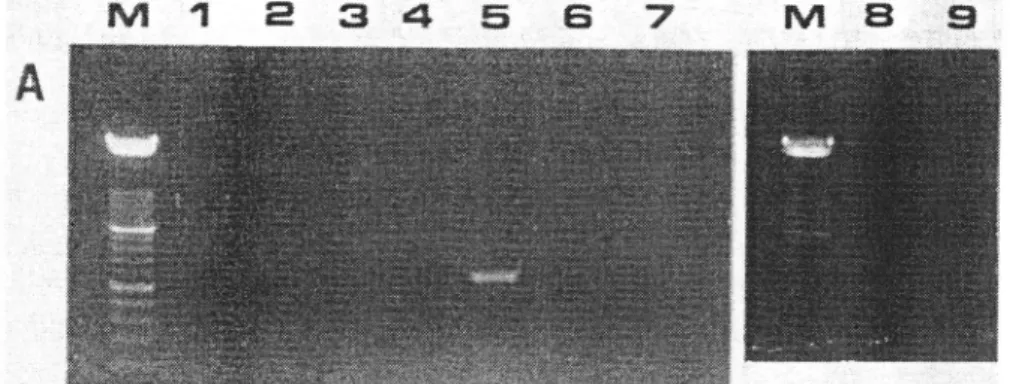
In the example showed in Figurel, the only fluorescence signal visible after agarose. electrophoresis
(Fig.
1a) correspondedto
amplification products generatedby
total RNA preparationof
ASGV
infectedC.
quinoa.By
colorimetric detectionafter
caplureof
amplification products by a biotin-labeled oligonucleotide, homologous to primer ASGV 1F,
positive signals are also observed from transcripts
of
ds-RNA preparationsfrom
ASGV-infected C. quinoa and apple tree, but not from comparable extracts from ACLSV- or ASPV-infected material (Fig. 1b), indicating the higher sensitivity of this detection method.
3.2. Colorimetric RT-PCR with capture and detection probes
The above method, although accurate and sensitil'e, presented limitations based on the need
of
complex template preparations (purified nucleic acids) and the useof
costly PCR nucleotide labeling mix; all the amplicons being labeled,it
also presented a potential riskof
detection
of
false positive results. To improve the efficiency and reduce the costs. longerprimers
(ASGV4F-ASGV4R:23
nt)
and biotin- and
digoxigenin-labeled probes (respectively30
and29 nt)
were selectedto
cope the variabilityof
partial sequences established from amplification products of the genomic RNA corresponding to a fragmentof
the 3'end of the gene coding for the viral replicase obtained for local isolates (Kummerta/., this symposium) and the sequence published for ASGV by Yoshikawa et al. (1992). For these probes particularly, attention has been given to avoid possible formation of secondary structures.
Different protocols have been used to perform the colorimetric detection of these RT-PCR amplification products obtained
rvith
specific primers ASGV4F-ASGV4R.The
more sensitive andthe only
protocol allowing detectionfrom
plant materialwith
low. virus concentrations included 5 successive steps as follows: (i) denaturation of RT-PCR amplified productsin
microtubes,(ii)
hybridizationof
the DlG-labeled probeto
its
complementary single-stranded sequence,(iii)
immobilizationof
the biotin-labeled probeto
streptavidin coated microtiter plate strips, (iv) hybridization of the preformed hybrid RI:PCR amplicor/ DlG-labeled probe with the immobilized biotin-labeled probe, and (v) colorimetric detectionwith anti-digoxigenin antibody conjugate and enzymatic substrate.
Although the detection probe only contains one molecule
of
digoxigenin,OD
signals aboveI
to 2 absorbance units were obtained.No
false positive results were observed forhealthy samples or from plants infected by ACLSV or ASPV.
As demonstrated elsewhere (Kummert et al., this s,v-mposium), the primers ASGV4F-4R allowed the detection of specific viral sequences, by a-qarose gel electrophoretic analysis
of
amplification products obtained
with
the TitanrM one tube KI-PCR sy-stem, directly fro.m d^idt;Jpi*i'tup
of
either ASGV-infected herbaceous hostsor
apple trees. When theiioau.tr
of suchiests were submitted to the 5 steps colorimetric detection protocols., positiveii;J;6.
U.."
obtained from ASGV-infected materials, sometimes even in the absenceof
uiiiUt. bands on electrophoretic analysis of these same PCR products.4. Conclusion
Although useful for detection and typing of human papillomavirus (Adams et a\.,1996), ttre simpteit protocol for KI-PCR ELISÀ, Ising DIG lâbéling of the amplification p^rod}1ts,
*â
tn.i
forward primer labeledwith
biotin,iould
not bJused
for
detectionof
ASGV iniection,due
to'its
lack
of
specificity$ositive
signals correspondingto
unspecific amplicons obtained for AC L SV-infected materiqJ).*^-Év
6*9
unlabeled primers for PCR amplifiôationwith
digoxigenin-labeled nucleotidemix-,'and u"biotin-laU.t.O probe
for
caphuêof
amplicons, theteit
became accurate and sensitive.io
reach the optimal specificity, needed to avoid false positive resu-lts in testing certifiedmultiplication
-âtériui,
â;
improved colorimetric deteciion method,usingj*o
,tp,..1fj'i;ù;là
probestr.sp..tiuely
witir biotin and digoxigenin) has beendwglope{ Coupled with^p.iÀ.it'eSCvqf
'liluted*a
ASCV4R,it
allows tËe détectiônof
ASGV infèctionfrom
crudesap from either leaf or bark tissue from apple trees.
Acknowledgments
This work was supported by the Ministry of Agriculture,
qG
VI.
Belgium. J. Daniels.and V.L.A. Marinho*è'..rp..tii'el..'-
post-docioral arid doctoral fellorvs of ETVBRAPA (Brazilian Enterprise for Agricultural Research).References
Adams V.,
Moll
C., Schmid lv{.. Rodriges C., Moos R., and Briner J.., 1996. Detection andty'ping'of human papillomav'irus in- biopsy and cytologic.al.specimens
bv
polymerasechain reaction and'restriction enz,'-me analy'sis,
-eihodiuitablè
for
semi automation. J' Med. Virol. 48: 161-170.Chang S., Puryear J., and Caimey J., 1993. A simple and efficient method for isolating RNA from pine trees. Plant Mol. Biol. Reporter 11: 113-116.
_Fenby N.S., Scott N.W., Slater
A.,
anà Ellion Ivf.C., 1995. PCR and non-isotopic labeling-
teéhniques for plant virus derection. Cell. Mol.
Biol
4I
639-652.Howell W.E.,
Mùk
G.1.. Hurn S.S., Foster J.A., and PostmanJ.D..
1996. Select lulaltts clones for rapid detection of apple stem grooving virus. Plant Dis. 80: 1200-1202.Kinard G.R., Siott S.W., and Barnett, O.\À'." 1996. betection of apple chlorotic leaf spot and apple stem grooving viruses using RT-PCR. Plant Dis. 80: 616-624.
KuniÀert J., R"ufflard "G., and de Al"meida Marinho V., I9g7 . Use of degenerate.primers, for RT-PCR detection of apple and pear tree viruses. In: Advances in the Detection of Plant
euthog.nt by Polymerir'. Cfruitr Reaction Commission of the European Communities' C'
Manceau and J. Spuk, eds. Luxembour-s. Pp. 34-48.
Lassne,
---rn'
D., 1995.Ôirltit.tio"ôf
-nXÀiyin.
eLlSAtechnique using extemal s.tandl{p.quaniitation-
;a
mRNA
by
PolymeraseChain
Reaciion: Nôn-radioactive PCRMethàds. KohÈr et al., eds.
Spriig..-i'.tlug,
Berlin, Heidelberg, l'{e.w'-Yor\.-lpll7
-.123 'Lister
R.M.,
Igi0.
Applâ stem^groËvingviiis.
CMVA/B
Deùriptionsof
Plant Viruses n"31. 4 p.fvfagome
H.,
YoshikawaN.,
TakahashiT.,
Ito-T.,
and MiyakawaT.,
1927'
Molecular-
Ï*iuUility
of
the genomeiof
capilloviruses from apple, Japanese pear. European pear,and citrus trees. Phytopathol. 87: 389-396.
McKenzie D.J., Mclean M.A., Mukerji S., and Green
M.,
1997.Improved RNA extraction-
from
woody
plantsfor
the
deteètionof
viral
pathogensby
reverse transcription-polymerase chàin reaction. Piant Dis. 88: 222-226.OËoJA.,
Dasi M.A., Candresse T., and CambraM.,
1996. Print capture PCR:a simple-and highly sensitive methodfor
detectionof
plum pox virus (PPV)in
plant tissues. Nucl. Acids Res. 24: 2192-2193.Rohwani A., Maningas M.A., Lile L.S., Daubert S.D., and Golino D.A., 1995.Development of a detection syslem for viruses of woody plants based on PCR analysis of immobiiized virions. Phytopathol. 85 : 347 -352.
Rowhani
e.,
ÉiaiAi L., Golino D.A., and Daubert S.D., 1996. Detection of viruses of w'oody host plants using colorimetric PCR. Phytopathql. 86;Sl
(Abstract 4A).Valverdè R.A., Naîneth S.T., and Jordan
R.L.
1990. Analysisof
double-stranded RNA forplant virus diagnosis. Plant Dis. 74: 255-258.
Wetzel T., Candrésse T., Macquaire G., Ravelonandro
M.,
and DunezJ.,
1992.A
highlysensitive immunocapture polymerase chain reaction method for plum pox virus detection. J. Virol. Meth. 39:27-37.
Wyatt S.D., and Brown J.K., 1996. Detection
of
subgroupIII
Geminivirus isolatesin
leafextracts
by
degenerate primers and polymerase chain reaction. Phytopathol.86:
1288-1293.Yoshikawa
N.,
Sasaki E., KatoM.,
and TakahashiT.,
1992. The nucleotide sequence of apple stem grooving capillovirus genome. Virology 191: 98-105.Ë $ # Ë
il
il
# .* Ë * T{
{ $ t d $ r ë rB
3,5 ? 05 0 o) (J et 6 E o 15 U'€
Absorbance reading after 1 5 hourincubation Absorbance reâding after overnight incubation,ffiÆ
@È@ 6-oN OO S a m p le sFigure
I
- Analysis of digoxignin-labeled tlvo-steps RT-PCR products obtained with primers ASGVIF-ASGVIR from: ds-RNApreparations of ASGV-infected C. quinoa isolate 10311;(lane 1) or from apple trees 10392 (lane 2), LP680 (lane 3) or LP679 (lane
4)
containing respectively ASGV ASPV or ACLSV; total RNA preparations of Ascv-infected C. quinoa isolateG
77; (lane 5). ASP\'-infectedÀ.
occidentalrs (isolate ASPV-J; (lane 6),ACLSV-infected C. quinoa (isolate 91300, Lane 7). or healthy C. quinoa (lane 8). Lane 9 = negative control (no cDNA added). A. Analysis b1' electrophoresis in ethidium bromide agarose gel; Lane
M
:
molecular r,veight marker (100 bp, Boehringer; lorver heary band:
500 bp). B. Absorbance values after captureof
the digoxigenin-labeled KI-PCR productsby
biotin-labeled oligonucleotide homologous to primer ASGVIF. and revelation wit the PCR-ELISAdetection