Some features of anisothermal solid-state transformations in alloy 718
11
0
0
Texte intégral
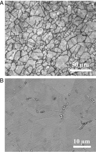
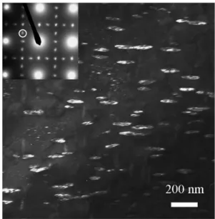
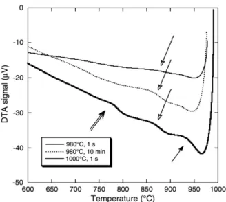
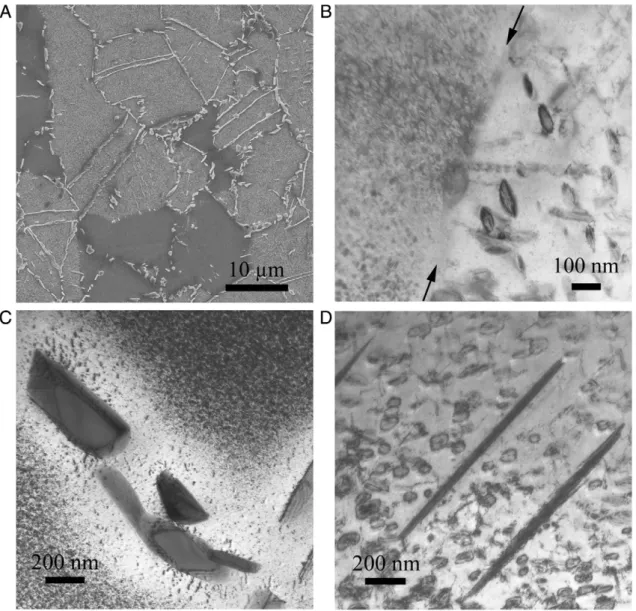
Figure
![Table 1 – Chemical composition of the alloy studied in the present work and nominal composition of alloy 718 [22].](https://thumb-eu.123doks.com/thumbv2/123doknet/3713184.110644/3.892.85.413.90.396/table-chemical-composition-alloy-studied-present-nominal-composition.webp)


+5

Documents relatifs