C O N S E N S U S S T A T E M E N T
Consensus Statements of the American College of Veterinary Internal Medicine (ACVIM) provide the veterinary community with up-to-date infor-mation on the pathophysiology, diagnosis, and treatment of clinically important animal diseases. The ACVIM Board of Regents oversees selection of relevant topics, identification of panel members with the expertise to draft the statements, and other aspects of assuring the integrity of the process. The statements are derived from evidence-based medicine whenever possible and the panel offers interpretive comments when such evidence is inadequate or contradictory. A draft is prepared by the panel, followed by solicitation of input by the ACVIM membership which may be incorporated into the statement. It is then submitted to the Journal of Veterinary Internal Medicine, where it is edited prior to publication. The authors are solely responsible for the content of the statements.
ACVIM consensus statement guidelines for the diagnosis,
classification, treatment, and monitoring of pulmonary
hypertension in dogs
Carol Reinero
1|
Lance C. Visser
2|
Heidi B. Kellihan
3|
Isabelle Masseau
4|
Elizabeth Rozanski
5|
Cécile Clercx
6|
Kurt Williams
7|
Jonathan Abbott
8|
Michele Borgarelli
9|
Brian A. Scansen
101
Department of Veterinary Medicine and Surgery, College of Veterinary Medicine, University of Missouri, Columbia, Missouri 2
Department of Medicine and Epidemiology, School of Veterinary Medicine, University of California, Davis, Davis, California 3
Department of Medical Sciences, School of Veterinary Medicine, University of Wisconsin, Madison, Wisconsin 4
Department of Sciences Cliniques, Faculté de Médecine Vétérinaire, Université de Montréal, Saint-Hyacinthe, Quebec, Canada 5
Department of Clinical Sciences, Cummings School of Veterinary Medicine, Tufts University, Medford, Massachusetts 6
Department of Clinical Sciences of Companion Animals and Equine, University of Liège, Liège, Belgium 7
Department of Pathobiology and Diagnostic Investigation, College of Veterinary Medicine, Michigan State University, East Lansing, Michigan 8
Department of Small Animal Clinical Sciences, College of Veterinary Medicine, University of Tennessee, Knoxville, Tennessee 9
Department of Small Animal Clinical Sciences, Virginia Maryland College of Veterinary Medicine, Blacksburg, Virginia 10
Department of Clinical Sciences, Colorado State University, Fort Collins, Colorado
Correspondence
*Carol Reinero, Department of Veterinary Medicine and Surgery, College of Veterinary Medicine, University of Missouri, 900 East Campus Drive, Columbia, MO 65211. Email: reineroc@missouri.edu
Abstract
Pulmonary hypertension (PH), defined by increased pressure within the pulmonary
vascu-lature, is a hemodynamic and pathophysiologic state present in a wide variety of
cardio-vascular, respiratory, and systemic diseases. The purpose of this consensus statement is
to provide a multidisciplinary approach to guidelines for the diagnosis, classification,
Abbreviations: 6MWT, 6-minute walk test; BOAS, brachycephalic obstructive airway syndrome; C-PH, combined postcapillary and precapillary pulmonary hypertension; CT, computed tomography; HFM, heart failure medications; Ipost-PH, isolated postcapillary pulmonary hypertension; LA, left atrial; LHD, left-sided heart disease; LHF, left-sided heart failure; MMVD, myxomatous mitral valve disease; PAH, pulmonary arterial hypertension; PAP, pulmonary arterial pressure; PAWP, pulmonary arterial wedge pressure; PCH, pulmonary capillary hemangiomatosis; PDE5i, phosphodiesterase 5 inhibitor; PE/PT/PTE, pulmonary emboli/pulmonary thrombi/pulmonary thromboemboli; PG, pressure gradient; PH, pulmonary hypertension; POCUS, point-of-care ultrasound; PR, pulmonary regurgitation; Pre-PH, precapillary pulmonary hypertension; PVD, pulmonary vascular disease; PVOD, pulmonary veno-occlusive disease; PVR, pulmonary vascular resistance; RA, right atrial; RHC, right heart catheterization; RV, right ventricular; TRV, tricuspid regurgitation velocity.
Carol Reinero, Lance C. Visser, Heidi B. Kellihan, Isabelle Masseau, Elizabeth Rozanski, Cécile Clercx, and Kurt Williams are consensus panel members. Jonathan Abbott, Michele Borgarelli, and Brian A. Scansen are advisory task force members.
This is an open access article under the terms of the Creative Commons Attribution License, which permits use, distribution and reproduction in any medium, provided the original work is properly cited.
© 2020 The Authors. Journal of Veterinary Internal Medicine published by Wiley Periodicals, Inc. on behalf of the American College of Veterinary Internal Medicine.
treatment, and monitoring of PH in dogs. Comprehensive evaluation including
consider-ation of signalment, clinical signs, echocardiographic parameters, and results of other
diag-nostic tests supports the diagnosis of PH and allows identification of associated
underlying conditions. Dogs with PH can be classified into the following 6 groups: group
1, pulmonary arterial hypertension; group 2, left heart disease; group 3, respiratory
dis-ease/hypoxia; group 4, pulmonary emboli/pulmonary thrombi/pulmonary thromboemboli;
group 5, parasitic disease (Dirofilaria and Angiostrongylus); and group 6, disorders that are
multifactorial or with unclear mechanisms. The approach to treatment of PH focuses on
strategies to decrease the risk of progression, complications, or both, recommendations to
target underlying diseases or factors contributing to PH, and PH-specific treatments. Dogs
with PH should be monitored for improvement, static condition, or progression, and any
identified underlying disorder should be addressed and monitored simultaneously.
K E Y W O R D S
echocardiography, pulmonary arterial hypertension, respiratory disease, tricuspid regurgitation velocity
1
|
I N T R O D U C T I O N
This article is the report of the American College of Veterinary Internal Medicine consensus panel on pulmonary hypertension (PH) in dogs. The panel first established a working definition of PH and then proposed a clini-cally applicable classification scheme for PH in dogs; with this framework as the basis, practical guidelines for diagnostic, treatment, and monitoring rec-ommendations were developed. Pulmonary hypertension is not a single dis-order, and a multidisciplinary approach is optimal. Therefore, the consensus panel was comprised of board-certified specialists in the areas of internal medicine, cardiology, emergency and critical care, diagnostic imaging, and anatomic pathology. The panel initially met in person to outline overall objectives and review the Delphi method1,2for consensus building. Review
of the human and veterinary medical literature, development of electronic revisions, and conference calls were used to draft the document and, using a modification of the Delphi method, build consensus for recommendations. An advisory panel of 3 additional cardiologists helped develop echocardio-graphic definitions of PH and guidelines for echocardioechocardio-graphic assessment of PH. After development of a draft of the document, 2 additional advisory panel members were included to provide input and rate each of diagnostic, therapeutic, and monitoring recommendations. The final tally of numbers of panelists (out of 7) and outside experts (out of 5) agreeing with each recom-mendation was noted, with comments to clarify reasons for any dissent.
2
|
D E F I N I T I O N O F P H
2.1
|
Hemodynamic definitions and terminology
Pulmonary hypertension is defined by abnormally increased pressure within the pulmonary vasculature. The criterion standard method for diag-nosis of PH is direct assessment of pulmonary arterial pressure (PAP) by right heart catheterization (RHC) and, in humans, PH has been defined asa mean PAP≥25 mm Hg at rest.3A schematic outlining the
pathophysiol-ogy of PH is shown in Figure 1. Increased PAP is not a defining character-istic of a specific clinical condition but rather an abnormal hemodynamic state associated with numerous, diverse disorders.4Increased PAP, in the absence of increased pulmonary vascular resistance (PVR), has several cau-ses requiring different management strategies and having different out-comes, including increased cardiac output, left-to-right shunts, and increased pulmonary arterial wedge pressure (PAWP) secondary to the left-sided heart disease (LHD). Increased PAP associated with increased PAWP (>15 mm Hg in humans5; a surrogate for left atrial [LA] or left ven-tricular filling pressure), is referred to as postcapillary PH. Postcapillary PH occurs most commonly in dogs with LHD that have increased LA pressure. It develops because LA hypertension increases the load on the right ventri-cle and indirectly necessitates the development of higher systolic right ventricular (RV) pressures. Chronic postcapillary PH can lead to pulmonary arterial vasoconstriction and pulmonary vascular disease (PVD), which increase PVR.6,7Thus, postcapillary PH can occur in isolation (isolated
postcapillary PH [Ipost-PH]) or can occur together with increased PVR (combined postcapillary and precapillary PH; C-PH) as a result of chronic, progressive LHD. In humans, C-PH currently is defined by a pressure dif-ference of≥7 mm Hg between diastolic PAP and PAWP.5An equivalent
pressure difference has not been established in dogs. Increased PAP also can be caused by increased PVR and structural pulmonary arterial changes associated with PVD due to a variety of other causes. Increased PAP asso-ciated with increased PVR in the absence of increased LA pressure defines precapillary PH (pre-PH).
3
|
E C H O C A R D I O G R A P H I C
A S S E S S M E N T O F P H
Echocardiography should be viewed as a clinical tool to help assess the probability that a dog has PH, rather than a definitive diagnostic
test for the presence of PH. Definitive diagnosis of PH requires RHC. Echocardiographic assessment of PH is based largely on characteristic cardiac changes that occur secondary to PH (ie, echocardiographic signs of PH) and by estimating PAP from spectral Doppler tracings. Because RHC rarely is utilized for definitive diagnosis of PH in dogs, veterinarians rely on echocardiography for diagnosis, classification, and management of dogs with PH.
However, clinicians should be mindful of limitations of the echocardiographic examination (particularly Dopp-ler echocardiography) and of inaccuracy,8 variability, and imprecision9,10potentially encountered when using
echocardiography to estimate PAP in individual dogs.
The panel has kept these echocardiographic shortcomings in mind when proposing guidelines and recommendations. Because many echo-cardiographic signs of PH have not been compared to RHC findings in
dogs, we have consulted current echocardiographic guidelines for humans11,12and the veterinary medical literature when relevant citations were available to establish these guidelines. The proposed criteria are intended to avoid misdiagnosis and inappropriate treatment of PH that might have lasting impact on the dog and its owner. Lastly, echocardio-graphic assessment of PH is just 1, albeit important, aspect in the overall clinical assessment of a dog with suspected PH. Echocardiographic ings always should be interpreted within the context of other clinical find-ings, especially the presence or absence of clinical signs suggestive of PH (Table 1) and right-sided heart failure status, as well as results of concur-rent diagnostic testing.
3.1
|
Echocardiographic signs of PH and
estimating PAP
The use of Doppler echocardiography to estimate PAP is crucial to the echocardiographic assessment of dogs with suspected PH. Assuming the F I G U R E 1 Development of pulmonary hypertension, defined as abnormally increased pressure within the pulmonary vasculature, results from increased pulmonary blood flow, increased pulmonary vascular resistance, increased pulmonary venous pressure, or some combination thereof. Normal pulmonary vasculature is comprised of thin-walled arteries, veins and capillaries; a low pressure, low vascular resistance, and high capacitance system. In homeostasis, blood ejected from the right ventricle (RV) into the pulmonary trunk is directed to the right and left
pulmonary arteries. The pulmonary arterial pressure remains low in homeostatic conditions by the dense network of pulmonary capillaries that can accommodate rapid transit of large volume of blood arriving from the pulmonary arteries. Following efficient gas exchange at the alveolar-capillary interface, oxygenated blood is then collected by the pulmonary venules that unite to form the veins, which eventually open into the left atrium. Upon complex interactions of genetic and environmental factors, most of which are still poorly understood in dogs, homeostasis of pulmonary circulation can be disturbed. These disturbances (summarized in boxes) can lead to an excessive increase in pulmonary blood flow, increased pulmonary vascular resistance, or increased pulmonary venous pressure. Additionally, there is interplay between these factors*: increased pulmonary blood flow or increased pulmonary venous pressure can lead to increased pulmonary vascular resistance due to pulmonary arterial vasoconstriction, pulmonary vascular disease/remodeling, or both. When the average pulmonary arterial pressure increases above a certain threshold (25 mm Hg is commonly used in humans), PH results. With sustained PH, the RV has to work harder against increases in pulmonary pressures to move blood through the pulmonary vasculature. As a consequence, the RV undergoes structural alterations. Over time, the progressive increase in RV workload can ultimately lead to RV dysfunction and failure, resulting in heart failure (ascites), low output signs, and death. ASD, atrial septal defect; EDD, endothelial-dependent dilatation; LV, left ventricle; NO, nitric oxide; PDA, patent ductus arteriosus; PH, pulmonary hypertension; VSD, ventricular septal defect
absence of RV outflow tract obstruction (eg, pulmonary valve stenosis), estimating systolic PAP involves quantifying peak tricuspid regurgitation velocity (TRV), and then derivation of the pressure gradient (PG) between the RV and right atrium using the simplified Bernoulli Equation (PG = 4× velocity [m/s]2). An estimate of right atrial (RA) pressure is added to the calculated PG to yield estimated systolic PAP. Validated methods to estimate RA pressure are unavailable in dogs, and therefore estimates of RA pressure are arbitrary and potentially flawed.8,10,13
Con-sequently, we recommend using only continuous wave Doppler measure-ment of TRV (versus estimated systolic PAP) as a key metric in determining PH probability as long as clinicians are aware that systolic PAP might be underestimated when severe RA hypertension is present. Measured TRV depends upon many factors including RV function and pericardial restraint14 in addition to PAP and PVR. Additionally, poor
patient cooperation and labored respiration affect measurements. Accu-rate interpretation of a TRV signal includes consideration of all factors impacting RV systolic pressure. A pulmonary regurgitation (PR) jet also can be used to estimate mean or diastolic PAP.11,15,16Applying the simplified
Bernoulli equation to spectral Doppler measurements of peak diastolic PR velocity provides an estimate of mean PAP.16Similarly, peak diastolic PR
jet velocity offers an estimate of diastolic PAP after adding an estimate of RA pressure.15Because tricuspid regurgitation has been utilized more
commonly, and clinicians conventionally have viewed PH in relation to estimates of systolic PAP, TRV can be considered the primary metric to estimate PAP and the key component to determine the probability of PH in at-risk dogs. Recommended echocardiographic criteria used to help determine the probability of PH are presented in Table 2.
Although specific techniques of echocardiographic image acquisi-tion and measurement are beyond the scope of this consensus state-ment, Doppler spectra should be well visualized and their shape and contour should comply with known hemodynamic principles. Care should be taken to align the cursor parallel to the direction of flow, T A B L E 1 Clinical findings suggestive of pulmonary hypertension
(PH) in dogsa
Findings strongly suggestive of PH
Findings possibly suggestive of PH Syncope (especially with exertion
or activity) without another identifiable cause
Tachypnea at rest
Respiratory distress at rest Increased respiratory effort at rest Activity or exercise terminating
in respiratory distress
Prolonged postexercise or post-activity tachypnea Right-sided heart failure
(cardiogenic ascites)
Cyanotic or pale mucous membranes
aIt should be noted that none of these clinical signs are specific solely for
PH and therefore other causes of clinical signs are not excluded. Although these clinical signs may be due to underlying respiratory disease, more pronounced clinical signs reflect more severe disease, with more severe disease likely to result in PH.
T A B L E 2 Echocardiographic probability of PH in dogs Peak tricuspid
regurgitation velocity (m/s)
Number of different anatomic sites of
echo signs of PHa Probability of PH
≤3.0 or not measurable 0 or 1 Low
≤3.0 or not measurable 2 Intermediate
3.0 to 3.4 0 or 1 Intermediate
>3.4 0 Intermediate
≤3.0 or not measurable 3 High
3.0 to 3.4 ≥2 High
>3.4 ≥1 High
a
See Table 3.
F I G U R E 2 Example measurements of tricuspid regurgitation velocity (TRV) spectra acquired using continuous wave Doppler. A, The solid arrow shows the recommended measurement of TRV. The dense outer edge of the velocity profile (brighter signal) is measured and measurement sof the fine linear signals has been avoided. The dotted arrow represents a measurement that includes the fine linear signals or“feathered edge,” which likely overestimates TRV. B, A TRV signal of good quality is shown. Notice the envelope is fully visible, the signal is not overgained (helps to avoid fine linear signals), the sweep speed is increased, and the scale along the vertical axis is nearly filled by the TRV signal. C, Shows 3 TRV signals, 1 of moderate quality (*) that is not completely filled in but can be measured by extrapolation and 2 others of poor quality (#) that are unreliable and should not be measured
optimize gain settings, and to measure regurgitant jets at the dense outer edge of the velocity profile while avoiding measurement of fine linear signals, also called the“feathered edge” (Figure 2).29-31Because
tricuspid regurgitation jets can be eccentric, nonstandard imaging planes might be necessary for visualization of tricuspid regurgitation and acquisition of TRV.
In addition to TRV, several echocardiographic signs of PH aid prob-ability assessment for PH. These signs involve 3 anatomic sites: (1) ven-tricles, (2) pulmonary artery (although pulmonary artery is the commonly used term in clinical practice, the preferred anatomic term for the main pulmonary artery is pulmonary trunk32and herein we
con-sider them synonymously), and (3) RA and caudal vena cava (Table 3). These echocardiographic signs of PH largely involve assessment of the ventricles (left ventricular and RV size and remodeling,19,21systolic flat-tening of the interventricular septum,17 and RV systolic
func-tion19,23-27,33), the pulmonary artery (pulmonary artery size17-19 and flow profile,13,17,28,34), and the RA and caudal vena cava (RA19,21and
caudal vena cava size19). Unfortunately, there is a lack of agreement on how best to assess (ie, measure and quantify) these echocardiographic variables in dogs with PH, and specific echocardiographic guidelines are beyond the scope of this consensus statement.
3.2
|
Echocardiographic probability of PH
with LHD
Recommendations in Tables 2 and 3 for echocardiographic assess-ment of PH pertain to the use of a probability-based approach for the assessment PH for all causes of PH (groups 1-6, Table 4). In addition to criteria in Tables 2 and 3, identifying dogs with PH sec-ondary to LHD requires fulfilling 2 additional echocardiographic criteria: (1) documentation of LHD (eg, mitral or aortic valvular dis-ease or left ventricular dysfunction) and (2) unequivocal LA enlargement. Because RHC and PAWP determination are
performed rarely, LA enlargement is utilized as a crude, but robust, surrogate for chronically increased PAWP and postcapillary PH. By definition, patients with PH secondary to LHD must have increased PAWP.5The panel recognizes it is possible to encounter acutely increased PAWP, and therefore iPost-PH secondary to LHD without LA enlargement caused by, for example, ruptured chordae tendineae in a dog with myxomatous mitral valve disease (MMVD). Sonographers should be cognizant of this uncommon exception. Chronically increased PAWP is a necessary component of clinically relevant C-PH secondary to LHD.5,6In other words, it is less likely for PH, and particularly C-PH, to be secondary to LHD unless unequivocal LA enlargement also is identified. Echocardio-graphic signs of PH such as decreased LV size or LV underfilling are not applicable to dogs with PH secondary to LHD because of the confounding effects of LV remodeling secondary to LHD. In dogs with PH secondary to LHD, criteria proposed in Tables 2 and 3 for high probability of PH are more likely to identify patients with C-PH. This approach is supported by clinical studies of dogs with MMVD demonstrating the clinical and prognostic relevance of TRV >3.5-3.7 m/s.82,152
3.3
|
Proposed clinical definition of PH
In the veterinary medical literature, the degree or severity of PH has been classified as mild, moderate, or severe. These categories are based on the PG derived from TRV (also called the tricuspid regurgitation PG) or estimated systolic PAP. Cutoffs used for these categories (mild, mod-erate, severe) are arbitrary, and the categories are potentially misleading or flawed. For example, based on the conventional definition of PH (based solely on estimated systolic PAP), a dog with severe PH can be free of clinical signs, whereas a dog with moderate PH could be in right-sided heart failure. Therefore, the panel does not advocate their use. Instead, severity of PH should be based on clinical findings (Table 1) and T A B L E 3 Anatomic sites of echocardiographic signs of PH used to help assess the probability of PH in dogs
Anatomic site 1: Ventricles Anatomic site 2: Pulmonary artery
Anatomic site 3: Right atrium and caudal vena cava
Flatting of the interventricular septum (especially systolic flattening)
Pulmonary artery enlargement (PA/Ao >1.017,18)
Right atrial enlargement19,20
Underfilling or decreased size of the left ventriclea
Peak early diastolic PR velocity >2.5 m/s Enlargement of the caudal vena cava19
Right ventricular hypertrophy (wall thickening, chamber dilation, or both)19,21
RPAD index <30%17,22
Right ventricular systolic dysfunction19,23-27
RV outflow Doppler acceleration time (<52-58 ms) or acceleration time to ejection time ratio (<0.30)17,18,28
Systolic notching of the Doppler RV outflow profile (caution: false positives are possible)
Abbreviations: PR, pulmonary regurgitation; RPAD, right pulmonary artery distensibility; RV, right ventricular.
outcome data from large (prospective) longitudinal studies, which unfor-tunately are unavailable. The panel recognizes the probabilistic approach to diagnosis of PH presents challenges, particularly regarding
standardized enrollment in future clinical studies of dogs with suspected PH. Therefore, we propose that the clinical definition of PH should include dogs with intermediate or high probability of PH, and specifically,
T A B L E 4 Proposed classification of pulmonary hypertension in the doga 1. Pulmonary arterial hypertension
1a. Idiopathic (IPAH)35-39,b; 40-45,c
1b. Heritable46,47
1c. Drugs and toxins induced48,49,d
1d. Associated with (APAH):
1d1. Congenital cardiac shunts17,18,35,41-43,46,47,50-75
1d2. Pulmonary vasculitis63,71,76-78
1d3. Pulmonary vascular amyloid deposition79
10. Pulmonary veno-occlusive disease (PVOD) or pulmonary capillary hemangiomatosis (PCH)80,81,e
2. Pulmonary hypertension due to left heart disease 2a. Left ventricular dysfunction
2a1. Canine dilated cardiomyopathy18,25,35,43,50,51
2a2. Myocarditis35
2b. Valvular disease 2b1. Acquired
2b1a. Myxomatous mitral valve disease10,13,17,18,20,24,25,34,35,41-43,50-52,82-89 2b1b. Valvular endocarditis
2c1. Congenital/acquired left heart inflow/outflow tract obstruction and congenital cardiomyopathies 2c1a. Mitral valve dysplasia18,52
2c2a. Mitral valve stenosis90
2c3a. Aortic stenosis43
3. Pulmonary hypertension secondary to respiratory disease, hypoxia or bothf
3a. Chronic obstructive airway disordersg
3a1. Tracheal or mainstem bronchial collapse18,85,91
3a2. Bronchomalacia91-93
3b. Primary pulmonary parenchymal disease 3b1. Interstitial lung disease (reviewed in94,95)
3b1a. Fibrotic lung disease18,28,40,91,92,96
3b1b. Cryptogenic organizing pneumonia/secondary organizing pneumonia97
3b1c. Pulmonary alveolar proteinosis98
3b1d. Unclassified interstitial lung disease (ILD)41,66,85,91,99,100
3b1e. Eosinophilic pneumonia/eosinophilic bronchopneumopathy66,92,101 3b2. Infectious pneumoniah: Pneumocystis91,102,103; Ehrlichia?104,105 3b3. Diffuse pulmonary neoplasia35,66,85,91,92,106
3c. Obstructive sleep apnea/sleep disordered breathing107,108,d; 91,92,109,i
3d. Chronic exposure to high altitudes110-112
3e. Developmental lung disease91
3f. Miscellaneous: bronchiolar disorders91; bronchiectasis91; emphysema91,113; pneumonectomy114
4. Pulmonary emboli/thrombi/thromboemboli (PE/PT/PTE)115-121
4a. Acute PE/PT/PTE8, 122-125,d
(Massive PE/PT/PTE with RV dysfunction or submassive PE/PT/PTE without RV dysfunction) 4b. Chronic PE/PT/PTE126,d,40
5. Parasitic disease (Dirofilaria or Angiostrongylus infection17,25,35,41,44,50,51,66,127-147)j
a tricuspid regurgitation PG cutoff of >46 mm Hg (TRV >3.4 m/s). This roughly equates to what has been conventionally referred to as at least moderate PH. Until there are echocardiographic variables that are repeatable and have acceptable diagnostic accuracy relative to RHC, this cutoff value could be used with an understanding of its limitations.
4
|
C O M P R E H E N S I V E C L I N I C A L
A S S E S S M E N T F O R P H
Diagnosis of PH requires comprehensive evaluation including consid-eration of signalment, clinical signs, echocardiographic parameters
supporting PH, and results of other diagnostic tests. Clinical signs reflect the degree of functional impairment and can be used to iden-tify dogs with clinical signs that are strongly or possibly suggestive of PH (Table 1). Attributing clinical signs directly to PH can be challeng-ing or impossible because of underlychalleng-ing disease that may dominate the clinical picture.
4.1
|
Clinical presentation
In dogs, PH occurs as a primary pulmonary vascular disorder or as a sequela to other cardiac, respiratory, or systemic diseases. Signalment, T A B L E 4 (Continued)
6. PH with multifactorial or unclear mechanismsk
6a. Disorders having clear evidence of 2 or more underlying groups 1-5 pathologies contributing to PHl
6b. Masses compressing the pulmonary arteries (eg, neoplasia, fungal granuloma, etc.) 6c. Other disorders with unclear mechanisms
aGiven the limitations of the veterinary literature (eg, single case reports or small case series, retrospective study design, frequent presence of confounding
comorbid conditions contributing to PH, lack of uniform and rigorous diagnostic testing to definitively rule out comorbid conditions, among others), not all panelists agree with provided references to support the disease as the cause of PH. Larger, prospective carefully designed studies will be required to provide the necessary evidence to further refine this classification scheme.
bIn the veterinary literature, when no underlying cause of PH has been found, PH is often assumed to be“idiopathic.” However, it is important to recognize
the difference between not finding a cause after an exhaustive diagnostic evaluation and calling a disease idiopathic after a cursory evaluation (see Figures 3– 7). The first 5 references are considered definitive studies as histopathology documents a pulmonary arteriopathy in the absence of a known cause.
c
The next 6 references are considered questionable support for IPAH; although no identified cause was found, the diagnostic evaluation may not have been reported or have been incomplete and histologic evaluation was not performed.
dExperimental canine studies. ePVOD and PCH can occur in tandem. f
In the peer-reviewed veterinary literature, many studies refer to“chronic respiratory/pulmonary disease” or “idiopathic” respiratory disease, or “chronic tracheobronchial disease” without definitive documentation of the specific underlying disorder.35,40-42,66,85,149Other listed“definitive” diagnoses may be
published without ruling out disease mimics in an exhaustive fashion (eg, thoracic radiography alone can be definitive for collapsing trachea but nondefinitive for bronchomalacia or fibrotic lung disease). Without a criterion standard definitive confirmation (eg, bronchoscopy for bronchomalacia or lung biopsy for pulmonary fibrosis), many of these respiratory diseases are likely inadequately characterized. Additionally, many dogs with disorders associated with PH in humans do not get a specific evaluation for PH; thus the group 3 disorders are likely grossly underestimated. Additionally, disorders which are not clearly documented or are undocumented to cause PH in the dog include pharyngeal collapse,150laryngeal collapse, laryngeal paralysis, and
epiglottic retroversion.
g
Although“chronic bronchitis” has been listed as a diagnosis in some canine reports,18,85this syndrome alone in the dog is unlikely to cause PH. The term chronic obstructive pulmonary disease (COPD) used in humans encompasses underlying and overlapping conditions such as chronic bronchitis and emphysema. Both cause airflow limitation and dyspnea in people. Canine chronic bronchitis by itself (ie, without concurrent bronchomalacia) does not cause airflow limitation leading to increased expiratory respiratory effort and emphysema is very rare in dogs, thus the term COPD is inappropriate to use in this species. Tracheal and mainstem bronchial collapse and bronchomalacia are common causes of obstructive airway disorders; however, referenced studies proving they cause PH are somewhat limited by many reported dogs having comorbid conditions also known to cause PH.
hAngiostrongylus and Dirofilaria are excluded from infectious causes of pneumonia as the pathophysiology of PH is usually multifactorial with these
parasitic infections. The term“pneumonia” by itself does not necessarily imply an infectious etiology and care must be taken when interpreting results of studies that do not specifically identify an organism but find compatible radiographic changes or inflammatory cells on airway lavage or
histopathology.35,51,66These cases may represent ILDs.
iBrachycephalic obstructive airway syndrome is listed under obstructive sleep apnea/sleep disordered breathing as the dog is a model for human disease.151
However, as this is a heterogeneous syndrome with multiple defects, clinical manifestations could also be classified under chronic obstructive airway disorders.
j
Dirofilaria and Angiostrongylus have been associated with endarteritis,17,25,35,41,44,50,51,66,127-142PE/PT/PTE,147inflammatory pulmonary parenchymal disease,143-146or all, as their mechanisms of PH.
kIn humans, hematologic disorders (eg, certain types of anemia, myeloproliferative disorders, and splenectomy), systemic disorders with lung involvement
(eg, sarcoidosis, Langerhans cell histiocytosis, vasculitis, etc), metabolic disorders (disorders of impaired cell metabolism, thyroid disease), and other diseases not well classified in another group (eg, compressive lesions such as lymphadenopathy, tumor or fibrosing mediastinitis obstructing the pulmonary arteries, etc) comprise the multifactorial, unclear mechanism group, or both.148As these analogous disorders with rare exception either do not occur in
dogs or if they occur, may not be documented to cause PH, additional research and modification of these group 6 disorders in dogs will likely be needed. This is a particularly poorly understood category and it is likely that other diseases will be added in the future with additional investigation.
l
To be classified as 6a, there must be identified diseases in more than 1 of the group 1-5 disorders (eg, group 2 MMVD and group 3b1 ILD) and not just 2 or more types of disease within a single disorder (group 3a1 tracheal collapse and group 3b1a fibrotic lung disease).
history, and clinical presentation can reflect the underlying cause. Congen-ital cardiac shunts and developmental lung disease are generally manifested in puppies, whereas acquired cardiac, respiratory, and systemic diseases usually present in adults. Dogs with MMVD and PH usually are older small breed dogs.18,35,50,51,109A breed predisposition for PH second-ary to interstitial lung disease has been suggested in West Highland White Terriers28and Pekingese dogs.99Historical data supporting exposure to Dirofilaria or Angiostrongylus in endemic regions warrant careful scrutiny for these parasites. Exertional syncope, labored respiration or overt respi-ratory distress, and exercise intolerance are noteworthy clinical signs fre-quently directly attributable to PH.40,52,153,154 Cough is commonly reported but more likely reflects an underlying respiratory disease.35
Car-diac auscultation may identify heart murmurs localized to the mitral valve, tricuspid valve or both; a diastolic heart murmur of PR occasionally is detected in patients with severe PH.52A split or loud second heart sound may be auscultated, particularly with severe PH.154A right-sided systolic
murmur,155right-sided heart failure (eg, a distended abdomen caused by cranial organomegaly or ascites), or both may be detected. Jugular vein distention and pulsation may be present. Cyanosis may occur secondary to primary pulmonary disease or congenital cardiac disease exhibiting right-to-left pulmonary-to-systemic shunting. Evaluation of the breathing pattern (increased inspiratory effort, increased expiratory effort, mixed inspiratory/expiratory effort, or paradoxical breathing pattern) may pro-vide clues to an underlying airway, parenchymal or pleural cavity disorder, especially when paired with audible noises present with or without the use of a stethoscope.156Pulmonary auscultation may identify muffled
sounds, crackles, wheezes, or increased bronchovesicular sounds. Crackles
may indicate interstitial lung disease (especially fibrotic lung disease), severe bronchomalacia, or pulmonary edema or exudate.94,157,158
5
|
C L A S S I F I C A T I O N O F P H
In dogs, the prevalence of PH is as yet unknown. There appears to be increased awareness of PH, likely because of increased recognition of clinical signs of PH and underlying disorders that have PH as a comor-bid condition and more widespread availability of echocardiography. Historically, LHD (eg, MMVD) had been among the most commonly identified causes of PH.7,13,18,35,41,42,50-52,83,109,152,159 However,
many disorders causing PH may go unrecognized, especially when echocardiography is not performed or when clinical signs attributable to the primary disease dominate the clinical picture.
We propose 6 groups of PH in dogs, loosely following the classification scheme used in humans with modifications148subdivided to reflect similar themes in some or all of the following: cause of PH, clinical presentation, hemodynamic characteristics, pathophysiology, and treatment. Such a clas-sification is intended to improve diagnostic evaluation and optimize thera-peutic management of dogs with PH. Finally, classification into well-defined groups should facilitate future clinical studies.160As in humans, it is
expected that evolution of this classification scheme will occur with increased recognition and understanding of underlying conditions.
The proposed clinical classification of PH in dogs comprises the fol-lowing 6 groups (Table 4): Group 1, pulmonary arterial hypertension (PAH); group 2, LHD; group 3, respiratory disease/hypoxia; group
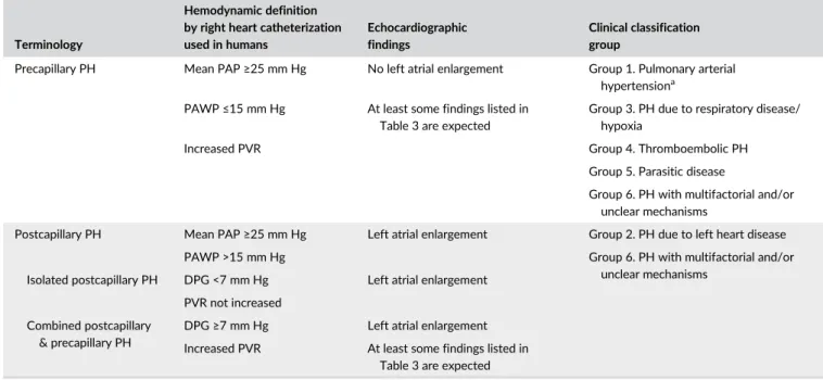
T A B L E 5 Terminology, hemodynamic definitions, and echocardiographic findings of PH together with the proposed clinical classification groups of pulmonary hypertension
Terminology
Hemodynamic definition by right heart catheterization used in humans
Echocardiographic findings
Clinical classification group
Precapillary PH Mean PAP≥25 mm Hg No left atrial enlargement Group 1. Pulmonary arterial
hypertensiona
PAWP≤15 mm Hg At least some findings listed in Table 3 are expected
Group 3. PH due to respiratory disease/ hypoxia
Increased PVR Group 4. Thromboembolic PH
Group 5. Parasitic disease
Group 6. PH with multifactorial and/or unclear mechanisms
Postcapillary PH Mean PAP≥25 mm Hg Left atrial enlargement Group 2. PH due to left heart disease
PAWP >15 mm Hg Group 6. PH with multifactorial and/or
unclear mechanisms Isolated postcapillary PH DPG <7 mm Hg Left atrial enlargement
PVR not increased Combined postcapillary
& precapillary PH
DPG≥7 mm Hg Left atrial enlargement
Increased PVR At least some findings listed in Table 3 are expected
Abbreviations: DPG, diastolic pressure gradient (diastolic PAP− mean PAWP); PAP, pulmonary arterial pressure; PAWP, pulmonary arterial wedge pressure; PH, pulmonary hypertension; PVR, pulmonary vascular resistance.
aCongenital cardiac shunts (group 1d1) exhibiting left-to-right shunting represents an exception. The PH may be primarily due to increased right heart
4, pulmonary emboli/pulmonary thrombi/pulmonary thromboemboli (PE/PT/PTE); group 5, parasitic disease (Dirofilaria and Angiostrongylus); and group 6, disorders that are multifactorial or with unclear mechanisms. Peer-reviewed references to support the classification are provided when possible, with the caveat that studies describing a diagnosis of a particular disease in a dog with PH do not necessarily prove that the disease caused PH. Refinement of this classification scheme will likely be needed in the future. When >1 possible cause for PH is present, consideration must be given to the likelihood that each individual underlying cause is contributing to PH. If the contribution to PH from a comorbid condition is minimal, PH should be classified according to the major disease, however, if there potentially are substantial contributions from ≥2 comorbid conditions, they should be placed in group 6, encompassing multifactorial mecha-nisms. For example, a dog with MMVD without LA enlargement and severe interstitial lung disease would be classified as group 3b1; a dog with stage C MMVD161and severe interstitial lung disease would be classified
as group 6a. This discrimination is important because group 6 disorders are likely to require multimodal treatment targeted at each underlying pathologic mechanism. The group 5 classification for PH in dogs repre-sents the largest deviation from the human medical literature, because humans are not affected by PH secondary to Dirofilaria or Angiostrongylus infection. Table 5 provides a summary of terminology, hemodynamic defi-nitions, and echocardiographic findings of PH with the corresponding pro-posed clinical classification of PH.
6
|
G U I D E L I N E S F O R D I A G N O S T I C
E V A L U A T I O N O F D O G S W I T H
S U S P E C T E D P H
6.1
|
General comments
Pulmonary hypertension is not a disease per se but rather a hemo-dynamic and pathophysiologic state present in a wide variety of diseases.53,154 Diagnostic testing in suspected cases thus must encompass 2 major goals: (1) to assess the probability of PH using echocardiography and (2) to determine the underlying cause of PH when possible. This information is critical to best guide therapeu-tic recommendations. In practherapeu-tice, there are 2 pathways of diag-nostic evaluation for PH in dogs. The first involves dogs presenting with a spectrum of clinical signs in which diagnostic testing targets identification of a primary cause of disease, with PH considered later in the evaluation. Consensus diagnostic rec-ommendations D1-D7 below are guidelines for when to pursue echocardiography to assess the probability of PH based on other test results. The second approach involves early assessment of the probability of PH using echocardiography. Because some clinicians identify intermediate or high probability of PH based on sugges-tive clinical signs, with echocardiography performed early in the diagnostic evaluation (specifically to investigate PH or for other reasons such as a heart murmur), diagnostic algorithms to help determine the underlying cause of PH (ie, groups 1-6) also are pro-vided (Figures 3–7).
6.1.1
|
Consensus recommendations for PH
diagnosis
D1. Echocardiography to assess the probability of PH should be consid-ered as an early diagnostic test in dogs with clinical findings suggestive of PH (Table 1)35,36,51,80,99,109,162after physical examination and
tho-racic imaging rule out another specific disorder not associated with PH.
• Consensus in 7/7 members of the panel and 5/5 advisory reviewers D2. Echocardiography to assess the probability of PH should be considered when thoracic radiography shows evidence of tortuous, blunted, or dilated pulmonary arteries; asymmetric radiolucent lung fields on dorsoventral or ventrodorsal views; patchy, diffuse alveolar infiltrates40; a bulge in the region of the pulmonary trunk or right-sided cardiac enlargement.52,154,163
• Consensus in 7/7 members of the panel and 5/5 advisory reviewers D3. Echocardiography to assess the probability of PH should be considered in dogs with clinical signs suggestive of PH (Table 1) and having ascites (modified transudate with noncardiac causes ruled out), a dilated caudal vena cava on point-of-care ultrasound (POCUS; dia-phragmaticohepatic view) or a dilated caudal vena cava and hepatic veins on abdominal ultrasonography.
• Consensus in 6/7 members of the panel and 4/5 advisory reviewers • Comment: One panelist recommended caution in interpretation of
POCUS right-sided cardiac markers (eg, large caudal vena cava, hepatic venous distension, ascites or gall bladder wall edema) because they had poor utility for discrimination of dogs with and without PH.164One advisory reviewer recommended the removal
of POCUS from D3 stating assessment of the caudal vena cava is challenging, subject to intra- and inter-individual variation and complicated by operator experience, patient positioning, and equipment.
D4. Echocardiography to assess the probability of PH may be con-sidered in dogs having spent time in endemic areas and that are, or have a history of, confirmed Dirofilaria positivity165or Angiostrongylus
positivity162with clinical signs (eg, for heartworm disease: coughing, respiratory distress, collapse, anemia, hyperbilirubinemia, exercise intol-erance or ascites; for angiostrongylosis: cardiac, respiratory, or neuro-logic signs or bleeding diathesis) or thoracic radiographic abnormalities (eg, right-sided heart enlargement; enlarged pulmonary trunk; enlarged, blunted, or tortuous pulmonary arteries; or pulmonary infiltrates) suspected in association with these parasites.
• Consensus in 7/7 members of the panel and 5/5 advisory reviewers Dogs with a condition associated with acute or chronic PE/PT/PTE are at risk for PH. Examples include immune-mediated hemolytic ane-mia, spontaneous hyperadrenocorticism, protein-losing nephropathy,
protein-losing enteropathy, sepsis, neoplasia, and disseminated intra-vascular coagulation.115-119,166-168 Additionally, although heartworm disease and angiostrongylosis cause PH by multifactorial mechanisms, an important contributor to increased PAP is parasitic embolism.
D5. Echocardiography to assess the probability of PH should be considered in dogs at high risk169for PE/PT/PTE that have developed clinical signs suggestive of PH particularly with evidence of hypoxemia and thoracic imaging that fails to identify another underlying cause for the respiratory signs.
• Consensus in 7/7 members of the panel and 5/5 advisory reviewers Thoracic computed tomography (CT) is a highly sensitive imaging modality. Especially when incorporated with single or multi-phase angiography, thoracic CT can provide supportive or definitive evi-dence for PVDs, pulmonary parenchymal diseases, and PE/PT/PTE, many of which can be associated with PH.
D6. If not already performed, echocardiography to assess the probability of PH should be considered when thoracic CT angiography shows≥1 of the following:
a. A pulmonary trunk-to-descending aorta ratio≥1.4170
b. Evidence of RA and RV enlargement
c. A decreased pulmonary vein-to-PA ratio; an increased pulmonary trunk-to-ascending aorta ratio, or an increased RV-to-LV ratio92 d. The presence of pulmonary arterial filling defects171
e. A mosaic attenuation pattern showing small vessels in a region of decreased attenuation (ie, hypoperfusion) on an inspiratory scan that fails to show accentuation of the mosaic attenuation pattern on an expiratory scan (ie, ruling out air trapping)172
f. Perivascular diffuse nodular to ill-defined patchy ground-glass opacity with a global distribution, compatible with pulmonary capillary hemangiomatosis (PCH) or pulmonary veno-occlusive disease (PVOD)80 • Consensus in 7/7 members of the panel and 5/5 advisory reviewers • Comments: Although measurement of RV173,174 (and perhaps RA)
enlargement evaluated on contrast CT scan is suspected to be a viable metric for PH, peer-reviewed studies in dogs with PH have not yet been published. Additionally, signalment and physical examination should be used to discriminate dogs with pulmonary valve stenosis that also may have findings a-c above.
F I G U R E 3 Algorithm demonstrating the overall diagnostic approach to the 6 groups of pulmonary hypertension in which echocardiography performed early in the clinical evaluation identifies intermediate or high probability PH. In addition to determining an intermediate or high probability of PH, echocardiography can also be used to support, confirm, or refute pathology in group 1d1, group 2, group 4, and group 5. The order in which diagnostic algorithms should be consulted are group 3 (Figure 4) and group 4 (Figure 5) with group 5 potentially being identified on this initial algorithm or within the group 3 algorithm. The group 1 algorithm (Figure 7) generally used after ruling out disorders in groups 2-6. Critical to appropriate use of the diagnostic algorithms is the understanding that dogs frequently have greater than 1 type of pathology contributing to PH either across groups (eg, a dog with MMVD with interstitial lung disease is encompassed in groups 2 and 3, respectively) or within a group (eg, a dog with tracheal collapse and fibrotic lung disease both fall within group 3). Clinical evaluation must drive the diagnostic approach and make sense in context of localizing disease and pursuit of comorbid conditions. For example, a small breed dog with left-sided heart failure that has inspiratory stridor in addition to rapid, shallow breathing should not have the diagnostic algorithm terminated after diagnosis of group 2c1a disease; instead, further evaluation for an upper airway defect such as extrathoracic tracheal collapse should be pursued.aThoracic
radiographs are frequently obtained before echocardiography and may provide additional findings supportive of underlying PH etiology.
bEvidence of an in situ PT in the main pulmonary artery may be noted on echocardiographic examination. LHD, left-sided heart disease; PH,
Although performed uncommonly as a primary diagnostic test for pulmonary disease, lung biopsy specimens may be acquired and sub-mitted for histologic examination and can be very valuable in identify-ing and characterizidentify-ing vascular pathology.
D7. Echocardiography to assess the probability of PH should be considered when histologic examination indicates evidence of wide-spread PVD:
a. The use of routine hematoxylin and eosin staining can help eluci-date the pathogenesis of PAH. Examples of vascular lesions include arterial or arteriolar medial smooth muscle hypertrophy or hyper-plasia, pulmonary arterial intimal hyperplasia or fibrosis, vascular thrombosis, or occlusion, and arterial plexiform lesions. Large num-bers of hemosiderophages may indicate pulmonary venous hyper-tension. Increased numbers of alveolar capillary endothelial cells suggest PCH or potentially severe pulmonary venous hypertension associated with markedly increased LA pressures.
b. Verhoeff-Van Gieson staining is a valuable additional technique that assists in distinguishing arteries and veins and can be very helpful in identifying affected veins in PVOD.
• Consensus in 7/7 members of the panel and 5/5 advisory reviewers • One panelist commented that adequately large lung biopsy specimens
and multiple (>2) lung biopsies may be needed to characterize the underlying disease process. The absence of lesions especially in small
biopsy specimens or in end-stage tissue (ie, fibrosis) may not allow the elimination of vascular pathology as a possibility. Additionally, the pathologist evaluating the lesions must be knowledgeable about PVDs.
7
|
T R E A T M E N T
Treatment of PH can be subdivided into strategies to decrease the risk of progression or complications (treatment consensus state-ments T1a-e), recommendations to target underlying diseases or fac-tors contributing to PH (treatment consensus statements T2-T12), and PH-specific treatments (treatment consensus statements T13-T24).
Interpretation of therapeutic recommendations is dependent on the level of evidence, degree of clinical impairment, and echo-cardiographic probability of PH. The strength of the recommenda-tion is higher when the primary literature is available regarding treatment of dogs with spontaneous PH (aside from single case reports) or, in the absence of these data, strong expert opinion, and the treatment statements are worded as “recommended.” When recommendations are extrapolated from humans, canine models, or weaker anecdotal experience of experts, the treatment statements are worded as“may be considered.” As reviewed in Tables 1 and 2, clinical findings are stratified as strongly or possibly suggestive of F I G U R E 4 Diagnostic algorithm for discrimination of group 3 respiratory disease/hypoxia in dogs. Proper interpretation relies on
confirmation that the comprehensive clinical picture can be explained solely by the“final diagnosis” (bold boxes); otherwise, continue to evaluate for PH in other subcategories of group 3 and in groups 1, 4, 5, and 6. BAL, bronchoalveolar lavage; CT, computed tomography; FNA, fine-needle aspiration; HW, heartworm; I/E, paired inspiratory/expiratory series; MSB, mainstem bronchial; OSA, obstructive sleep apnea; PAH, pulmonary arterial hypertension; PH, pulmonary hypertension; PTE, pulmonary thromboembolism
PH and echocardiographic evidence of PH as low, intermediate, or high probability.
7.1
|
Strategies to decrease the risk of progression
or complications of PH
T1. Several guidelines yet untested in randomized clinical trials, seem prudent, especially in dogs with high probability of PH:
a. Exercise restriction
b. Prevention of contagious respiratory pathogens using vaccina-tion175 and parasitic disease (eg, Dirofilaria and Angiostrongylus) control using chemoprophylaxis in endemic areas
c. Avoidance of pregnancy (because of potential to exacerbate PH and because of the possibility of transmission of genetic contributors)
d. Avoidance of high altitude and air travel
e. Avoidance of nonessential wellness procedures (eg, dental cleanings) and elective surgery requiring general anesthesia
• Consensus in 7/7 members of the panel and 5/5 advisory reviewers
7.2
|
Recommendations to target underlying
diseases or factors contributing to PH
Long-term supplemental oxygen has yet to be evaluated as supportive treatment using randomized clinical trials in people with PH but gen-erally is recommended.12A recent large observational study showed
benefit in PAH.176At home, oxygen treatment is feasible in dogs and could be considered, especially if there appears to be a positive clinical response. Additional studies are warranted.
7.2.1
|
Group 1 PAH
For the majority of group 1 dogs, there is no effective primary treat-ment and PH-specific treattreat-ment is the major means of managetreat-ment (see T13-T15 below).
T2. Shunt closure or occlusion is recommended in dogs in group 1d1, provided the shunt is hemodynamically relevant (ie, cardiac remodeling is present or likely to develop) and the shunt is exclusively from left to right or becomes so upon administration of pulmonary vasodilators.
T3. It is recommended that dogs in group 1d1 exhibiting right-to-left shunting and having erythrocytosis and clinical signs be F I G U R E 5 Diagnostic algorithm for discrimination of group 4 pulmonary emboli/thrombi/thromboemboli in dogs. Proper interpretation relies on confirmation that the comprehensive clinical picture can be explained solely by the“final diagnosis” of PE/PT/PTE; otherwise, continue to evaluate for PH in groups 1 and 6.aRisk factors include but are not limited to hypercoagulability (eg, CBC, serum biochemical profile, UA, UP:C,
TEG, D-dimers), pulmonary arterial mass, endothelial injury (eg, IV catheter, polytrauma with immobility), and evidence of air or fat emboli.bIdeally
triphasic angiography is recommended.cVentilation-perfusion scans using nuclear scintigraphy can also be used to document PE/PT/PTE but are
not commonly performed. CT, computed tomography; PH, pulmonary hypertension, UA, urinalysis; UP:C, urine protein:creatitine ratio; TEG, thromboelastography
F I G U R E 6 Diagnostic algorithm for determination of group 6 (multifactorial and/or unclear mechanisms) in dogs. Confirm that the comprehensive clinical picture can be explained solely by the“final diagnosis” (bold boxes). Otherwise, consider hematologic, systemic, and metabolic disorders of unclear mechanism that have been identified in humans with PH148and if not present or likely to be causative of PH, continue to evaluate for PH in group 1. Each disease identified must be addressed in the overall treatment plan.aEach will need to be addressed
independently when considering optimal treatment. CT, computed tomography; HW, heartworm; LHD, left heart disease; PA, pulmonary artery; PH, pulmonary hypertension; PE/PT/PTE, pulmonary emboli/thrombi/thromboemboli
F I G U R E 7 Diagnostic algorithm for discrimination of group 1 pulmonary arterial hypertension in dogs. The approach to group 1 disorders generally requires ruling out groups 2-6 disorders first. Importantly, histologic changes associated with the pulmonary vasculature in group 1a-c are not pathognomonic and can occur secondary to primary cardiac and respiratory disease. Histopathology can provide definitive diagnosis for group 1d2, 1d3, and 10disorders.aExtrapolated from humans; not definitively proven for canine PVOD/PCH.bConfirmed on histopathology. PAH, pulmonary arterial hypertension; PH, pulmonary hypertension, PDE5, phosphodiesterase 5; PVOD/PCH, pulmonary veno-occlusive disease/pulmonary capillary hemangiomatosis
treated by periodic phlebotomy, typically with fluid replacement.54 Hydroxyurea can be considered as an alternative to decrease red cell volume.177
• Consensus in 7/7 members of the panel and 5/5 advisory reviewers
7.2.2
|
Group 2 PH secondary to LHD
Treatment strategies for targeting the underlying disease in group 2 patients are centered around identifying and, if possible, reversing the cause of LHD, decreasing postcapillary PH (ie, lowering LA pres-sure) and, if present, treating heart failure. Management strategies for specific LHD and left-sided heart failure (LHF) are beyond the scope of this consensus statement. However, readers are encouraged to consult the ACVIM consensus guidelines for the management of MMVD,161which includes management strategies for LHF and pre-clinical MMVD treatment, and the veterinary literature evaluating pharmacotherapy to delay the onset of heart failure in dogs with com-mon LHD such as MMVD178,179and dilated cardiomyopathy.180
T4. Because dogs with PH secondary to LHD, by definition, have postcapillary PH (with or without pre-PH), the use of phosphodiester-ase 5 inhibitors (PDE5i) is not recommended as first line treatment.
• Consensus in 7/7 members of the panel and 5/5 advisory reviewers
7.2.3
|
Group 3 PH secondary to respiratory
disease, hypoxia, or both
Group 3 dogs have diverse respiratory diseases, and an exhaustive discussion of specific treatments is beyond the scope of this consen-sus statement. Treatment of the underlying respiratory disorder should decrease the severity of clinical signs, improve the perceived quality of life, and attenuate, halt, or delay the progression of pathol-ogy leading to further impairment in respiratory function.
T5. General strategies of value that are recommended include weight loss in obese patients, environmental modifications to improve air quality and optimize humidity, and reduction of recognized triggers of clinical signs. In obese patients, weight loss can decrease clinical signs by increasing thoracic wall compliance and decreasing extrathoracic and intra-abdominal adipose tissue.181Although not documented in dogs to
date, morbid obesity may cause severe but reversible PH in people,182,183underscoring its importance in the general management
strategy. Environmental changes, reduction of specific triggers of bar-king, anxiety, and excitement, as well as the use of a harness instead of a neck collar can help decrease the stimulus to cough.184
• Consensus in 7/7 members of the panel and 5/5 advisory reviewers T6. In group 3a disorders, recommended treatment is primarily symptomatic and includes cough suppression, sedation, oxygen sup-plementation, and, when present, control of secondary infection and
inflammation. Specific examples may include, but are not limited to, glucocorticoids, opioids or other sedatives, antimicrobials, and antitus-sives.181,185-188For management of severe tracheal collapse, place-ment of an intraluminal stent can be considered.189,190
• Consensus in 7/7 members of the panel and 5/5 advisory reviewers T7. The group 3b disorders are diverse, some with specific treat-ments and some in which viable treatment options do not exist.
a. Within group 3b1, fibrotic lung disease to date has no effective treatments, likely reflecting end-stage lesions and lack of under-standing of specific triggers.94,158In some cases of fibrotic lung dis-ease, PO or inhaled corticosteroids may relieve cough, particularly in the presence of concurrent bronchial changes.191,192Dogs with cryptogenic organizing pneumonia receiving early and aggressive treatment with immunosuppressive doses of glucocorticoids may have a good prognosis.94Whole lung lavage has been described to
treat pulmonary alveolar proteinosis.193Corticosteroids are the pri-mary treatment for eosinophilic lung disease.95,185,186
b. In dogs with group 3b2 disorders in which infection underlies pathology, appropriate antimicrobials are recommended. For exam-ple, pneumocystis pneumonia should be treated with high-dose trimethoprim-sulfonamide with or without an anti-inflammatory dose of corticosteroids.102,194
c. In dogs with group 3b3 diffuse pulmonary neoplasia, consultation with a veterinary oncologist is recommended because options are limited and for most cancers (aside from lymphoma), and prognosis is grave.
• Consensus in 7/7 members of the panel and 5/5 advisory reviewers T8. In group 3c, dogs with brachycephalic obstructive airway syn-drome (BOAS) and other causes of upper airway obstruction that have been less clearly documented to cause PH, early recognition and med-ical or surgmed-ical management is recommended. In BOAS, the upper air-way obstruction components that can be surgically corrected (eg, elongated soft palate, stenotic nares, aberrant rostral and caudal turbi-nates, everted saccules) should be surgically treated early in life to minimize the progression of clinical signs and avoid possible develop-ment of PH.195-199Although the link to development of PH is unclear,
concurrent management of alimentary tract disease contributing to respiratory disease is prudent, even in the absence of overt dysphagia, vomiting, and regurgitation.198,200
• Consensus in 7/7 members of the panel and 5/5 advisory reviewers
7.2.4
|
Group 4 PH secondary to PE/PT/PTE
Treatments for underlying causes of PE/PT/PTE are beyond the scope of this consensus statement and are reviewed elsewhere.201A recent consensus statement on the rationale use of antithrombotics is relevant.202T9. In dogs with PH caused by suspected or confirmed PE/PT/ PTE, prompt treatment with antithrombotic agents should be insti-tuted. Heparin (low molecular weight or unfractionated) and PO direct anticoagulants (eg, rivaroxaban, apixaban) may be preferred over PO antiplatelet agents (eg, clopidogrel, aspirin).
• Consensus in 7/7 members of the panel and 5/5 advisory reviewers T10. In dogs with PH caused by acute PE/PT/PTE and having overt RV dilatation and systolic dysfunction associated with systemic hypotension and collapse, immediate use of systemic or local tissue plasminogen activator (with or without concurrent endovascular or surgical thrombectomy) may be considered, with an understanding of the potential risks and appropriate access to intensive 24-hour monitoring.
• Consensus in 7/7 members of the panel and 5/5 advisory reviewers
7.2.5
|
Group 5 PH secondary to parasitic disease
(Dirofilaria or Angiostrongylus infection):
T11. The reader is referred to guidelines for management of heart-worm disease165and angiostrongylosis.203
• Consensus in 7/7 members of the panel and 5/5 advisory reviewers
7.2.6
|
Group 6 PH with multifactorial or unclear
mechanisms
Treatment of the group 6a disorders should focus on identifying and addressing individual pathology contributing to PH when possible (see above recommendations).
T12. When feasible in group 6b dogs, medical, endovascular, or surgical treatment to address the compressive mass lesion (eg, treat-ment of blastomycosis, radiation therapy for heart base masses, intra-vascular stents) is recommended.
• Consensus in 7/7 members of the panel and 5/5 advisory reviewers
7.3
|
PH-specific treatment
Excessive pulmonary arterial vasoconstriction secondary to a variety of pulmonary arterial endothelial insults develops via the nitric oxide, endothelin, or prostacyclin pathways.204In people, recommendations for PH-specific treatment focus on maximizing vasodilatory response by targeting multiple pathways concurrently. In dogs, initial simulta-neous targeting of all pathways is uncommon because of the lack of evidence, feasibility, cost, and quality of life issues with repeated med-ication administration. In dogs, the first-line treatment of PH consists of PDE5i that specifically target and augment the vascular nitric oxide
pathway. The PDE5i are intended to target pre-PH by decreasing PVR. Dose escalation of PDE5i or other PH-specific medications may be considered if the patient becomes refractory and if severe clinical signs warrant more aggressive treatment.
Most of the peer-reviewed veterinary medical literature regarding dogs with PH has evaluated the PDE5i, sildenafil.40,42,43,52,80,84,205-207These
studies suggest benefit with improvement of clinical signs,52 quality of life,52,84,207 exercise capacity,84 and decreased
echocardiographically-estimated PAP compared with baseline,42,84,207but TRV might not decrease after PDE5i treatment despite observed clinical benefits.52This outcome
may occur because pulmonary blood flow might increase as PVR decreases, thus resulting in little change in PAP. Additionally, aforementioned limita-tions of echocardiography to estimate PAP also may play a role. Sildenafil has a short half-life, ideally necessitating q8 hour dosing,208,209which
repre-sents a disadvantage. Rectal administration of sildenafil can be considered when PO dosing is not feasible.209More recently, tadalafil has emerged as
an appealing alternative210with a longer half-life, allowing for q24h dosing, improved compliance and, in some cases, lower cost. In a randomized double-blinded study comparing sildenafil and tadalafil in dogs with PH, PDE5 inhibition was safe and improved quality of life without demonstrat-ing superiority of 1 PDE5i over the other.210
Treatment strategies for PH are highly dependent on cause and chronicity of PH. Some PH-specific treatments (eg, pulmonary artery vasodilators such as PDE5i) might lead to acute pulmonary edema in some dogs with PH. Therefore, pulmonary artery vasodilators in some specific situations such as dogs with PH associated with congenital car-diac shunts (group 1d1) or secondary to LHD (group 2) warrants caution. In dogs with PH associated with congenital cardiac shunts, increased PAP can be caused primarily by increased blood flow through the pulmo-nary vasculature, reactive pulmopulmo-nary vasoconstriction, or may occur sec-ondary to PVD. Often it is difficult using echocardiography to discern the relative impact of each of these factors on estimated PAP. Dogs in group 1d1 without substantially increased PVR exhibit left-to-right (systemic-to-pulmonary) shunting and will benefit from closure or occlusion of the shunt rather than a pulmonary artery vasodilator. Dogs in group 1d1 with increased PVR might benefit from a pulmonary artery vasodilator, partic-ularly if they are exhibiting bidirectional or right-to-left (pulmonary-to-systemic) shunting and erythrocytosis. Closure will not be possible if PVR exceeds systemic vascular resistance. In some cases, shunt flow may reverse (ie, become left-to-right) after administration of a pulmonary artery vasodilator, thus permitting safer closure or occlusion of the shunt.55However, a pulmonary artery vasodilator might induce
pulmo-nary edema in some dogs in this scenario if they have“reactive” or “responsive” pulmonary arteries (or arterioles) and substantial irrevers-ible PVD has not developed.
Similarly, dogs in group 2 may (C-PH) or may not (Ipost PH) have increased PVR (Table 5). In addition to treatment specifically targeting the LHD and LHF, some dogs with C-PH might benefit from a PDE5i in an attempt to alleviate clinical signs. However, similar to dogs in group 1d1, their vascular reactivity or responsiveness to a pulmonary vasodilator is difficult to predict, and pulmonary edema also may ensue. The mechanism of inducing pulmonary edema is similar in dogs in groups 1d1 and 2. In both situations, a pulmonary artery vasodilator
might increase right heart cardiac output, acutely increasing pulmo-nary venous return to the LA and subsequently increase LA and thus pulmonary venous and capillary pressures, resulting in pulmonary edema. In scenarios in which a PDE5i is initiated (see below), close monitoring for development of pulmonary edema is strongly rec-ommended. Ideally, such monitoring should be done in a veterinary hospital. Resting or sleeping respiratory rate and effort should be closely monitored. Pulmonary edema should be ruled out (eg, by tho-racic radiography) if sleeping or resting respiratory rates are consis-tently >30-40 breaths/min211,212or if respiratory distress is observed.
Because of the risk of inducing pulmonary edema in this context, some clinicians advise starting with a conservative dosage of pulmo-nary vasodilator medications (eg, sildenafil 0.5 mg/kg PO q8h).
Unless otherwise stated, consensus recommendations for PH-specific treatment below assume that clinical findings suggestive of or associated with PH are appar-ent (Table 1) and the proposed clinical definition of PH has been fulfilled, that is, intermediate or high probabil-ity of PH is present (Tables 2, 3).
The panel does not advocate PH-specific treatment in dogs with-out clinical signs or findings suggestive of PH.
7.3.1
|
Group 1 PAH
Insufficient data are available in the veterinary medical literature to follow consensus recommendations for humans. In treatment-naive humans, management begins with acute vasoreactivity testing (gener-ally using a short acting vasoactive agent such as nitric oxide) to deter-mine the likelihood of response to initial therapeutic trials.213 Empirical treatment is advocated in dogs.
T13. A PDE5i is recommended for group 1a, 1b, and 1c because without treatment the prognosis is guarded to grave and no specific treatment is available for underlying disease. Anecdotal experience suggests a subpopulation of dogs with suspected group 1a-c disorders may have a good clinical response to sildenafil.
• Consensus in 7/7 members of the panel and 5/5 advisory reviewers Dogs with group 1 PAH associated with occlusive cellular or fibrotic vascular occlusive lesions or both (ie, group 1d2, 1d3, and 10) may have a lesser contribution from defects in vasomotor tone. Once PH has been diagnosed, patients with these disorders tend to have short survival periods, no cure and poor response to typi-cal PH-specific treatments. A lack of clinitypi-cal trials assessing treat-ment response is challenging because these diseases are rare, advanced, and frequently lack a definitive antemortem diagnosis.
T14. Using PDE5i in dogs with group 1’ disorders may result in fatal acute pulmonary edema as seen in some humans; this may occur because higher blood flows are not accommodated by the fixed
downstream obstruction in the veins, capillaries, or both. Although insufficient evidence of a similar phenomenon exists in the veterinary medical literature,80the recommendation is that PDE5i be initiated in the hospital in dogs with known or suspected PVOD and PCH, with close monitoring for development of acute pulmonary edema. The drug should be withdrawn immediately if this complication occurs. Because PVOD and PCH are most likely to be definitively diagnosed postmortem, a high index of suspicion antemortem will be required to maintain appropriate vigilance.
• Consensus in 7/7 members of the panel and 5/5 advisory reviewers In dogs in group 1d1 exhibiting right-to-left (pulmonary-to-sys-temic) shunting, morbidity and mortality generally are thought to be linked to effects of chronic hypoxemia and erythrocytosis rather than heart failure. The use of PDE5i may attenuate both PH and clinical signs and help manage secondary erythrocytosis.206In the panelists' experience, survival of dogs with right-to-left shunting is highly vari-able, and untreated dogs have been observed to remain free of clinical signs and survive for prolonged periods of time.
T15. A PDE5i may be considered in dogs in group 1d1 exhibiting bidirectional or right-to-left shunting in an attempt to improve clinical signs and help manage erythrocytosis. In this scenario, hematocrit might serve as an objective variable to monitor response to the PDE 5i. In-hospital monitoring when PDE 5i treatment is initiated is advisable for the reasons discussed above.
• Consensus in 7/7 members of the panel and 5/5 advisory reviewers
7.3.2
|
Group 2 PH secondary to LHD
Treatments for pre-PH (ie, pulmonary artery vasodilators) in people with LHD are controversial and not routinely advised because of the risk of adverse events and lack of compelling data showing benefit.214Although addressing the underlying LHD or LHF is most important, PH-specific treatment in dogs with group 2 disease may be considered as adjunctive in selected cases in an attempt to alleviate clinical signs. The following recom-mendations assume cardiogenic pulmonary edema has been ruled out (eg, by thoracic radiography) because a PDE5i should be administered only to dogs free of acute or decompensated LHF (cardiogenic pulmonary edema). T16. Heart failure medications (HFM) and a PDE5i are rec-ommended for dogs with clinical (eg, jugular venous distension, fluid wave on abdominal palpation, or auscultable pleural fluid line) and ultra-sonographic (abdominal or pleural effusion without another cause along with RA enlargement, caudal vena caval distension, hepatic venous dis-tension or hepatomegaly) evidence of right-sided heart failure.
• Consensus in 7/7 members of the panel and 5/5 advisory reviewers T17. Addition of a PDE5i may be considered in dogs with exer-tional syncope without another identifiable cause that have failed to respond to other treatments for preclinical LHD (eg, pimobendan).