Study of the NHE6/RACK1 interaction : design of a peptidic inhibitor directed towards prevention of hypoxia-induced chemoresistance
Par
Pierre-Paul Pelletier Programme d’immunologie
Mémoire présenté à la Faculté de médecine et des sciences de la santé en vue de l’obtention du grade de maître ès sciences (M. Sc.)
en immunologie
Sherbrooke, Québec, Canada Juillet, 2017
Membres du jury d’évaluation
Pre Claire Dubois, Service d’immunologie, Département de pédiatrie Pr Subburaj Ilangumaran, Service d’immunologie, Département de pédiatrie
Pr Jean-Luc Parent, Service de rhumatologie, Département de médecine
R
ÉSUMÉÉtude de l’interaction NHE6/RACK1 : développement d’un inhibiteur peptidique visant à prévenir la chimiorésistance induite par l’hypoxie
Par
Pierre-Paul Pelletier
Programme d’immunologie, Département de pédiatrie
Mémoire présenté à la Faculté de médecine et des sciences de la santé en vue de l’obtention du diplôme de maitre ès sciences (M.Sc.) en immunologie, Faculté de médecine et des sciences de la santé, Université de Sherbrooke, Sherbrooke, Québec, Canada, J1H 5N4 Les cellules tumorales exposées à l’hypoxie sont connues pour développer une résistance accrue aux traitements de chimiothérapie. Ce phénomène est attribué à plusieurs facteurs. Tout d’abord, peu de drogue en circulation atteignent les régions hypoxiques tumorales mal perfusées. Le métabolisme des cellules tumorales en hypoxie est altéré de façon à créer un milieu extracellulaire sur-acidifié qui nuit à la perméabilité membranaire de certaines drogues. Des mécanismes cellulaires tels que l’arrêt du cycle cellulaire, l’inhibition de l’apoptose et l’expression de pompes à efflux s’additionnent aux facteurs tissulaires de résistance. En 1996, il fut observé que des cellules MCF7 résistantes à la doxorubicine possédaient des compartiments intra-vésiculaires plus acides que leurs homologues sensibles à la doxorubicine. Selon le modèle de protonation, séquestration et sécrétion (PSS), proposé pour expliquer cette découverte, les drogues qui agissent à titre de bases faibles peuvent être séquestrées dans des compartiments acides suite à leur protonation et être sécrétées par la suite. Nos résultats ont démontré que la culture en hypoxie mène à une acidification des compartiments intra-vésiculaires accompagnée d’une augmentation de la résistance aux traitements d’agents chimiothérapeutiques de type anthracycline dans des lignées cellulaires de divers types de cancer. L’imagerie confocale nous a permis d’observer une localisation extranucléaire de la doxorubicine qui co-localise de façon partielle avec un marqueur endosomal, la transferrine. Cette étude cherchait d’abord à évaluer l’implication de l’échangeur endosomal NHE6 dans l’acidification intravésiculaire produite par l’hypoxie. De plus, le projet visait à concevoir une stratégie pour contrer cette acidification et améliorer la réponse aux anthracyclines. Les résultats obtenus démontrent que l’hypoxie affecte la localisation de NHE6 ce qui contribue à l’acidification intravésiculaire des endosomes. Le changement de localisation de NHE6 s’effectue suite à une interaction directe et transitoire avec la protéine d’échafaudage RACK1. L’expression d’un peptide de compétition dérivé d’une séquence de 62 aa de NHE6, nous permet de contrer la délocalisation de l’échangeur, de réduire l’ampleur de l’acidification intravésiculaire et d’atténuer la résistance aux anthracyclines causés par l’hypoxie. Une analyse in-silico reposant sur la conservation de résidus fonctionnels, les propriétés biochimiques des interfaces d’interaction protéine-protéine transitoires, ainsi qu’un protocole de raffinement d’arrimage moléculaire, a permis de mettre en lumière une courte portion de la région d’interaction susceptible de permettre l’ancrage de NHE6 à travers une cavité à la surface de RACK1.
S
UMMARYStudy of the NHE6/RACK1 interaction : design of a peptidic inhibitor for the prevention of hypoxia-induced chemoresistance
By
Pierre-Paul Pelletier Immunology Program
Thesis presented to the Faculty of Medicine and Health Sciences for the obtention of a Master’s Degree in Sciences (M.Sc.) in Immunology, Faculty of Medicine and Health
Sciences, Université de Sherbrooke, Sherbrooke, Québec, Canada, J1H 5N4
Tumor cells exposed to hypoxia are known to develop resistance to chemotherapy. Several components have been shown to contribute to such resistance. Previous studies indicate that only a small amount of the drug in circulation is able to penetrate poorly perfused hypoxic areas of tumors. Hypoxia alters the metabolism of tumor cells and leads to acidosis of the extracellular fluid thus altering the ability of various drugs to cross lipid bilayers. Compounded with resistance factors observed in living tissues are cellular mechanisms such as cell cycle arrest, inhibition of apoptosis and the expression of drug efflux pumps. In 1996, it was observed that doxorubicin resistant MCF7 cells have more acidic intra-vesicular compartments than their doxorubicin sensitive counterparts. According to the protonation, sequestration and secretion (PSS) model, which was proposed as an explanation for the aforementioned discovery, drugs that act as weak bases can be sequestered in acidic compartments following protonation and eventually be secreted. Our results obtained in several cancer cell lines have shown that the incubation of the cells under hypoxic conditions leads to an acidification of intra-vesicular compartments and an increased resistance to anthracycline. Using confocal imaging we were able to visualize extranuclear doxorubicin spots that partially co-localized with the transferrin endosomal marker. The initial goal of this study was to evaluate the implication of the endosomal Na+/H+ exchanger, NHE6, in
intra-vesicular acidification induced by hypoxia. Additionally, the project aimed to elaborate a strategy to counter this acidification and to improve the response to anthracycline drugs. Our results demonstrate that hypoxia affects NHE6 localization which contributes to intra-vesicular acidification. NHE6 change of localization takes place through a direct transient interaction with the scaffold protein RACK1. Expression of a competition peptide derived from a 62 aa sequence of NHE6, was used to block NHE6 delocalization, to reduce the magnitude of intra-vesicular acidification and to attenuate the effect of anthracycline resistance caused by hypoxia. An in-silico analysis based on the conservation scores of functional residues, biochemical properties of transient protein-protein interfaces, as well as a molecular docking refinement protocol, has allowed us to detect a short segment of the region of interaction that may be necessary for NHE6 anchorage on a surface cavity of RACK1.
Résumé ... iii
Summary ... iv
Table Of Contents ... v
List of figures ... viii
List of Tables ... ix
List of Abbreviations ... x
1. Introduction... 1
1.1 Clinical Consequences of the Chemoresistance Problematic Faced in Oncology .. 1
1.1.1 Chemoresistance in Solid Tumors ... 7
1.1.1.1 Increased Interstitial Fluid Pressure and Tumor Hypoxia ... 7
1.1.1.2 Tumor Acidosis and Metabolism ... 13
1.1.2 Cellular and Molecular Mechanisms of Chemoresistance ... 19
1.1.2.1 Drug Detoxification ... 19
1.1.2.2 Multidrug Resistance Efflux Pumps ... 20
1.1.2.3 Defects and Imbalances in Cell Death and Survival Pathways ... 20
1.1.2.4 Compartmentalization and the PSS Model ... 22
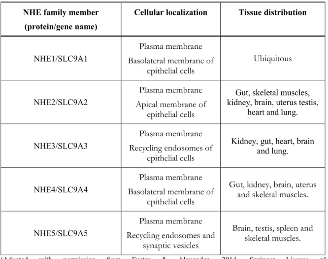
1.2 Regulators of pH Equilibrium in Tumor Cells ... 24
1.2.1 Plasmalemmal Sodium/Hydrogen Exchangers ... 26
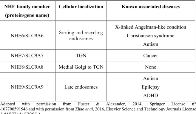
1.2.2 Intracellular Sodium/Hydrogen Exchangers ... 28
Hypothesis ... 36
2. Material and Methods ... 37
2.1 Antibodies and Reagents ... 37
2.2 Cell Culture and Hypoxic Incubation ... 37
2.3 Plasmid Constructs and Expression in Human Cell Lines ... 38
2.4 Intracellular pH Measurement ... 39
2.5 Intracellular Localization of Doxorubicin ... 39
2.6 Immunofluorescence... 40
2.7 Cell Surface Biotinylation ... 41
2.8 Complementary Confocal Microscopy Protocols... 42
2.9 Western Blotting and co-Immunoprecipitation ... 43
2.11 Protein Disorder Propensity Analysis ... 45
2.12 3D Molecular Visualization ... 45
2.13 3D Peptide Folding and Molecular Docking Refinement Protocol ... 45
2.14 Statistical Analysis ... 46
3. Results ... 47
3.1 Exposition to Hypoxia Induces Formation of a Complex Between Transiently Expressed NHE6 and RACK1. ... 47
3.2 Cellular Expression of a Peptide Derived from the SLC9A6 Gene. ... 49
3.3 Assessment of the Functional Effects from Expression of the NHE6527-588 Peptide. ... 50
3.3.1 Expression of the NHE6527-588 Peptide Prevents Formation of an Hypoxia-Inducible Complex between Transiently Expressed NHE6 and Endogenous RACK1. ... 50
3.3.2 Hypoxia Favors NHE6 Plasmalemmal Localization in Human Cancer Cell Lines; this Trend is Countered by Expression of the NHE6527-588 Peptide. ... 52
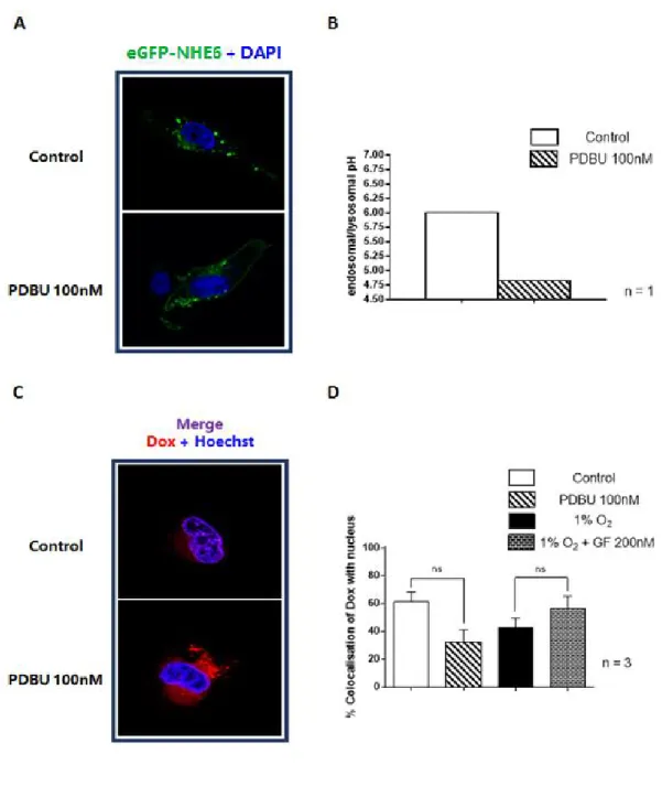
3.3.3 The Acidification of the Endosomal/Lysosomal pH in Cells Exposed to Hypoxic Conditions is Attenuated by Expression of the NHE6527-588 Peptide. ... 54
3.3.4 Localization of Doxorubicin in the Nucleus is Diminished in Hypoxia; Expression of NHE6527-588 Peptide Restores the Nuclear Levels of Doxorubicin. ... 56
3.4 Activation of PKC Produces Effects Similar to Hypoxia on NHE6 Localization, Endosomal/Lysosomal pH Modulation and Doxorubicin Distribution. ... 58
3.5 Reactive Oxygen Species Produced in Hypoxia Correlate Positively with Extra-Nuclear Distribution of Doxorubicin. ... 60
3.6 Multiple Sequence Alignment of Human NHE6, 7 & 9 with Orthologs from Other Species Reveal a Strongly Conserved Sequence within the RACK1 Binding Domain. ... 62
3.7 Analysis of NHE6 RACK1 Binding Domain as a Segment Involved in Inducible Transient Protein-Protein Interactions... 67
3.7.1 A Segment of the Human NHE6 RACK1 Binding Domain is Enriched in Neutral Polar Residues. ... 67
3.7.2 The Human NHE6 RACK1 Binding Domain has Characteristics of an Intrinsically Disordered C-terminal Region and Contains Several Possible Anchoring Residues. ... 68
3.8 Overlapping Residues Forming a Cavity at the Surface of RACK1 and Possibly Involved in the Interaction with NHE6 were Identified Through Two Alternative Methods. ... 71 3.9 NHE6 Segment Rich in Potential Anchorage Residues around Y-539 is
Predicted to be Involved in Forming an Energetically Favourable Complex with RACK1. ... 74 4. Discussion ... 79 References ... 87
Figure 1. Physical characteristics of the tumor microenvironment ... 14
Figure 2. Subcellular localization of doxorubicin under hypoxia ... 23
Figure 3. Three-dimensional structure of the RACK1 scaffold protein ... 33
Figure 4. Sequences of interaction with RACK1 amongst human NHE proteins ... 34
Figure 5. Co-Immunoprecipitation of RACK1 with Transiently Expressed NHE6-HA in Cells Subjected to Hypoxia ... 49
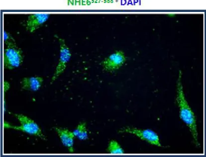
Figure 6. Immunofluorescence image of the NHE6527-588 peptide ... 50
Figure 7. Co-Immunoprecipitation of RACK1 and NHE6-GFP in MDA-MB-231 cells overexpressing the NHE6527-588 peptide. ... 52
Figure 8. Subcellular localization of NHE6 in hypoxia. ... 54
Figure 9. Measurement of endosomal/lysosomal pH in living cells. ... 56
Figure 10. Subcellular distribution of doxorubicin in stably expressing NHE6 (527-588) HT1080 cells. ... 57
Figure 11. Effect of PKC activity modulation on NHE6 localisation, endosomal/lysosomal pH and doxorubicin distribution. ... 59
61 Figure 12. ROS production in hypoxia and correlation with doxorubicin distribution in live HT1080 cells. ... 62
Figure 13. Phylogenetic tree of human NHE proteins divided into subfamilies ... 63
Figure 14. MSA analysis of a subfamily of intracellular Na+/H+ exchangers. ... 67
Figure 15. Neutral polar residue enrichment in transient interfaces. ... 68
Figure 16. Protein segment disorder propensity predicted using two alternative methods. . 71
Figure 17. Identification of potential protein binding sites at the surface of RACK1. ... 74
Figure 18. Simulation of peptide-protein interaction through molecular docking refinement protocol. ... 76
Figure 19. Examination of peptide-protein molecular docking results for buried residues. 78 Figure 20. Proposed model of cellular events in hypoxia-induced NHE6 translocation to the plasma membrane. ... 82
Table 1: Types of cancer in which clinical practice indicates primary chemotherapy for advanced cases and in which adjuvant therapy is indicated following surgery. ... 2 Table 2: Calculated Log P values for lysosome and cytoplasm partitioning of common drugs
used for cancer treatment ... 18 Table 3: Localization and tissue distribution of human NHE plasmalemmal proteins ... 27 Table 4: Cellular location and diseases associated with human NHE intracellular proteins ... 29 Table 5: Listing of antibodies used for IF and their dilutions ... 41 Table 6: Intracellular NHE sequences included in the dataset used for MSA. ... 65
ABC ALDH ALL AMAS BCRP BSA CA CDKN2A CFTR ChK ClC CML CSC c-SNARF DAPI DMSO ECF ECM EGFR EMDR EMEM EMT FAK FBS GF GNB2L1 GPCR ATP-binding cassette Aldehyde dehydrogenase Acute lymphoblastic lymphoma
Analysis of Multiply Aligned Sequences Breast cancer resistance protein
Bovine serum albumin Carbonic anhydrase
Cyclin-dependent kinase inhibitor 2A Cystic fibrosis transmembrane regulator Checkpoint kinase
Chloride channel
Chronic myelogenous leukemia Cancer stem cells
Carboxy seminaphtharhodafluor 4’,6’-diamidino-2-phenylindole Dimethyl sulfoxide
Extracellular fluid Extracellular matrix
Epidermal growth factor receptor Environment mediated drug resistance Eagle’s minimum essential medium Epithelial-mesenchymal transition Focal adhesion kinase
Fetal bovine serum GF109203X
Guanine nucleotide-binding protein subunit beta-2-like-1 G-protein coupled receptor
GSH GTPase HA HCC HER2 HIF HPTS HRE HRP IFP IGF1R ID IF IP IRS I_sc LDH LOX MBP MCT MDR MD_raw MMP MORFs MSA NADPH NHE PAGE PBS PDB PDBU Glutathione
Guanosine triphosphate cyclohydrolase Hemagglutinin
Hepatocellular carcinoma
Human epidermal growth factor-2 Hypoxia inducible factor
8-Hydroxypyrene-1,3,6-Trisulfonic Acid (or pyranine) Hypoxia responsive element
Horseradish peroxidase Interstitial fluid pressure
Insulin-like growth factor 1 receptor Intrinsically disordered
Immunofluorescence Immunoprecipitation Insulin receptor substrate Interface energy score Lactate dehydrogenase Lysyl oxidase
Maltose binding protein Monocarboxylate transporter Multidrug resistance
Meta-Disorder predictor Matrix metalloproteinase Molecular recognition features Multiple sequence alignment
Nicotinamide adenine dinucleotide phosphate Sodium-hydrogen exchanger
Polyacrylamide gel electrophoresis Phosphate-buffered saline
Protein Data Bank Phorbol 12,13-dibutyrate
PDGFR PFK-1 P-gp PHDs pHe pHi PI3K PFA PKC PKM2 PRMs PSS PTEN PVDF pVHL RACK1 RCSB ROS SDS Stat3 TAF TBS TFEB TGN TIGAR TNBC V-ATPase
Platelet-derived growth factor receptor Phosphofructokinase-1 P-glycoprotein HIF-specific prolyl-hydroxylases Extracellular pH Intracellular pH Phosphatidylinositol 3-kinase Paraformaldehyde Protein kinase C Pyruvate kinase M2 Proline-rich motifs
Protonation, sequestration and secretion Phosphatase and tensin homolog
Polyvinylidene fluoride membrane
Von Hippel-Lindau tumor suppressor protein Receptor for activated C kinase 1
Research Collaboratory for Structural Bioinformatics Reactive oxygen species
Sodium dodecyl sulfate
Activator of transcription factor 3 Tumor-associated stromal fibroblasts Tris-buffered saline
Transcription factor EB Trans-Golgi network
TP53-inducible glycolysis and apoptosis regulator Triple negative breast cancer
1.1 Clinical Consequences of the Chemoresistance Problematic Faced in Oncology Several projects from which rose precursors to modern chemotherapy took place during World War II, including the nitrogen mustards, alkylating agents that were the first chemotherapeutic agents used to treat cancer (DeVita and Chu, 2008). The early compounds used for chemotherapy were typically biological compounds isolated from living organisms that would later be chemically modified to give rise to chemotherapeutic classes with a large number of synthetic analogs. Examples of such include: analogs of folic acid, the first antifolates developed and tested during the 1940’s (DeVita and Chu, 2008), 5-flurouracyl, a modified uracil nucleobase (DeVita and Chu, 2008), both vincristine and vinblastine, plant alkaloids isolated from Catharanthus roseus (Moudi et al, 2013) and both daunorubicin and doxorubicin, anthracyclines that were isolated from the gram-positive soil bacteria Streptomyces peucetius (Di Marco et al, 1981). These projects would eventually lead to the development of compounds that became regularly used in the 1960’s for cancer treatment in the clinic alongside surgery, hormone therapy and radiotherapy (DeVita and Rosenberg, 2012). The latest forms of cancer treatment to have made their appearance in clinical practice are immunotherapy and targeted therapy in the late 1990’s (DeVita and Rosenberg, 2012).
Following early success in the late 1990’s and early 2000’s of the Bcr-Abl tyrosine kinase inhibitor Imatinib (Gleevec) used to treat chronic myelogenous leukemia (CML), targeted therapies used in personalized medicine have been widely acclaimed as the future of cancer therapy because of high success rates and few side effects (DeVita and Chu, 2008). However targeted therapies present their own set of challenges: they rely heavily on biomarkers for diagnostic purposes, each therapy can only be used for a limited subset of patients presenting the specific molecular target and they have limited potential for treatment of cancers with intratumor heterogeneity (Curigliano and Criscitiello, 2014). Steady progress has been made in the development and usage of targeted therapies over the last two decades, however research to identify practical biomarkers and to fully understand the consequences of tumor heterogeneity is still in its early stages. Therefore, it remains to be seen to which extent
targeted therapy can rise to the future challenges of oncology (Fisher et al, 2013). First-line treatments for solid tumors such as surgery and radiotherapy have limited benefits when used alone for tumors in advanced stages with poorly defined borders and in which metastases have occurred (Kirkwood et al, 2013). Likewise, these techniques can rarely be used effectively to treat cancers such as leukemia and lymphoma that do not depend on the formation of solid tumors (Greaves, 2016). For all of these reasons, despite high toxicity, chemotherapy remains one of the most commonly used forms of treatment in oncology. For many types of cancers primary chemotherapy offers the best option to improve progression free survival although it is not used for curative purposes (Table 1) (DeVita and Chu, 2008). Combination chemotherapy is used effectively for the treatment of several types of leukemia and lymphoma, it is also used as neoadjuvant therapy to reduce tumor size before surgical resection and as adjuvant therapy to reduce the likelihood of local or metastatic recurrence post-surgery (DeVita and Chu, 2008). Adjuvant chemotherapy used in a post-surgical setting generally yields greater benefit than equivalent primary therapy used in cases of unresectable tumors (Kirkwood et al, 2013). Cancer staging and use of prognosis factors is becoming increasingly precise and intricate to help guide the choice of therapy regimen that yields the best risk/benefit ratio for each individual patient.
Table 1: Types of cancer in which clinical practice indicates primary chemotherapy for advanced cases and in which adjuvant therapy is indicated following surgery.
Type of cancer Primary
chemotherapy chemotherapy Adjuvant
Anaplastic astrocytoma √ Bladder √ Breast √ √ Cervical √ √ Colorectal √ √ Esophageal √ Gastric √ √
Head and Neck √ √
Melanoma √
Nasopharyngeal √
Non-small cell lung √ √
Osteogenic sarcoma √
Ovarian √ √
Pancreatic √
Prostate √
(Adapted with permission from DeVita and Chu, 2008, American Association for Cancer Research License n° 4107690780581)
Cancer treatments that fall under the umbrella term of chemotherapy are drugs that inhibit cancer growth by impairing mitosis in fast dividing cells. They are thus cytotoxic compounds that differ from targeted therapies, hormone therapy and immune therapy because of their lack of specificity and of their significant toxicity. Although cytotoxic drugs are the preferred treatment in many cases as described above and have shown better effects than alternatives for prolonging remission, it remains that response rates are low and that development of resistance as manifested by relapse is nearly universal. Treatment regimens that include anthracycline such as doxorubicin have represented one of the most viable options for decades for the treatment of aggressive tumors. As for all the classes of chemotherapeutic agents, resistance regularly arises and deeply compromises the response to such an adjuvant therapy regimen.
Chemoresistance has been observed to occur across all types of cancers irrespectively of the tissue of origin. It has been categorized under two large categories based upon the response to the initial therapy; cancer that does not respond to initial therapy is deemed intrinsic resistance whereas cancer that returns after an initially successful therapy is called acquired resistance (Lippert et al, 2011). Examples of cancers that display intrinsic resistance, which is also called primary resistance, include acute lymphoblastic lymphoma (ALL) where approximately 20% of adult patients exhibit resistance when they first undergo treatment (Thomas et al, 1999). Patients with adenocarcinoma that undergo the prescribed treatment of
chemotherapy with surgery are 50 to 70% likely to relapse within a year and in most of those cases a phenotype of chemoresistance is acquired (Castells et al., 2012). Breast cancer is by far the most common form of cancer in women worldwide, affecting approximately 1 of 8 women at one point during their lifetime and accounting for about 14% of new cancer cases (Ferlay et al, 2015; Verwey et al, 2016). Aside from being highly prevalent, breast cancer is a type of cancer that is known to have frequent recurrences in the form of treatment resistant tumors during the years that follow surgery with neoadjuvant or adjuvant therapy (Arnason and Harkness, 2015). It has been estimated that 40% of breast cancer patients undergoing conventional treatment will eventually relapse and in the majority of those cases the returning tumors will be from metastasis; this is at least partially due to development of resistance to either chemotherapy or radiation therapy (Smalley, 2013). Although conventional therapy is usually effective for the treatment of a primary tumor it is much less so for treatment of resistant secondary tumors at the sites of metastasis (Verwey et al, 2016). Treatment outcome thus depends largely on tumor aggressiveness and metastatic potential. Breast cancer classification based on the presence or absence of the estrogen, progesterone and human epidermal growth factor-2 (HER2) receptors gives insight into the treatment approach to be used and into the aggressiveness of the tumor (Verwey et al, 2016). Triple negative breast cancer (TNBC) tumors are the most challenging to treat because they are insensitive to hormone therapy, are the most aggressive and are the most likely to recur in the form of resistant tumors from metastatic sites (O’Reilly et al, 2015). Effective treatments for TNBCs are currently lacking and urgently needed; predictive factors to guide treatment for this type of cancer also need to be developed and refined (Rakha & Chan, 2011).
Two alternate models have been postulated to explain the origin of chemotherapy resistant cells (Zharedine and Borden, 2013). In the first model, a small number of quiescent cancer stem cells (CSC) are thought to have properties that enable them to initiate tumor formation and to provoke resistance (Nguyen et al, 2012). According to the second model, called environment mediated drug resistance (EMDR), the tumor microenvironment plays a large role in modulating the entrance of tumor cells into a dormant state (Meads et al, 2009). This dormant state allows them to survive long enough under selective pressure to undergo genetic alterations that confer drug resistance even when exiting the dormant state (Meads et al,
2009). Proponents for either model have put together strong supporting arguments in both cases and although most investigators in the field of chemoresistance prefer to explain their findings according to one model, there is no reason why they could not be complementary. For the remainder of this dissertation, we have chosen to rationalize our findings under the lens of the EMDR model although we recognize that resistance arising as described in the CSC model may also have occurred.
Extrinsic microenvironment factors leading to chemoresistance that fall under the scope of the EMDR model can be either physical factors, soluble factors or can be linked to cell-adhesion mediators. Known physical factors include hypoxia and acidosis whereas known soluble factors include cytokines, chemokines and growth factors; cell-adhesion mediators include changes to integrins and other cell-adhesion molecules that often take place during epithelial-mesenchymal transition (EMT) (Meads et al, 2009).
Because the development of human cancer usually involves genomic instability that leads to an accumulation of point mutations and epigenetic modifications, a majority of tumors are comprised of highly heterogeneous populations of cells (Caulin and Maley, 2011. Epithelial breast carcinomas are a particularly striking example in which it has been reported that high intra-tumor diversity was found in 97% of tumors examined (Wild et al, 2000). Mutations acquired in cancer sub-populations can provide survival advantages for certain contexts, such as under therapeutic pressure. In such contexts, heterogeneous tumors obey the Darwinian law of evolution by natural selection; tumor clones that have undergone favourable adaptations are able survive therapy whereas all or most other clones are eliminated (Greaves, 2007). Although therapy might initially appear to have been successful because of tumor shrinkage, a niche of highly resistant cells in a dormant state (Meads et al, 2009) may remain and can eventually repopulate the tumor to cause relapse.
The problematic behind chemoresistance acquired under therapeutic selective pressure is compounded by the fact that favourable adaptations to one therapy will also negatively affect the efficacy of ensuing therapies. This phenomenon is associated with poor treatment outcome and prognosis, and it is recognized as multidrug resistance (MDR) (Ullah, 2008).
Specific mechanisms of MDR and examples will be described in a later section; for the scope of this thesis, we will discuss MDR in the context of chemotherapy resistance although it also occurs in response to other distinct compounds such as for antibiotics.
Cancer resistance to therapy is not singly limited to chemotherapy; resistance to targeted therapies and to immunotherapy have been observed and described. Unlike for chemotherapy, mechanisms of resistance to these therapies tend to involve specific molecular targets and pathways and do not typically give rise to MDR. The targeting of oncogenes around which cancer cells develop oncogene addiction has become a popular strategy for the design of therapies (Weinstein and Joe, 2008). That is because in many cases cancer cells become over reliant on these oncogenes and cannot survive if they become inactivated. In the case of tyrosine kinase oncogenes, mutations to gatekeeper residues of the kinase domain has been shown to lead to constitutive activation of the enzyme which renders targeted therapies ineffective; such examples have notably been demonstrated for BCR-ABL fusion protein, for epidermal growth factor receptor (EGFR), and for platelet-derived growth factor receptor (PDGFR) isoforms α and β (Azam et al, 2008). In other cases, targeted pathways that were initially believed to be essential for tumor survival can effectively be substituted for by a parallel pathway. One such example is the emergence of the insulin-like growth factor 1 receptor (IGF1R) pathway following treatment with phosphatidylinositol 3-kinase (PI3K) inhibitors that gives rise to PI3K inhibitor resistant cell lines (Isoyama et al, 2012).
Novel cancer therapy regimens are now being engineered to attempt to overcome the ever-present problematic of chemoresistance. Improved understanding of molecular events leading to resistance has been imperative for the recent advances in this field. As proposed by Hall et al (2009), an effective strategy to overcome MDR could rely on identifying the new “Achilles’ heel” of resistant tumors; a concept the authors refer to as collateral sensitivity (CS). According to this principle, adaptations that have made tumor cells resistant to one type of therapy may also hypersensitize them to other types of therapies. CS has certainly been observed and exploited for targeted therapies where PI3K inhibitor resistant cell lines have been treated in conjugation with selective IGF1R inhibitors for much more convincing outcomes (Isoyama et al, 2012). The concept of CS builds on the mathematical model of
tumor resistance to chemotherapy proposed by Coldman and Goldie (1983), in which being able to pinpoint alternating non-cross-resistant chemotherapy would be the optimal route for treatment. Therapies with specific cellular targets can now be used along with chemotherapy to exploit CS, a strategy that appears to be the most promising avenue for the development of future combination therapies.
1.1.1 Chemoresistance in Solid Tumors
Up to now, most of the research on resistance to chemotherapy has been focused on molecular adaptations of cancer cells leading up to cellular mechanisms of chemoresistance (Yu & Tannock, 2012). Many of the proposed therapeutic strategies that have emerged from these studies have however neglected to consider the effects that tumor microenvironment characteristics can have on hindering therapeutic efficacy (Wojtkowiak et at, 2012). Indeed, physiological factors can exacerbate resistance to chemotherapy by limiting the amount of drug in the circulation that reaches and penetrates tumor cells (Yu & Tannock, 2012). Physical microenvironment factors also act as stressors that, as per the EMDR model, select for molecular adaptations that favour survival under these harsh physical conditions and that coincidentally also confer increased resistance to chemotherapy (Doktorova et al, 2015). Although increased interstitial fluid pressure (IFP), nutrient deprivation, hypoxia and acidosis, might at first glance appear to act separately on resistance to chemotherapy, there is increasing evidence of the interplay that exists between these common physical factors of the tumor microenvironment. This section summarizes the current state of research regarding how extrinsic tumor microenvironment factors affect chemoresistance and highlights known interactions between these factors.
1.1.1.1 Increased Interstitial Fluid Pressure and Tumor Hypoxia
A fundamental difference between solid tumors and normal tissues is the increased rate of cancer cell proliferation. Because the rate of proliferation exceeds the capacity of existing and newly formed vasculature to provide a proper supply of nutrients and oxygen, it is a common feature of solid tumors to contain areas impoverished in nutrients and oxygen (Chan & Giaccia, 2007). In fact, O2 has a diffusion limit of about 100-200 μm around tumor blood
capillaries which means that hypoxic regions usually start forming in tumors that reach 1-2 mm in diameter (Egeblad et al, 2010). It has been estimated that nearly 40% of all breast tumors contain important hypoxic areas associated with increased resistance to chemotherapy and radiotherapy (Ward et al, 2013). Oxygen concentration in these areas can drop below 0.3% whereas the oxygen concentration of normal breast tissues is typically around 9% (Chun et al, 2006). A correlation between hypoxia, chemoresistance and resistance to radiotherapy has also been observed in several other types of solid tumors (Jia & Nan, 2011). Resistance to radiotherapy is due, at least in part, directly to low oxygen concentrations; the mechanism of action of radiotherapy requires the formation of cytotoxic free radicals in a reaction with oxygen to damage the integrity of DNA and halter proliferation (Pajonk et al, 2010). Along with other factors, this phenomenon can increase resistance to radiation doses by 2-3-fold. Hypoxia is also recognized to enhance the invasive potential of tumors. In this regard, hypoxia was shown to contribute to local invasion and to increase incurrence of metastasis at distant sites (Jia & Nan, 2011). Studies examining transcription of miR-210, GLUT1 and carbonic anhydrase IX (CAIX) as well as protein expression of GLUT1 and CAIX in TNBC associated with a poor prognosis reveal that the hypoxic response is likely stronger than for other types of breast cancers (Ward et al, 2013). It is thus no surprise that tumor hypoxia has become a strong indicator of poor clinical prognoses for many types of cancers (Doktorova et al, 2015).
As a tumor expands within its host tissue, it exerts pressure onto the surrounding stroma that resists the increase in volume (Stylianopoulos, 2016). This phenomenon results in a stiffening of the tumor due to densification of the extra-cellular matrix (mostly hyaluronan and collagen) and stroma (Yu & Tannock, 2012). The mechanical stress that is generated by tumor expansion also compresses intratumoral and surrounding blood and lymphatic vessels (Stylianopoulos, 2016). As perfusion from blood vessels and lymphatic vessel fluid drainage are hindered, IFP is invariably elevated. This increased IFP poses a major barrier to chemotherapy by reducing the ability of drugs to penetrate tumor tissue (Yu & Tannock, 2012). Tumor capillaries can eventually collapse if their internal pressure is exceeded by IFP; tumor vasculature collapse further limits delivery of chemotherapy, of oxygen and of nutrients. As a whole, solid tumor vasculature has been shown to be more disorganized than
for normal tissues and the distance between functional capillaries of tumors is wider than for normal tissues. Since the tumor vasculature is constantly changing, the tumor microenvironment becomes a continuously fluctuating landscape of oxygen and nutrient gradients. Therapeutic strategies targeting the extracellular matrix (ECM) and stromal cells are being explored to help normalize the IFP and to improve chemotherapy delivery as well as restore normal vasculature architecture in tumors (Yu & Tannock, 2012). These strategies include disruption of Hedgehog signaling to deplete tumor-associated stromal fibroblasts (TAF) (Olive et al, 2009) or treatment with PEGPH20, an enzyme that degrades hyaluronan (Yu & Tannock, 2012).
The most studied mediators of the hypoxic response have undoubtedly been the Hypoxia inducible factor (HIF) transcription factors; dimeric complexes formed of a HIF1α, HIF2α or HIF3α labile subunit and a stable HIF1β (also called ARNT) which as a complex can bind hypoxia responsive elements (HRE) and act as important transcriptional regulators (Keith et al, 2012). HIF alpha subunits are labile under normal oxygen tension where they are modified by HIF-specific prolyl-hydroxylases (PHDs) and targeted for proteasomal degradation via E3ubiquitin ligase von Hippel-Lindau tumor suppressor protein (pVHL). Levels of HIF1α and HIF2α expression and ensuing activity of the mature dimeric complex are primarily controlled through this post-translational mechanism where PHDs require oxygen as a substrate to allow specific recognition of HIFα subunits by pVHL. Low oxygen tension thus leads to stabilization of HIFα subunit and modulation of HRE containing genes. Up to now, more than 100 genes controlled by HIF-1 have been identified and new targets are constantly being added to the list (Yu et al, 2017); it is thus understandable that most of the research on the hypoxic response has been focused on HIF1α which also happens to be ubiquitously expressed across human tissues unlike its HIFα counterparts. The remainder of the discussion on HIF transcription factors will thus focus on HIF1α; the dimeric protein containing HIF1α will be henceforth referred to as HIF-1.
Genes modulated by HIF-1 fall under a broad range of normal and abnormal cellular processes such as proliferation, cell cycle control, glycolysis, angiogenesis, erythropoiesis, invasion, migration and chemoresistance. Many of the genes identified thus far have been
associated with either cancer progression or maintenance (Ward et al, 2013). HIF-1α has been found to be commonly overexpressed across a wide range of malignant tumor types (Barar & Omidid, 2013). The list of genes modulated by HIF-1 is too extensive to be properly represented in this section but a few examples will be briefly presented to illustrate how HIF-1 can be linked to cancer progression and maintenance.
Several oncogenic driver mutations have been shown to increase HIF-1 irrespective of oxygen tension (Ward et al, 2013). Those include mutations leading to constitutive activation of Ras GTPases regulating MAPK/ERK pathway of growth and differentiation, of Src tyrosine kinase or of kinases from the PI3K/AKT/mTor pathway of cell cycle regulation. Likewise, activation of growth factor receptors such as IGF1R, EGF, HER2 and c-Met has also been shown to elevate levels of HIF-1 (Jia & Nan, 2011). A strong positive correlation with activity of these oncogenes as well as association with loss of function of phosphatase and tensin homolog (PTEN) and cyclin-dependent kinase inhibitor 2A (CDKN2A) tumor suppressor genes indicate a role for HIF-1 as a regulator of cancer growth and progression (Barar & Omidi, 2013).
Many of the cellular functions affected by the expression of HIF-1 favour cancer maintenance by contributing to a pro-survival phenotype. Among these, angiogenesis has repeatedly been shown to be associated with stabilization of HIF-1α (Bos et al, 2005). Cell cycle deregulation and activation of growth pathways alone would not allow a tumor to grow beyond a few millimetres in diameter due to poor vascularization and appearance of necrotic zones in the de-oxygenated core of the tumor (Yu & Tannock, 2012). By promoting angiogenesis under low, but not yet lethal oxygen tensions, HIF-1 allows renewal of blood vessels and continued growth of the tumor. These new vessels do little to re-establish proper drug perfusion in the tumor core as they are of poor integrity and form and abnormal architecture (Maugeri-Saccà et al, 2011).
Glucose metabolism is another function that is significantly altered by HIF-1 expression. HIF-1 is indeed one of the key factors that lead to increased reliance on glycolysis by cancer cells, a metabolic shift widely known as the “Warburg effect”. It should be noted that
although we focus here on the role of HIF-1 in promoting glycolysis, the Warburg effect denotes a wide-ranging observation of glycolysis reliance by cancer cells that go beyond the effect of hypoxia and HIF-1. HIF-1 acts as a regulator of glycolysis by promoting the expression of pyruvate kinase M2 (PKM2) (Porporato et al, 2011), which acts as a rate-limiting enzyme of this metabolic pathway as well as by promoting the expression of glucose transporters GLUT1 and GLUT4 (Marin-Hernandez et al, 2009). Since glycolysis does not strictly require oxygen for generation of adenosine triphosphate (ATP), this metabolic shift allows extended survival in regions impoverished in oxygen. The pro-survival effects of HIF1α however extend far beyond cancer cells exposed to hypoxia; in reality, HIF-1 also affects the metabolism of neighbouring cells under normoxia. That is because HIF-1 also controls the expression of lactate dehydrogenase (LDH) which is responsible for converting pyruvate, the end-product of glycolysis, into lactate (Semenza, 2009) as well as the ratio between plasma membrane monocarboxylate transporters 1 (MCT1) and 4 (MCT4) that respectively regulate its import and export (Ullah et al, 2006). The effect is a symbiotic relationship in which lactate is exported from tumor cells in hypoxia to be imported by tumor cells in normoxia and reutilized for oxidative phosphorylation; this mechanism allows for efficient glucose metabolism and optimized energy production.
Although cancer progression and maintenance are often discussed separately due to the different cellular processes involved, these sometimes involve common signaling pathways. Such is the case for the PI3K/AKT/HIF1α that has been shown to be involved with EMT and chemoresistance for breast cancer (Semenza, 2003), hepatocellular carcinoma (HCC) (Jiao & Nan, 2012) and other types of carcinomas. The important role of HIF1α stabilization in cancer maintenance is illustrated through the concomitant expression of several markers of EMT and MDR strongly correlated with poor treatment outcome. In the first case, lysyl oxidase (LOX) (Rebucci & Michiels, 2013) , SNAIL, vimentin and N-cadherin come to mind (Imai et al, 2003), whereas BCRP, MDR-1 and c-MYC are some of the key markers identified to be associated with both hypoxia sustained expression and resistance to chemotherapy (Davies et al, 2014).
Specifically, LOX is a family of enzymes that catalyzes a reaction forming aldehydes from the lysine residues side chain amines of ECM proteins (Mayorca-Guiliani & Erler, 2013). Follows from this reaction spontaneous formation of cross-linkages between elastin and collagen fibers as part of the maturation process of ECM; through this mechanism, LOX has been shown to promote the formation of metastatic niches in several cancer types (Bennewith & Dedhar, 2011). LOX enzymes have additionally been shown to act in invasion and migration in early stages of the metastatic cascade (Erler et al, 2006). These effects of the LOX enzymes are due, at least in part, to LOX-induced stabilization of the SNAIL protein, which is a critical regulator of EMT (Sahlgren et al, 2008). SNAIL protein contributes to the loss of the epithelial phenotype in carcinomas undergoing EMT as it acts as a transcriptional repressor of the epithelial marker E-cadherin. Amongst the other important HIF-1-dependent genes that facilitate invasion and metastasis are the type IV collagen degrading metalloproteinases MMP2 and MMP9 that act by weakening the integrity of basement membranes (Gilkes & Wirtz, 2014).
Many of the pro-survival characteristics observed in cells exposed to hypoxia have been associated to HIF1-induced genes that reprogram tumor cells to acquire stem cell-like characteristics (Crowder et al, 2014). Reprogramming of CSC genes through HIF proteins contributes to chemoresistance in several ways including by increasing genomic instability, by modifying the cell cycle, by favouring glycolysis (Crowder et al, 2014) or by controlling expression of multidrug efflux pumps (Jiao & Nan, 2012). Although HIF-1 is the most studied response to hypoxia and was most often reported as a mediator of chemoresistance and resistance to radiotherapy, it would be misleading to equate the pro-survival phenotype induced by hypoxia with HIF effects. A prominent example of a complementary mediator of chemoresistance prevalent in the hypoxic microenvironment would be the loss or inactivation of the P53 cell cycle regulator. The functional form of P53 indeed acts as a negative regulator of the hypoxic response by promoting proteasomal degradation of HIF proteins (Ward et al, 2013). HIF-independent stress response pathways as well as dysregulated reactive oxygen species (ROS) and redox mechanisms are also becoming increasingly associated with the hypoxia response and MDR (Crowder et al, 2014). The relative contribution of the different elements of the hypoxic response towards chemoresitance remains largely elusive and will
undoubtedly continue to be clarified in the coming years. Current evidence suggests that oxidative stress is more severe in areas of intermittent hypoxia and that this type of hypoxia, as opposed to chronic or acute hypoxia, is mainly responsible for resistance in solid tumors (Kwee, 2014). Further investigation into cancer redox mechanisms and MDR is thus warranted. The molecular mechanisms associated with the chemoresistance mediators mentioned herein will be described further along in this introduction.
1.1.1.2 Tumor Acidosis and Metabolism
As briefly mentioned earlier, lactate, as a by-product of glycolytic tumor cells, is exported through MCT4 to the extracellular fluid (ECF) (Draoui & Feron, 2011). Lactate will then be progressively shuttled into non-glycolytic cells including endothelial tumor cells to be used in oxidative phosphorylation (Ullah et al, 2006). As recognized by A. Weinberg and accepted by most of cancer researchers, metabolic reprogramming, distinguished by an increased rate of glucose metabolism through glycolysis, is now among the list of the hallmarks of cancer (Hanahan & Weinberg, 2011). Since increased glycolysis generates protons as by-products at a fast rate (Alfarouk et al, 2014); they tend to accumulate in cancer cells particularly under anaerobic conditions where protons cannot react with molecular oxygen to form water (Chiche et al, 2010). One of the ways by which tumor cells prevent acidification of intracellular pH is through MCT (primarily MCT4) symporter secretion of H+ ions with
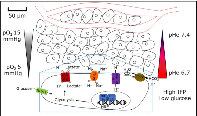
lactate (Draoui & Feron, 2011).Proton extrusion from cancer cells however far exceeds what is needed to maintain intercellular pH equilibrium; in fact, proton export from cancer cells through both primary and secondary active transport machinery leads to proton gradient reversal across the plasma membrane (Alfarouk et al, 2014). Examples of other important transporters that mediate proton export from tumors include: V-ATPase proton pump, CAIX and sodium-hydrogen exchange 1 (NHE1). Whereas normal tissues have extracellular pH (pHe) around 7.4 and intracellular pH (pHi) around 7.2, pHe of tumors in acidosis is typically in the range of 6.6-6.8 and pHi in the range of 7.3-7.5 (Barar & Omidi., 2013). As distance from blood vessel increases and oxygen tensions drop, pHe also progressively diminishes and can go as low as pH 6.0 (Gatenby et al, 2017). Figure 1 depicts some of the physical characteristics of the tumor microenvironment.
Figure 1. Physical characteristics of the tumor microenvironment
Depiction of oxygen concentration and pH gradients typical of the tumor microenvironment and of the main transporters involved in creating and maintaining tumor acidosis.
It has been argued that exposition to the harsh tumor microenvironment, that includes physical factors such as hypoxia and acidosis, selects specifically for tumor cells that display extensive proton export machinery (Wojtkowiak et at, 2012). The early stage at which pH reversal occurs during carcinogenesis indeed supports the idea that this adaptation plays a major role in cancer progression (Reshkin et al, 2014). In fact, cancer cells that cannot regulate their pHi have been shown to be less proliferative and to be less likely to form tumors (Ward et al, 2013).
The cancer cell’s response to acidosis remains poorly understood; that is in part because findings from past studies have often been confounding. Four major cellular alteration features have however stood out in the literature: these are metabolic reprogramming, defense against ROS, triggering of autophagy and alterations in endo-lysosomal functions (Lamonte et al, 2013; Wojtkowiak et at, 2011 and Zhitomirsky & Assaraf, 2015).
Reversal of pH gradient in cancer cells has at times been reported to contribute to a switch from oxidative phosphorylation to glycolysis just has it has been reported to inhibit glycolysis. The foremost claim that the activity of glycolytic enzymes such as lactate dehydrogenase and phosphofructokinase-1 (PFK-1) is increased due lo lower intracellular pH (Calderon-Montano et al, 2011) whereas the later claim that these genes are down-regulated as a result of lactic acidosis (Chen et al, 2008). The divergence in these findings suggests that distinct cellular machinery must be important in determining the exact metabolic response to acidosis; the reason behind these seemingly conflicting results may lie with the P53 status of cells being studied. In fact, the P53 protein has been shown to be activated by tumor acidosis when functional and to reduce glycolysis through TP53-inducible glycolysis and apoptosis regulator (TIGAR) (Lamonte et al, 2013). Additionally, the same study showed that P53 activated through acidosis contributes to the activity of two other important metabolic pathways: the oxidative branch of the pentose phosphate pathway (PPP) necessary for novel ribose synthesis and glutaminolysis which is parallel pathway to glycolysis for ATP generation by cancer cells for their elevated energy requirements. These two pathways have in common the fact that they both increase the reductive potential of the cells, which leads us to the next feature of the acidosis response: defense against ROS. Consequently, the overall level of the reducing equivalent nicotinamide adenine dinucleotide phosphate (NADPH) is raised and serves to counteract the similar increase in ROS due to the stress generated by acidosis (Lamonte et al, 2013). Neutralization of oxidative stress under such harsh microenvironmental conditions is necessary to ensure prolonged tumor cell survival. Because the P53 dependent changes outlined above also coincide with decreased proliferation; it is unclear if the sum of these adaptations plays a role that is tumor suppressive or if they contribute to cancer maintenance. Increased pentose phosphate activity as induced by acidosis, has previously been associated with brain metastasis (Chen et al, 2007) and chemoresistance (Tamada et al, 2012). Importantly, an enhanced reductive potential, manifested through increased NADPH replenishment, is a recurring theme with other environmental stresses such as hypoxia, glucose deprivation and matrix detachment; it thus seems to be a key necessary feature for cancer cell extended survival in the tumor microenvironment.
Also consistent with the cellular responses to acidosis mentioned above, is the triggering of autophagy in response to starvation (Wojtkowiak et al, 2012). It appears that autophagy in this context is required as an adaptation to obtain biomolecules from recycled organelles to offset metabolic pathways skewed towards catabolism. Alteration of the endo-lysosomal compartments is the final feature of the response to acidosis and is closely related to the process of autophagy, which entails fusion of lysosomes and phagosomes to form phagolysosomes (Hosogi et al, 2014). Different studies in fact suggest tumor acidosis has the effect of increasing the number of lysosomes (Avnet et al, 2016), lowering their pH value (Zhitomirsky & Assaraf, 2015) and triggering the secretion of lysosomal components into the cytoplasm (Steffan et al, 2009). Low lysosomal pH in cancer as well as concomitant occurrence of autophagy, and transcription factor EB (TFEB) associated lysosomal biogenesis have been linked to prolonged survival under tumor microenvironment stress (Salerno et al, 2014) (Zhitomirsky & Assaraf, 2015). Aside from the induction of phagocytosis which was already discussed; release of lysosomal components and acidification of lysosomal compartments have both been shown to contribute to increased survival of tumor cells. Not only does low pHe acts on lysosomal trafficking to increase lysosomal exocytosis (Steffan et al, 2009) but it also increases the expression of lysosomal proteases such as MMP2, MMP9 and Cathepsin B (Wojtkowiak et at, 2011). In an acidic environment, these enzymes promote invasion of the ECM which ultimately also contributes to migration and survival. Early reports showed that low pHe can inhibit gap junction (Ruch et al, 1990), this finding taken with others that show low pHe favors mesenchymal type-like morphology (Amith et al, 2016) suggest that tumor acidosis may play a role in deconstruction of cell junctions to further contribute to cancer progression through EMT (Lamouille et al, 2014).
A steep gradient between the intracellular, extracellular and endo-lysosomal pH has been shown to contribute to decreased tumor responsiveness to chemotherapy (Wojtkowiak et al, 2011) and radiotherapy treatments (Ohtsubo et al, 2001). Although it appears that the characteristic of resistance to radiotherapy is mostly linked to the selection of tumor cells with defective P53 necessary for adaptation to chronic acidosis (Williams et al, 1999), chemoresistance is in turn largely due to the impact that altered pH has on the tissue and cell
distribution of the therapeutics themselves. Although the phenomena of impaired tissular and cellular distribution of chemotherapy has been observed on several occasions over the last few decades, it is only recently that it has become well understood and integrated under one overarching framework. The work of Robert J. Gillies and Yehuda J. Assaraf, among others, clearly explains how drug distribution is affected through inter-compartment pH gradients. Both authors have described a phenomenon coined as “ion trapping” (Wojtkowiak et al, 2011) that explains how the pH gradient acts as a physiological barrier for drugs or other molecules. This phenomenon is of relevance because for many drugs there exists a large difference in permeability between the nonionized species of a drug, that are more permeant, and the ionized species that can be much more impermeant. In the case of tumor acidosis, this phenomenon negatively impacts the capacity of weak base chemotherapeutics to effectively permeate through the cell membrane and ultimately reach their intended therapeutic target which generally is the cell nucleus. Even the small proportion of weak base therapeutics that does penetrate the plasma membrane is not guaranteed to reach the nucleus as it is also likely to passively diffuse and become trapped in acidic organelles such as lysosomes (Zhitomirsky & Assaraf, 2015). Similarly, molecules that enter cells through endocytosis can become entrapped in late endosomes or lysosomes as they are sorted to acidic lysosomes through the endo-lysosomal system. This phenomenon however cannot be generalized to all chemotherapeutic drugs as some drugs do not have ionizable species, and thus pH would not alter their membrane permeability, and others are weak acids which means the pH gradient found in tumor acidosis would positively affect their ability to cross the plasma membrane (Wojtkowiak et al, 2011).
In fact, based on the chemical structure of each drug and the pH value of cellular compartments, it is possible to calculate a partitioning coefficient on a logarithmic scale called Log P to describe the distribution of the equilibrium between two phases, one being the lysosomes and the other being the cytoplasm. Such calculated Log P values (Zhitomirsky & Assaraf, 2015) for common chemotherapeutic drugs are shown in table 2. Drugs with Log P values close to 1, such as is the case for doxorubicin and Mitoxantrone, would thus be partitioned at a ratio of 10 to 1 in favor of acidic compartments such as lysosomes as opposed to the cytoplasm.
Table 2: Calculated Log P values for lysosome and cytoplasm partitioning of common drugs used for cancer treatment
Drug Log P Lysosome trapping
Lapatinib 4.64 [+] Gefitinib 3.75 [+] Vincristine 3.13 [+] Sunitinib 2.93 [+] Pyrimethamine 2.75 [+] Daunorubicin 1.73 [+] Mitoxantrone 1.19 [+] Doxorubicin 0.92 [+] Pemetrexed 0.73 [-] 5-Fluorouracil -0.66 [-]
Adapted with permission from Zhitomirsky & Assaraf, 2015.
Both extracellular space and intracellular compartments have been shown to entrap large amounts of chemotherapeutic drugs and to limit their effectiveness (Gerweck, 2006) (Zhitomirsky & Assaraf, 2015); the importance of both parallel occurrences should be taken into consideration while studying drug resistance since the extracellular space represents a much larger volume but lysosomes can reach much lower pH values neighboring pH 5 or lower. The phenomenon of “ion trapping” should certainly be taken into consideration in selecting the best therapeutic approaches for cancer treatment and for future design of cancer therapeutics (Wojtkowiak et al, 2011).
1.1.2 Cellular and Molecular Mechanisms of Chemoresistance
As recognized previously, the tumor microenvironment has a crucial role to play in the development of chemotherapy and is clearly an important driver in selecting molecular determinants for resistance to chemotherapy. This section provides an overview of specific cellular and molecular determinants of chemoresistance as currently recognized in the literature.
1.1.2.1 Drug Detoxification
Mechanisms of drug detoxification constitute a rather disparate group of processes that all share the characteristic of limiting chemotherapeutic drug activity; these processes can take an active form where drugs are inactivated by being metabolized or via chemical modifications, or they can take a passive form where the necessary activation mechanisms are repressed. A prime example of detoxification through a metabolic enzyme would be the ability of aldehyde dehydrogenases (ALDH), important biomarkers of CSC to remove various toxic aldehydes generated as intermediary metabolites of several chemotherapeutic drugs such as cyclophosphamides (Tomita et al, 2016). High expression of ALDH enzymes in breast CSCs has been strongly associated with increased resistance to chemotherapy and radiotherapy; effects that can be prevented through inhibition of ALDH activity (Croker & Allan, 2012). ALDH plays an analogous role in normal stem cells for protection against cellular stress and improvement of survival outcomes. Another common mechanism contributing to chemoresistance is the conjugation of an anionic group such as glutathione (GSH), glucuronate or sulfanate, to chemotherapeutic drugs (Homolya et al, 2003). Such chemical modifications transform many drugs in such a way that they become substrates to ATP-binding cassette (ABC) transporters that can then export such drugs outside cancer cells. These multidrug resistance efflux pumps will be discussed in more details in an incoming section. Finally, some chemotherapeutic drugs need to first be activated to become cytotoxic; cytarabine needs to undergo a series of phosphorylation to cytarabine triphosphate in order to be fully active (Sampath et al, 2006). Cancer cells have been known to adapt to cytarabine treatment by downregulating kinases responsible for phosphorylating the drug (Bardenheuer et al, 2005).
1.1.2.2 Multidrug Resistance Efflux Pumps
Transporters responsible for drug efflux and development of MDR mostly belong to the ATP-binding cassette (ABC) protein superfamily that can be subdivided into three subfamilies that each have different drug substrate specificities (Glavinas et al, 2004). ATP hydrolysis is the general mechanism used by ABC transporters to pump a variety of drugs outside of cells or inside vesicular compartments to lower intracellular drug concentrations (Nakanishi and Ross, 2012). Fueling ABC transporters represents a large burden for cancer cell metabolic needs, as approximately two ATP molecules are consumed to export each molecule of the substrate and ATP hydrolysis continues to a certain extent even in the absence of any substrate. There is yet no agreed upon naming convention for ABC transporters and each protein transporter tends to be referred to under several different names. The P-glycoprotein (P-gp), also called MDR-1, is expressed through the ABCB1 gene, whereas the Breast cancer resistance protein (BCRP) is expressed through the ABCG2 or MXR gene. These first two protein subfamilies mainly recognize and export large positively charged amphiphilic compounds whereas the final subfamily of ABC transporters the multidrug resistance proteins (MRPs), expressed through the ABCC gene, recognize neutral hydrophobic molecules or certain soluble anionic compounds (Glavinas et al, 2004). P-gp is known to transport and to lead to resistance against drugs such as vinca alkaloids, anthracyclines and paclitaxel (Gottesman et al.,2002). MCRP also transports anthracyclines and also recognizes topoisomerase inhibitors and mitoxantrone. Finally, MRP1 can transport many classes of chemotherapeutic compounds including vinca alkaloids and anthracyclines, although these compounds often need to have first been conjugated with glutathione (GSH), glucuronide or sulfate (Yin and Zang, 2011). There is evidence that certain selective pressures, such as cancer drug exposition, lead to differential P-gp localization (Petriz et al, 2004), which when targeted to the lysosomal membrane can lead to lysosomal drug sequestration (Chapuy et al, 2008).
1.1.2.3 Defects and Imbalances in Cell Death and Survival Pathways
There exist a multitude of adaptations that cancer cells may undergo involving modifications to cell death and survival pathways that ultimately result in increased resistance to chemotherapy. As these have been extensively described in the literature, to summarize these
adaptations could represent an entire review on its own; as such the current section merely seeks to acknowledge their vast role in EMDR and to provide a few examples. Of course, it isn’t possible to discuss alterations to apoptotic pathways without mentioning selection of inactivating mutations to P53, s staple of apoptotic proteins that has wide ranging impacts on cellular function and that is correlated with de-novo resistance to doxorubicin in breast cancer (Arnason and Harkness, 2015). Alternatively, selective pressures may by contrast select for activating mutations to anti-apoptotic proteins such a B cell lymphoma 2 (Bcl-2) protein (Teicher, 2006). In many cases the effects of environmental pressures such as hypoxia and high pHi can prevent activation of G1/S checkpoint and activate checkpoint kinases 1 & 2 (ChK1 & ChK2) that coordinate the DNA damage responses with the cell cycle to facilitate progression through the G2 phase (Maugeri-Saccà et al, 2011). Hypoxia can also negatively affect several DNA damage response pathways including homologous recombination, non-homologous end joining and mismatch repair (Bristow and Hill, 2008); hypoxia may also result in telomere shortening and chromosomal instability (Verdun and Karlseder, 2007). Modifications to cell death and survival pathways may result from selection by exposition to a particular drug; however once acquired these adaptations are likely to cause resistance to a wide-ranging spectrum of drugs.
1.1.2.4 Compartmentalization and the PSS Model
The rationale behind the cellular mechanism of weak base therapeutics compartmentalization has previously been covered through the explanation of the lysosomal ion-trapping phenomenon. It must be noted that ion-trapping is in no way exclusive to lysosomes as it has also been observed to occur in other acidic cellular compartments such as endosomes (Lee & Tannock, 2006). Description of ion-trapping as a contributing factor to chemoresistance was first described in 1996 where following the initial observation that doxorubicin resistant MCF7 breast cancer cells have more acidic intra-vesicular compartments than their doxorubicin sensitive counterparts, the protonation, sequestration and secretion (PSS) model was proposed by Sanford Simon (Schindler et al, 1996) to rationalize increased resistance to doxorubicin. The first two step of the process are consistent with ion-trapping as previously described and the third step refers to secretion through efflux pumps or through simple exocytosis.
Cellular or molecular mechanisms leading up to compartmentalization of weak base therapeutics by cancer cells remain to be described. Doxorubicin is often used as model drug in experimental design involving microscopy as it has the advantageous property of emitting natural fluorescence (Lucien et al, 2014). This property allowed our laboratory to observe doxorubicin extranuclear spots reminiscent of the ion-trapping phenomenon (Figure 2) in HT1080 cells (Lucien et al, 2017). Similar findings had previously been obtained by other researchers (Lee & Tannock, 2006), but our results were the first indication that the tumor microenvironment, in this case hypoxia, could consistently increase drug sequestration. The current study will explore ramifications of this key finding and how improved understanding of this apparent mechanism of chemoresistance can be exploited in a novel therapeutic approach.
Figure 2. Subcellular localization of doxorubicin under hypoxia
A) Representative images of sub-cellular distribution of doxorubicin in live HT1080 cells under normoxia and hypoxia. The nucleus was stained by incubating cells with Hoechst 33342. Cells at 1% O2 were kept under hypoxia for 4h. B) The percentage of doxorubicin
1.2 Regulators of pH Equilibrium in Tumor Cells
Distinct metabolic signatures of tumor cells and increased in energy demands contribute to create a harsh tumor microenvironment characterized by highly acidic ECF. These conditions exert major selective pressures on tumor cells that force them to adapt to such adverse variations. Expression and activity of a number of proton channels and exchangers can be modulated by cancer cells in order to survive tumor acidosis. Regulators of tumor cell pH equilibrium and their known functions in the process are described below.
The MCT protein family responsible for the transport of monocarboxylate substrates such as pyruvate, lactate and ketone bodies is constituted of 14 known members, but only MCT1-4 have been confirmed to have a proton linked transporter activity (Halestrap, 2012). Although each of these transporters may contribute to either efflux or influx of protons, MCT1 was shown to be mostly located in well oxygenated regions of xenograft tumors where it is involved in proton influx (Sonveaux et al, 2008) whereas MCT4 is better adapted to facilitate efflux (Dimmer et al, 2000). The activity of these transporters is driven by the concentration gradient of the monocarboxylate substrates, such as lactate, on which proton transport is dependent (Wilson et al, 2009). In the case lactate and proton transport, lactic acid is transiently formed during transport across the plasma membrane to be readily dissociated in the ECF. Although MCT1 and MCT4 have by far received the most attention in cancer research, MCT2 has also been found to be expressed in brain, prostate and colon cancer (Spugnini et al, 2015). Evidence of import and export duality of the MCT transporters in tumor goes a long way to support the theory of metabolic symbiosis in tumors. Interestingly, in-vivo inhibition of MCT1 was shown to limit tumor growth (Sonveaux et al, 2008). This finding may be a result of lactate metabolism being replaced by glycolysis in MCT1 expressing cells which would limit the pool of fuel sources the tumor as a whole can use. Several MCT inhibitors are currently being developed or undergoing clinical trial; one example would be the 7ACC2 compound which acts as a specific inhibitor of lactate import by MCT1 and MCT4 transporters (Draoui et al, 2014). Another family of proteins that is critical for inter and intracellular pH regulation is that of carbonic anhydrases (CAs); a family of 14 known zinc metalloenzymes that catalyze the reaction from one molecule of carbon dioxide and of water, two oxidative phosphorylation by-products, to a bicarbonate anion and