HAL Id: hal-02512403
https://hal.archives-ouvertes.fr/hal-02512403
Submitted on 19 Mar 2020HAL is a multi-disciplinary open access archive for the deposit and dissemination of sci-entific research documents, whether they are pub-lished or not. The documents may come from teaching and research institutions in France or abroad, or from public or private research centers.
L’archive ouverte pluridisciplinaire HAL, est destinée au dépôt et à la diffusion de documents scientifiques de niveau recherche, publiés ou non, émanant des établissements d’enseignement et de recherche français ou étrangers, des laboratoires publics ou privés.
THE N-TERMINAL DOMAIN OF ANNEXIN 2
SERVES AS A SECONDARY BINDING SITE
DURING MEMBRANE BRIDGING
Malik Zibouche, Michel Vincent, Françoise Illien, Jacques Gallay, Jesus
Ayala-Sanmartin
To cite this version:
Malik Zibouche, Michel Vincent, Françoise Illien, Jacques Gallay, Jesus Ayala-Sanmartin. THE N-TERMINAL DOMAIN OF ANNEXIN 2 SERVES AS A SECONDARY BINDING SITE DURING MEMBRANE BRIDGING. Journal of Biological Chemistry, American Society for Biochemistry and Molecular Biology, 2008, 283 (32), pp.22121-7. �10.1074/jbc.M801000200�. �hal-02512403�
THE N-TERMINAL DOMAIN OF ANNEXIN 2 SERVES AS A SECONDARY BINDING SITE DURING MEMBRANE BRIDGING
Malik Zibouche1, 2, Michel Vincent3, 4, Françoise Illien1, 2, Jacques Gallay3, 4, and Jesus Ayala-Sanmartin1, 2.
From 1INSERM UMRS893, CdR Saint-Antoine, Paris, F-75012 ; 2Université Pierre et Marie Curie,
CHU Saint-Antoine, Paris, F-75012 ; 3CNRS UMR8619 IBBMC, Orsay, F-91405; 4Université
Paris-Sud, Orsay, F-91405
Running head: The Annexin A2 N-terminal domain associates with membranes
Address correspondence to: Jesus Ayala-Sanmartin, INSERM UMRS893, CdR Saint Antoine, 27 rue Chaligny, 75012 Paris, France. Tel: 33 1 40 01 13 24. Fax: 33 1 40 01 13 90. E-mail:
jayala@chusa.jussieu.fr, and Jacques Gallay, IBBMC, UMR8619 CNRS Université Paris-Sud Bâtiment 430, F-91405 Orsay. Tel.: 33 1 69 15 48 42; fax: 33 1 69 85 37 15; e-mail:
jacques.gallay@u-psud.fr
Annexin A2 (AnxA2) is a Ca2+- and acidic phospholipid- binding protein involved in many cellular processes. It undergoes Ca2+ -mediated membrane bridging at neutral pH and has been demonstrated to be involved in an H+-mediated mechanism leading to a novel AnxA2-membrane complex structure. We used fluorescence techniques to characterize this H+-dependent mechanism at the molecular level, in particular the involvement of the AnxA2 N-terminal domain. This domain was labeled at Cys8 either with acrylodan or pyrene-maleimide fluorescent probes. Steady-state and time-resolved fluorescence analysis for acrylodan and fluorescence quenching by doxyl-labelled phospholipids revealed direct interaction between the N-terminal domain and the membrane. The absence of pyrene excimer suggested that interactions between N-termini are not involved in the H+-mediated mechanism. These findings differ from those previously observed for the Ca2+-mediated mechanism. Protein titration experiments showed that the protein concentration for half-maximal membrane aggregation was twice for Ca2+-mediated compared to H+ -mediated aggregation, suggesting that AnxA2 was able to bridge membranes either as a dimer or as a monomer, respectively. A N-terminally deleted AnxA2 was two to three times less efficent than the wild type protein for H+-mediated membrane aggregation. We propose a model of AnxA2-membrane assemblies, highlighting the different roles of the N-terminal domain in the H+- and Ca2+ -mediated membrane bridging mechanisms.
Annexins (Anx) belong to a family of Ca2+-dependent phospholipid-binding proteins
and have various membrane-related functions
(1,2). These proteins have two structural domains: a C-terminal core with a conserved structure and a N-terminal domain variable in sequence and length (3). The core domain is formed by four repeats (or eight for AnxA6) containing Ca2+ binding sites. Each repeat
consists of five α-helices connected by short loops. The four repeats are organized into a slightly curved oblate shape with a convex face harboring the Ca2+ binding sites, allowing contact
with membrane phospholipids. The variable N-terminal domain, facing the concave surface at the opposite side of the membrane bilayer, contains interaction sites for other protein partners. This domain harbors a number of posttranslational modifications and regulates the membrane-activity properties of the core domain.
Anx proteins not only bind the surface of model and biological membranes but can also assemble into different structures on these surfaces. Some of these proteins including AnxA5 can assemble into lateral structures forming two-dimensional networks on the membrane surface (4-6). Others, including AnxA1 and AnxA2 form membrane junctions with or without protein ligands attached to their N-terminal segment (7-11). The classical mechanism described for these two processes involves a primary Ca2+-dependent membrane
binding step leading to conformational changes of the core domain, in particular of repeat III (12-18). The N-terminal domain of Anxs also plays an important role in the specific membrane aggregative properties. In particular, the N-terminal domain of AnxA1 and AnxA2 interacts strongly with proteins of the S100 family, resulting in a several-fold increase in the Ca2+ sensitivity of membrane aggregation (17). Recently, we showed that the N-terminal domain of monomeric AnxA2 was also involved in the Ca2+-dependent membrane bridging at neutral
pH: we demonstrated interactions between the N-terminal domains of two adjacent AnxA2 molecules, which were not in direct contact with the membrane phospholipids (19).
Besides this classical Ca2+-dependent
mechanism, Anx proteins also bind both model and cellular membranes independently of calcium ions (20,21) or in a proton-dependent manner (22-27). This Ca2+-independent binding is associated with an increased hydrophobicity of Anx at acidic pH (28,29), and may involve a significant conformational change of the core generating a transmembrane form of the protein as suggested for AnxB12 (23,30). This hypothesis has been extended to AnxA5 (24) and AnxA6 (25), but not for AnxA1 and A2. AnxA2 undergoes an H+-dependent conformational
change of repeat III, very similar to that in the presence of Ca2+ at neutral pH, but the H+
-mediated binding at the membrane surface did not involve further structural modification of the core domain (17). The monomeric AnxA2 started to form bridges in vitro in the absence of Ca2+ at
pH 6 and attained the maximal bridging at pH 5.2. However, the organization of the membrane junctions at acidic pH differed from that at neutral pH (26).
We investigated the molecular structure of these novel membrane junctions previously observed by cryo-electron microscopy (26) for monomeric AnxA2 at pH 4. In particular we studied the dynamics of the N-terminal domain in the free soluble form of the protein at acidic pH in the absence of calcium and the conformational and environment changes of this domain that may occur during membrane bridging, using steady-state and time-resolved fluorescence techniques. We specifically labeled the reactive cysteine Cys8 in the N-terminus by thiol-reactive fluorescent markers (19,31). We used 6-acryloyl-2-dimethylaminonaphthalene (acrylodan), a polarity-sensitive probe (32,33) to detect environment and mobility changes of the N-terminal domain and the excimer-sensitive probe N-(1-pyrene)maleimide (pyrene-maleimide) (34), to detect potential interactions between N-termini that we previously showed for the Ca2+-dependent membrane bridging mechanism at neutral pH (19). We propose a model, based on our findings, for the organization of AnxA2 in Ca2+- and H+-mediated membrane bridging.
Experimental Procedures
Chemicals- Acrylodan and
N-(1-pyrene)maleimide were purchased from Molecular Probes (Invitrogen, France). Type III-B egg α-phosphatidylcholine (PC) and brain L-α-phosphatidyl-L-serine (PS) were obtained from Sigma-Aldrich, France. Dr. Claude Wolf (CHU Saint-Antoine, Paris) kindly provided 1-
palmitoyl-2-stearoyl(n-doxyl)-sn-glycerophosphatidylcholine (n-doxyl PC) derivatives labelled with a doxyl group at positions n = 5, 7 and 12 of the sn2 chain.
Protein preparation and labelling with acrylodan and pyrene maleimide- AnxA2 produced in S. cerevisiae was purified as previously described
(35). Protein purity was estimated as >95% by gel electrophoresis. The N-terminally deleted AnxA2 (ΔN-AnxA2) was prepared as described in (10). The only reactive cysteine of AnxA2, Cys8, was labelled with acrylodan or pyrene maleimide using the method of Johnsson et al. (31) previously described in (19). The efficiency of acrylodan labelling was 50% and that of pyrene-maleimide labelling was 83% (see (19)). Labelled proteins were stored at -20°C. Membrane aggregative properties did not appear to differ between labelled and unlabelled proteins.
LUV preparation and aggregation- Large
unilamellar vesicles (LUV) (PC/PS: 75/25 weight ratio) were prepared by extrusion as previously described (36). Briefly, lipids were mixed together in chloroform. The solvent was removed from the mixture under a stream of nitrogen. Residual solvent was removed under vacuum for one hour. Lipids were then resuspended in buffer A (40 mM Hepes pH 7, 30 mM KCl, 1 mM EGTA) at a final concentration of 1 mg ml-1, by vortexing vigorously. The
multilamellar liposomes were then extruded by passing the suspension 21 times through a polycarbonate membrane with 0.1 μm pores (Avestin, Canada). Free calcium concentration, expressed as pCa (-log [Ca2+]), was controlled by
EGTA buffering. Vesicle aggregation was monitored in buffer A in the presence of calcium, or in buffer B (40 mM Acetate pH 4, 30 mM KCl, 1mM EGTA) for pH 4 experiments, by turbidimetry at 340 nm, as previously described (37).
Steady-state fluorescence measurements-
Fluorescence emission spectra were recorded with a Cary Eclipse spectrofluorimeter with slit width of 10 nm and 5 nm for excitation and emission, respectively. Samples were contained in microcuvettes (120 μl).
Time-resolved fluorescence measurements-
Fluorescence intensity and anisotropy decays were obtained from the polarized Ivv(t) and Ivh(t)
components measured by the time-correlated single-photon counting technique. A diode laser (LDH 405 from Picoquant, Berlin-Adlershof, Germany; maximal emission at 392 nm) operating at 10 MHz was used as an excitation source. A Hamamatsu fast photomultiplier (model R3235-01) was used for detection. Emission wavelength was selected with a Jobin-Yvon H10 monochromator (band width 16 nm) and a Schott KV418 cut-off filter. Each experimental decay Ivv(t) and Ivh(t) was stored on
a 2 K plug-in multichannel analyzer card (Ortec Trump-PCI 2k, Ametek France), using Maestro-32 software. Automatic data sampling was controlled by the microcomputer. The instrumental response function (FWHM ~600 ps) was collected automatically by measuring the light scattering of a glycogen solution during 30 s at the excitation wavelength, alternating with the parallel and perpendicular components of polarized fluorescence decay, each over a total of 90 s. Samples were contained in microcuvettes (120 μl).
Fluorescence intensity decay analyses were performed with maximum entropy method (MEM), using a multiexponential model,
) exp( i i i t
τ
α
−∑
, as previously described(38). A classical anisotropy model,
∑
−i i
iexp( t
θ
)β
, in which any rotationalcorrelation time (θ) is coupled with each lifetime (τ), was used to resolve polarized fluorescence decays (39). Calculations were carried out with a set of 150 or 100 independent variables (equally spaced on a logarithmic scale) for intensity and anisotropy, respectively. The programs, including the MEMSYS 5 subroutines (MEDC Ltd, Cambridge, UK), were written in double-precision FORTRAN 77.
Fluorescence quenching by doxyl-PCs-
Quenching by doxyl-PCs (distearoylphosphatidylcholine (PC) labelled
with a doxyl group bound at C5, C7 and C12 on the sn2 acyl chain) was carried out with LUV prepared with 10, 20 or 30% molar fractions of labelled PC, 25% PS and supplemented with unlabelled PC. The distance, Zcf, from the centre
of the bilayer can be estimated according to the expression:
where F1 and F2 are the fluorescence intensities
of the fluorophore in the presence of the shallow and of the deeper quenchers, respectively; L21 is
the vertical distance between the shallow and the deep quenchers; Lc1 is the distance between the
shallow quencher and the centre of the bilayer; and C is the concentration of the quenchers in molecules per unit area (40). The distance of the quenchers from the centre of the bilayer was 12.15 Å for 5-doxyl PC and 5.85 Å for 12-doxyl PC (41).
RESULTS
Conformational dynamics of the acrylodan-labelled AnxA2 N-terminal segment in solution at pH 4. The steady-state fluorescence emission
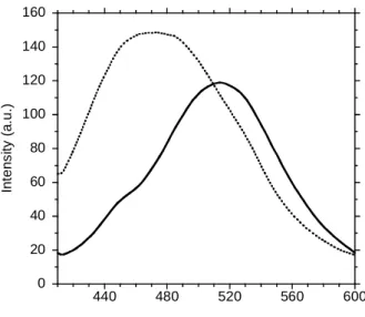
spectrum of Annexin A2 labelled with acrylodan (AnxA2acryl) at pH 4 peaked at 515-520 nm,
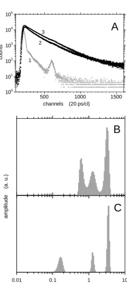
consistent with previously reported values at neutral pH. Thus, the environment of acrylodan bound to Cys8 in the N-terminal segment remained highly polar in mild acidic conditions (Fig. 1). The fluorescence intensity decay was not monoexponential (Fig. 2A). MEM analysis of the data showed the presence of three lifetime populations (Fig. 2B & C), probably attributable to the existence of three local conformers: we did not detect any sign of excited-state reaction, such as fluorescence build-up at long emission wavelength (data not shown). The centre value and amplitude of each lifetime peak are presented in Table 1. The lifetime values are similar to those measured at neutral pH, showing that mild acidification does not alter the interactions between acrylodan and its microenvironment. However, we observed an H+-induced redistribution of the long and the intermediate lifetime amplitudes, involving a significant increase of the amplitude average lifetime by ~15%: thus, mild acidification seemed to induce a redistribution of local conformers.
Fluorescence anisotropy decay of AnxA2acryl showed rapid dynamics of the AnxA2
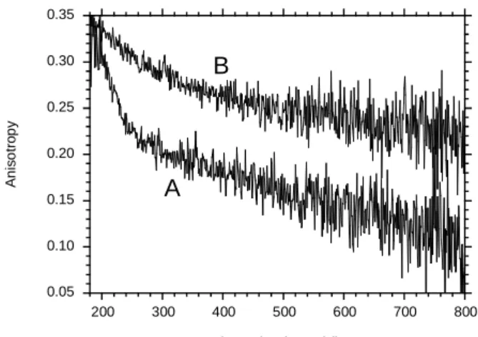
N-terminal segment at pH 4, consistent with that observed at neutral pH. The experimental anisotropy decay curve decreased rapidly at first, and then more slowly (Fig. 3). Two time constants were calculated: a subnanosecond component and a much longer component (Table
2). The short decay time probably reflects the local, partially restricted motion of acrylodan around its linker. The semi-angle value of the wobbling-in-cone subnanosecond rotation ωmax was large. The longest rotational correlation time value probably corresponds to the Brownian rotational motion of the protein. This value is significantly smaller at pH 4 than at pH 7 (19), consistent with the more compact state of the protein at mild acidic pH in solution (26).
Conformational dynamics of the acrylodan-labelled AnxA2 N-terminal segment upon membrane bridging at pH 4. Interaction of
AnxA2acryl with negative phospholipid membranes at mild acidic pH, shifted the fluorescence emission spectrum of acrylodan to shorter wavelengths (from 515 nm to 470 nm for L/P=100) (Fig. 1). The amplitude of the shift (45 nm) is significantly larger than that observed at pH 7 in the presence of calcium (30 nm) (19), suggesting that the microenvironment of acrylodan is more apolar when the protein interacts with the membrane in mild acidic conditions than at neutral pH in the presence of calcium. The fluorescence intensity decay changed slightly upon membrane bridging (Fig. 2 & Table 1), indicating a local conformational change of the N-terminal segment during these processes.
The AnxA2-induced membrane bridging caused a significant decrease of the local dynamics of the N-terminal segment compared to the protein in solution, as shown by fluorescence anisotropy decay (Fig. 3). MEM analysis computed two rotational correlation time populations (Table 2). A nanosecond rotational motion appeared and the longest rotational correlation time value increased substantially. However, the initial anisotropy value At=0 was significantly lower than the intrinsic anisotropy value in vitrified media (42), thus subnanosecond rotation was probably present. The semi-angle of the acrylodan subnanosecond wobbling-in-cone rotation ωmax also decreased considerably (Table 2), confirming a much larger restriction of movement than that previously observed for the Ca2+-dependent membrane bridging mechanism
at neutral pH (19).
Location of the AnxA2 N-terminal segment in bridged membranes in mild acidic conditions.
We showed above that when monomeric AnxA2acryl binds to LUV in mild acidic
conditions, favouring membrane aggregation, the
fluorophore moved to a hydrophobic environment. Direct interactions of the N-terminus with the membrane, as previously described for AnxA1 (22,43-45) may underlie these observations. To determine the extent of AnxA2acryl-membrane interactions, quenching
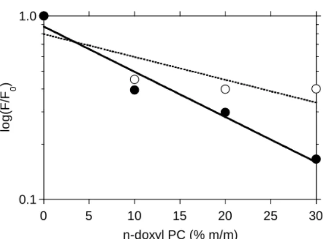
experiments were performed with LUV (PC/PS 75/25) containing n-doxyl PC at different mole fractions (10, 20 and 30) (40,46). The various n-doxyl PCs showed high levels of quenching efficiency (up to 80%), depending on their mole fractions (Fig. 4). These findings demonstrated that acrylodan bound to Cys8 on the N-terminal segment of the protein interacted strongly with the membrane lipids. The distance of the acrylodan moiety from the centre of the bilayer was estimated from the quenching efficiency values of the shallow 5-doxyl PC and that of the deeper 12-doxyl PC derivatives to be 10.7 Å. Ca2+-induced membrane bridging by monomeric
AnxA2 at neutral pH involves interactions between their N-terminal segments, previously demonstrated by disulfide bond formation (47), and by the formation of excimers using AnxA2 labelled by N-pyrene maleimide on Cys8 (19). However, in the absence of calcium and at pH 4, we did not observe the spectral signature corresponding to excimer formation (emission peak at 455 nm) (34). for the AnxA2-membrane complex, suggesting that N-terminal domain dimerisation is not required for membrane bridging in mild acidic conditions (Fig. 5).
Molecular organisation of AnxA2 in membrane bridges in mild acidic pH. In membrane bridging
experiments, the rise in turbidity (vesicles aggregation) was greater at pH 4 in the absence of calcium than at pH 7 in its presence. We previously viewed membrane bridges by cryo-electron microscopy in mild acidic conditions and found that monomeric AnxA2 was mainly organized in a single protein layer (26), whereas at pH 7, in the presence of calcium mainly two protein layers were observed, indicating 'face-to-face' dimerization of AnxA2 molecules (11). Thus, two molecules appeared to be necessary for membrane bridging in the presence of calcium at neutral pH, but only one molecule was required at mild acidic pH. It would thereby follow that protein concentration needed to attain half maximal extent of aggregation at pH 4 must be two-fold lower than that at pH 7 in the presence of calcium. To test this hypothesis, the extent of aggregation was measured as a function of protein concentration. Indeed, the protein
concentration giving half maximal aggregation at pH 4 was 2.5±0.2 μg ml-1 and 5.1±0.2 μg ml-1 at
pH 7 in the presence of calcium (pCa 3) (Fig. 6A). The magnitude of cooperativity of Ca2+
-induced membrane aggregation at pCa 3 (two molecules) was greater (Hill number 0.90) than at pH 4 (one molecule) (Hill number 0.34). The protein titration experiments suggested that at acidic pH, membrane bridging required only one molecule of AnxA2, and the quenching experiments by n-doxyl PC that the N-terminal domain was in contact with the membrane. To test the role of the N-terminal domain contact with the membrane in bridging, we compared the abilities of the complete and the N-terminally deleted AnxA2 (ΔN-AnxA2) abilities for membrane aggregation. As shown in Fig. 6B, ΔN-AnxA2 is able to bridge membranes, but the extent of aggregation is two to three-fold lower than the aggregation mediated by the complete protein. This indicates that the contact of the N-terminal domain with the membrane is not strictly necessary but is important to enhance the capacity of the protein to form membrane bridges at acidic pH.
DISCUSSION
The annexin family of membrane binding proteins binds negatively charged phospholipid membranes through either Ca2+-dependent or
Ca2+-independent/H+-dependent mechanisms
(20,24,26,29). The classical Ca2+-dependent
binding and aggregation mechanisms have been extensively studied (see (2) for a review). Several aspects of the H+-dependent binding have been
studied for annexins A1, A2, A5, A6, and B12. Two types of membrane interaction have been proposed: insertion of the protein into the membrane bilayer, resulting in a transmembrane form of the protein (in particular AnxB12 (30,48,49)), and the association at the membrane surface without large structural changes of the core domain (shown for AnxA2 (26)). A modulatory role of the AnxA1 N-terminal domain in H+-mediated binding has also been suggested (22).
In this study, we investigated the dynamics of the AnxA2 N-terminal domain and its localization with respect to the membrane during the H+ -mediated membrane bridging. We previously demonstrated that AnxA2 aggregated membranes in the pH range (6-5.2) (26), a pH range in agreement with that published for AnxA2 in cells (50). The experiments in this study were
performed at pH 4 to avoid data misinterpretation due to mixed AnxA2 populations (soluble, bound and aggregative forms), and to favor 100% of the protein in membrane bridges. The N-terminal domain was labeled on Cys8 by thiol-specific fluorescent probes: acrylodan and pyrene-maleimide. The characteristics of the N-terminal domain during H+-mediated membrane bridging were compared with those previously observed for the Ca2+-mediated processes.
In solution, the N-terminal tail of AnxA2 at pH 4 was as flexible as at pH 7 with Ca2+. We did not detect any H+-induced conformational change of this domain similarly to our previous observations with Ca2+ (19). This is in contrast to
the core domain, where we observed a H+- (26)
and a Ca2+-induced restricted mobility (17) for
the sole tryptophan residue (W212) in repeat III. The two domains of the AnxA2 molecule behave therefore independently.
The environment and dynamics of acrylodan bound to the N-terminal domain of the protein in H+-induced membrane bridges differed from
those previously observed in Ca2+-induced
membrane bridges: the environment was less polar and the subnanosecond mobility more restricted than in the Ca2+-induced
membrane-bridged state. Furthermore, the n-doxyl PC quenchers also behaved differently in the two conditions: in the H+-driven membrane-bridged
mechanism, highly efficient quenching revealed strong interactions between the N-terminal domain and the membrane, whereas a low level of quenching in the Ca2+-induced state indicated
the absence of these strong interactions (19). Additionally, the geometry of the junctions differed in these two conditions: the Ca2+
-induced mechanism involves interactions between N-termini, demonstrated previously by formation of excimers with N-pyrene-maleimide-labelled monomeric AnxA2 (19), that were not observed in this study, thereby suggesting that such interactions did not occur in the H+
-mediated membrane bridging mechanism at mild acidic pH.
The distribution of protein charges can provide information about the topology of the H+-dependent membrane junctions. Anx A2 is mainly neutral with a theoretical isoelectric point of 7.56 but the distribution of the charged residues is not homogeneous. The core exhibits strong positively charged regions on its convex face, with many Lys residues (49 in repeat I; 340, 206 and 249 in repeat III; 279, 281, 313 and 324 in repeat IV) and some Arg residues (205 and
245 in repeat III) exposed at the surface. These residues can make electrostatic bridges with the negatively charged PS headgroups, thereby replacing the Ca2+ bridges as some of these Lys
residues are close to the Ca2+-binding loops. The
flanking and the convex surfaces of repeat I also have a strong positive charge density, probably favoring interaction between the N-terminal segment and the negatively charged membrane surface. The N-terminal segment (residues 1 to 30) has a net charge of +5 at pH 4. The first 14 residues of this segment are able to behave like an amphipathic α-helix in different conditions such as in water/TFE mixtures (51), and when complexed with S100A10 (p11) (52). It is thus possible that the N-terminal domain may fold as an amphipathic helix on the membrane surface in a similar way as when complexed with S100A10 (p11). This helix could interact with the membrane surface through salt bridges involving His4 and Lys9. In such conformation, acrylodan bound to Cys8 would be located on the hydrophilic side of the helix (52). Its spectroscopic properties showed however that it should be located inside the membrane bilayer. This potential amphipathic helix could therefore lie at the membrane/water interface, with its hydrophilic side located at a shallow position in this interface, thus acrylodan would then be in contact with the acyl chains, the hydrophobic side of the helix facing the concave face of the protein.
We propose a model incorporating previous findings and those from this study, implicating two mechanisms for the monomeric AnxA2 organization during membrane bridging (Fig. 7): one being Ca2+-mediated and the other H+
-mediated. In cells, levels of H+ and Ca2+ are
interconnected, Ca2+ gradients being important
cofactors of pH-induced effects (50). Our model proposes a balance between these ions that may favor either mode of organization, determined by the physiological conditions and the cellular compartments. Both mechanisms require the presence of acidic phospholipids for interaction of the protein core with the membrane, mediated by either Ca2+- or salt bridges. The N-terminal
domain appeared to have a different modulating role in each process, probably related with its electrical charge. In the classical Ca2+-mediated
mechanism at neutral pH, two N-terminal domains interact, without direct contact with the membrane surface (Fig. 7A) (19). These N-terminal interactions stabilize a complex between two AnxA2 molecules that form the Ca2+
-mediated membrane bridges, consistent with previous observations by cryo-electron microscopy (11) and with our aggregation experiments in the present study. Our model for H+-dependent membrane bridging, consistent with cryo-electron microscopy studies (26), proposes that these bridges are formed with only one AnxA2 molecule located between two juxtaposed membranes, with the core interacting with one membrane surface on its convex side and the N-terminal domain interacting with the second membrane surface on the concave side, as previously suggested for AnxA1 (22,43) (Fig. 7B). It is possible that some residues on the concave side of the molecule could be involved also in the interactions since the ΔN-AnxA2 was partly able to perform membrane aggregation. Notice that in the presence of calcium and at neutral pH, Lambert et al. (11) observed mainly two protein stripes for AnxA1 and A2, but they also observed some bridges with only one protein stripe. These structures may thus coexist and are not exclusive for one calcium or proton concentration. Other factors such as ionic strength or temperature may be involved in determining the organization of Anx membrane bridges. In both cases, core-core contacts may contribute to the stabilization of the structure as shown by cross linking of AnxA1 (43) and AnxA2 (53). Two possible orientations of the AnxA2 core are schematized. The structure depicted at the bottom of Fig. 7B is consistent with the observation of dimers in the crystal structure of the complete AnxA2 (54).
Finally, the physiological role of the monomeric AnxA2 has been suggested by studies on membrane exocytosis with no S100A10 protein (55) and in particularly acidic organelles (56) such as endosomes in which AnxA2 may have an impact in endosomal membranes fusion. Therefore, the proposed models for AnxA2-mediated membrane bridging are likely to reflect physiological events in cells and this may underlie the calcium-independent membrane binding observed in endosomes (20).
REFERENCES
1.
Rescher, U., and Gerke, V. (2004) J Cell Sci 117, 2631-2639
2.
Gerke, V., Creutz, C. E., and Moss, S. E. (2005) Nat Rev Mol Cell Biol 6, 449-461
3.
Gerke, V., and Moss, S. E. (2002) Physiol Rev 82, 331-371
4.
Mosser, G., Ravanat, C., Freyssinet, J. M., and Brisson, A. (1991) J Mol Biol 217,
241-245
5.
Pigault, C., Follenius-Wund, A., Schmutz, M., Freyssinet, J. M., and Brisson, A.
(1994) J Mol Biol 236, 199-208
6.
Oling, F., Santos, J. S., Govorukhina, N., Mazeres-Dubut, C., Bergsma-Schutter, W.,
Oostergetel, G., Keegstra, W., Lambert, O., Lewit-Bentley, A., and Brisson, A. (2000)
J Mol Biol 304, 561-573.
7.
Drust, D. S., and Creutz, C. E. (1988) Nature 331, 88-91
8.
Wang, W., and Creutz, C. E. (1994) Biochemistry 33, 275-282
9.
Bitto, E., and Cho, W. (1999) Biochemistry 38, 14094-14100
10.
Ayala-Sanmartin, J. (2001) Biochem Biophys Res Commun 283, 72-79.
11.
Lambert, O., Gerke, V., Bader, M. F., Porte, F., and Brisson, A. (1997) J Mol Biol
272, 42-55
12.
Pigault, C., Follenius-Wund, A., Lux, B., and Gerard, D. (1990) Biochim Biophys Acta
1037, 106-114
13.
Sopkova, J., Renouard, M., and Lewit-Bentley, A. (1993) J Mol Biol 234, 816-825.
14.
Concha, N. O., Head, J. F., Kaetzel, M. A., Dedman, J. R., and Seaton, B. A. (1993)
Science 261, 1321-1324.
15.
Sopkova, J., Gallay, J., Vincent, M., Pancoska, P., and Lewit-Bentley, A. (1994)
Biochemistry 33, 4490-4499
16.
Sopkova, J., Vincent, M., Takahashi, M., Lewit-Bentley, A., and Gallay, J. (1999)
Biochemistry 38, 5447-5458.
17.
Ayala-Sanmartin, J., Vincent, M., Sopkova, J., and Gallay, J. (2000) Biochemistry 39,
15179-15189
18.
Sopkova-De Oliveira Santos, J., Vincent, M., Tabaries, S., Chevalier, A., Kerboeuf,
D., Russo-Marie, F., Lewit-Bentley, A., and Gallay, J. (2001) FEBS Lett 493, 122-128
19.
Ayala-Sanmartin, J., Zibouche, M., Illien, F., Vincent, M., and Gallay, J. (2008)
Biochim Biophys Acta 1778, 472-482
20.
Jost, M., Zeuschner, D., Seemann, J., Weber, K., and Gerke, V. (1997) J Cell Sci 110,
221-228
21.
Ayala-Sanmartin, J., Henry, J., and Pradel, L. (2001) Biochim Biophys Acta 1510,
18-28.
22.
Rosengarth, A., Wintergalen, A., Galla, H. J., Hinz, H. J., and Gerke, V. (1998) FEBS
Lett 438, 279-284.
23.
Langen, R., Isas, J. M., Luecke, H., Haigler, H. T., and Hubbell, W. L. (1998) J Biol
Chem 273, 22453-22457
24.
Isas, J. M., Cartailler, J. P., Sokolov, Y., Patel, D. R., Langen, R., Luecke, H., Hall, J.
E., and Haigler, H. T. (2000) Biochemistry 39, 3015-3022
25.
Golczak, M., Kicinska, A., Bandorowicz-Pikula, J., Buchet, R., Szewczyk, A., and
Pikula, S. (2001) Faseb J 15, 1083-1085.
26.
Lambert, O., Cavusoglu, N., Gallay, J., Vincent, M., Rigaud, J. L., Henry, J. P., and
Ayala-Sanmartin, J. (2004) J Biol Chem 279, 10872-10882
27.
Monastyrskaya, K., Babiychuk, E. B., Hostettler, A., Rescher, U., and Draeger, A.
(2007) Cell Calcium 41, 207-219
28.
Genge, B. R., Wu, L. N., Adkisson, H. D. t., and Wuthier, R. E. (1991) J Biol Chem
266, 10678-10685
29.
Kohler, G., Hering, U., Zschornig, O., and Arnold, K. (1997) Biochemistry 36,
8189-8194.
30.
Kim, Y. E., Isas, J. M., Haigler, H. T., and Langen, R. (2005) J Biol Chem 280,
32398-32404
31.
Johnsson, N., Marriott, G., and Weber, K. (1988) Embo J 7, 2435-2442
32.
Weber, G., and Farris, F. J. (1979) Biochemistry 18, 3075-3078
33.
Prendergast, F. G., Meyer, M., Carlson, G. L., Iida, S., and Potter, J. D. (1983) J Biol
Chem 258, 7541-7544
34.
Galla, H. J., and Hartmann, W. (1980) Chem Phys Lipids 27, 199-219
35.
Ayala-Sanmartin, J., Gouache, P., and Henry, J. P. (2000) Biochemistry 39,
15190-15198
36.
Lamaziere, A., Burlina, F., Wolf, C., Chassaing, G., Trugnan, G., and
Ayala-Sanmartin, J. (2007) PLoS ONE 2, e201
37.
Lamaziere, A., Wolf, C., Lambert, O., Chassaing, G., Trugnan, G., and
Ayala-Sanmartin, J. (2008) PLoS ONE 3, e1938
38.
Vincent, M., Brochon, J. C., Merola, F., Jordi, W., and Gallay, J. (1988) Biochemistry
27, 8752-8761.
39.
Vincent, M., and Gallay, J. (1991) Eur Biophys J 20, 183-191
40.
Chattopadhyay, A., and London, E. (1987) Biochemistry 26, 39-45
41.
Abrams, F. S., and London, E. (1993) Biochemistry 32, 10826-10831
42.
Gallay, J., Vincent, M., Li de la Sierra, I. M., Munier-Lehmann, H., Renouard, M.,
Sakamoto, H., Barzu, O., and Gilles, A. M. (2004) Eur J Biochem 271, 821-833
43.
Bitto, E., Li, M., Tikhonov, A. M., Schlossman, M. L., and Cho, W. (2000)
Biochemistry 39, 13469-13477.
44.
Rosengarth, A., Gerke, V., and Luecke, H. (2001) J Mol Biol 306, 489-498.
45.
Hu, N. J., Bradshaw, J., Lauter, H., Buckingham, J., Solito, E., and Hofmann, A.
(2008) Biophys J 94, 1773-1781
46.
London, E., and Feigenson, G. W. (1981) Biochemistry 20, 1932-1938
47.
Ayala-Sanmartin, J., Cavusoglu, N., Masliah, J., and Trugnan, G. (2004) Annexins 1,
19-25
48.
Langen, R., Isas, J. M., Hubbell, W. L., and Haigler, H. T. (1998) Proc Natl Acad Sci
U S A 95, 14060-14065
49.
Ladokhin, A. S., and Haigler, H. T. (2005) Biochemistry 44, 3402-3409
50.
Monastyrskaya, K., Tschumi, F., Babiychuk, E. B., Stroka, D., and Draeger, A. (2008)
Biochem J 409, 65-75
51.
Hong, Y. H., Won, H. S., Ahn, H. C., and Lee, B. J. (2003) J Biochem (Tokyo) 134,
427-432
52.
Rety, S., Sopkova, J., Renouard, M., Osterloh, D., Gerke, V., Tabaries, S.,
Russo-Marie, F., and Lewit-Bentley, A. (1999) Nat Struct Biol 6, 89-95
53.
Faure, A. V., Migne, C., Devilliers, G., and Ayala-Sanmartin, J. (2002) Exp Cell Res
276, 79-89.
54.
Rosengarth, A., and Luecke, H. (2004) Annexins 1, 129-136
55.
Chasserot-Golaz, S., Vitale, N., Sagot, I., Delouche, B., Dirrig, S., Pradel, L. A.,
Henry, J. P., Aunis, D., and Bader, M. F. (1996) J Cell Biol 133, 1217-1236
56.
Morel, E., and Gruenberg, J. (2007) PLoS ONE 2, e1118
FOOTNOTES
We would like to thank Dr. C. Wolf for kindly providing the n-doxyl PC derivatives. This work was supported by CNRS and INSERM funding.
The abbreviations used are: Acrylodan, 6-acryloyl-2-dimethylaminonaphthalene; Anx, annexin; AnxA2acryl, Annexin A2 labelled with acrylodan on Cys8; ΔN-AnxA2, Annexin A2 deleted of the 29 first residues; LUV, large unilamellar vesicles; MEM, maximum entropy method; n-doxyl PC, 1-palmitoyl-2-stearoyl(n-doxyl)-sn-glycerophosphatidylcholine (n= 5, 7 or 12); P11, S100A10 protein; PC, egg L-α-glycerophosphatidylcholine; PS brain L-α-glycerophosphatidyl-L-serine; Pyrene-maleimide, N-(1-pyrene)maleimide.
TABLES Table 1
The effect of AnxA2acryl binding to LUV (PC/PS 75/25) at pH 4 on fluorescence intensity decay,
measured by MEM of acrylodan. Excitation wavelength: 392 nm; emission wavelength: 510 nm for AnxA2 and 480 nm for AnxA2-LUV. Protein concentration 0.5 μM.
Sample τ1 (ns) α1 τ2 (ns) α 2 τ3 (ns) α 3 <τ> (ns) a AnxA2 0.58 ± 0.11 0.22 ± 0.03 1.28 ±0.17 0.30 ± 0.06 3.37 ± 0.05 0.48 ± 0.03 2.13 ± 0.03 AnxA2 LUV L/P=100 0.17 ± 0.02 0.32 ± 0.05 1.30 ± 0.09 0.22 ± 0.04 3.59 ± 0.06 0.47 ± 0.06 2.01 ± 0.17 AnxA2 LUV L/P=150 0.18 ± 0.01 0.33 ± 0.01 1.32 ± 0.0.2 0.19 ± 0.01 3.62 ± 0.06 0.48 ± 0.02 2.05 ± 0.01
a amplitude averaged lifetime :
i i i
τ
α
τ
>=∑
< Table 2The effect of AnxA2acryl binding to LUV (PC/PS 75/25) at pH 4 on the fluorescence anisotropy decay
of acrylodan; MEM analysis.
Sample θ1(ns) β1 θ2(ns) β2 At=0 = Σβi ωmax (°) a AnxA2 0.14 ± 0.03 0.191 ± 0.036 15 ± 2 0.232 ± 0.001 0.423 31 ± 1 AnxA2 LUV L/P=100 2.2 ± 0.4 0.038 ± 0.004 42 ± 4 0.290 ± 0.013 0.328 16 ± 1 AnxA2 LUV L/P=150 1.4 ± 0.1 0.041 ± 0.008 37 ± 1 0.292 ± 0.007 0.333 15 ± 1
a The semi-angle of the wobbling-in-cone subnanosecond motion was calculated as ω
max = arccos{1/2[(1+8(Σβω /A0)1/2)1/2-1]}, with βω = Σβi (with associated θi > 1 ns); The intrinsic anisotropy A0 = 0.370 measured in vitrified medium was used (42). AnxA2acryl concentration: 0.5 μM.
FIGURE LEGENDS
Fig. 1. Fluorescence spectra of acrylodan in AnxA2acryl. Spectrum from 0.5 μM protein in solution at
pH 4 shows a maximum peak at about 515 nm (plain line). A blue shift is observed (peak at 470 nm) in the presence of phospholipid membranes (dotted line). The lipid protein molar ratio L/P is 100. Fig. 2. Time-resolved fluorescence intensity decay and lifetime distribution of AnxA2acryl. A) Time-resolved fluorescence decay. Instrumental response function (curve 1); experimental decays of: AnxA2acryl at pH 4 in solution (curve 2); AnxA2acryl bound to LUV (PC/PS 75/25) L/P = 100 at pH 4 (curve 3). Experimental conditions are shown in the legend of Table 1. B,C) MEM reconstructed excited state lifetime distribution of AnxA2acryl at pH 4. B) AnxA2acryl in solution. C) AnxA2acryl associated to LUV (PC/PS 75/25) L/P = 100. Numerical values are shown in Table 1.
Fig. 3. Experimental fluorescence anisotropy decay of AnxA2acryl at pH 4. AnxA2acryl (curve A),
AnxA2acryl bound to LUV (PC/PS 75/25) (L/P = 100) (curve B). Experimental conditions as in Table
1.
Fig. 4. Quenching of AnxA2acryl by doxyl PCs. Acrylodan fluorescence intensity was measured with
LUV with different mole ratios of PC labelled with the quencher doxyl, at carbon positions 5 (○) and 12 (●) of the acyl chain, as described in the methods section.
Fig. 5. Fluorescence emission spectra of AnxA2pyr at pH 4. 0.5 μM protein in buffer (plain line). 0.5 μM protein bound to LUV (PC/PS 75/25) L/P = 100 (dotted line). Excitation wavelength: 350 nm. Spectra normalized at 378 nm emission.
Fig. 6. Annexin 2-mediated membrane aggregation. A) LUV aggregation measured by turbidimetry at pH 4 (●) and at pH 7 with calcium (1 mM) (○) as a function of AnxA2 concentration. B) Protein concentration dependent LUV aggregation at pH 4 for AnxA2 (●) and ΔN-AnxA2 (□). Means of three independent experiments.
Fig. 7. Models for Annexin 2 membrane bridging. Schematic representation of the model for monomeric AnxA2 in membrane bridges. Two modes of organization of the protein are proposed: the classical calcium-dependent (A on the left) and the proton-dependent mechanisms (B on the right). Both modes are in equilibrium and the mode of organization of the membrane bridge is determined mainly by the balance in H+ and Ca2+ concentrations. Ca2+ favours the organization at the left and H+
Figure 1 0 20 40 60 80 100 120 140 160 440 480 520 560 600 In te ns it y ( a. u .) emission wavelength (nm)
Figure 2 100 101 102 103 104 105 500 1000 1500 co u n ts channels (20 ps/cl) 1 2 3 0.01 0.1 1 10
excited state lifetime (ns)
am pl it u d e (a. u. )
A
B
C
Figure 3 0.05 0.10 0.15 0.20 0.25 0.30 0.35 200 300 400 500 600 700 800 An is o tro p y channels (20 ps/cl) A B
Figure 4 0.1 1.0 0 5 10 15 20 25 30 lo g(F /F 0 ) n-doxyl PC (% m/m)
Figure 5 0 100 200 300 400 500 400 440 480 520 In te n s it y ( a .u .) emission wavelength (nm)
Figure 6 0 5 10 15 20 0 20 40 60 80 100 AnxA2 (μg/ml) LU V a ggr e ga ti on ( % ) 0 5 10 15 20 0.00 0.02 0.04 0.06 0.08 0.10 0.12 0.14 Protein (μg/ml) L U V ag g reg at io n ( O D)
A
B
Figure 7