HAL Id: hal-01632392
https://hal.archives-ouvertes.fr/hal-01632392
Submitted on 20 Oct 2018
HAL is a multi-disciplinary open access
archive for the deposit and dissemination of
sci-entific research documents, whether they are
pub-lished or not. The documents may come from
teaching and research institutions in France or
abroad, or from public or private research centers.
L’archive ouverte pluridisciplinaire HAL, est
destinée au dépôt et à la diffusion de documents
scientifiques de niveau recherche, publiés ou non,
émanant des établissements d’enseignement et de
recherche français ou étrangers, des laboratoires
publics ou privés.
Carbonate-containing apatite (CAP) synthesis under
moderate conditions starting from calcium carbonate
and orthophosphoric acid
Doan Pham Minh, Ngoc Dung Tran, Ange Nzihou, Patrick Sharrock
To cite this version:
Doan Pham Minh, Ngoc Dung Tran, Ange Nzihou, Patrick Sharrock. Carbonate-containing
ap-atite (CAP) synthesis under moderate conditions starting from calcium carbonate and
orthophos-phoric acid.
Materials Science and Engineering:
C, Elsevier, 2013, 33 (5), p.
2971-2980.
Carbonate-containing apatite (CAP) synthesis under moderate conditions starting
from calcium carbonate and orthophosphoric acid
Doan Pham Minh
a,⁎
, Ngoc Dung Tran
a, Ange Nzihou
a, Patrick Sharrock
baUniversité de Toulouse, Mines Albi, CNRS, Centre RAPSODEE, Campus Jarlard, F-81013 Albi cedex 09, France bUniversité de Toulouse, SIMAD, IUT Paul Sabatier, Avenue Georges Pompidou, 81104 Castres, France
a b s t r a c t
Keywords: Apatite One-step synthesis Calcium carbonate Orthophosphoric acidThe synthesis of carbonate-containing apatite (CAP) from calcium carbonate and orthophosphoric acid under moderate conditions was investigated. In all cases, complete precipitation of orthophosphate species was ob-served. The reaction temperature influenced strongly the decomposition of calcium carbonate and therefore the composition of formed products. The reaction temperature of 80 °C was found to be effective for the com-plete decomposition of calcium carbonate particles after 48 h of reaction. Infra-red spectroscopy (IR), nuclear magnetic resonance (NMR), thermogravimetry/mass spectroscopy (TG–MS) coupling, and X-ray diffraction (XRD) characterizations allowed the identification of the composition of formed products. By increasing the reaction temperature from 20 °C to 80 °C, the content of A-type CAP increased and that of B-type CAP de-creased, according to the favorable effect of temperature on the formation of A-type CAP. The total amount of carbonate content incorporated in CAP's structure, which was determined by TG–MS analysis, increased with the reaction temperature and reached up to 4.1% at 80 °C. At this temperature, the solid product was mainly composed of apatitic components and showed the typical flat-needle-like structure of CAP particles obtained in hydrothermal conditions. These results show an interesting one-step synthesis of CAP from calcium car-bonate and orthophosphoric acid as low cost but high purity starting materials.
1. Introduction
The principal mineral composition of bones and teeth consists of apatite-based compounds, and among them calcium hydroxyapatite (Ca-HA, Ca10(PO4)6(OH)2) is considered as the main component. In
addi-tion, bones and teeth also contain substituted apatites with the presence of other elements including Na+, Mg2+, Cl−, F−and CO
3
2−[1]. Calcium
carbonate-containing apatite (CAP) is considered as the major impurity, which is formed by the replacement of PO43−and/or OH−groups in
Ca-HA's structure by CO32−groups[2,3]. Ca-HA has been considered as
a prominent biomaterials for bone and dental tissue reconstitution for its excellent bioactivity with hard tissues, but recently, substituted Ca-HA such as CAP has been found to be more effective with a shorter post-operative rehabilitation program[4–6]. Thus, CAP base materials at-tract much attention and the awareness of the contribution of CAP to bone and dental enamel health is increasing[7].
Much work has been devoted to the preparation of CAP, which gen-erally consists of the precipitation of orthophosphate anions by calcium cations in aqueous solution in the presence of CO32−anions[8–10], or
the heating of a calcium phosphate at high temperature under a flux of CO2[11,12]. By solution reactions, a mixture of water soluble salts
is usually used to enhance the insertion of CO32−anions in the apatitic
structure. This leads to supplementary washing steps for the elimina-tion of counter ions in order to purify final solid products. Washing steps may be arduous when nano-particles of calcium phosphates are formed, accompanied by the formation of waste by-products containing all counter ions. The heating route relates to a multi-step synthesis in rigorous conditions of temperature and pressure. By this way, a mixture of calcium phosphate (for example, tricalcium phosphate (Ca3(PO4)2),
or calcium pyrophosphate (Ca2P2O7)), lime (CaO) and/or calcium
car-bonate (CaCO3) is heated under CO2atmosphere[12–14].
The present study reports a simple one-step synthesis of CAP from calcium carbonate and orthophosphoric acid under moderate conditions of temperature and pressure. Calcium carbonate was chosen as the source of both calcium cations and carbonate anions, and orthophosphoric acid was chosen as the most effective orthophosphate for the dissolution of calcium carbonate powder particles. Their wide availability and low cost also constitute advantages for an eventual in-dustrialization of the synthesis process.
2. Materials and methods
Calcite powder (CaCO3, 98%) with particles of volume-mean
diam-eter of 23.3 μm from Fisher Scientific and orthophosphoric acid (H3PO4, 85 wt.% in water) from Merck were used as received, without
further modification. The synthesis was carried out in a 250 mL stain-less steel reactor (Top Industrial). The reactor was equipped with an
⁎ Corresponding author. Tel.: +33 563493258; fax: +33 563493043. E-mail address:doan.phamminh@mines-albi.fr(D. Pham Minh).
electrical heating jacket and a magnetic stirrer. For each reaction, 10 g of CaCO3and 45 mL of water were fed into the reactor. After closing,
6.9 g of H3PO4was quickly injected into the reactor by an injection
valve and the reaction started by adjustment of the stirring rate at 800 rpm. During the reaction, the reactor was thermostated at the se-lected temperatures in the range of 20–80 °C for 48 h. After reaction, solid phase was separated from liquid phase by filtration using 0.45 μm filter paper. Then the solid products were dried at 50 °C for 48 h before further characterizations.
Different analysis and characterization techniques were used for studying both solid and liquid phases. Elemental analysis was carried out using inductively coupled plasma atomic emission spectroscopy (ICP-AES) with a HORIBA Jobin Yvon Ultima 2 apparatus. X-ray diffraction (XRD) of the solids was performed using a Philips PANalytical X'pert Pro MPD diffractometer with a Cu Kα (1.543 Å) radiation source.
Thermogravimetry (TG) was performed in a TA Instruments SDTQ600 an-alyzer with a heating rate of 5 °C min−1under air flux (100 mL min−1).
Exhaust gases from the TG analyzer outlet were analyzed by a Pfeiffer Vacuum OmniStar GSD 320 mass spectrometer which acquires mass spectra from 0 to 200 amu (atomic mass unit). Diffuse reflection infrared Fourier transform (DRIFT) spectra were recorded on a Shimadzu FTIR 8400S spectrometer on ground solid powders.31P and1H NMR spectra
were registered on a Bruker Avance 400, operating at 10 kHz for both
31P and1H NMR at room temperature (21 °C). The
adsorption–desorp-tion isotherm was determined with a MICROMETRICS ASAP 2010 using nitrogen as gas adsorbate with the data collection from relative pressure (P/P°) of 0.03 to 0.99. True density of the solid powders was measured by helium pycnometry using an Accupyc 1330 (Micromeritics). Scanning electron microscopy (SEM) was measured on a Philips XL30 ESEM appa-ratus (FEI Company). Particle size distribution was measured by laser scattering in a Mastersizer 2000 (Malvern Instruments Ltd., Malvern, UK) in the range from 0.020 to 2000 μm.
CAP obtained at 80 °C with the highest calcium carbonate decom-position was formed into cylindrical disks using a hydraulic press, with no additives. The disks were immersed in simulated body fluid (Tris–SBF-27 mM) made according to the method of Jalota et al.
Fig. 2. Content of residual calcium carbonate in the solid products after 48 h of reaction. Fig. 1. Quantity of the carbon dioxide at the end of the reaction estimated from gas phase using the ideal gas law and final pressure as a function of the reaction temperature.
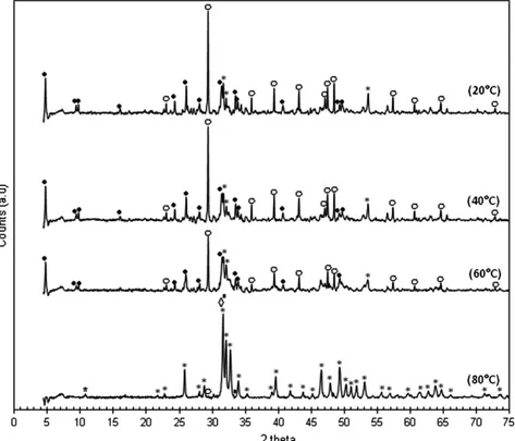
Fig. 3. XRD patterns of the solid products; (•) OCP (JCPDS standard no. 00-026-1056); (*) Ca-HA (JCPDS standard no. 01-072-1243); (▪) B-type CAP (JCPDS standard no. 00-004-0697); (◊) A-type CAP (JCPDS standard no. 00-035-0180); and (○) calcite (JCPDS standard no. 00-047-1743).
[15]. Following seven days of incubation at 37 °C, the samples were rinsed with distilled water, dried at 37 °C and observed with SEM analysis technique in order to examine the growth of new calcium phosphate on the surface of CAP disks.
3. Results and discussion
3.1. Orthophosphate precipitation and calcium carbonate dissolution When H3PO4was injected into the suspension of CaCO3, the
reac-tion started immediately as observed by the evolureac-tion of the pressure in the batch reactor. In all cases, the pressure quickly increased for the first 10 min. Then it slowly increased and stabilized up to the end of the reaction. In the temperature range investigated, the partial pres-sure of water vapor was close to the atmospheric prespres-sure (approxi-mately 1 bar) so the increase of the pressure could be attributed to the formation of the carbonic gas as shown in Eq.(1).
CaCO3þ H3PO4→Calciumphosphates þ H2O þ CO2 ð1Þ
Carbon dioxide from Eq.(1)could be in gas phase or dissolved in liquid phase. The quantity of carbon dioxide in gas phase could be es-timated using the ideal gas law and taking into account the volume of the reactor occupied by the solid and liquid phases and the partial pressure of water vapor at the corresponding temperature (Fig. 1).
Both final pressure and quantity of carbon dioxide in gas phase lin-early increased with the reaction temperature. At 80 °C, 88.9 mmol of carbon dioxide was released in gas phase which indicates that most ini-tial calcium carbonate introduced in the reactor (100 mmol) was decomposed.
The filtrate recovered from the reaction mixture was acidified with concentrated nitric acid to avoid any further precipitation or transfor-mation of calcium cations with orthophosphate and/or carbonate an-ions. Then, dissolved calcium and phosphorus contents in the filtrate were analyzed by ICP-AES technique. In all cases, there was less than
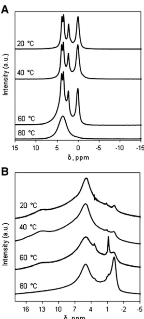
Fig. 4.31P NMR (A) and1H NMR (B) spectra of the dried solid products.
Fig. 5. IR spectra of the solid products; dotted tick: peak position of C\O stretching of A-type CAPs (880 cm−1); and continuous tick: peak position of C\O stretching of B-type CAPs
1% of the initial calcium and phosphorus introduced in the reactor which existed in soluble forms. This means that the precipitation of orthophosphoric acid into solid calcium phosphates was complete and
all calcium cations released from Eq.(1) completely re-precipitated too. ICP-AES analysis of the bulk solids after mineralization with con-centrated nitric acid showed that the molar ratio of Ca/P was about 1.8. This is slightly higher than the stoichiometric value of Ca-HA and will be discussed in more details in the NMR and IR characterizations section.
The content of residual calcium carbonate in the solid products was quantified by TG analysis (Section 3.4) and the obtained results are reported inFig. 2. As expected, the decomposition of calcium carbonate increased with the increase of reaction temperature. At 80 °C, the disap-pearance of calcium carbonate was nearly complete after 48 h of reaction. The solid products were then characterized by different physico-chemical techniques to identify the intermediates present.
3.2. XRD characterization
XRD characterization was performed in order to investigate the crystalline phases present in the solid products using JCPDS stan-dards. The results are presented inFig. 3.
As shown inFig. 3, unreacted calcium carbonate, having the main diffraction peak at 29.40°, was clearly observed in the solid products synthesized within the reaction temperature range of 20–60 °C. On the other hand, only trace amounts of this starting reactant was ob-served in the solid product obtained at 80 °C which confirmed the fa-vorable effect of the reaction temperature on the decomposition of initial calcium carbonate particles. The yield of calcium carbonate de-composition strongly influenced the crystalline structure of the solid calcium phosphates formed. When the decomposition of calcium car-bonate was nearly complete (80 °C), all diffraction peaks could be
Table 1
IR peak positions and corresponding assignment[19–22]; Y and N: with and without the presence of absorption band, respectively.
Wavelength,
cm−1 Solid Assignment
20 °C 40 °C 60 °C 80 °C
3600 Y Y Y Y O\H (water), ν1 mode 1645 Y Y Y N H\O\H, ν3 mode 1545 N N Y Y C\O, ν3 mode 1450 N N Y Y C\O, ν3 mode 1415 N N Y Y C\O, ν3 mode 1296 Y Y Y N P\(OH) stretching 1194 Y Y Y N P\(OH) stretching 1120 Y Y N N PO4, ν3 mode 1076 Y Y Y N PO4, ν3 mode 1055 Y Y Y N PO4, ν3 mode 1038 Y Y Y N PO4, ν3 mode 1018 Y Y Y Y PO4, ν3 mode 960 Y Y Y Y PO4, ν1 mode 916 Y Y Y N HPO4, ν4 mode
870 Y Y Y Y C\O, ν2 mode (B-type CAPs or calcite)
880 N N Y Y C\O, ν2 mode (A-type CAPs) 711 Y Y Y N C\O, ν4 mode
633 Y Y Y Y O\H (Ca-HA), libration HPO4, ν4 mode
603 Y Y Y Y PO4, ν4 mode
559 Y Y Y Y PO4, ν4 mode
530 Y Y Y N HPO4, ν4 mode
attributed to Ca-HA (JCPDS standard no. 01-072-1243) and calcium carbonate-containing apatites of both B-type (JCPDS standards nos. 00-019-0272 and 00-004-0697) and A-type (JCPDS standard no. 00-035-0180), labeled thereafter CAPs. However, it was difficult to distinguish these apatitic compounds because their main diffraction peaks are all in the 2 theta range of 31.5–31.8°.
When the dissociation of calcium carbonate was incomplete (20–60 °C), different calcium phosphates co-existed. Octacalcium bis(hydrogenphosphate) tetrakis(phosphate)pentahydrate (OCP, Ca8(HPO4)2(PO4)4·5H2O), which has the main diffraction peak at
2 theta of 4.7°, was the main apatitic calcium orthophosphate. Its signal decreased with the increase of the reaction temperature in-dicating its transformation into more stable apatites. Ca-HA, which has the main diffraction peak at 2 theta of 31.7°, was also formed but with low crystallinity as shown by its weak broad peaks. Other calcium orthophosphates such as CAPs, dicalcium phosphate anhydrous (DCPA, JCPDS standard no. 00-003-0398) could be also present at much lower contents.
In all cases, the final pH of the reaction mixture was around 8, which was favorable for the formation of apatitic compounds.
3.3. NMR and IR characterizations
To enhance the results of XRD characterization, NMR and IR analyses of the solid products were investigated.Fig. 4(A) presents the31P NMR
spectra of the dried solid products. The three solid products obtained at 20, 40 and 60 °C showed four characteristic chemical shifts of OCP[16]. On the other hand, the solid product synthesized at 80 °C showed only one chemical shift at 3.7 ppm, which was characteristic for Ca-HA based products[17].
Fig. 4(B) shows the1H NMR spectra of the dried solid products. The
chemical shift at about 13 ppm was characteristic for the HPO42−ions
located in the apatitic layer of the OCP's structure [18]. This shift appeared only in the solid products synthesized at 20–60 °C with uncompleted dissociation of calcium carbonate, and was not found in the product synthesized at 80 °C with complete conversion of calcium carbonate. The shift at about−0.1 ppm was characteristic for OH−
ions present in Ca-HA's structure[17]. This peak was much more intense in the product synthesized at 80 °C than in the other products. All these results confirmed those of XRD analysis, that OCP and Ca-HA were the main components of the solid products synthesized at
20°C
40°C
60°C
80°C
A
B
20–60 °C and 80 °C, respectively. Finally, the peak with chemical shift at about 5 ppm (Fig. 4(B)) could be attributed to adsorbed water, which was present in all four solid products[19].
Fig. 5shows IR spectra of all four solid products, and most significant peaks in the wavelength range of 4000–500 cm−1are summarized in
Table 1. In the wavelength range of 4000–1700 cm−1, there was only
a weak broad peak at about 3600–3000 cm−1(not presented), which
was assigned to O\H stretching of molecular water.
According to the results of XRD analysis, the remaining calcium carbonate in the solid products was characterized by the peak at 711 cm−1, which was not present in the solid obtained at 80 °C.
This last one also lacked the peak at 1645 cm−1, assigned to
20 °C (L)
20 °C (R)
40 °C (L)
40 °C (R)
60 °C (L)
60 °C (R)
80 °C (L)
80 °C (R)
H\O\H bending of molecular water in the structure of OCP, which confirmed the absence of OCP as shown by XRD (Fig. 3). The weak absorption band at 633 cm−1, assigned to the vibrational mode of
OH−groups indicated the presence of stoichiometric Ca-HA. The
bi-modal peak at 1450 cm−1/1415 cm−1and the peak at 870 cm−1
were characteristic for B-type CAPs (replacement of PO43−groups by
CO32−groups). On the other hand, A-type CAPs (replacement of OH−
groups by CO32− groups) were characterized by the peaks at
1545 cm−1and 880 cm−1. Thus, B-type CAPs could be present in all
four solid products while A-type CAPs could only be found in the solids obtained at 60 °C and 80 °C. So, the increase of reaction temperature was favorable for both the decomposition of calcium carbonate and the formation of CAPs, particularly A-type CAPs. The presence of B-type CAP explained why the molar ratio of Ca/P of the bulk solids was slightly higher the stoichiometric value of pure Ca-HA. Finally, the absorption bands in the wavelength ranges of 650–500 cm−1and
1300–910 cm−1 were assigned to the vibrations of orthophosphate
groups. Classical vibration bands of HPO42−groups in OCP structure
and PO43−in Ca-HA or CAP structure could clearly be observed.
3.4. TG–MS coupling
The thermal behavior of the solid products was investigated using TG analysis. TG and DTG curves obtained in the temperature range of 25–1200 °C are shown in Fig. 6(A) and (B), and the formation of
water and carbon dioxide during the thermal analysis, which was qual-itatively measured by MS analysis, are presented inFig. 6(C) and (D).
All changes in mass as a function of heating temperature could be at-tributed to the decarbonation and dehydration which were well con-firmed by MS analysis. The thermal decomposition of OCP, which was the principal intermediate, is generally complex with different dehydra-tions in the temperature ranges of 30–80 °C; 80–170 °C and 200–300 °C [23]. These weight losses were not observed for the solid synthesized at 80 °C which was in coherence with the result of XRD, NMR and IR char-acterizations. The first weight loss at 30–80 °C was attributed to the elimination of physically absorbed surface moisture which should not be significant after drying. The weight loss at 170–200 °C, which was only observed for the solid synthesized at 20 °C, could be due to the de-hydration of DCPD to form DCPA. This indicated that DCPD could also be an intermediate of the reaction as previously observed in the reaction of limestone with orthophosphoric acid[24]. DCPA was then condensed into calcium pyrophosphate (Ca2P2O7) at 400–500 °C. The clearest
weight loss at 600–720 °C was due to the decarbonation of residual cal-cium carbonate. Its content decreased with the increase of reaction temperature, as discussed in Fig. 2. Two next weight losses at 740–850 °C and 850–1200 °C were attributed to the decarbonation and dehydroxylation of CAPs, as accompanied with the release of CO2
and H2O, detected by MS analysis. According to the results of IR analysis,
the decarbonation of B-type CAPs, which were present in all four solids, happened at 740–850 °C. On the other hand, the decarbonation of
A-type CAPs, which were only formed at 60 °C and 80 °C, occurred at higher temperatures in the range of 850–1200 °C. The decarbonation of the solid synthesized at 80 °C was not finished at 1200 °C which indi-cated its high thermal stability.
All changes in mass in the temperature range investigated were en-dothermic as found by differential scanning calorimetry (DSC, not presented).
3.5. SEM and particle size distribution
In order to investigate the surface morphology of particles, SEM ob-servation was performed.Fig. 7presents SEM micrographs of the solid products at low magnification and starting calcium carbonate powder. This initial reactant had a high regularity in particle dimension (Fig. 7(A)) with a smooth and non-porous structure (Fig. 7(B)), which was in coherence with its low specific surface area (b2 m2g−1). On
the other hand, all four solid products had a variation of particle sizes in large ranges from hundreds of nm to hundreds of μm, as usually ob-served previously[25–27].
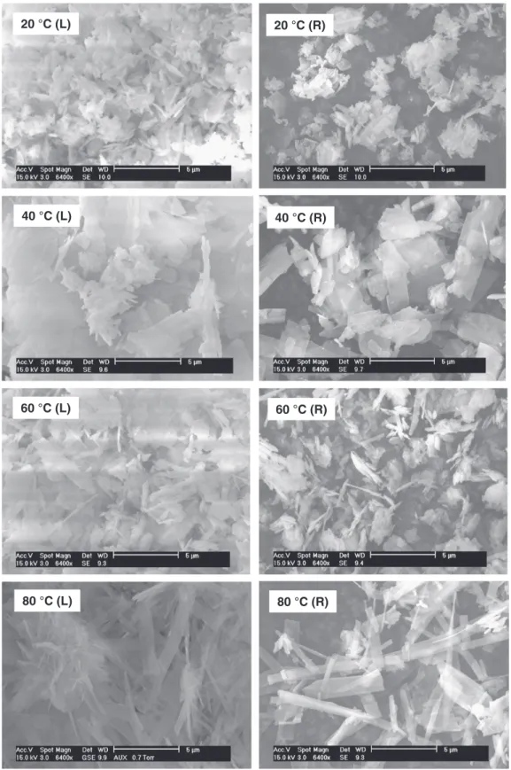
Fig. 8presents SEM micrographs of particles larger than 50 μm (L) and smaller than 5 μm (R) at the same magnification of 6400 times (5 μm scale). For all solid products, large particles seem to be a result of agglomeration of corresponding fine particles. This has been ob-served for the precipitation of calcium orthophosphates in aqueous solution[28–30]. Taking into account the rapid increase of pressure in the batch reactor during the first minute of reaction, it is assumed that the first step of the reaction must be the decomposition of calci-um carbonate particles in acid medicalci-um, accompanied by the release of carbonic gas and the precipitation of calcium cations (Ca2+) by
orthophosphate species to form primary fine calcium phosphate particles at nanometric scale. Then, primary fine calcium phosphate particles agglomerated during the reaction time to form larger particles.
Change in morphology of particles as a function of reaction temper-ature can also be observed inFig. 8. On the left-hand side, larger aggre-gates seem to have a porous structure. On the right-hand side, primary particles had a compact appearance and at higher magnification, no macro-pores could be observed (Supplementary data 1). At 20 °C, par-ticles of sheet structure frequently appeared. They seemed to be more numerous at 40 °C and could be attributed to OCP particles[21,31] which were in coherence with XRD results (Fig. 3). Some needle-like particles also appeared at these temperatures. At 60 °C, the frequency of particles of sheet structure decreased and that of needle-like particles considerably increased. Particularly, the change was distinct at 80 °C where most particles were of flat-needle-like morphology (more details in Supplementary data 2). This morphology was characteristic for apatitic compounds formed under hydrothermal conditions, as ob-served in previous works[32–34].
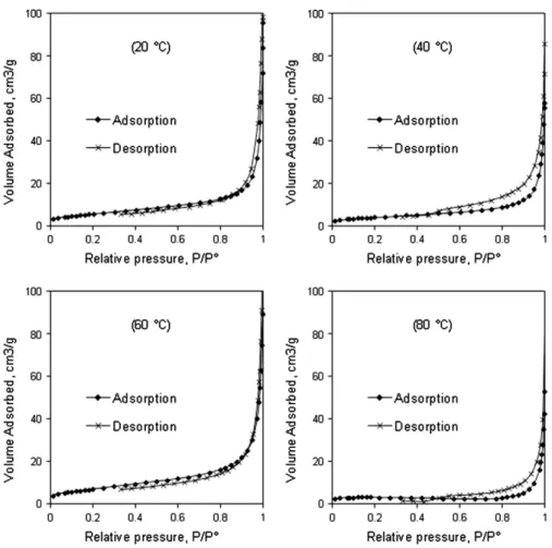
Fig. 10. Adsorption–desorption isotherms of the solid products.
Table 2
Specific surface area (SBET) and true density (D) of the solid products.
Product SBET, m2g−1 D, g cm−3 Initial CaCO3 b2 2.675 Product at 20 °C 15 2.670 Product at 40 °C 14 2.677 Product at 60 °C 25 2.784 Product at 80 °C 12 2.991
Particle size distribution was also analyzed in order to better un-derstand the formation of particles (Fig. 9).
According to the observation inFig. 7(A), starting calcium carbonate had a narrow particle size distribution from 7 μm to 50 μm. On the other hand, solid calcium phosphate products showed large ranges of particle sizes from about 0.2 μm to about 270 μm. For the solid products synthe-sized at 20–60 °C, there was more than 93% of number of particles found in the range of 0.2 μm to 1 μm. On the other hand, the volumes corre-sponding to this fraction of particle size were found at less than 7%. For the solid synthesized at 80 °C, 97% of the number of particles was found in the range of 1.5 to 10 μm, which corresponded to only 3% of cu-mulative volume. This means that a deep agglomeration of fine particles took place during the synthesis under the conditions investigated. 3.6. Adsorption–desorption isotherm measurements and true density
SEM micrographs at high magnification showed that large solid particles could have macro-pores but this technique did not allow the observation of smaller pore sizes. To better understand the poros-ity of the obtained materials, adsorption–desorption isotherm mea-surements were carried out.Fig. 10 shows isotherm plots for the reaction products.
All four solids showed similar adsorption–desorption isotherms of type II solids according to the IUPAC classification of physisorption iso-therms[35]. These results indicate that the solid products had only macro-porous structure which confirmed the results of SEM observation. From the results of isotherm measurements, specific surface areas were obtained using BET method (SBET). True density was measured
also and the results are presented inTable 2.
The two solid products synthesized at 20 °C and 40 °C had the same specific surface area, in agreement with similar chemical composition with about 20–22% of the remaining calcium carbonate. At 60 °C, the decomposition of calcium carbonate was more advanced (Fig. 2) and
the resulting solid had a higher specific surface area. At 80 °C, despite a complete decomposition of calcium carbonate, the specific surface area of the reaction product was smallest, which was due to the forma-tion of larger particles by agglomeraforma-tion (Fig. 9). In parallel with the in-crease of the reaction temperature, the reaction evolved to apatitic phases which were generally denser than the initial calcium carbonate [36]. This explains the increase of true density as a function of reaction temperature. Clogged porosity may be also formed during the precipita-tion of apatitic compounds since carbon dioxide was released from the decomposition of calcite. This porosity may be destroyed with the in-crease of the temperature leading to denser products.
3.7. Bioactivity test
SEM observations showed the CAP disks to be composed of flat-needle-like crystals of approximately one micrometer width and sev-eral micrometers length (Fig. 11). Following incubation in Tris–SBF-27 mM, the crystals were covered by new calcium phosphate deposits re-sembling those described in earlier works[15,37]. The underlying CAP crystals can hardly be recognized inFig. 11(B). The non-compacted crys-tals are loosened inFig. 11(D) as a result of water rinsing. Kokubo et Takadama[38]described the formation of such coatings as related to sur-face bioactivity appropriate for bone bonding activity. Possible improve-ments of the proposed bioactivity test have been proposed[39]. 3.8. Discussion
In all our reactions using calcium carbonate and orthophosphoric acid as starting reactants, only traces of soluble calcium cations and or-thophosphate species were observed to be present in the liquid phase. This indicates that orthophosphate species precipitated from solution into poorly soluble solid phases. The decomposition of calcium carbon-ate was also complete at 80 °C after 48 h of reaction. This resulted in the
Fig. 11. SEM images of CAP synthesized at 80 °C: (A) disk just prepared; (B) disk after soaking in Tris–SBF-27 mM solution at 37 °C for 7 days; (C) dried CAP powder; and (D) CAP powder after soaking in Tris–SBF-27 mM solution at 37 °C for 7 days.
final formation of “pure apatitic products” which did not require further washing for the elimination of soluble counter ions. This represents real advantages of the present synthesis process in comparison with other routes for CAP synthesis, discussed in theIntroductionsection[12–14], since water-rinsing is usually a limiting step for the purification of pow-der products.
From TG curves, the carbonate contents in apatitic structure or in CaCO3form could be both estimated quantitatively (Table 3). In parallel
with the decomposition of the initial calcium carbonate, the carbonate content (CO2wt.%) of CAP increased. The formation of A–B-type CAP
(replacement of both PO43−and OH−groups by CO32−groups) was
fa-vorable at 80 °C, where the reaction reached the complete decomposi-tion of starting calcium carbonate.
4. Conclusions
One-step synthesis of carbonate-containing apatite (CAP) has been investigated under moderate conditions, using calcium carbonate and orthophosphoric acid as ubiquitous starting materials. Simple and well known starting materials are advantageous for pharmaceutical produc-tions. The reaction temperature was found to be the key parameter for both the decomposition of initial calcium carbonate particles and the for-mation of CAP. At 80 °C, which was lower than most hydrothermal con-ditions, the decomposition of calcium carbonate was complete. At this temperature, the final solid product was composed of only apatitic phases including Ca-HA and A–B type CAP. At lower temperatures, other calcium phosphate phases were formed, in particular OCP. In all cases, only traces of soluble calcium cations and orthophosphate species were found, which shows the complete transformation of the starting materials into the desired product. Carbon dioxide, formed in gas phase, was the only by-product of the synthesis process. CAP obtained at 80 °C was found to be bioactive for the growth of calcium phosphate on its surface after soaking in a simulated body fluid (Tris–SBF-27 mM). Supplementary data to this article can be found online athttp:// dx.doi.org/10.1016/j.msec.2013.03.023.
Acknowledgments
The authors gratefully acknowledge our colleagues, Nathalie Lyczko, Christine Rolland, Sylvie Delconfetto, Philippe Accart and Yannick Coppel, for the different characterization measurements.
References
[1] R. Murugan, S. Ramakrishna, K. Panduranga Rao, Mater. Lett. 60 (2006) 2844. [2] H. Morgan, R.M. Wilson, J.C. Elliott, S.E.P. Dowker, P. Anderson, Biomaterials 21
(2000) 617.
[3] K. Onuma, Prog. Cryst. Growth Charact. Mater. 52 (2006) 223.
[4] C.V.M. Rodrigues, P. Serricella, A.B.R. Linhares, R.M. Guerdes, R. Borojevic, M.A. Rossi, M.E.L. Duarte, M. Farina, Biomaterials 24 (2003) 4987.
[5] E.S. Thian, Z. Ahmad, J. Huang, M.J. Edirisinghe, S.N. Jayasinghe, D.C. Ireland, R.A. Brooks, N. Rushton, W. Bonfield, S.M. Best, Biomaterials 29 (2008) 1833. [6] Y. Doi, T. Shibutani, Y. Moriwaki, T. Kajimoto, Y. Iwayama, J. Biomed. Mater. Res.
39 (4) (1998) 603.
[7] M.E. Fleet, X. Liu, Biomaterials 26 (2005) 7548. [8] I. Khattech, M. Jemal, Thermochim. Acta 95 (1985) 119.
[9] J.P. Lafon, E. Champion, D. Bernache-Assollant, J. Eur. Ceram. Soc. 28 (2008) 139. [10] F. Bel Hadj Yahia, M. Jemal, Thermochim. Acta 505 (2010) 22.
[11] J.C. Elliott, Studies in Inorganic Chemistry 18: Structure and Chemistry of the Ap-atites and Other Calcium Orthophosphates, Elsevier, Amsterdam–London–New York–Tokyo, 1994. 191–199.
[12] M.E. Fleet, Biomaterials 30 (2009) 1473.
[13] M.E. Fleet, X. Liu, J. Solid State Chem. 174 (2003) 412.
[14] L.M. de Oliveira, A.M. Rossi, R.T. Lopes, Radiat. Phys. Chem. 61 (2001) 485. [15] S. Jalota, S.B. Bhaduri, A.C. Tas, Mater. Sci. Eng. C 28 (2008) 129.
[16] Y.H. Tseng, J. Zhan, K.S.K. Lin, C.Y. Mou, J.C.C. Chan, Solid State Nucl. Magn. Reson. 26 (2004) 99.
[17] C. Jager, T. Welzel, W. Meyer-Zaika, M. Epple, Magn. Reson. Chem. 44 (2006) 573. [18] J.C. Elliott, Studies in Inorganic Chemistry 18: Structure and Chemistry of the Ap-atites and Other Calcium Orthophosphates, Elsevier, Amsterdam–London–New York–Tokyo, 1994. 21–22.
[19] R.M. Wilson, J.C. Elliott, S.E.P. Dowker, R.I. Smith, Biomaterials 25 (2004) 2205. [20] B. Mihailova, B. Kolev, C. Balarew, E. Dyulgerova, L. Konstantinov, J. Mater. Sci. 36
(2001) 4291.
[21] R. Horvathova, L. Muller, A. Helebrant, P. Greil, F.A. Muller, Mater. Sci. Eng. C 28 (2008) 1414.
[22] A. Antonakos, E. Liarokapis, T. Leventouri, Biomaterials 28 (2007) 3043. [23] J.C. Elliott, Studies in Inorganic Chemistry 18: Structure and Chemistry of the
Ap-atites and Other Calcium Orthophosphates, Elsevier, Amsterdam–London–New York–Tokyo, 1994. 9–29.
[24] D.W. Kim, I.S. Cho, J.Y. Kim, H.L. Jang, G.S. Han, H.S. Ryu, H. Shin, H.S. Jung, H. Kim, K.S. Hong, Langmuir 26 (1) (2010) 384.
[25] M. Yoshimura, P. Sujaridworakun, F. Koh, T. Fujiwara, D. Pongkao, A. Ahniyaz, Mater. Sci. Eng. C 24 (2004) 521.
[26] W.L. Suchanek, P. Shuk, K. Byrappa, R.E. Riman, K.S. TenHuisen, V.F. Janas, Bioma-terials 23 (2002) 699.
[27] Y. Guo, Y. Zhou, D. Jia, H. Tang, Microporous Mesoporous Mater. 118 (2009) 480. [28] R. Rodriguez-Clemente, A. Lbpez-Macipe, J. Gbmez-Morales, J. Torrent-Burges,
V.M. Castafio, J. Eur. Ceram. Soc. 18 (1998) 1351.
[29] R. Morsya, M. Elsayed, R. Krause-Rehberg, G. Dlubek, T. Elnimr, J. Eur. Ceram. Soc. 30 (2010) 1897.
[30] W.L. Suchanek, K. Byrappa, P. Shuk, R.E. Riman, V.F. Janas, K.S. TenHuisen, J. Solid State Chem. 177 (2004) 793.
[31] S. Ishihara, T. Matsumoto, T. Onoki, T. Sohmura, A. Nakahira, Mater. Sci. Eng. C 29 (2009) 1885.
[32] M.I. Dominguez, J. Carpena, D. Borschnek, M.A. Centeno, J.A. Odriozola, J. Rose, J. Hazard Mater. 150 (2008) 99.
[33] A. Kasioptas, T. Geisler, C. Perdikouri, C. Trepmann, N. Gussone, A. Putnis, Geochim. Cosmochim. Acta 75 (2011) 3486.
[34] K. Ioku, S. Yamauchi, H. Fujimori, S. Goto, M. Yoshimura, Solid State Ionics 151 (2002) 147.
[35] K.S.W. Sing, D.H. Everett, R.A.W. Haul, L. Moscou, R.A. Pierotti, J. Rouquerol, T. Siemieniewska, Pure Appl. Chem. 57 (4) (1985) 603.
[36] H. Morga, R.M. Wilson, J.C. Elliott, S.E.P. Dowker, P. Anderson, Biomaterials 21 (2000) 617.
[37] Y.R. Duan, Z.R. Zhang, C.Y. Wang, J.Y. Chen, X.D. Zhang, J. Mater. Sci. Mater. Med. 15 (2004) 1205.
[38] T. Kokubo, H. Takadama, Biomaterials 27 (2006) 2907. [39] M. Bohner, J. Lemaitre, Biomaterials 30 (2009) 2175. Table 3
Carbonate contents (CO2wt.%) determined from TG curves.
Solid Decarbonation (CO2wt.%)
B-type CAP form
(740–850 °C) A-type CAP form(850–1250 °C) CaCO(600–720 °C)3form
20 °C 0.27 0 9.20
40 °C 0.49 0 9.77
60 °C 1.52 0.01 5.41 80 °C 0.76 4.05a 0.18
aFrom TG–MS coupling analysis in the temperature range of 25–1350 °C for a