Environmental Microbiology (2002) 4(12), 898–911
© 2002 Blackwell Science Ltd
Blackwell Science, LtdOxford, UKEMIEnvironmental Microbiology1462-2912Blackwell Science, 20024Original ArticlePseudomonas aeruginosa population structureJ.-P. Pirnay et al.
Received 13 February, 2002; revised 29 April, 2002; accepted 29 April, 2002. *For correspondence. E-mail [email protected]; Tel. (+32) 2359 0221; Fax (+32) 2359 0399.
Pseudomonas aeruginosa
displays an epidemic
population structure
Jean-Paul Pirnay,1,2 Daniel De Vos,1,3
Christel Cochez,1 Florence Bilocq,2
Alain Vanderkelen,2 Martin Zizi,4 Bart Ghysels1
and Pierre Cornelis1*
1Laboratory of Microbial Interactions, Department of
Immunology, Parasitology, and Ultrastructure, Flanders Interuniversity Institute of Biotechnology, Brussels Free University, Paardenstraat 65, B-1640 Sint-Genesius-Rode, Belgium.
2Science Department, Belgian Military Medical Service,
Queen Astrid Military Hospital, B-1120 Neder-Over-Heembeek, Belgium.
3Department of Infectious Diseases, Innogenetics N.V.,
B-9052 Ghent, Belgium.
4Department of Physiology, Faculty of Medicine and
Pharmacy, Brussels Free University, B-1090 Brussels, Belgium.
Summary
Bacteria can have population structures ranging from the fully sexual to the highly clonal. Despite numerous studies, the population structure of Pseudomonas aeruginosa is still somewhat contentious. We used a polyphasic approach in order to shed new light on this issue. A data set consisting of three outer mem-brane (lipo)protein gene sequences (oprI, oprL and
oprD), a DNA-based fingerprint (amplified fragment length polymorphism), serotype and pyoverdine type of 73 P. aeruginosa clinical and environmental iso-lates, collected across the world, was analysed using biological data analysis software. We observed a clear mosaicism in the results, non-congruence between results of different typing methods and a microscale mosaic structure in the oprD gene. Hence, in this network, we also observed some clonal complexes characterized by an almost identical data set. The most recent clones exhibited serotypes O1, 6, 11 and 12. No obvious correlation was observed between these dominant clones and habitat or, with the excep-tion of some recent clones, geographical origin. Our results are consistent with, and even clarify, some
seemingly contradictory results in earlier epidemio-logical studies. Therefore, we suggest an epidemic population structure for P. aeruginosa, comparable with that of Neisseria meningitidis, a superficially clonal structure with frequent recombinations, in which occasionally highly successful epidemic clones arise.
Introduction
Pseudomonas aeruginosa is noted for its metabolic ver-satility and its exceptional ability to adapt to and colonize a wide variety of ecological environments (water, soil, rhizosphere, animals) (Goldberg, 2000). It is also known for its capacity to cause disease in cystic fibrosis, burn, cancer and ventilated intensive care patients. Infections
caused by P. aeruginosa are difficult to treat because of
its inherent resistance to antibiotics. There seems to be
a consensus about the fact that P. aeruginosa clinical
isolates are genotypically, chemotaxonomically and func-tionally indistinguishable from environmental isolates.
Römling et al. (1994) reported that the most frequently
(28%) identified clone in cystic fibrosis patients was also detected at a relatively high frequency (21%) in aquatic environments, suggesting a common recent origin of
these strains. Rahme et al. (1995) demonstrated the
infectivity of a P. aeruginosa strain in both plant and animal
models. Foght et al. (1996) observed that P. aeruginosa
strains isolated from a gasoline-contaminated aquifer were indistinguishable, by molecular biological
tech-niques, from clinical isolates. Alonso et al. (1999) reported
that both oil-contaminated soil isolates and clinical
iso-lates of P. aeruginosa show pathogenic and
biodegrada-tive properties. However, the population structure of P.
aeruginosa is still under discussion. Denamur et al. (1993)
and Picard et al. (1994) suggested that the population
structure of P. aeruginosa was panmictic, but highlighted
the need for caution in inferring bacterial population struc-ture from any single class of genetic marker. Comparative sequencing of six genes in 19 environmental and clinical P. aeruginosa isolates revealed a high frequency of recombination and a net-like population structure (Kiewitz
and Tümmler, 2000). Ruimy et al. (2001) used
random-amplified polymorphic DNA (RAPD) typing to study the
genetic diversity of P. aeruginosa pneumonia,
and pneumonia are not caused by specific clones.
Recently, Lomholt et al. (2001) observed an epidemic
population structure for a P. aeruginosa population
iso-lated mainly from patients with keratitis and their environ-ment. They found evidence for an epidemic clone that is pathogenic to the eye and is characterized by a distinct combination of virulence factors. The above-mentioned studies were, however, somewhat biased, as the studied P. aeruginosa populations were often sampled in a rela-tively small region (mainly one country) and/or focused on a single pathology or specific environment and/or were analysed by only one method. With the call of van Belkum
(1996) and Vandamme et al., 1996) for a polyphasic
approach in mind, we integrated all phenotypic and geno-typic data available to us in a consensus type of clustering to study aspects of the population genetics and
epidemi-ology of P. aeruginosa. A data set, consisting of the
nucle-otide sequences of three outer membrane protein genes
(oprI, oprL and oprD), amplified fragment length
polymor-phism (AFLP) pattern analysis, serotype and pyoverdine
type, was combined for 73 P. aeruginosa isolates,
col-lected from 18 countries, from clinical and environmental
habitats. The oprI (249 bp) and oprL (504 bp) genes are
coding for the outer membrane lipoproteins I (Cornelis et al., 1989a) and L (Lim et al., 1997) of P. aeruginosa.
The oprI gene is conserved among the fluorescent
pseudomonads and was found to be useful as a comple-mentary phylogenetic marker for the classification of rRNA
group I pseudomonads (De Vos et al., 1998). The oprL
gene is conserved in P. aeruginosa and has proved to be
a useful detection and identification target molecule (De Vos et al., 1997; Pirnay et al., 2000; Jaffe et al., 2001). The P. aeruginosa oprD gene (1323 or 1329 bp) codes for a specialized pore protein, OprD, which allows selective permeation of basic amino acids and their structural ana-logues such as the carbapenem antibiotic imipenem (Trias
and Nikaido, 1990). Analysis of the oprD gene from 55
clinical and environmental isolates revealed important sequence variability and a microscale mosaic structure resulting from multiple recombinational events (Pirnay et al., 2002).
AFLP analysis is a genotyping method based on the selective amplification of a subset of DNA fragments
gen-erated by restriction enzyme digestion (Vos et al., 1995).
This technique has proved to be highly discriminatory and reproducible, which allows the compilation of
standard-ized patterns in a database (Janssen et al., 1996;
Savelk-oul et al., 1999).
Pseudomonas aeruginosa strains have been divided into serotypes since 1926 (Aoki, 1926). Since then, many investigators have formed their own serotyping schemata,
which has made serological study of P. aeruginosa very
used the international serogrouping schema for P.
aerug-inosa, comprising 17 groups based on the heat-stable major somatic antigens, for clinical serotyping. The
sero-typing of P. aeruginosa strains, using this standardized
schema, allows us to compare the evolutionary relation-ships between our isolates, provided by the two DNA-based techniques, with earlier published epidemiological data.
To satisfy their need for iron, pseudomonads generally produce high-affinity fluorescent peptidic siderophores,
called pyoverdines (PVDs) (Meyer, 2000). In P.
aerugi-nosa, three PVDs (I, II and III), easily differentiated by
isoelectric focusing (IEF), have been observed so far, only
one being produced by a given strain (Cornelis et al.,
1989b; Meyer et al., 1997). The combined results obtained
in this study are in agreement with earlier epidemiological studies and clearly indicate that the population structure of P. aeruginosa is epidemic.
Results
Sequence analysis of oprI, oprL and oprD genes
The oprI, oprL and oprD sequences of the studied P.
aeruginosa population were aligned and clustered using
UPGMA. Alleles were assigned numbers according to their
position in the alignment (Fig. 1). The oprI and oprL genes
showed sequence variability comparable with that of housekeeping genes, as was to be expected because both genes code for a structural outer membrane lipoprotein. In
the dendrograms (UPGMA), based on the similarity of the
oprI and oprL genes of the studied P. aeruginosa
popula-tion, supplemented with other members of the rRNA group
I pseudomonads (Fig. 2), P. aeruginosa forms a sharply
delineated species. Strains LMG 10643 and, to a lesser extent, strains LMG 5031 and Br680 diverge from the rest. With the exception of isolate LMG 10643, all mutations in
oprI and oprL were silent, often occurring at the third
position of the codon. The oprD gene, on the other hand,
showed high sequence variability, a mosaic structure and multiple non-silent mutations, typical of a gene that is
under strong selection for diversity (Fig. 1). The oprD gene
of strains LMG 10643 and Lw1048 could not be amplified by polymerase chain reaction (PCR).
AFLP analysis
The AFLP patterns of the P. aeruginosa strains and one
Pseudomonas pseudoalcaligenes strain were normalized
and clustered using UPGMA. By applying the criteria for
differentiation of P. aeruginosa by AFLP (Speijer et al.,
900 J.-P. Pirnay et al.
electrophoresis (Tenover et al., 1995), five clusters of
related isolates (with ≥80% homology) were identified
(Fig. 3).
Serotype determination
Sixty-one out of the 73 strains could be serotyped. Six strains were non-agglutinable, and six were polyagglutin-able. The predominant serotypes were O1 (12.3%, 9/73), 6 (10.9%, 8/73), 11 (15.1%, 11/73) and 12 (9.6%, 7/73) (Table 1).
Pyoverdine typing by IEF
A majority of the P. aeruginosa strains (37/73) produced
or were growth stimulated by type II PVD (Table 1). Fifteen isolates produced type I PVD and 14 type III. A few iso-lates failed to produce enough PVD to allow analysis by IEF. The presence of the receptor for a pyoverdine was therefore determined by a growth stimulation assay. In some cases, growth was stimulated by more than one PVD (Table 1). When this was the case, the pyoverdine that gave the strongest growth stimulation was considered as the cognate one, and the others are indicated between brackets.
Combined analysis
The data obtained from sequence analysis, AFLP
analy-sis, serotyping and PVD typing of the 73 P. aeruginosa
isolates was combined and analysed using BIONUMERICS
biological data analysis software. In the dendrogram from the composite data set (Fig. 4), we identified a limited
number of phylogenetic groups with ≥80% similarity.
Some subclusters even showed >90% similarity. We also observed unique isolates, some of which diverged consid-erably from the rest of the population. There is also evi-dence that the relation among the isolates was distorted by recombination. We observed a network of relationships between all analysed parameters (Table 1) and non-congruence between experiments (Fig. 5).
Discussion
The observation of clones in many bacterial populations has led to the assumption that bacteria reproduce clonally. It was long supposed that point mutations are the major source of genetic variation in bacteria, whereas recombi-national exchanges were considered to be rare. This view has changed in recent years. Maynard Smith et al. (1993) used multilocus enzyme electrophoresis (MLEE) to dem-onstrate that bacterial population structure ranges from the panmictic or fully sexual, with random association between loci (e.g. Neisseria gonorrhoeae), to one that is
clonal, with non-random association of alleles, resulting in the frequent recovery of only a few of the possible multilo-cus genotypes (e.g. Salmonella enterica). Intermediate types of population structure were also reported. Neisseria meningitidis, for example, displays what the authors have called an ‘epidemic’ structure. Although the population is sexual in the long term, some epidemic clones show significant association between loci. Recently, Feil et al. (2001) used multilocus sequence typing (MLST) (Maiden et al., 1998) to examine the extent and significance of recombination in six bacterial pathogens. In four species (N. meningitidis, Streptococcus pneumoniae, Streptococ-cus pyogenes and StaphylococStreptococ-cus aureus), they observed a lack of congruence between gene trees, supported by high ratios of recombination to point mutation. In contrast, for Haemophilus influenzae and pathogenic isolates of Escherichia coli, there was some congruence between gene trees, suggesting lower rates of recombination.
In this work, data obtained by four different typing meth-ods, performed on a large batch of unrelated clinical and environmental P. aeruginosa isolates, are combined using biological data analysis software in order to get some insights into the population structure of P. aeruginosa.
The lack of congruence among experiments (Fig. 5) is most easily explained as the consequence of multiple recombinational events that have eliminated the phyloge-netic signal in each tree. This view is supported by the observation of a microscale mosaic structure in the oprD gene (Fig. 1), which supplies direct evidence for recombi-nation. The non-congruence between the AFLP dendro-gram and the trees based on sequence analysis could be expected, as sequence diversity is generally caused by single nucleotide polymorphisms, whereas differences in macrorestriction fragment patterns are mainly the result of insertions and/or deletions (Kiewitz and Tümmler, 2000). Although bacterial species in which recombination appears to be common are naturally transformable, there seems to be no obvious correlation between the degree of recombination and the transformability of species. Feil et al. (2001) showed that the naturally transformable H. influenzae showed by far greater congruence between gene trees than the non-transformable species S. aureus and S. pyogenes. They suggested that recombina-tional exchanges in S. aureus and S. pyogenes are pre-sumably mediated by phage transduction, the effect of which is as great as that of transformation. P. aeruginosa is considered not to be competent for natural transforma-tion. In this context, it is interesting that Ripp et al. (1994) suggested that environmentally endemic bacteriophages are formidable transducers of naturally occurring microbial communities of P. aeruginosa.
Hence, the population structure of P. aeruginosa is not fully sexual. In the dendrogram based on the comparison of the composite data set (Fig. 4), we clearly observed
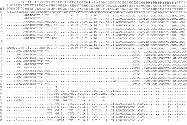
Fig. 1. Polymorphic sites detected in the different alleles of the oprI, oprL and oprD genes of 73 P. aeruginosa isolates. Only the sites that differ from the sequence of the allele of PAO1 (no. 23) are shown. Alleles are arranged according to their positions in the dendrogram of an UPGMA alignment. The number of representatives for each allele is shown on the right. Defective oprD mutations and alleles with unusually high sequence variability are excluded. Gaps are represented by *.
902 J.-P. Pirnay et al.
Fig. 1. cont.
seven distinct clonal complexes (CCs) with ≥80%
similar-ity. Most CCs contain strains from geographically and ecologically different sites, suggesting high rates of migra-tion and a remarkable nutrimigra-tional versatility, acquired through recombination or another evolutionary mecha-nism. CC A, for example, contains a blood, wound and urine isolate from three distant cities in the USA, a blood isolate from Congo, a sputum, throat and plant rhizo-sphere isolate from Belgium and a burn wound isolate from Turkey. The variability within each geographic region was nearly as great as within the whole population. In Belgium and The Netherlands, for example, members of CCs A, B, C, E, F and G were isolated.
Clones are transient and, over time, recombination will obliterate the evidence of association. The nearly identical data set of the members of subclusters a, c, d, e and g
(Table 1), resulting in ≥90% homology in the composite
data dendrogram (Fig. 4), is evidence of a recent, explo-sive increase in these clones. These recent clones exhibit serotypes O1, 6, 11 and 12. This observation is in agree-ment with earlier epidemiological studies. A study of the
serotypes of 2952 P. aeruginosa isolates showed a predominance of serotypes O1, 6 and 11 (Bert and Lambert-Zechovsky, 1996), and serotypes O11 and 12 are frequently associated with multidrug-resistant epi-demic strains (Farmer et al., 1982; Grattard et al., 1993; Elaichouni et al., 1994; Richard et al., 1994; Tassios et al., 1998; Dubois et al., 2001). Serotyping of 7089 P. aerugi-nosa strains, isolated in 16 Belgian hospitals in the period from 1977 to 1986, revealed a steady increase in P. aerug-inosa O12 isolates from 2% in 1982 to 22% in 1986 (Allemeersch et al., 1988). The majority of these O12 iso-lates showed the same distinctive pyocin and phage types, suggesting a high degree of homogeneity within the O12 strains in Belgium. A multicentre European study provided evidence for a common O12 P. aeruginosa strain in Europe (Pitt et al., 1989). Yet, not all O12 isolates belong to clone c. Serotype O12 clinical isolates Bo546 and Br680 are positioned far away from clone c in the composite data dendrogram (Fig. 4). Evidence of geno-typic heterogeneity among P. aeruginosa serotype O12 outbreak isolates has been reported (Bingen et al., 1996).
Although CCs are globally distributed, recent clones are, logically, less widespread (Table 1). Clone d, for example, consists of 10 isolates of a major clone (called clone C) common to patients and aquatic environments in Germany, previously identified by Römling et al. (1994). The occasional clustering of strains of distant geogra-phical origin in recent clones (e.g. strain PAO29 in clone c) illustrates the efficient dispersal of P. aeruginosa clones, probably aided by increased mobility of the human population.
The close genetic relation among the isolates of each clone was also detected by AFLP analysis. This shows that AFLP can be used, for example in clinical settings, to recognize epidemic P. aeruginosa clones over the short term (10 to maybe hundreds of years).
No significant correlation could be made between the type of PVD produced and the habitat. Recently, De Vos et al. (2001) reported a prevalence of type II PVD isolates in cystic fibrosis patients, but suggested that their might be a correlation between the pyoverdine type and the (clinical) origin of the P. aeruginosa isolates. The fact that several P. aeruginosa strains are able to use more than one PVD type (Table 1) could be the result of recombina-tional events involving PVD receptors. Strain LMG 10643 did not produce, nor was able to use, any of the three PVDs. This, together with the aberrant oprI and oprL sequences, makes us doubt that this isolate is a true P. aeruginosa. The clustering of isolates with different sero-types is not necessarily the result of recombinational events. Kobayashi et al. (1994) demonstrated that anti-pseudomonal drugs were able to induce changes in sero-type, and possible evidence of a bacteriophage-mediated
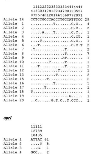
Fig. 2. Sequence similarity trees (UPGMA) based on the comparison of the oprI and oprL nucleotide sequences of the 73 studied P. aeruginosa isolates, supplemented with members of the rRNA group I pseudomonads. Sequences of P. aeruginosa and P. pseudoalcaligenes isolates were determined in this study, P. fluorescens (pf0-1), P. syringae (pv. tomato) and P. putida (KT2440) sequences were retrieved from the unfinished genomic sequence database (http://www.ncbi.nlm.nih.gov/cgi-bin/Entrez/genom_table_cgi) using BLASTN software; all other sequences were retrieved from the NCBI GenBank (http://www.ncbi.nlm.nih.gov/). Percentages of similarity are shown above the dendrogram.
serotype O5 to O16 conversion (Newton et al., 2001) was found in the clustering of reference strain PAO1 and strain LMG 14083 in CC B (Fig. 4).
Not that long ago, the ability to identify clusters of iso-lates with an identical data set from different countries and habitats over a period of time would have been taken as evidence of a clonal structure. The multiple associations of serotypes O11 and 12 with infection and epidemics and the frequent recovery of only a few of all the possible serotypes (O1, 6 and 11) superficially suggest that the P. aeruginosa population is clonal. Our results show an epi-demic population structure for P. aeruginosa, comparable with that of N. meningitidis (Feil et al., 2001), a population composed of a limited number of widespread clones, which originated, through selection, from a background of a large number of relatively rare and unrelated geno-types that are recombining at a high frequency. These adaptive clones are abundant and widespread in nature
and are therefore expected to predominate in the patient population.
Future investigations should be directed at factors that play a role in the selective advantages of these highly successful clones in the environment as a whole, instead of restricting analysis to patients and the hospital environ-ment. The cause of the association between virulence and fitness is still unclear (Groisman and Ochman, 1994).
It should be noted that MLST focuses exclusively on housekeeping genes because of selective neutrality. Anal-ysis of these genes provides a more realistic impression of the effect of recombination. In contrast, oprD recombi-nants can be selectively favoured if, for example, they confer resistance to carbapenem antibiotics. However, we chose to include the oprD sequence data for the following reasons: (i) OprD-related resistance to carbapenems is mainly achieved by non-recombinational events such as point mutations (Pirnay et al., 2002); (ii) it provides direct
904 J.-P. Pirnay et al.
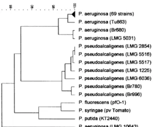
Fig. 3. Normalized AFLP patterns and dendrogram for 73 different P. aeruginosa isolates and one P. pseudoalcaligenes isolate (Br780). Cluster analysis was performed with BIONUMERICS software using the Pearson correlation and UPGMA. Percentages of similarity are shown above the dendrogram. Clusters with ≥80% similarity (according to Tenover et al., 1995; Speijer et al., 1999) are indicated by Roman numerals. Clusters I, II and III contain isolates with slightly <80% homology.
environmental isolates from other parts of the world, as well as housekeeping gene sequence data, is currently under way. We also feel that the exchange of standardized data between laboratories and the creation of interna-tional reference databases of typed microorganisms should be encouraged. It will enable the efficient monitor-ing of changes in microbial populations.
Experimental procedures
Bacterial strains and growth conditions
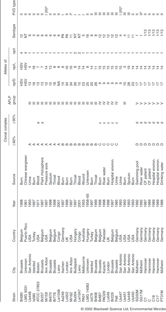
A total of 73 P. aeruginosa clinical and environmental iso-lates, collected worldwide, mainly in the late 1980s and 1990s, with some earlier isolates, were examined. The geo-graphical origin, isolation site and time of all P. aeruginosa isolates are listed in Table 1.
The P. aeruginosa strains used in this study were kindly provided by: Dr A. T. McManus, US Army Institute of Surgical Research, TX, USA; Dr L. Ménesi, General Hospital St Istvan, Budapest, Hungary; Dr A. Vanderkelen, Queen Astrid Military Hospital, Neder-Over-Heembeek, Belgium; Dr J. A. Clark, Queen Mary's University Hospital, London, UK; Dr A. F. Vloemans, Rode Kruis Ziekenhuis, Beverwijk, The Netherlands; Dr T. Taddonio, University of Michigan, MI, USA; Dr A. Radke, Klinik für Verbrennungs- und Plastische Wiederherstellungschirurgie, Aachen, Germany; Professor R. Konigova, Charles University Hospital, Prague, Czech Republic; Dr R. G. Tompkins, Burns Institute, Shriners Hospital for Children, Boston, MA, USA; Dr B. Tümmler, Medizinische Hochschule, Hannover, Germany; Dr M. Caneira, Hospital de Santa Maria, Lisbon, Portugal; Profes-sor A. Boudabous, Science Faculty, Tunis, Tunisia; Dr M. Mergeay, Environmental Technology Expertise Centre, Mol, Belgium; Dr A. E. Lim, Jr., St Scholastica's College of Health Sciences, Tacloban City, Philippines; Professor O. Hadjiiski, Scientific Institute of Emergency Medicine Pirogov, Sofia, Bulgaria; Professor K. Taviloglu, University of Istanbul, Istanbul, Turkey; Dr W. D. H. Hendriks, Zuiderziekenhuis, Rotterdam, The Netherlands; Dr G. Wauters, University of Louvain, Brussels, Belgium; Dr O. Vandenberg, Universitair Ziekenhuis St-Pieters, Brussels, Belgium. Strain PAO-1 was kindly provided by Dr C. K. Stover (PathoGenesis Corpora-tion, Seattle, WA, USA). Strain ATCC 27853 was purchased from Gibson Laboratories. P. aeruginosa strains LMG 2107, 5031, 10643 and 14083–5 and P. pseudoalcaligenes strains LMG 1225, 2854, 5516, 5517 and 6036 were purchased from the BCCM/LMG bacteria collection. Unless otherwise indicated, strains were grown on Luria–Bertani broth medium (Gibco BRL Life Technologies) at 37°C on a rotary shaker (150 r.p.m.).
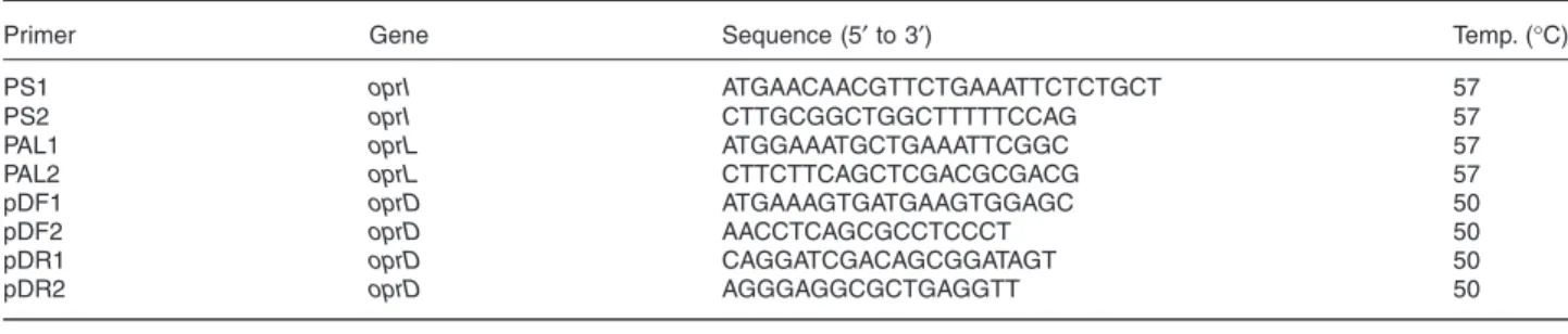
water, 5 µl of 10× PCR buffer (500 mmol l−1 KCl and 100 mmol l−1 Tris-HCl, pH 8.3), 4 µl of a deoxynucleotide mix-ture (dGTP, dTTP, dATP and dCTP; 2 mmol l−1 each), 5 µl of MgCl2 (2.5 mmol l−1), 5 µl of a primer mixture (PS1/2 for oprI,
PAL1/2 for oprL or pDF1/R1 for oprD; 10 µmol l−1 each), 5 µl of template DNA and 0.5 µl of AmpliTaq DNA polymerase (5 U µl−1). All PCR reagents and primers were ordered from PE Applied Biosystems. The amplification was performed in a GeneAmp® PCR system 2400 (PE Applied Biosystems).
The amplification programme was set at 50 cycles of dena-turation at 94°C for 30 s, annealing at 50°C or 57°C, accord-ing to the primers (Table 2), for 30 s and elongation at 72°C for 1 min. For the amplification of P. pseudoalcaligenes oprL genes, the annealing temperature was lowered to 55°C. The reaction mixture was put on a 1.5% (w/v) agarose gel for electrophoresis and visualization of the PCR product after staining with ethidium bromide on a transilluminator. The DNA bands corresponding to the amplified oprI, oprL and
oprD genes were excised from the agarose gel with a clean
scalpel. DNA was extracted from the gel slice using the QIAEX II gel extraction kit (Westburg) according to the man-ufacturer's recommendations. Purified PCR fragment (5 µl) was used as a template in the sequencing reaction. PCR primers were used for sequencing. Sequencing of the coding and anticoding strand of the oprD PCR products necessitated two additional internal primers, pDF2 and pDR2 (Table 2). DNA sequencing used an ABI 377 automated fluorescence sequencer (PE Applied Biosystems) and the ABI Prism®
BigDye™ Terminator cycle sequencing ready reaction kit (PE Applied Biosystems) as detailed in the manufacturer’s proto-cols. The oprD gene of isolate Be128 was sequenced directly from genomic DNA. PCR and sequencing were performed in duplicate in order to be able to detect eventual PCR mistakes. Sequences were aligned and clustered using the unweighted pair group method using arithmetic averages (UPGMA) and
BIONUMERICS software (Applied Maths).
AFLP
AFLP used an ABI 377 automated fluorescence sequencer (Applied Biosystems) and the AFLP microbial fingerprinting kit (Applied Biosystems) as detailed in the manufacturer’s protocols. The enzymes used were T4 DNA ligase, EcoRI and Tru9I (all purchased from Roche Diagnostics). The primer pair used was EcoRI-0[FAM]/MseI-C. GeneScan-500[ROX] internal standard (Applied Biosystems) was co-electrophoresed with each sample in order to allow an accurate calculation of fragment lengths and correction for variation rates and gel distortions. Normalization and frag-ment sizing were carried out using GENESCAN software (Applied Biosystems). Band patterns were imported into
906 J.-P. Pirnay et al.
Ta
b
le 1.
Proper
ties of the isolates analysed in this study
. Str ain City Countr y Y ear Source Clonal comple x AFLP group Alleles of: Serotype PVD type ≥ 80% ≥ 90% oprD oprL oprI Br680 Br ussels Belgium 1998 Bur n HSV HSV 4 1 2 II LMG 5031 Unkno wn Puer to Rico 1961 Chinese e v erg reen HSV HSV 4 N T II Us448 San Antonio USA 1993 Ur ine A III 13 4 1 9 II Is580 Istanb ul T u rk e y 1997 Bur n A III 16 18 1 3 II A TCC 27853 Boston USA 1971 Blood A a III 13 7 1 6 II Br257 Br ussels Belgium 1997 Plant rhiz osphere A a III 13 7 1 6 II Mi159 Ann Arbor USA 1997 Pressure sore A III 12 14 1 6 I (III) a Br670 Br ussels Belgium 1998 Sputum A III 15 13 1 6 II Br776 Br ussels Belgium 1998 Throat A III 18 8 1 6 II Lw1048 Lwiro Congo 2001 Blood A III NA 8 1 6 II Aa249 Aachen Ger man y 1997 Bur n B III 15 8 1 P A I Lo053 London UK 1996 Bur n B III 34 16 1 P A II Mi162 Ann Arbor USA 1997 Bur n B III 34 16 1 1 1 I a Bu004 Budapest Hungar y 1997 Throat B III 26 8 2 NT I Lw1047 Lwiro Congo 2001 Blood B III 15 14 2 N T I P A01 Melbour ne A ustr alia 1955 W ound B III 23 14 1 5 I LMG 14083 Unkno wn Hungar y 1958–65 Unkno wn B III 25 8 1 16 II Is579 Istanb ul T u rk e y 1997 Bur n B III 20 14 1 8 II Br908 Br ussels Belgium 1999 Throat B III 26 11 1 8 III Br667 Br ussels Belgium 1998 Bur n C c IV 9 15 1 1 2 III PA O29 Kar achi P akistan 1998 Riv er w ater C c IV 9 1 5 1 12 III Lo049 London UK 1996 Bur n C c IV 9 15 1 1 2 III Br229 Br ussels Belgium 1997 Hospital en viron. C c IV 9 1 5 1 12 III Pa 6B russels Belgium 1985 Ur ine C c IV 9 1 5 1 12 III Us447 San Antonio USA 1993 Ur ine 9 1 5 1 4 I (III) a Us451 San Antonio USA 1993 Bur n III 10 3 1 4 III Us449 San Antonio USA 1993 Sputum 28 19 1 1 1 III Us450 San Antonio USA 1993 Bur n 2 3 1 4 1 11 II SG50M Mülheim Ger man y 1992 Swimming pool D d V 1 7 1 4 1 1 II SG17M Mülheim Ger man y 1992 Riv er w ater D d V 1 7 1 4 1 1 II a C1 Hanno v e r Ger man y 1987 CF patient D d V 1 7 1 4 1 1/13 II C Hanno v e r Ger man y 1989 CF patient D d V 1 7 1 4 1 1/13 II C18 Hanno v e r Ger man y 1989 Hospital en viron. D d V 1 7 1 4 1 1/13 II C17 Hanno v e r Ger man y 1989 Hospital en viron. D d V 1 7 1 4 1 1/13 II PT31M Mülheim Ger man y 1986 Dr inking w ater D d V 1 7 1 4 1 1/13 II a. Siderotyped b y PVD-induced g ro wth stim ulation. Non-cognate g ro wth-stim
ulating PVDs are indicated betw
een br ac k ets . b.
Agglutinated with the antiser
a mix E (O2+5+15+16), b
ut not with either of the separ
ate mono v alent antiser a. CF , cystic fibrosis; en viron., en vironment; HSV , un
usually high sequence v
a riability; NA, no amplification in PCR; NP , no p y o v erd in production or uptak e; NT , non-typeab le; P A, poly agglutination.
C19 Hanno v e r Ger man y 1989 CF patient D d V 1 7 1 4 1 C2 Hanno v e r Ger man y 1988 CF patient D d V 1 7 1 4 1 C13 Hanno v e r Ger man y 1985 CF patient D d V 1 7 1 4 1 Pr335 Pr ague Cz ech Repub lic 1997 Hospital en viron. D V 12 14 2 Li012 Lisbon P o rtugal 1997 CF patient D V 12 17 1 Br642 Br ussels Belgium 1998 Hospital en viron. E e I 3 16 1 Ro124 Rotterdam The Nether lands 1997 Bur n E e I 3 1 6 1 LMG 2107 Canas Puer to Rico 1938 Shallo w w ell E I 5 1 1 T u863 T unis T unisia 1998 Ear 11 20 1 LMG 14085 Unkno wn Hungar y 1958–65 Unkno wn 13 14 2 Bo548 Boston USA 1992 Bur n 2 7 8 2 Li010 Lisbon P o rtugal 1997 CF patient 22 6 1 Li009 Lisbon P o rtugal 1997 CF patient 19 13 1 So095 Sofia Bulgar ia 1997 Bur n 2 6 8 1 Be128 Be v erwijk The Nether lands 1997 Sputum F 3 4 1 4 1 T uD199 T unis T unisia 1998 Sputum F 6 1 1 Mi151 Ann Arbor USA 1997 Bur n F 8 1 4 1 Br692 Br ussels Belgium 1998 Bur n G g II 3 0 1 4 1 Is573 Istanb ul T u rk e y 1997 Bur n G g II 2 9 1 4 1 Aa245 Aachen Ger man y 1997 Bur n G g II 2 9 1 4 1 So092 Sofia Bulgar ia 1997 Bur n G II 31 14 1 Bu007 Budapest Hungar y 1997 Bur n G II 29 14 1 Pr317 Pr ague Cz ech Repub lic 1996 Bur n 2 9 1 4 1 Be136 Be v erwijk The Nether lands 1996 Sputum 1 2 1 T uD47 T unis T unisia 1998 Ascite 4 1 4 1 PA O23 Kar achi P akistan 1998 Riv er w ater 34 14 1 Lo050 London UK 1996 Bur n 1 4 1 4 1 Li004 Lisbon P o rtugal 1997 CF patient 8 1 4 1 Br906 Br ussels Belgium 1999 Nose 2 5 1 Br735 Br ussels Belgium 1998 Bur n 3 3 1 1 Bo546 Boston USA 1992 Bur n 2 1 9 3 So099 Sofia Bulgar ia 1997 Bur n 2 4 1 2 2 PhD W6 T acloban City Philippines 1993 W ound 12 10 2 Be133 Be v erwijk The Nether lands 1996 Bur n 4 2 1 Bo559 Boston USA 1997 Bur n 3 16 2 Aa246 Aachen Ger man y 1997 Bur n 3 2 6 1 LMG 14084 Bucharest Romania 1960–64 W ater 7 1 1 LMG 10643 Pusakanegar a Indonesia 1990 Or yza sativ a lea v e s N A HSV HSV Str ain City Countr y Y ear Source Clonal comple x AFLP group Alleles of: ≥ 80% ≥ 90% oprD oprL oprI a. Siderotyped b y PVD-induced g ro wth stim ulation. Non-cognate g ro wth-stim
ulating PVDs are indicated betw
een br ac k ets . b.
Agglutinated with the antiser
a mix E (O2+5+15+16), b
ut not with either of the separ
ate mono v alent antiser a. CF , cystic fibrosis; en viron., en vironment; HSV , un
usually high sequence v
a riability; NA, no amplification in PCR; NP , no p y o v erd in production or uptak e; NT , non-typeab
subtraction, filtering: arithmetic average and band search: minimum profiling 0.5% relative to maximum value) and clus-ter analysis (similarity coefficient: Pearson correlation, den-drogram type: UPGMA, optimization: 0%, position tolerance: 1%, uncertain bands were ignored).
Serotyping
Isolates were serotyped by slide agglutination according to the international serogrouping schema for P. aeruginosa (Liu
et al., 1983), using a panel of 16 type O monovalent antisera
(Sanofi Diagnostics Pasteur).
Pyoverdine typing by IEF
PVD-IEF was carried out according to the method developed by Koedam et al. (1994), as described previously (Meyer
et al., 1997). IEF was performed on Ampholine-PAG plates
(pH 3.5–9.5; Pharmacia). The following reference strains were included: PAO-1, representative of PVD type I; ATCC 27853, representative of PVD type II; and strain Pa6, a clin-ical isolate representing PVD type III (Meyer et al., 1997).
Pyoverdine typing by PVD-induced growth stimulation The effect on bacterial growth of each of the three known PVDs was tested as described previously (Meyer et al.,
stimulation (no growth), slight stimulation (growth, diameter <10 mm) and good stimulation (thick growth, diameter >15 mm). Pyoverdines produced by the type strains PAO1 (PVD type I), ATCC 27853 (PVD type II) and Pa6 (PVD type III) were purified as described previously (Meyer et al., 1997).
Data analysis
The entire data set, consisting of oprI, oprL and oprD sequences, AFLP pattern, serotype and pyoverdine type of 73 P. aeruginosa isolates, was analysed and combined using
BIONUMERICS (Applied Maths) biological data analysis
soft-ware. Similarity values were taken from the individual exper-iments and multiplied by weights (AFLP: 35, oprD: 11, oprL: 6, serotype: 5, oprI: 2, and PVD type: 1). These weights were an educated guess and designed to compensate for the bias caused by the differences in discriminatory capacity between the experiments in this study. In other words, the weights are proportional to the supposed discriminatory capacity of the different typing methods. A dendrogram from the composite data set was obtained using UPGMA. Congruence between experiments was calculated using the Pearson product-moment correlation coefficient.
Acknowledgements
We thank all the collectors and providers of the bacterial isolates used in this study. This work was supported by the ‘Alphonse and Jean Forton Fund’, VZW ‘De Vrienden van de Huidbank’ and grant JSM-R and T G98/02 from the Belgian Department of Defence. Parts of this paper were presented
Table 2. Primers for PCR and sequencing.
Primer Gene Sequence (5′ to 3′) Temp. (°C)
PS1 oprI ATGAACAACGTTCTGAAATTCTCTGCT 57
PS2 oprI CTTGCGGCTGGCTTTTTCCAG 57
PAL1 oprL ATGGAAATGCTGAAATTCGGC 57
PAL2 oprL CTTCTTCAGCTCGACGCGACG 57
pDF1 oprD ATGAAAGTGATGAAGTGGAGC 50
pDF2 oprD AACCTCAGCGCCTCCCT 50
pDR1 oprD CAGGATCGACAGCGGATAGT 50
pDR2 oprD AGGGAGGCGCTGAGGTT 50
Fig. 4. Dendrogram (UPGMA) based on the comparison of the composite data set consisting of oprI, oprL and oprD nucleotide sequences, AFLP
pattern, serotype and pyoverdine type of 73 P. aeruginosa isolates. Letters indicate clusters or clonal complexes with ≥80% (caps) and subclusters with ≥90% similarity. Some clusters contain isolates with slightly less homology. Percentages of similarity are shown above the dendrogram. and AFLP patterns, and a dendrogram derived from that matrix. The
congruence between experiments was calculated using the Pearson product-moment correlation coefficient.
910 J.-P. Pirnay et al.
at the Pseudomonas 2001 conference, Brussels, Belgium, September 17–21.
References
Allemeersch, D., Beumer, J., Devleeschouwer, M., De Maeyer, S., Dony, J., Godard, C., et al. (1988) Marked increase of Pseudomonas aeruginosa serotype O12 in Belgium since 1982. Eur J Clin Microbiol Infect Dis 7: 265– 269.
Alonso, A., Rojo, F., and Martinez, J.L. (1999) Environmental and clinical isolates of Pseudomonas aeruginosa show pathogenic and biodegradative properties irrespective of their origin. Environ Microbiol 1: 421–430.
Aoki, K. (1926) Agglutinatorische einteilung von Pyocyaneus-bazillen welche bei verschiedenen menschenerkran-gungen nach gewiesen wurden. Zentralbl Bakteriol
Parasitenkd Infectionskr Hyg Abt 1 Orig 98: 186–195.
van Belkum, A. (1996) Current trends in typing of bacterial strains for medical purposes. Zentralbl Bakteriol 1045: 249–252.
Bert, F., and Lambert-Zechovsky, N. (1996) Comparative distribution of resistance patterns and serotypes in
Pseudomonas aeruginosa isolates from intensive care
units and other wards. J Antimicrob Chemother 37: 809– 813.
Bingen, E., Bonacorsi, S., Rohrlich, P., Duval, M., Lhopital, S., Brahimi, N., et al. (1996) Molecular epidemiology provides evidence of genotypic heterogeneity of multidrug-resistant Pseudomonas aeruginosa serotype O:12 out-break isolates from a pediatric hospital. J Clin Microbiol 34: 3226–3229.
Cornelis, P., Bouia, A., Belarbi, A., Guyonvarch, A., Kammerer, B., Hannaert, V., and Hubert, J.C. (1989a) Cloning and analysis of the gene for the major outer mem-brane lipoprotein from Pseudomonas aeruginosa. Mol
Microbiol 3: 421–428.
Cornelis, P., Hohnadel, D., and Meyer, J.M. (1989b) Evi-dence for different pyoverdine-mediated iron uptake sys-tems among Pseudomonas aeruginosa strains. Infect
Immun 57: 3491–3497.
Cornelis, P., Anjaiah, V., Koedam, N., Delfosse, P., Jacques, P., Thonart, P., and Neirinckx, L. (1992) Stability, fre-quency and multiplicity of transposon insertions in the pyoverdine region in the chromosomes of different fluores-cent pseudomonads. J Gen Microbiol 138: 1337–1343. De Vos, D., Lim, A., Jr, Pirnay, J.P., Struelens, M.,
Vandenvelde, C., Duinslaeger, L., et al. (1997) Direct detection and identification of Pseudomonas aeruginosa in clinical samples such as skin biopsy specimens and expec-torations by multiplex PCR based on two outer membrane lipoprotein genes, oprI and oprL. J Clin Microbiol 35: 1295– 1299.
De Vos, D., Bouton, C., Sarniguet, A., De Vos, P., Vauterin, M., and Cornelis, P. (1998) Sequence diversity of the oprI gene, coding for major outer membrane lipoprotein I, among rRNA group I pseudomonads. J Bacteriol 180: 6551–6556.
De Vos, D., De Chial, M., Cochez, C., Jansen, S., Tümmler, B., Meyer, J.M., and Cornelis, P. (2001) Study of
pyover-dine type and production by Pseudomonas aeruginosa isolated from cystic fibrosis patients: prevalence of type II pyoverdine isolates and accumulation pyoverdine-negative mutations. Arch Microbiol 175: 384–388.
Denamur, E., Picard, B., Decoux, G., Denis, J.B., and Elion, J. (1993) The absence of correlation between allozyme and rrn RFLP analysis indicates a high gene flow rate within human clinical Pseudomonas aeruginosa isolates.
FEMS Microbiol Lett 110: 275–280.
Dubois, V., Arpin, C., Melon, M., Melon, B., Andre, C., Frigo, C., and Quentin, C. (2001) Nosocomial outbreak due to a multiresistant strain of Pseudomonas aeruginosa P12: efficacy of cefepime-amikacin therapy and analysis of β-lactam resistance. J Clin Microbiol 39: 2072–2078. Elaichouni, A., Verschraegen, G., Claeys, G.,
Devleeschouwer, M., Godard, C., and Vaneechoutte, M. (1994) Pseudomonas aeruginosa serotype O12 outbreak studied by arbitrary primer PCR. J Clin Microbiol 32: 666– 671.
Farmer, J.J., III, Weinstein, R.A., Zierdt, C.H., and Brokopp, C.D. (1982) Hospital outbreaks caused by Pseudomonas
aeruginosa: importance of serogroup O11. J Clin Microbiol 16: 266–270.
Feil, E.J., Holmes, E.C., Bessen, D.E., Chan, M.-S., Day, N.P.J., Enright, M.C., et al. (2001) Recombination within natural populations of pathogenic bacteria: short-term empirical estimates and long-term phylogenetic conse-quences. Proc Natl Acad Sci USA 98: 182–187.
Foght, J.M., Westlake, D.W.S., Johnson, W.M., and Ridgway, H.F. (1996) Environmental gasoline-utilizing isolates of
Pseudomonas aeruginosa are taxonomically
indistinguish-able by chemotaxonomic and molecular techniques.
Micro-biology 142: 2333–2340.
Goldberg, J.B. (2000) Pseudomonas: global bacteria. Trends
Microbiol 8: 55–57.
Grattard, F., Gaudin, O.G., Pozzetto, B., Ros, A., and Mbida, A.D. (1993) Genotypic homogeneity of nosocomial
Pseudomonas aeruginosa O12 strains demonstrated by
analysis of protein profiles, DNA fingerprints and rRNA gene restriction patterns. Eur J Clin Microbiol Infect Dis 12: 57–61.
Groisman, E.A., and Ochman, H. (1994) How to become a pathogen. Trends Microbiol 2: 289–294.
Jaffe, R.I., Lane, J.D., and Bates, C.W. (2001) Real-time identification of Pseudomonas aeruginosa direct from clinical samples using a rapid extraction method and polymerase chain reaction (PCR). J Clin Lab Anal 15: 131–137.
Janssen, P., Coopman, R., Huys, G., Swings, J., Bleeker, M., Vos, P., et al. (1996) Evaluation of the DNA fingerprinting method AFLP as a new tool in bacterial taxonomy.
Micro-biology 142: 1881–1893.
Kiewitz, C., and Tümmler, B. (2000) Sequence diversity of
Pseudomonas aeruginosa: impact on population structure
and genome evolution. J Bacteriol 182: 3125–3135. Kobayashi, L., Hasegawa, M., Miyazaki, S., Nishida, M., and
Goto, S. (1994) In vitro and in vivo changes of serotype in
Pseudomonas aeruginosa isolates by anti-pseudomonal
drugs. J Antibiot (Tokyo) 47: 72–79.
Koedam, N., Wittouck, E., Gaballa, A., Gillis, A., Höfte, M., and Cornelis, P. (1994) Detection and differentiation of
Survey of heat-stable, major somatic antigens of
Pseu-domonas aeruginosa. Int J Syst Bacteriol 33: 256–264.
Lomholt, J.A., Poulsen, K., and Kilian, M. (2001) Epidemic population structure of Pseudomonas aeruginosa: evi-dence for a clone that is pathogenic to the eye and that has a distinct combination of virulence factors. Infect
Immun 69: 6284–6295.
Maiden, M.C.J., Bygraves, J.A., Feil, E., Morelli, G., Russell, J.E., Urwin, R., et al. (1998) Multilocus sequence typing: a portable approach to the identification of clones within pop-ulations of pathogenic microorganisms. Proc Natl Acad Sci
USA 95: 3140–3145.
Maynard Smith, J., Smith, N.H., O'Rourke, M., and Spratt, B.G. (1993) How clonal are bacteria? Proc Natl Acad Sci
USA 90: 4384–4388.
Meyer, J.M. (2000) Pyoverdines: pigments, siderophores and potential taxonomic markers of fluorescent Pseudomonas species. Arch Microbiol 174: 135–142.
Meyer, J.M., Stintzi, A., De Vos, D., Cornelis, P., Tappe, R., Taraz, K., and Budzikiewicz, H. (1997) Use of siderophores to type pseudomonads: the three Pseudomonas
aerugi-nosa pyoverdine systems. Microbiology 143: 35–43.
Newton, G.J., Daniels, C., Burrows, L.L., Kropinsky, A.M., Clarke, A.J., and Lam, J.S. (2001) Three-component-mediated serotype conversion in Pseudomonas
aerugi-nosa by bacteriophage D3. Mol Microbiol 39: 1237–1247.
Picard, B., Denamur, E., Barakat, A., Elion, J., and Goullet, P. (1994) Genetic heterogenicity of Pseudomonas
aerugi-nosa clinical isolates revealed by esterase electrophoretic
polymorphism and restriction fragment length polymor-phism of the ribosomal RNA gene region. J Med Microbiol 40: 313–322.
Pirnay, J.P., De Vos, D., Duinslaeger, L., Reper, P., Vandenvelde, C., Cornelis, P., and Vanderkelen, A. (2000) Quantitation of Pseudomonas aeruginosa in wound biopsy samples: from bacterial culture to rapid ‘real-time’ poly-merase chain reaction. Crit Care 4: 255–261.
Pirnay, J.P., De Vos, D., Mossialos, D., Vanderkelen, A., Cornelis, P., and Zizi, M. (2002) Analysis of the
Pseudomo-nas aeruginosa oprD gene from clinical and environmental
isolates. Environ Microbiol doi: 10.1046/j.1462-2920.2002.00281.x.
Pitt, T.L., Livermore, D.M., Pitcher, D., Vatopoulos, A.C., and Legakis, N.J. (1989) Multiresistant serotype O12
of multiply resistant strains. J Infect Dis 170: 377–383. Ripp, S., Ogunseitan, O.A., and Miller, R.V. (1994)
Trans-duction of a freshwater microbial community by a new
Pseudomonas aeruginosa generalized transducing phage,
UT1. Mol Ecol 3: 121–126.
Römling, U., Wingender, J., Müller, H., and Tümmler, B. (1994) A major Pseudomonas aeruginosa clone common to patients and aquatic habitats. Appl Environ Microbiol 60: 1734–1738.
Ruimy, R., Genauzeau, E., Barnabe, C., Beaulieu, A., Tibayrenc, M., and Andremont, A. for the P. aeruginosa Study Group (2001) Genetic diversity of Pseudomonas
aeruginosa strains isolated from ventilated patients with
nosocomial pneumonia, cancer patients with bacteremia, and environmental water. Infect Immun 69: 584–588. Savelkoul, P.H.M., Aarts, H.J.M., de Haas, J., Dijkshoorn, L.,
Duim, B., Otsen, M., et al. (1999) Amplified-fragment length polymorphism analysis: the state of an art. J Clin
Microbiol 37: 3083–3091.
Speijer, H., Savelkoul, P.H.M., Bonten, M.J., Stobberingh, E.E., and Tjhie, J.H.T. (1999) Application of different geno-typing methods for Pseudomonas aeruginosa in a setting of endemicity in an intensive care unit. J Clin Microbiol 37: 3654–3661.
Tassios, P.T., Gennimata, V., Maniatis, A.N., Fock, C., and Legakis, N.J. (1998) Emergence of multidrug resistance in ubiquitous and dominant Pseudomonas aeruginosa sero-group O:11. J Clin Microbiol 36: 897–901.
Tenover, F.C., Arbeit, R.D., Goering, R.V., Micklesen, P.A., Murray, B.E., Persing, D.H., and Swaminathan, B. (1995) Interpreting chromosomal DNA restriction patterns pro-duced by pulsed-field gel electrophoresis: criteria for bac-terial strain typing. J Clin Microbiol 33: 2233–2239. Trias, J., and Nikaido, H. (1990) Outer membrane protein D2
catalyzes facilitated diffusion of carbapenems and penems through the outer membrane of Pseudomonas aeruginosa.
Antimicrob Agents Chemother 34: 52–57.
Vandamme, P., Pot, B., Gillis, M., De Vos, P., Kersters, K., and Swings, J. (1996) Polyphasic taxonomy, a consensus approach to bacterial systematics. Microbiol Rev 60: 407– 438.
Vos, P., Hogers, R., Bleeker, M., Reijans, M., van de Lee, T., Hornes, M., et al. (1995) AFLP: a new concept for DNA fingerprinting. Nucleic Acids Res 21: 4407–4414.