Université de Sherbrooke
DNA damage induced by low energy electrons (LEEs)
By
Surakarn Choofong
Department of Nuclear Medicine and Radiobiology
Thesis presented at the Faculty of medicine and health sciences for the obtention of Master degree diploma (M.Sc.) in Radiation Sciences and Biomedical Imaging
Sherbrooke, Québec, Canada April, 2016
Membres du jury d’évaluation
J.Richard Wagner, Co-director, Departement of Nuclear Medicine and Radiobiology, Faculty of Medicine and health Sciences, Université de Sherbrooke Léon Sanche, Co-director, Departement of Nuclear Medicine and Radiobiology, Faculty of
Medicine and health Sciences, Université de Sherbrooke
Jean-Paul Jay-Gerin, Evaluator of internal program, Departement of Nuclear Medicine and Radiobiology, Faculty of Medicine and health Sciences, Université de Sherbrooke
Klaus Klarkov, Evaluator of external program, Departement of pharmacology-physiology, Faculty of Medicine and health Sciences, Université de Sherbrooke
i
R
ESUMEDommages à l'ADN induits par les électrons de basse énergie Par
Surakarn Choofong
Département de médecine nucléaire et de radiobiologie
Mémoire présenté à la Faculté de médecine et des sciences de la santé en vue de l’obtention du diplôme de maitre ès sciences (M.Sc.) en sciences des radiations et imagerie biomédicale, Faculté de médecine et des sciences de la santé, Université de Sherbrooke,
Sherbrooke, Québec, Canada, J1H 5N4
L'objectif majeur de ce projet étude est d'étudier les lésions d'ADN induites par les rayons X mous (1,5 keV) et des électrons de faible énergie (˂ 30 eV) à partir d'un nouveau système d'irradiation créé par le groupe du Pr. Sanche. De minces couches d'ADN double brin sont déposées soit sur du verre ou sur les substrats de tantale. Celles-ci sont irradiées sous une température et pression environnante, mais dans une atmosphère de N2. Les bases relâchées (cytosine, la thymine, l'adénine et la guanine) et les produits de modification de base (8-oxo-7,8-dihydro-2'-désoxyguanosine, 5-hydroxyméthyl-2'-désoxyuridine, 5-formyl-2'-désoxyuridine, 5,6-dihydrothymine et 5,6-dihydrouridine) sont analysés et quantifiés par LC-MS/MS. Nos résultats révèlent un plus grand rendement de dommages dans les échantillons déposés sur le tantale que celles sur le verre. Cette différence peut être expliquée par l’interaction des électrons de faible énergie qui sont photo émis des substrats métalliques. D'après les résultats obtenus, la libération de bases est un produit majeur en comparaison avec la modification de bases. Ceci provient, en particulier, surtout des purines qui libèrent la base a un niveau trois fois plus grand que la modification de la base. Une voie proposée, conduisant à la libération de base, implique la formation d'ions négatifs transitoires (TNI), suivie par l'attachement d'électrons dissociatifs (DEA) à la liaison N-glycosidique. En outre, les produits de modification de base sont composés en deux grands types de modifications chimiques. L’un des produits est l’oxydation du groupe méthyle de la thymine, qui probablement consiste de en d'hydrure (-H-) par l'intermédiaire de DEA. Alors que l’autre modification chimique est la formation de 5,6-dihydropyrimidine qui implique l'addition d'hydrure à la double liaison du 5,6-pyrimidine.
Mots clés: dommage à l'ADN, la libération de base, la modification de base, electrons de basse energie, LC-MS/MS
ii
S
UMMARYDNA damage induced by low-energy electrons (LEEs) By
Surakarn Choofong
Department of Nuclear Medicine and Radiobiology
Thesis presented at the Faculty of medicine and health sciences for the obtention of Master degree diploma (M.Sc.) in Radiation Sciences and Biomedical Imaging,
Faculty of medicine and health sciences, Université de Sherbrooke, Sherbrooke, Québec, Canada, J1H 5N4
The major objective of our study is to investigate DNA damage induced by soft X-rays (1.5 keV) and low-energy electrons (˂ 30 eV) using a novel irradiation system created by Prof. Sanche’s group. Thin films of double-stranded DNA are deposited on either glass and tantalum substrates and irradiated under standard temperature and pressure surrounded by a N2 environment. Base release (cytosine, thymine, adenine and guanine) and base modifications (8-oxo-7,8-dihydro-2’-deoxyguanosine, 5-hydroxymethyl-2’-deoxyuridine, 5-formyl-2’-deoxyuridine, 5,6-dihydrothymidine and 5,6-dihydro-2’-deoxyuridine) are analyzed and quantified by LC-MS/MS. Our results reveal larger damage yields in the sample deposited on tantalum than those on glass. This can be explained by an enhancement of damage due to low-energy electrons, which are emitted from the metal substrate. From a comparison of the yield of products, base release is the major type of damage especially for purine bases, which are 3-fold greater than base modifications. A proposed pathway leading to base release involves the formation of a transient negative ion (TNI) followed by dissociative electron attachment (DEA) at the N-glycosidic bond. On the other hand, base modification products consist of two major types of chemical modifications, which include thymine methyl oxidation products that likely arises from DEA from the methyl group of thymine, and 5,6-dihydropyrimidine that can involve the initial addition of electrons, H atoms, or hydride ions to the 5,6-pyrimidine double bond.
Keywords: DNA damage, base release, base modification, low-energy elctrons, LC-MS/MS
iii
CONTENTS
Résumé ... i
Summary ... ii
Contents ... iii
List of figures and schemes ... iv
List of tables ... v
List of abreviations ... vi
1 Introduction ... 1
1.1 Radiation and human life ... 1
1.2 Non-ionizing and ionizing radiation ... 2
1.3 The effect of ionizing radiation on biological systems ... 3
1.4 Secondary low-energy electrons (LEEs) production ... 6
1.4.1 LEEs generated via ionizaion ... 6
1.4.2 LEEs mechanisms ... 9
1.5 How to detect DNA damage ... 13
1.5.1 Acid hydrolysis of DNA ... 13
1.5.2 Enzymatic digestion of DNA ... 14
1.5.3 DNA lesions detected by liquid chromatography/ electrospray tandem mass spectrometry (LC/MS) ... 14
1.5.4 Hydroperoxides detection ... 15
1.5.5 DNA damage detected by capillary and pulse field gel electrophoresis ... 16
1.5.6 Electrochemical process ... 17
1.6 Priciple of LC-MS/MS ... 18
1.7 DNA damage induced by LEEs studies ... 20
1.7.1 An experimental setup for LEEs production ... 20
1.7.2 Types of DNA bombarded with LEEs ... 22
1.7.3 Studies of LEEs-induced DNA damage ... 24
1.8 Hypothesis/problem ... 28
1.8.1 Objective ... 28
iv
3 Discussion ... 65
3.1 Electron transfer process ... 65
3.2 Unpublished results ... 67
Conclusions and perspectives ... 73
Acknowledgements ... 75
List of references ... 76
L
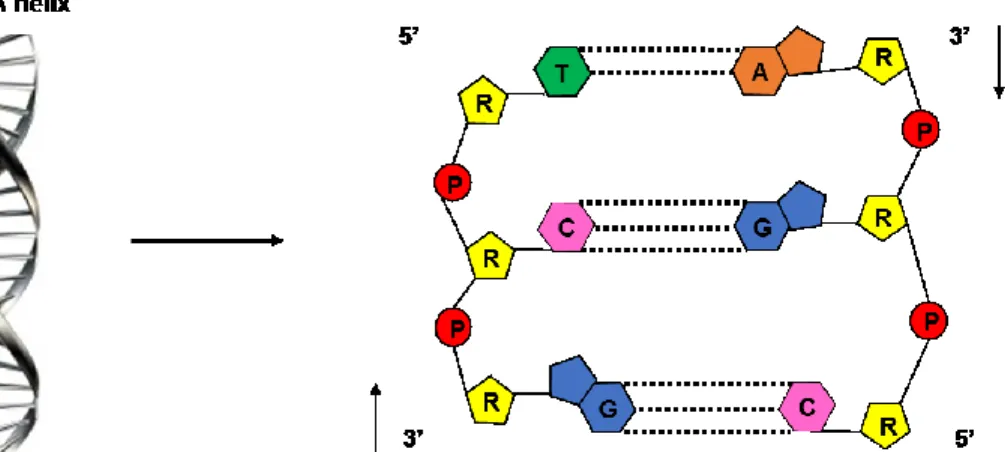
IST OF FIGURES AND SCHEMES Introduction Figure 1. Electromagnetic spectrum separated in non-ionizing and ionizing radiation ... 3Figure 2. DNA structure ... 4
Figure 3. Mechanism of molecular damage via ionizing radiation ... 6
Figure 4. Two major types of resonance; shape resonance and core-excited resonance in a molecule AB. E refers to the incoming electron energy ... 10
Figure 5. The decay channels of TNI of DNA base at an initiated energy of electron (E0) . 12 Figure 6. LC-MS/MS: Multiple reaction monitoring (MRM) ... 19
Figure 7. A schematic of the experimental setup for irradiating DNA sample with X-rays photons ... 21
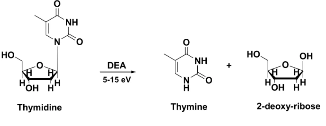
Figure 8. N-gylcosidic bond cleavage in thymidine induced by LEE. ... 66
Article Figure 1. Calibration curves for base release (A) and base modifications (B) ... 41
Figure 2. Analysis of base release by LC-MS/MS carried out by multiple-reaction monitoring in the positive mode (Table 1). The natural products (solid line) and isotope standards (dash line) are shown for the analyses of cytosine (Cyt), thymine (Thy), adenine (Ade) and guanine (Gua). ... 43 Figure 3. Analysis of base modifications by LC-MS/MS carried out by multiple-reaction monitoring in the positive mode (Table 1). The natural products (solid line) and isotope standards (dash line) are shown for the analyses of 8-oxo-7,8-dihydroguanine (8oxoG), 5-hydroxymethyl-2’-deoxyuridine HmU), 5-formyl-2’-deoxycytidine
(5-v ForU), 5,6-dihydro-2’-deoxythymidine (5,6-dHT) and 5,6-dihydro-2’-deoxyuridine (5,6-dHU) ... 44 Figure 4. Graphs representing the total formation of products in DNA films deposited on glass and tantalum substrates as a function of photon fluences. A) base release of guanine; B) base modification giving 5,6-dihydro-2’-deoxyuridine (5,6-dHU). ... 46 Figure 5. Proposed pathways of base release in DNA by X-rays and LEEs ... 51 Figure 6. Proposed pathways for the formation of 5,6-dHU by X-rays and LEEs. ... 54 Figure 7. Proposed pathways for the formation of 5-HmU and 5-ForU by X-rays and LEEs
... 56
L
IST OF TABLESArticle
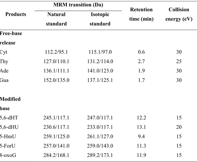
Table 1. LC-MS/MS parameters for the analysis of DNA damage. ... 40 Table 2. Yields of Base Release Products from DNA ... 47 Table 3. Yields of Base Modification Products from DNA ... 49
Discussion
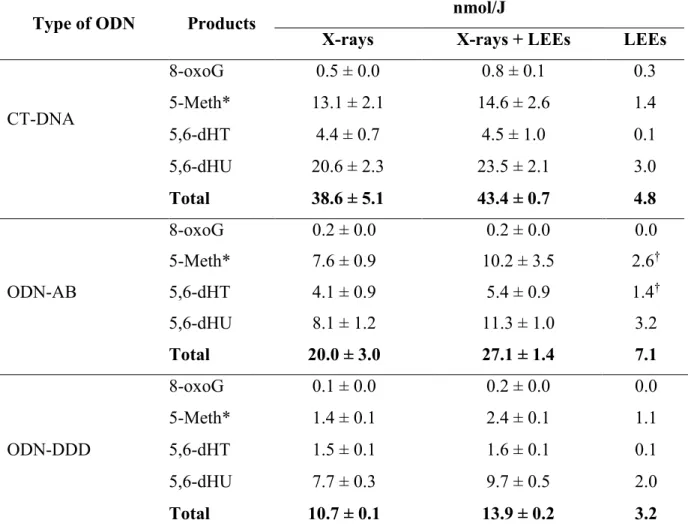
Table 1. Oligonucleotide sequences ... 68 Table 2. The G values (nmol/J) with various types of ODN from X-rays (glass substrate), X-rays plus LEEs (tantalum substrate) and LEEs only (subtracted between tantalum and glass) for free bases release in electrical ground state ... 69 Table 3. The G values (nmol/J) with various types of ODN from X-rays (glass substrate), X-rays plus LEEs (tantalum substrate) and LEEs only (subtracted between tantalum and glass) for bases modification in electrical ground state ... 72
vi
L
IST OF ABREVIATIONS A Ade C °C CGE C-O bond cm Cyt DDD DEA DFT 5,6-dHT 5,6-dHU DNA DSB dsDNA E E0 e- Ea e-(aq) e c-E. coli ESIMS et- eV Fapy Fapy-A Adenine Adenine Cytosine Degree celciusCapillary gel electrophoresis Sugar-phosphate bond Centimeter
Cytosine
Dickerson-Drew dodecamer oligonucleotide Dissociative electron attachment
Density functional theory 5,6-dihydro-2’-deoxythymidine 5,6-dihydro-2’-deoxyuridine Deoxyribonucleic acid Double strand break(s)
Double stranded deoxyribonucleic acid Electron energy, electrical field (equation 1) Incident electron energy
Electron
Activation energy
Solvated electron/ hydrated electron Continuum electron
Escherichia coli
Electrospray ionization mass spectrometry Transter electron
Electon volt
2,6-diamino-4-hydroxy-5-N-methylformamidopyrimidine
vii Fapy-G Fe2+ Fe3+ 5-ForU FWHM G GC/MS Gua h •H H2 HF HmU 5-HmU H2O* H2O+ H2O2 H3O+ HOMO HPLC IR k keV LBL LC/MS LC-MS/MS LEE(s) LET LUMO M MeV 2,6-diamino-4-hydroxy-5-N-methylformamidopyrimidine-guanine Ferrous ion Ferric ion 5-formyl-2’-deoxyuridine Full width half maximum Guanine
Gas chromatography/ mass spectrometry Guanine Hour(s) Hydrogen radical Hydrogen gas Hydrogen fluoride Hydroxymethyluracil 5- hydroxymethyl-2’-deoxyuridine Excited water Water cation Hydrogen peroxide Hydronium ion
Highest occupied molecular orbital High performance liquid chromatography Infrared
A factor (equation 1)
Kilo electron volt (103 electron volt) Layer-by-layer
Liquid chromatography/ mass spectrometry
Liquid chromatography-mass spectrometry/ mass spectrometry Low-energy electron(s)
Linear energy transfer
Lowest unoccupied molecular orbital Molar
viii min N2 NaBH4 nm N2O O2 ocDNA O(1D) ODNs •OH 8-OH-A 5-OHC 8-OH-G 5-OHU O(3P) 8-oxoA 8-oxodA 8-oxodG 8-oxoG P PAGE PCR PFGE pT pTp pTpTp Q QQQ QTOF R ROS Minute(s) Nitrogen gas Soudium borotetrahydride Nanometer(n) (10-9 meter) Nitrous oxide Oxygen gas
Open circular deoxyribonucleic acid (plasmid DNA form) Oxygen atom in the singlet 1D excited state
Oligodinucleotides Hydroxyl radical 8-hydroxyl-adenine 8-hydroxycytosine 8-hydroxyl-guanine 8-hydroxyuracil
Oxygen atom in the triplet 3P ground state 8-oxo-7,8-dihydroadenine
8-oxo-7,8-dihydro-2-deoxyadenosine 8-oxo-7,8-dihydro-2-deoxyguanosine 8-oxo-7,8-dihydro-2-guanine
Phosphate group
Polyacrylamide gel electrophoresis Poly chain reaction
Pulse field gel electrophoresis Thymidine-5'-monophosphate Thymidine-3' ,5' -diphosphate 5'-pTpTp-3'
Net polyanion charge (equation 1) Triple quadrupole in mass spectrometer Quadrupole time-of-flight
Deoxyribose sugar, universal gas constant (equation 1) Radical oxidative species
ix S SATP scDNA S. cerevisiae SE SSB ssDNA SWCNTs T Thy TNI TOF Tp TpT TTT TXT UHV UV V
Fragment size (equation 1)
Standard ambient of temperature and pressure
Supercoiled deoxyribonucleic acid (plasmid DNA form) Saccharomyces cerevisiae
Secondary electrons Single strand break(s)
Single stranded deoxyribonucleic acid Single-walled carbon nanotubes Thymine, temperature (equation 1) Thymine
Transient negative ions Time-of-flight
Thymidine-3' -monophosphate Thymine-phosphate-thymine 5'-TpTpT-3'
5'-TpXpTp-3' (X= T, C, A, G) Ultra high vacuum
Ultraviolet
1
I
NTRODUCTION1.1 Radiation and human life
It has been over 200 years ago since the 19th century that we discovered electromagnetic radiation. The discovery of X-rays by Wilhelm Roentgen, the German physicist, is well-known and led to an explosion of applications in research and medicine. Sunlight is one form of radiation which is produced by a natural process, also known as background radiation. On the other hand, artificial radiation made by humans is widely used in medical processes,communication, industrial and commercial activities. Although, people may think that the radiation is dangerous and thus try to avoid it as much as possible. The sufferings from nuclear bomb explosions in Hiroshima and Nagasaki in 1945, the radioactive release in Chernobyl in 1986 and a high release of radioactivity due to a major earthquake in Fukushima in 2011 are disasters that become a nightmare for everybody.
It might be said that the effect of radiation is a “two-edged sword”, it can be good if we use it in the right way or it can be bad if we do not know how to control it. To understand the effects of radiation and the mechanisms of action on bio-organisms especially within cellular DNA is the principal way to assess the risk of exposure. We cannot live without radiation, it exists all around us. One said that “Life on earth has developed with an ever present background of radiation. It is not something new, invented by the wit of man: radiation has always been there.” (Hall, 1989).
2 1.2 Non-ionizing and ionizing radiation
Radiation is distinguished in two categories by energy: non-ionizing and ionizing radiations (Figure 1). Non-ionizing radiation is apparently low-energy, which does not have enough energy to ionize atoms or molecules. Types of non-ionizing radiation include visible light, infrared light, radiowaves and microwaves. They may be considered to have less energy than ionizing radiation, however, overexposure to non-ionizing radiation can damage tissues, for example, too much ultraviolet (UV) light from sunbathing can cause skin cancer and even moderate amounts of exposure can cause sunburn.
Types of ionizing radiation or high-energetic radiation includes X-rays and gamma rays (electromagnetic waves) as well as accelerated charged particles and heavy ions. After the radiation is absorbed by material, the energy can change atoms or molecules in term of two pathways: excitation and ionization. Firstly, electronic excitation occurs when the electron of any shell of the atom is promoted to an empty orbital lying at a higher energy. The second pathway depends on whether the energy is sufficient to remove an electron from atoms or molecules. The loss of an electron can lead to chemical changes in cells. With each ionization event, secondary electrons (SEs) are produced. Furthermore, if the SEs have enough energy to excite or ionize molecules, in turn they can create further excitations and ionizations before dissipating their energy to the environment (thermalization).
3 Figure 1. Electromagnetic spectrum separating non-ionizing and ionizing radiation.
1.3 The effect of ionizing radiation on biological systems
Ionizing radiation induces a variety of damages in cells by the direct ionization of macromolecules (the direct effect), and indirectly, by electronic excitation and ionization of water molecules leading to hydroxyl radicals (OH), hydrated electrons (e) and hydrogen atoms (H), which subsequently react with biological molecules usually in a very short time to lead to damage (the indirect effect) (O’Neill and Fielden, 1993; O’Neill et al., 2002). In turn, the above species can react with oxygen to give other oxidizing species known collectively as reactive oxygen species. The biological effect depends on the radiation energy, which is measured by the average energy deposited per unit length of the radiation track, called linear energy transfer (LET). Ionizing radiation can produce sparsely ionizations (low LET via X-rays and gamma rays) or dense ionizations (high LET via neutrons and alpha particles) along the track in a non-homogeneous manner within cells. High LET radiation is much more destructive to cells than low LET because it transfers the energy into small volumes (spurs). High LET radiation produces multiple damaged sites
4 that are difficult to repair by cells. The longterm biological effect of radiation can be divided into two types of injuries: somatic and germline. Somatic injury appears in the organism when exposed to ionizing radiation. It can show damage in a short time like a sickness or spending long time like cancer. For germline injury, the reproductive cells are exposed to ionizing radiation can transfer the genetic effects to the next generation, which can be in the form of birth abnormalities or cancer. Deoxyribonucleic acid (DNA) serves as the carrier of genetic information, which is beneficial for understanding the fundamental prominence to all living organisms (Swiderek,2006). DNA is composed of purine (guanine and adenine) and pyrimidine (cytosine and thymine) bases, connected through a ribose sugar ring to a phosphate group (Figure 2). Adenine pairs with thymine and guanine pairs with cytosine by hydrogen bonds taking the form of a right handed helix. Hydrogen bonding between bases on opposite strands and π stacking between adjacent bases on the same strand give stability to the DNA polymer.
5 DNA damage is the most influential cause of cell killing after exposure to ionizing radiation (Karapetyan et al., 2013). Most studies have reported that ionizing radiation, such as certain wavelengths of UV, X-rays, ɣ-rays, and so on, can induce a broad variety of DNA damage, including single strand breaks (SSB), double strand breaks (DSB), modifications in purine and pyrimidine bases, sugar damage, apurinic/apyrimidic sites, removal of bases, and cross-linking of DNA with DNA and adjacent proteins (Shikazono et al., 2009; Sutherland et al., 2000; von Sonntag, 1987; von Sonntag, 2006; Horan, 1999; Sanche, 2009). The most important biological effects of ionizing radiation are due to the formation of a cluster of DNA damage consisting of two or more damaged sites within 10-20 base pairs that are created by a single radiation track (O’Neill et al., 10-2002). Normally, damaged DNA is repaired to sustain cell survival and survival of the organism. In case the damage is not efficiently repaired, it may be converted into mutations that in turn cause cancer and other genetic disorders.
The time scale of events concerned the radiolysis of water during the time living cells are exposed to radiation divived into three stages that must be taken into account (Platzman, 1958). A physical stage occurs by deposition of radiation energy and the formation of initial products (H2O•+, e- and H2O*elec) in highly nonhomogeneous track structure geometry within less than 10-15 seconds, followed by a physicochemical stage in 10-15 to 10-6 seconds, mostly in 10-12 seconds, that produces the stable molecules (H
2O and H2) and chemical reactive species, e.g., free atoms and radicals (H-, OH-, H2O•-, e-, H•, OH•, O(1D), O(3P)) formed in thermal equilibrium in the bulk medium with reactions and reorganization of initial products. Lastly, the nonhomogeneous chemical stage takes place when the various reactive species diffuse and react with another species or with dissolved
6 solutes until completely all spurs or track reactions have occurred (Meesungnoen and Jay-Gerin, 2011). Then at longer times, from a few seconds to years, the biological stage leads to mutation and carcinogenesis (Figure 3).
Figure 3. Mechanism of molecular damage via ionizing radiation.
1.4. Secondary low-energy electrons (LEEs) production
1.4.1 LEEs generated via ionization
Electromagnetic radiation in the form of photons (X-rays, gamma rays or UV) and fast charged particles produce secondary electrons that arise from the ejection of electrons from molecules during the initial ionization process. There are two dominant processes when energetic photons are absorbed by molecules. In the case of low energy photons (less than 0.5 MeV), an electron from an inner shell of the molecule is ejected by gaining energy of the photon to overcome the binding energy. The kinetic energy is transferred to the
7 ejected electron; this main process is known as photoelectric absorption. For high energy photons (approximately 0.7-10 MeV), the photon usually transfers some energy to the electron, which has higher energy than the binding energy of this electron, and thereby, it becomes a fast high energy electron, and as well, the initial photon is deflected with reduced energy and can react by other interactions. In this main mechanism, the Compton process dominates. Compton process or Compton scattering is the inelastic scattering of a photon by a charged particle, usually an electron, resulting in a decrease in energy of the photon (X-rays or gamma rays) and absorbed electron ejects from the atom. Compared to photoelectric effect, Compton scattering occurs when the energy of photon is higher than photoelectric effect. Many fast electrons can be generated and either excite or ionize other molecules to obtain a large number of the secondary LEEs. In both processes, LEEs are obtained carrying most of the energy absorbed by the photon that is transformed to kinetic energy.
Along the ionizing radiation track, many reactive species are formed including radicals, ions, excited molecules and secondary LEEs. Secondary LEEs are created in large amounts of approximately 4 ⨯ 104 per 1 MeV of initial absorbed energy. Most of the energy distribution of LEEs lies below 30 eV with a most probably energy of around 9–10 eV (LaVerne and Pimblott, 1995; Pimblott and LaVerne, 2007; Henke et al., 1981). This energy distribution of SEs has also been observed by the Monte Carlo simulation of water radiolysis which is in the similarily range in agreement with the previous studies (Cobut et al., 1998; Autsavapromporn, 2006; Mirsaleh Kohan et al., 2013). In this energy range, all electrons undergo thermalization within a probable distance of 10 nm. It has been investigated in electrons with initial kinetic energy of ~10 eV by Monte Carlo simulation
8 on the distance of thermalization in liquid water at 25°C (Meesungnoen et al., 2002). Kai et al. also reported the thermalization length and spatial distribution of electrons in liquid water using a dynamic Monte Carlo code (Kai et al., 2015). They found that traveling distance of electrons is distributed from 2 to 14 nm when they reach thermal equilibrium within 10-700 fs, these results are similar to those calculated results with an incident energy of 5 eV proposed by Ritchie et al. (Ritchie et al., 1994). Furthermore, it is considered to be the initial volume of deposited energy by high energy radiation, known as a spur. Non-thermal reactions can produce reactive species, excited atoms or molecules and LEEs within 10-15 seconds (Sanche, 2002). Thus, it can be concluded that LEEs are one of many quantitatively reactive species included water radiolysis species to initiate subsequent chemical reactions.
Secondary LEEs with energy under 30 eV can cause damage to DNA by forming a transient negative ion (TNI) of DNA components; i.e. sugar ring, phosphate group and base. In the case of direct damage to DNA, SSB and DSB can be produced by the decay of TNI states into dissociative electronically excited states or by dissociative electron attachment (DEA) (Huels et al., 2003; Boudaiffa et al., 2000). Furthermore, in cells mostly surrounded by water, LEEs can interact with water molecules close to DNA and the reactive species from water radiolysis products can be formed to induce indirect DNA damage (Alizadeh et al., 2013). It has been proposed by Michael and O’Neill that human genome damage induced by high energy radiation occurs in a ratio of about 1/3 by direct and 2/3s by indirect processes (Michael and O’Neill, 2000). On the other hand, another experiment based on ultrafast electron-transfer to DNA suggests that the ratio of damage
9 caused by direct and indirect pathways is 2/3 and 1/3, respectively (Nguyen et al., 2011; Bald et al., 2012).
1.4.2 LEEs mechanisms
In order to understanding how LEEs induce damage to DNA and cause strand breaks and other fragments, the chemical mechanisms associated with this damage should be taken into consideration. The results of many experiments have also been published using a variety of measurements, in particular, by characterizing the products formed from LEEs bombardment in monolayer and multilayer DNA films (Alizadeh and Sanche, 2012; Sanche, 2005; Zheng and Sanche, 2013), and by observing the formation of fragments of DNA using high resolution electron energy loss spectrometry (Panajotovic et al., 2007; Bazin et al., 2010; Levesque et al., 2005) and also mass spectrometry (Arumainayagam et al., 2010; Sanche, 2009; Baccarelli et al., 2011). Interestingly, other novel techniques have been employed to measure chemical changes in samples, such as atomic force microscope (Keller et al., 2012), Raman spectroscopy (Sidorov and Orlando, 2013) and atmospheric pressure plasma jets (Han et al., 2013).
The proposed mechanisms of bond cleavage observed in DNA have been proposed on the basis of numerous studies. TNI is the most important feature explaining how LEEs induce DNA damage (Allan, 2007; Panajotovic et al., 2007); it is distinguished by two major types of interactions: shape resonances and core-excited or Feshbach resonances (Figure 4). For shape resonances, a single particle resonance, an electron (E ˂ 3 eV) temporarily occupies an unfilled orbital (such as a lowest unoccupied molecular orbital or
10 LUMO which is an electron in excited state that jumps from the ground state after the energy of photon is high enough to absorb by electron in the HOMO) of the ground state of DNA components. For core-excited resonance (E > 3 eV), the previously unfilled orbital becomes occupied with two electrons and the transitory anion is created. The incoming electron is captured by the positive electron affinity of the excited state of the molecule due to the higher energy of the electron (E > 3 eV), giving a configuration consisting of an electron in a LUMO and a hole in the highest occupied molecular orbital or HOMO of the molecule.
Figure 4. Two major types of resonance: shape resonance and core-excited resonance in a molecule AB. E refers to the incoming electron energy.
Furthermore, a core-excited resonance with an electron energy around 10 eV can play a significantly important role in DNA fragmentation via DEA. This is supported by studies in which the yield of products is highest in this electron energy range (Pan et al., 2003; Zheng et al., 2006). If the incoming electron has non-zero angular momentum when it is captured, it is defined as a core-excited shape resonance (Alizadeh et al., 2015).
11 According to TNI decay, a chemical bond can fragment into a stable anion and a radical such that the electron goes to one side of the chemical bond while the other side becomes a neutral radical. Based on theoretical and experimental studies, the energy of electrons inferior to 3 eV are able to cleave the C-O bond of the phosphate and sugar moiety at the 3’ and 5’ positions (Zheng et al., 2005; Berdys et al., 2004) The lowest π* resonance states associated with the incoming electron of the base unit can transfer to the anti-bonding σ* orbital of C-O bond in the DNA backbone. The excess electron of the C-O bond is unstable leading to cleavage of the C-O bond by a DEA pathway. In addition, another experiment explained the effect of electron energy above 3 eV. It was found that C-O bond breaking decreased upon bombardment with electrons of 6 and 10 eV next to an abasic site in oligonucleotide tetramers presumably by inhibition of electron transfer from the abasic site (Zheng et al., 2006). However, the precise mechanism involving bond cleavage above 3 eV electrons by TNI decay is still to be determined.
The proposed decay channels of TNI states of DNA leading to strand breaks, base release and other products (base damage) are described in Figure 5 (Zheng et al., 2005). They are separated in three channels, which refer to (i) the elastic channel (E = E0), (ii) DEA channel and (iii) the inelastic channel (E
«
E0).12 Figure 5. The decay channels of TNI states of DNA bases with an initial electron energy (E0). Pathway 1, the shape resonancecan occur on a base due to the low incoming electron energy (E0 ˂ 0.5 eV) compared to the excitation energy. Excess energy of the electron can be transfered (et-) to the phosphate group within DNA or re-emitted into the elastic continuum (ec-) as shown in pathway 3. The et- that is found on a sugar-phosphate bond can lead to C-O bond cleavage at the 3’ and 5’ positions. In pathway 2, core-excited resonances are likely involved due to the fact that the excitation energy is higher than the threshold, which leads to fragmentation of the nucleobase or to modification of the base via DEA. For the inelastic pathway (E0 > 5 eV), the excited state of the base can be formed together with cleavage of the C-O bond and release of an additional LEEs. Reprinted (adapted) with permission from Zheng et al., 2005. Copyright 2005 American Chemical Society.
13 1.5 How to detect DNA damage
DNA damage has been studied in a variety of organisms, it can be caused by endogenous reactive oxygen and nitrogen species that are generated by biochemical processes as well as by environmental toxins that are absorbed by the body. There are many types of DNA damages and several DNA repair strategies to correct the damage and avoid genetic changes. Therefore, the development and application of methods to detect DNA damage is important.
1.5.1 Acid hydrolysis of DNA
DNA base damage remains attached to the DNA backbone through sugar-phosphate moieties. For analysis, it is necessary in certain methods to cleave the linkage-glycosidic bond by hydrolysis before analysis of the modified base moieties. A common method is to treat DNA with 60-88% formic acid at 140 °C for 2 h. It is worth noting that 60% formic acid provided a higher degree of hydrolysis than 88% formic acid, although the 5-hydroxymethyluracil (5-HmU) yield is lower with the lower acid concentration (Douki et al., 1996). Alternatively, 35% hydrogen fluoride (HF) in pyridine at 37 °C for 2 h is a milder condition to avoid the degradation of 5-HmU (Douki et al., 1996). In addition, Fapy (2,6-diamino-4-hydroxy-5-N-methylformamidopyrimidine) derivatives, A and Fapy-G withstand concomitant degradation much better when using HF (Douki et al., 1997). However, all these procedures are very severe and the products may be transformed by strong acid under these conditions, for example, cytosine and uracil glycols do not
14 withstand acid and are released as 5-hydroxycytosine OHC) and 5-hydroxyuracil (5-OHU), respectively (Douki et al., 1996; Dizdaroglu et al., 1986).
1.5.2 Enzymatic digestion of DNA
Specific N-glycosylase enzymes have been considered for the detection of DNA damage because they induce a strand break next to the damaged site (Dodson and Lloyd, 2002). However, this enzymatic excision of nucleobases damage may cause some problems, for example, nuclease P1 enzyme acts on 8-oxoG in DNA, which is released as the modified nucleotide (Maccubbin et al., 1992). Other lesions by the enzymatic excision are observed in 8-oxoG or AP site (Beckman et al., 2000), but it has a large variation in the yields of 8-oxoG and concomitant discussions as to the best technique for this DNA lesion detection. In addition, the enzymatic excision of 8-oxoA by Ogg1 from S. cerevisiae is productive when it is paired with only cytosine (Girard et al., 1998). DNA Enzymatic hydrolysis technique has been suggested as a good method for HmU determination (Teebor et al., 1984; Frenkel et al., 1985). Recently, the development of using specific enzyme is required to active only in specific DNA sites. This advantage can prove the appropriate detection without causing a problem to non-specific DNA sites.
1.5.3 DNA lesions detected by liquid chromatography/ electrospray tandem mass spectroscopy (LC/MS)
Nowadays, LC/MS is a sensitive and specific technique for the detection of oxidative DNA damage. A drawback is that certain products are not retained enough on
15 reversed phase liquid chromatography columns generally used to separate the mixture of modifications before MS analysis (especially modifications of guanine). The soft ionization property of electrospray provides one to assess most kinds of DNA adducts including those with bulky chemicals (Rindgen et al., 1995) and UV induced dimeric pyrimidine photoproducts (Douki and Cadet, 2001). The detection of the cis-syn and trans-syn I cyclobutane thymine dimers is also possible by this technique (Douki et al., 2000). In addition, this technique is suitable to quantify 8-oxo-7,8-dihydroadenine (8-oxoA), 5,6-dihydroxy-5,6-dihydrothymine and 5-hydroxyuracil as the modified nucleoside from isolated and cellular DNA exposed to gamma rays (Frelon et al., 2000).
1.5.4 Hydroperoxides detection
Hydrogen peroxide reacts fasterwith molybdate activated iodide reagent than with organic hydroperoxides (Allen et al., 1952). Using this reagent, however, iodine is formed, which can react with other products. This problem can be resolved by using HPLC to remove this impurity prior to analysis. To quantify hydroperoxide products, a slow reaction with heating is required to avoid underestimating the yields of hydroperoxide products due to incomplete reactions. Moreover, ferrous ion (Fe2+) can react with hydroperoxides in acid to form ferric ion (Fe3+) as in the related method termed Fricke dosimetry. This technique can be combined with HPLC to identify hydroperoxides formed upon •OH-attack of thymidine in aqueous solution (Wagner et al, 1990).
16 1.5.5 DNA damage detected by capillary and pulse field gel electrophoresis
Capillary gel electrophoresis (CGE) has a relatively short time of analysis, a high degree of automation and reproducibility together with a good resolving power for dsDNA fragments (Valenzuela et al., 2000). The mobility of DNA fragments can be explained by the following equation (1):
ln1 𝑉 = ln 𝑘 𝐸𝑆+ 𝐸𝑎 𝑅𝑇 (1) where the velocity of the fragment (V), a factor (k) is related to the net polyanion charge (Q), the fragment size (S), the electrical field (E), the activation energy for viscous flow (Ea), the universal gas constant (R) and temperature (T).
In the case of pulsed field gel electrophoresis (PFGE), DNA is treated with a restriction enzyme to create small fragments of DNA in order to improve their resolution by electrophoresis in acrylamide or agarose gel. The pulse length and orientation of the electric field is modulated such that DNA with a small fragment will migrate farther than larger fragments. Sutherland et al. developed a gel electrophoresis/ quantitative imaging method to estimate the amount of clustered damage in large DNAs (Sutherland et al., 2000). They found that the high energy of charged particle (Fe26+, 1 GeV/ nucleon) induced oxidized pyrimidine clusters, which were recognized by treatment with a specific E. coli protein that cleaves oxidizied pyrimidines (Endo III) giving DSBs in human cells. This technique is very sensitive for the analysis of DSBs in DNA damage, it can detect damaged DNA after exposed to very low dose (1 mGy) of ionizing radiation (Rothkamm and Lobrich, 2003).
17 1.5.6 Electrochemical process
The detection of DNA damage can be achieved by electrochemistry, which is a sensitive and selective technique, and the device can be miniaturized and used at low cost (Jelen et al., 2002; Rahman et al., 2005). DNA is an electroactive and surface active substance yielding analytically valuable electrochemical signals. Certain DNA bases (cytosine, guanine and adenine) pass through redox steps at the mercury electrodes, in particular, guanine and adenine are easily oxidizable at carbon and other solid electrodes. These signals correspond to the changes in DNA structure (Fojita et al., 2004). Certain lesions, such as thymine cyclobutyl dimers, can be detected when they are associated with distortions of the DNA double helix (Fojita et al., 2002). Native DNA (double stranded) and denatured DNA (single stranded) are responsive to this technique using mercury electrodes, which can determine small amounts of ssDNA in dsDNA analyses (Cahova-Kucharikova et al., 2005). Furthermore, He and Bayachou proposed the development of DNA sensors by the fabrication of new DNA/Single-Walled Carbon nanotubes (SWCNTs) particles using the layer-by-layer (LBL) technique on oxidized side walls of SWCNTs (He and Bayachou, 2005). They found that these particles can be used as DNA carriers for sensitive electrochemical detection of DNA chemical damage, including NO-induced DNA damage. Chang and co-workers also developed conducting polyaniline nanotube array for the ultrasensitive biosensor detection of DNA hybridization (Chang et al., 2007). This DNA electrochemical biosensor technique demonstrated an extremely high sensitivity that could detect the presence of a target nucleotide at a concentration down to 10-15M.
18 1.6 Principle of LC-MS/MS
LC-MS and LC-MS/MS are the analytical chemistry techniques which combine the physical separation capabilities of liquid chromatography (LC) with the mass analysis capabilities of mass spectrometry (MS). LC separates chemical compounds by conventional chromatography on the column that usually will be reverse phase chromatography. The analyte binds to the column by hydrophobic interactions in the presence of hydrophilic solvent (e.g. water) and is eluted off by more hydrophobic solvent (e.g. methanol or acetonitrile). The analytes appear at the end of the column, the solvent is evaporated and analytes are ionized using electrospray ionization. The MS detector can detect only ionized analytes, scan the molecules by mass and show the separation of all ions that have different masses by a mass spectrum (John Innes Centre, n.d.). MS is a microanalytical and physical analysis technique, which allows (i) selectively the detection and identification of molecules of interest by measuring their masses and (ii) the characterization of their chemical structures. MS analyzer is used for analysis of the precursor ion generated in the source (e.g. iontrap, quadrupole or time-of-flight (TOF)). MS/MS is the combination of two mass analyzers in one MS instrument, the first MS filters for precursor ion followed by fragmentation of the precursor ion with a neutral gas. The fragmented ions are then separated and measured in a second MS analyzer. The advantages of MS/MS is the increased sensitivity in QQQ due to the noise reduction and it provides more structural information on the analyte based on the fragmentation pattern (Kellner, 2015). In mass analysis, m and z stand for mass and charge of the detected ions, respectively. The number of electrons removed is the charge number (positive ions), m/z represents mass divided by charge number and the horizontal axis in a mass spectrum is expressed by a m/z ration
19 (Wysocki et al., 2005). Most of the ions passing through MS will have a charge of 1+, that means the m/z ratio will be the same as the mass of ion. So, the basic principle of MS is the separation ions in the gas phase according to the m/z, whereas, MS/MS measures the m/z of the fragment ions.
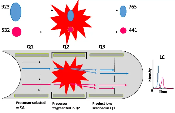
In our experiment, we detected the DNA damage products by using LC-MS/MS with multiple reaction monitoring (MRM) mode (Figure 6).
Figure 6. LC-MS/MS: Multiple reaction monitoring (MRM).
MRM has two stages of mass filters in a triple quadrupole MS. Firstly, the precursor ion (ion of interest) is preselected in Q1 filter and passed through the pressurized collision
20 cell (Q2) to fragment with a neutral gas. The second stage, the possible fragment ions are filtered by mass analyzer in Q3, which measures a pre-set of known fragment ions also called transition ions.
1.7 DNA damage induced by LEEs studies
1.7.1 An experimental setup for LEEs production
The general design of the irradiation systems used to study LEEs-induced DNA damage in condensed phase is separated into five parts (i) the most important part is an electron gun that generates the LEEs beam. Electrons are emitted in a variable region between a few eV to hundreds of eV with a full width half maximum (FWHM) of 0.5 eV. (ii) the chamber holding the electron gun and an oil free turbo molecular pump for pumping from atmospheric pressure to ultra high vacuum (UHV) range (around 10-9 torr). For UHV, it is important to protect the surface and avoid contamination. (iii) the LEE target is comprised of DNA target molecules or their components in condensed phases. (iv) the substrate, which is made up of a metal substrate, for example, gold and tantalum. In the case of gas phase studies, DNA samples are vaporized and interact with electrons emitted inside the chamber. Gas phase experiments have the disadvantage that the sample may undergo decomposition during heating required for evaporation. Thus, it is limited to small molecules that have a low boiling point. The samples have to be deposited onto the metal substrate and brought to lyophilization. (v) lastly, the module that performs product analysis after LEE bombardment. This can be located either inside or outside the UHV
21 chamber, for example, product analysis can be carried out by gel electrophoresis, MS, HPLC, LC/MS, GC/MS and so on.
The experimental setup to irradiate DNA with 1.5 keV Al K⍺ X-ray photons surrounded by a gas (nitrogen or oxygen) under atmospheric pressure is shown in Figure 6 (Alizadeh and Sanche, 2012).
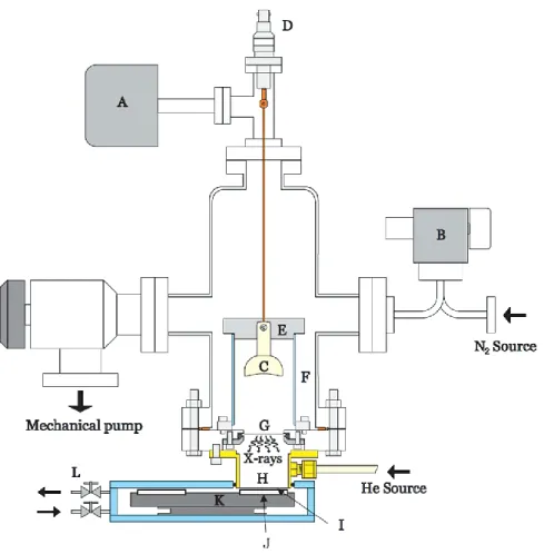
Figure 7. A schematic diagram of the experimental setup for irradiating DNA samples with X-rays photons. Reprinted with permission from Alizadeh and Sanche, 2012. Copyright 2012 American Chemical Society.
22 This apparatus is comprised of a stainless steel chamber evacuated by a mechanical pump to a pressure of less than 5 mTorr and connected to a baraton (A). An adjustable leak valve (B) is connected to a nitrogen gas source. 3.4 kV is applied to a concave aluminium cathode (C) with a negative potential passing through a high voltage electrical feed through (D) to create an electrical discharge. The nitrogen gas pressure is important to control and stabilize the plasma current. Electrons from the discharge hit the aluminium thin foil, thereby emitting X-ray photons into the He-filled side enclosed volume (H), through a thin foil of mylar (I), and then through a cylindrical chamber. The latter chamber is the place that holds the DNA thin film samples, which are deposited on different substrates on top of an aluminum plate holder (J). The sample holders are put at different positions of a rotating disk (K) that can rotate the DNA sample like a merry-go-round into position for irradiation by X-rays with various radiation doses. The flow of gas is adjusted at the gas circulation valves (L).
1.7.2 Types of DNA bombarded with LEEs
In order to study LEEs-induced DNA damage, oligonucleotide DNA and plasmid DNA are widely used for investigation due to their similarily to natural DNA macromolecules (Boudaiffa et al., 2000; Zheng et al., 2005; Li et al, 2010). In case of oligonucleotides, a defined sequence and length can be obtained by chemical synthesis from a commercial vendor. Short oligonucleotide products can be analyzed and quantified by HPLC-UV and GC/MS as well as by LC-MS/MS. For electrospray ionization MS (ESIMS), e.g. LC-MS/MS, works on soft ionization technique whereas GC/MS works on hard ionization technique. These are the basic differences between these two methods.
LC-23 MS/MS is useful for non-volatile compounds, vitamins, amino acids, proteins and peptides having molecular weight in kilo Dalton. It is very much useful for studies purity and impurity profiles in drugs. So these tool has widely applications in pharmaceutical industries. GC/MS is useful for the identification of volatile compounds having molecular weight less than 1200 amu, realistically, it is around 500 amu but it can be up to 600-800 amu with chemical derivatization. It is useful in petro chemical, pesticides industries and in field of perfumery. Furthermore, the analyte used for LC-MS/MS must be soluble in the mobile phase, while analyte for GCMS must be volatile and does not decompose upon heating. Comparison between LC-MS/MS and GC/MS, LC-MS/MS provides high specificity, sensitivity, high through put and devoid of several derivatization and complex sample preparation required by the GC-MS. Long oligonucleotides can be analyzed by polyacrylamide gel electrophoresis (PAGE) that separates DNA fragments according to their length and charge. Moreover, DNA fragments can be synthesized by either polymerase chain reaction (PCR) or the method of solid phase synthesis. Oligonucleotide can be prepared in either single or double stranded conformations, which is associated with a minimum of 6-10 complementary nucleotide base pairs in order to achieve the stable formation of DNA duplex. Hydrogen bonds between G-C and A-T are broken by heat or chemical agents, which denature double stranded oligonucleotides into two single stranded oligonucleotides. Conversely, double stranded DNA can be formed by renaturation of single stranded DNA in a slow cooling down system. Thus, oligonucleotides are suitable to study DNA damage induced by ionization radiation.
Plasmid DNA is extracted from microorganisms (i.e. E. coli) and it forms macromolecules of up to 150000 base pairs in a native supercoiled form. Strand breaks can
24 be quantified by gel electrophoresis that separate three forms of plasmid DNA; supercoiled DNA (scDNA) found in natural plasmid DNA, open circular DNA (ocDNA) arising from a single strand break and linear DNA arising from a double strand breaks. In agarose gel electrophoresis, the migration order is scDNA > linear DNA > ocDNA.
1.7.3 Studies of LEEs-induced DNA damage
The interaction of LEEs with DNA molecules has been studied for many years. The basic interactions of LEEs with nucleobases, deoxyribose derivatives, oligonucleotides and plasmid DNA have been examined (Ptasinska et al., 2005; Huels et al., 1998; Aboul-Carime et al., 2001; Breton et al., 2004; Huels et al., 2004; Lepage et al., 1998; Ptasinska et al., 2004; Park et al., 2006; Ray et al., 2005; Martin et al., 2004). In addition, there have been numerous calculations with density functional theory (DFT) and theoretical simulations to predict LEEs interactions with DNA (Bao et al., 2006; Berdys et al., 2004; Gu et al., 2005; Kumar and Sevilla, 2007; Li et al., 2006; Schymann and Laaksonen, 2008; Simons, 2007). Certain non-volatile radiation products trapped on the surface have been identified especially strand break products and base reduction products.
The interaction of thymidine with LEEs has been investigated showing cleavage of the N-glycosidic bond of thymidine leading to the release of thymine as a major product (Zheng et al., 2004). Also, Zheng and co-workers published the formation of products from two oligonucleotide tetramers (CGTA and GCAT) with monoenergetic LEEs (Zheng et al., 2005). They found several products including non-modified nucleobase, nucleoside and nucleotide fragments due to rupture of the phosphodiester bonds and N-glycosidic bonds
25 within each tetramer. A mechanism of non-modified base products was suggested involving initial electron attachment to nucleobase moieties followed by electron transfer to the sugar-phosphase backbone and subsequent dissociation of the phosphodiester bond. The phosphodiester bond breaks with the negative charge located on the phosphodiester bond leading to non-modified fragments with an intact terminal phosphate group. This pathway is supported by theoretical studies that follow the energetics of electron transfer and sugar-phosphate backbone cleavage (Simons, 2006). From these studies, it is important to underline that non-thermalized electrons (LEEs) captured by DNA bases may play a crucial role in the induction of DNA damage in living cells.
Li and co-workers examined the effect of terminal phosphate and base moieties on LEEs induced DNA damage analyzed by LC-MS/MS (Li et al., 2008). They used DNA model compounds composed of monomers (pT, Tp, pTp), dinucleotides (pTpT, TpTp, pTpTp) and trinucleotides (TpTpT). They found that the terminal phosphate groups in monomers and dinucleotides increases the damage by 2-3-fold but that it decreases N-glycosidic (N-C) linkage and sugar-phosphate (C-O) bond cleavage by 2-10-fold. The capture of LEEs directly on the terminal phosphate appears to be an important factor to induce DNA damage; however, it does not contribute to N-C and C-O bonds cleavage. Furthermore, Li and co-workers investigated the effect of base sequence in oligonucleotide trinucleotides after irradiation with LEEs (Li et al., 2010). The trinucleotides included TXT where X can be one of four normal DNA bases (C, T, A, G). After bombardment with LEEs of 10 eV, the total damage within trimers decreased by 2-fold in the following order: TTT > TCT > TAT > TGT and non-modified base release occurred to a great extent at T bases. Thus, they concluded that initial LEEs capture and subsequent bond cleavage
26 through a transient negative anion depended on the sequence and electron affinity of the DNA base. This was prevalent for short oligonucleotides containing T bases, which are the most electronegativity DNA base.
There are several reports in the literature about LEEs-induced damage to plasmid DNA. Boudaiffa and co-workers bombarded dried plasmid DNA films with electrons in the range of 3 eV to 1.5 keV (Boudaiffa et al., 2002). They measured the total effective cross-section of about 4 ⨯ 10-15 cm2 and effective range of about 13 nm for the destruction of scDNA within this energy range. Furthermore, they also reported the yields of DNA damage with electron exposure between 3-100 eV. They found that the yields of damage did not depend on the ionization cross-section but that the yields varied with the incident electron energy under 15 eV. LEEs-induced DNA damage was dependent on initial kinetic energy lower than the threshold of ionization of 15 eV (Boudaiffa et al., 2000). For electrons with energies below 3 eV, the only strand breaks detected were SSB while no DSB were observed (Martin et al., 2004). This is consistent with the capture of electrons by molecules with the formation of a TNI state that decomposes into many fragments by DEA processes (Illenberger and Momigny, 1992).
The results of a study on the dependence of LEEs-induced DNA damage on the average dry film thickness in the range of 2-80 nm were presented (Alizadeh and Sanche, 2011). This experiment allowed the G value of the dry films with a thickness of 10 nm, which are too thin to allow for an effective absorption of LEEs generated in the system. It is supported by certain reports that LEEs have thermalization distances on the order of 10 nm in biological materials which define the initial deposition volume (Nikjoo and
27 Lindborg, 2010; Goodhead and Nikjoo, 1990). Furthermore, Alizadeh and Sanche reported the formation of SSB and DSB induced by soft X-rays (1.5 keV) and LEEs (0-30 eV) in dry and humid thin films of plasmid DNA irradiated under different levels of oxygen at standard ambient temperature and pressure (SATP) (Alizadeh and Sanche, 2013a). They calculated the G value for X-ray and LEEs-induced DNA damage under a variety of conditions by changing the humidity and atmosphere from N2 to O2. They also found that the formation of DSB increased by 4.5- and 11.8-fold for X-rays and LEEs, respectively. The results indicated that H2O, O2 and N2O promoted the formation of SSB and DSB in plasmid DNA. X-ray photons can excite and also ionize these molecules to form into superoxide species, which enhance DNA damage. In contrast, LaVerne and Pimblott proposed the electron energy-loss distributions in solid DNA by using Monte Carlo simulations (LaVerne and Pimblott, 1995). They found that ineleastic mean free paths for electrons of 25 eV are around 2-fold greater in liquid water compared to solid DNA. This means the electrons in liquid water can travel longer distance in the same volume and electron energy comparable with dry DNA. Thus, this result may have a confliction idea to the previously mentioned studies that revealed the dry films with a thickness of 10 nm, which are too thin to allow for an effective absorption of LEEs generated in the soft X-ray system.
28 1.8 Hypothesis/problem
Sanche and co-workers built a novel instrument to irradiate DNA samples under SATP conditions. They studied by gel electrophoresis for the formation of strand breaks in plasmid DNA. They had not observed base release and modified base in DNA analyzed by LC-MS/MS technique. In this work, we extend our studies with the soft X-ray instument to investigate the formation of DNA damage. It would be possible to detect and to determine the yield of several structurally distinct types of damage by LC-MS/MS.
1.8.1 Objective
The main objective of the present project is to investigate the mechanism of LEEs-induced DNA damage and extract the component of LEEs-LEEs-induced processes from the total effect of ionizing radiation. To this end, dried DNA thin films are deposited on either glass or tantalum substrates to be exposed to soft X-rays (1.5 keV) under SATP surrounded by N2 atmosphere. The release of DNA bases (Cyt, Thy, Ade and Gua) and base modification products of Cyt, Thy and Gua are detected by LC-MS/MS. A considerable amount of work in this project involved the development of suitable analytical methods to precisely measure DNA damage by LC-MS/MS.
29
A
RTICLE1
Base release and modification in solid-phase DNA exposed to low-energy electrons
Surakarn Choofong, Pierre Cloutier, Léon Sanche, J. Richard Wagner.
Submitted to Radiat. Res. March 30, 2016
I did all the experiments in this paper and wrote the first version of the manuscript. Prof. J. Richard Wagner taught me how to use the instruments of HPLC and LC-MS/MS. Mr. Pierre Cloutier helped me to work with the X-ray irradiation source and repaired it when it had problems. Prof. Leon Sanche and Prof. J. Richard Wagner are my joint supervisors and helped me to revise the manuscript for submission.
30 Résumé
L’ionisation génère un grand nombre d'électrons secondaires de faible énergie (LEEs) avec une énergie d'environ 10 eV, qui peuvent rompre les liaisons dans l'ADN par l'attachement d'électrons dissociatif (DEA), et conduire à des lésions d'ADN. Dans la présente étude, nous mesurons les dommages de radiation sur l’ADN sèche induits par les rayons X (1,5 keV) seul sur un substrat de verre et les rayons X en combinaison avec LEEs en surplus (énergie moyenne de 5,8 eV) émis à partir d'un substrat de tantale sous une atmosphère de N2 et des conditions ambiantes standards de température et de pression. Les cibles comprennent l'ADN de thymus de veau et de double brin d’oligonucléotides synthétiques. Nous avons développé des méthodes d'analyse pour mesurer la libération de bases d’ADN non modifiées à partir de l'ADN, et la formation de plusieurs modifications de base par LC-MS/MS avec dilution isotopique pour la quantification précise. Les résultats montrent que le rendement de bases non modifiés, ainsi que des modifications de base augmentent de 20 à 30% lorsque l'ADN est déposé sur un substrat en tantale par rapport à celle d'un substrat en verre. La quantité de bases libérées qui suit l’ordre Gua> Ade> Ta ~ Cyt, correspond bien à plusieurs études théoriques indiquant que Gua est le site le plus suceptible pour la division sucre-phosphate. La formation de lésions d'ADN par LEE est expliquée par DEA conduisant à la libération des bases non modifiées impliquant la division initial des liens N1-C1', C3'-O3, ou C5’-C5’. Le rendement des modifications de bases est inférieur à la libération des bases non modifiées. Les principales modifications de base induites par LEE comprennent 5,6-dihydrothymine (5,6-dHT), 5,6-dihydrocytosine (5-dHU), 5-hydroxyméthyluracile (5-HmU) et 5-formyluracile (5-ForU). La formation de modifications de base par LEE peut être expliquée par DEA et la séparation de la liaison du lien C-H groupe méthyle de Thy (donnant 5-HmU et 5-ForU), et par des réactions secondaires des atomes d'hydrogène et d'anions hydrure qui sont générés par réactions primares des LEE suivies par une réaction ultérieure avec Cyt et Thy (donnant 5,6-dHU et 5,6-dHT).
Mots clés: rayonnements ionisants, lésions de l'ADN, la spectrométrie de masse, électrons secondaires
31 Abstract
Ionization generates a large number of secondary low-energy electrons (LEEs) with a most probable energy of about 10 eV, which can break bonds in DNA by dissociative electron attachment (DEA), and lead to DNA damage. In the present study, we investigate radiation damage to dry DNA induced by X-rays (1.5 keV) alone on a glass substrate and X-rays in combination with extra LEEs (average energy of 5.8 eV) emitted from a tantalum substrate under an atmosphere of N2 and standard ambient conditions of temperature and pressure. The targets include calf-thymus DNA and double stranded synthetic oligonucleotides. We developed analytical methods to measure the release of non-modified DNA bases from DNA and the formation of several base modifications by LC-MS/MS with isotopic dilution for precise quantification. The results show that the yield of non-modified bases as well as base modifications increase by 20-30% when DNA is deposited on a tantalum substrate compared to that on a glass substrate. The order of base release (Gua>Ade>Thy~Cyt) agrees well with several theoretical studies indicating that Gua is the most susceptible site toward sugar-phosphate cleavage. The formation of DNA damage by LEE is explained by DEA leading to the release of non-modified bases involving the initial cleavage of N1-C1’, C3’-O3’, or C5’-C5’ bonds. The yield of base modifications was lower than the release of non-modified bases. The main LEE-induced base modifications include 5,6-dihydrothymine (5,6-dHT), 5,6-dihydrocytosine (5-dHU), 5-hydroxymethyluracil (5-HmU) and 5-formyluracil (5-ForU). The formation of base modifications by LEE can be explained by DEA and cleavage of the C-H bond of the methyl group of Thy (giving 5-HmU and 5-ForU) and by secondary reactions of H atoms and hydride anions that are generated by primary LEE reactions followed by subsequent reaction with Cyt and Thy (giving 5,6-dHU and 5,6-dHT).
32 INTRODUCTION
Ionizing radiation, such as vacuum UV, X-rays, γ-rays, induces a wide variety of DNA damage, including single strand breaks (SSB), double strand breaks (DSB), modifications of purine and pyrimidine bases, sugar damage, apurinic/apyrimidic sites, base release, DNA-DNA and DNA-protein cross-links (1-8). The effects of ionizing radiation arise from either the indirect effect, which involves the generation of reactive species such as hydroxyl radicals by the radiolysis of water, or the direct effect, which involves the absorption of radiation energy directly by the target molecule leading to its excitation or ionization. In the initial energy-deposing events, ionization produces a large number of secondary low-energy electrons (LEEs), which have a most probable energy of below 10 eV (9).The later species can cause both indirect and direct mediated damage to DNA. LEEs with energy below 15 eV can temporarily attach to specific orbitals localized on DNA and lead to the formation of molecular transient negative ions (TNIs) (10). These metastable molecular anions can dissociate into an anion and a neutral radical (e- + AB AB*- A + B-) (10) on a short time scale by a process known as dissociative electron attachment (DEA).
There are about 3 × 10⁴ electrons generated per MeV of deposited energy with a kinetic energy below 30 eV (11). Previously, LEEs were shown to induce fragmentation of the DNA backbone by the formation of SSB or DSB upon irradiation of supercoiled plasmid molecules under ultrahigh vacuum followed by gel electrophoresis as a sensitive method for the detection of conformational changes (12-18). The type of damage was identified in greater detail by chemical analysis of LEEs-induced products remaining within the irradiated DNA films. Zheng et al.(12, 19, 20) reported two main reactions: the release
33 of non-modified bases and the formation of strand breaks using short oligonucleotides as models of LEEs-induced DNA damage. The formation of strand breaks involved direct cleavage of the phosphodiester bond leaving a non-modified phosphate group at one terminus and a modified as yet unidentified residue on the other terminus. Based on these results, we have proposed that LEEs are initially captured by DNA bases giving a transient negative ion (TNI) which is then transferred to an antibonding orbital of either the N1-C1 bond leading to dissociation of this bond and prompt base release or to an antibonding orbital of the sugar-phosphate moiety leading to cleavage of the C-O bond either at the C5’ or C3’ positions of the deoxyribose ring. These reactions have extensive support from thereoretical investigations (21-23). Similar studies using pure sources of LEEs have identified several base modifications induced by the reaction of LEEs with TTT including 5,6-dihydrothymine (5,6-dHT), 5-hydroxymethyluracil HmU) and 5-formyluracil (5-ForU) (24, 25).
The role of LEEs-induced DNA damage during the exposure of cells to ionizing radiation remains difficult to evaluate. Because LEEs are very reactive and have a short interaction range (~ 10 nm), electron beams can only be formed under vacuum conditions. Thus, irradiation of solid DNA films with such beams greatly restrict the chemical reactions of primary reactive species. However, it is essential to probe the initial chemical steps that are common under both extreme and ambient conditions in order to extrapolate the mechanisms of LEEs-induced DNA damage to those occuring in cells. With this approach in mind, we have devised a novel system to demonstrate the reactivity of LEEs under standard ambient conditions of temperature and pressure (SATP) (26). This system also allows for the introduction of primary reactants, such as H2O and O2, which can change the course of reaction of the initial radical and ionic species believed to be
34 generated by LEEs reactions. In this system, liquid samples of DNA are freeze-dried as a thin film onto either glass and tantalum substrates. Upon exposure to X-rays (1.5 keV) under SATP, the formation of DNA damage on the glass substrate can be attributed solely to the interaction of X-ray photons, whereas damage on the tantalum substrate can be attributed to both the direct interaction of X-rays and the X-ray-induced emission of LEEs from the tantalum substrate. The emitted electron distribution exhibits a peak at 1.4 eV and an average energy of 5.85 eV. For comparision, we first reported the enhancement of SSB, DSB, and DNA-DNA crosslinks induced by LEEs and 1.5 kev X-rays when supercoiled plasmid DNA was deposited on a tantalum substrate under vaccum conditions (27). Afterwards, the supercoiled plasmids were irradiated in air (28) and under a nitrogen atmosphere at SATP (29). More detailled studies with supercoiled plasmid DNA have been performed by introducing specific primary reactants during irradiation. Interestingly, the addition of H2O, O2 and N2O into this system greatly enhanced the formation of SSB and DSB, particularly when DNA was irradiated when on a tantalum surface with the emission of LEEs (29-33). Here, we continue similar investigations using linear double-stranded DNA from calf thymus and oligonucleotides, as a target under SATP conditions. Instead of analyzing conformational modifications to DNA as in previous investigations, we identify by liquid chromatography (LC) and tandem mass spectrometry (MS/MS) specific products remaining on the surface of the irradiated glass and tantalum substrates; these include base release and base modifications.