O
pen
A
rchive
T
oulouse
A
rchive
O
uverte
(OATAO)
OATAO is an open access repository that collects the work of some Toulouse
researchers and makes it freely available over the web where possible.
This is
an author's
version published in:
https://oatao.univ-toulouse.fr/23063
Official URL :
https://doi.org/10.1016/j.otsr.2017.10.015
To cite this version :
Any correspondence concerning this service should be sent to the repository administrator:
[email protected]
Marot, Vincent and Bayle-Iniguez, Xavier and Cavaignac, Étienne and Bonnevialle, Nicolas
and Mansat, Pierre and Murgier, Jérôme Results of non-operative treatment of olecranon
fracture in over 75-year-olds. (2018) Orthopaedics & Traumatology: Surgery & Research, 104
(1). 79-82. ISSN 1877-0568
OATAO
Results
of non-operative treatment of olecranon fracture in over
75-year-olds
V. Marot , X. Bayle-Iniguez , E. Cavaignac , N. Bonnevialle , P. Mansat , J. Murgier
∗Département de chirurgie orthopédique et traumatologique, hôpital Pierre-Paul-Riquet, CHU de Toulouse, place du Docteur-Baylac, TSA 40031, 31059 Toulouse cedex 9, France
Keywords: Olecranon fracture Non-operative treatment Elderly patient
a b s t r a c t
Introduction: Surgery is the gold-standard treatment of displaced olecranon fracture, but is associated with numerous complications, especially in the elderly. Functional results of non-operative treatment in this population have never been analyzed in a prospective study.
Study hypothesis: Non-operative treatment of isolated olecranon fracture with stable elbow-joint in over 75-year-olds gives functional results comparable to those of surgery as reported in the literature, with fewer complications.
Material and methods: A prospective study analyzed functional results of non-operative treatment of isolated closed Mayo I and II olecranon fracture with stable elbow, in patients aged≥ 75 years. The principal assessment criterion was functional recovery on the Mayo Elbow Performance Score (MEPS) and QuickDASH at 6 months.
Results: Twenty-two fractures in 21 patients were included. Mean MEPS was 95.26/100 (range, 85–100), and mean QuickDASH 4.3 (range, 0–29.55). Eighteen fractures showed osteoarthritis of the olecranon. There were no cases of elbow instability. There were no complications.
Discussion: Non-operative treatment of olecranon fracture in patients aged≥ 75 years provided excellent functional results at 6 months, without associated complications.
Type of study: Single-center prospective observation cohort study. Level of evidence: IV.
1. Introduction
Olecranon fracture accounts for 10% of upper-limb fractures[1].
Mean age is 50 years for males and 63 years for females[2]. Surgery
is often performed in elderly patients: internal fixation by
tension-band wiring, plate or intramedullary nail[3], or fragment excision
and triceps reinsertion[4]. However, surgery is not risk-free,
espe-cially in the elderly, where risk of scar disunion and infection is greater due to fragile skin cover. Moreover, material migration is more frequent in case of osteoporosis, and may lead to disassembly
[5,6]. Fixation may lead to various iatrogenic complications, with
rates of 30% in the general population[7,8]. Assessments of surgical
treatment in the general population reports Mayo Elbow
Perfor-mance Score (MEPS) ranging from 79.8[9]to 97[10]and the Quick
disabilities of the arm, shoulder and hand (QuickDASH) score from
3.3[9]to 14[11]depending on the surgical technique[12].
∗ Corresponding author.
E-mail address:[email protected](J. Murgier).
Non-operative treatment is already used by some authors
[13,14], and may be a valid alternative when displacement is not severe. There are presently no guidelines on treatment in elderly patients. To our knowledge, there are no prospective assessments of non-operative treatment of olecranon fracture.
1.1. Study hypothesis
Non-operative treatment of isolated stable olecranon fracture in over 75-year-olds gives good functional results comparable to those of surgery as reported in the literature, with fewer complica-tions.
1.2. Objectives
The main study objective was to assess the functional results of non-operative treatment in over 75-year-olds admitted to our department for olecranon fracture without severe displacement. The secondary objective was to assess functional sequelae of ole-cranon fracture in over 75-year-olds managed non-operatively.
Table 1
Results.
Number of patients
Age MEPS QuickDASH ROM at 6 months VAS at
6 months 1.5 months 3 months 6 months 1.5 months 3 months 6 months Extension Flexion
22 88.8 92.89 (80–100) 95.26 (85–100) 95.26 (85–100) 11.13 (0–29.55) 4.3 (0–29.55) 4.3 (0–29.55) − 15◦± 8◦ (5◦–30◦) 135◦± 6◦ (130◦–140◦) 1 (0–3)
2. Material and methods
A single-center prospective continuous observational study was performed from January to October 2016.
2.1. Patients
All patients aged≥ 75 years admitted to or seen in emergency in the orthopedic department of the Pierre-Paul-Riquet hospital of Toulouse, France, for isolate, closed, stable olecranon fracture with-out severe displacement (Mayo types 1 and 2)[15]were included. Exclusion criteria comprised known history of severe cognitive dis-order or refusal to participate.
2.2. Treatment
Non-operative treatment was founded on 2 weeks’ elbow-to-body sling-and-swathe immobilization in a comfortable position
(70–90◦ flexion and neutral pronation-supination), with
associ-ated analgesia. Immobilization was then removed definitively and rehabilitation was initiated: firstly analgesic physiotherapy associ-ated to unrestricted below-threshold passive elbow mobilization, then work on all active ranges of motion at 6 weeks, associated to muscle reinforcement beginning at 8 weeks, and ending with proprioception exercises.
2.3. Endpoints
The main endpoint was functional recovery at 6 months
post-trauma on MEPS and QuickDASH[16,17]. Complications were also
inventoried.
The main assessment was performed at 6 months post-trauma, in consultation with a single investigator (VM). Follow-up was classical, with control consultations at 6 weeks and 3 months post-trauma, including MEPS and QuickDASH.
Fractures were classified according to displacement and stabil-ity on the Mayo Clinic system: a double-entry table with 3 rows (I: non-displaced fracture; II: stable displaced fracture; III: unsta-ble displaced fracture) and 2 columns (a: single fracture line; b:
comminution)[15].
Any complications were also noted. 2.4. Statistics
Sample size: our department’s recruitment of elderly patients with olecranon fracture averages 20 per year. For a single-center prospective observational pilot study, the recommended number of patients to treat was 20.
3. Results
By the end of the inclusion period, 22 fractures in 21 patients were included. There was female predominance: 18/21 (82%); mean age was 88.8 years (range: 77–95 years). Fracture types were: 6 Mayo I-a, 2 Mayo I-b, 10 Mayo II-a, and 4 Mayo II-b. Nine of the 22 fractures (41%) involved the dominant side. There was no loss
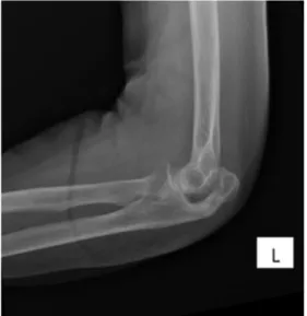
Fig. 1. Non-union fracture.
Fig. 2. Healed fracture.
to follow-up. Three patients were initially recruited but excluded due to severe cognitive disorder.
Functional scores at 6 months were: mean MEPS, 95.26/100 (range: 85–100), with 16 excellent and 6 good results; mean
Quick-DASH, 4.3 points (range: 0–29.55) (Table 1). Mean range of motion
at 6 months were:−15◦± 8◦ (range, 5–30◦) extension, 135◦± 6◦
(range: 130–140◦) flexion, and full pronation-supination.
Mean pain score on a 0-10 visual analog scale (VAS) was 1 (range: 0–3). Eighteen of the 22 fractures (82%) showed non-union (Figs. 1 and 2). There were no cases of elbow instability at last follow-up.
There were no local (cutaneous, functional impotence, etc.) or general complications (loss of autonomy, failure to thrive, etc.).
Table 2
Functional results of different treatments in the literature.
Authors Treatment Number of patients Age (years) Fracture Follow-up (months) Scores Extension deficit MEPS QuickDASH
Tarallo et al.
Tension band wiring 33 51.82 Mayo II-a, II-b 33 88.3 12.4 −9.7
Plate fixation 45 49.38 Mayo II-a, II-b 33 89.1 10.7 −7.8
Delsole et al.
Tension band wiring 23 64.5 Mayo I-a, I-b, II-a, II-b 13.5 97 −3.5
Hook plate 25 65.76 Mayo I-a, I-b, II-a, II-b 14.4 93.6 −8.6
Schliemann et al.
Tension band wiring 13 Mayo II-a 43 92 14
Plate fixation 13 Mayo II-a 43 77 12.5
Chen et al.
N-T memory connector 20 47.8 Mayo II-a, II-b, III-a, III-b 36 87 3.3 −4.2
Plate fixation 20 48.9 Mayo II-a, II-b, III-a, III-b 36 79.8 4 −2.9
Veras et al. Non-operative 12 81.8 Parker I 15.2 −7.5
4. Discussion
Functional results for non-operative treatment of olecranon fracture in≥ 75-year-olds were excellent and comparable to those of surgery.
Extension deficit was systematic, but without impact on quality of life, as seen from the scores. This is in agreement with Morrey et al., who reported a functional range of elbow motion of 30–130◦ for everyday activity[18].
It was interesting to find that olecranon non-union, present in 18 cases, was always well tolerated. Thus, can be explained by the well-known biomechanical phenomenon of olecranal patellization,
which restores active extension of the elbow[19]. In the present
cohort, fractures mainly involved the non-dominant side, which may have contributed to the lack of loss of autonomy which is
typi-cal of this type of fracture[2], as seen from the MEPS and QuickDASH
scores. Even so, patients with dominant-side fracture also showed good tolerance of treatment.
There have been previous studies of non-operative management
of olecranon fracture[13] [8,19], but all were retrospective.
New-man et al.[14]reported 3 weeks’ cast immobilization in 45–90◦
flexion for strictly non-displaced fracture. For Veras et al.[13], 8
out of 12 patients had good clinical results, with a mean−7.5◦
extension deficit, and 9 cases of non-union, in a series with a mean age of 81.8 years and a mean 15 months’ follow-up. Duckworth
et al.[19]reported a mean QuickDASH score of 2.9, Oxford Elbow
Score of 47% and 91% satisfaction in 23 patients with a mean age of
76 years and a mean 6 years’ follow-up. Gallucci et al.[8]reported
1/10 pain on VAS, a mean−15◦extension deficit and 22 non-unions
in 28 patients with a mean age of 82 years and a mean 16 months’ follow-up.
Compared to the literature on surgical treatment in the
gen-eral population, the present results are comparable or better[20],
as seen from Table 2. Tarallo et al. [21] reported poorer
func-tional results, with slightly less extension deficit, at 33 months’ follow-up in 33 patients with a mean age of 51 years, presenting with Mayo II-a or II-b fracture, managed by tension-band wiring. The same authors likewise found poorer functional scores but less extension deficit at 33 months’ follow-up in 45 patients with a mean age of 49 years presenting with Mayo II-a and II-b fracture
treated by plate fixation. Delsole et al.[10]reported a slightly higher
mean MEPS and less extension deficit, at 13.5 months’ follow-up in 23 patients with a mean age of 64 years presenting with Mayo I-a, I-b, II-a and II-b fracture, managed by tension-band wiring. The same authors reported a slightly lower MEPS and less extension deficit at 14.4 months’ follow-up in 25 patients with a mean age of 66 years presenting with Mayo I-a, I-b, II-a and II-b, fracture treated
by plate fixation. Schliemann et al.[11]reported lower MEPS and
QuickDASH at 43 months’ follow-up in 13 patients presenting with presenting with Mayo II-a fracture, managed by tension-band wiring. The same authors reported lower MEPS and QuickDASH at
43 months’ follow-up in 13 patients presenting with presenting with Mayo II-a fracture, managed by plate fixation.
Surgical complications can occur: painful protrusion of fixation
material under the skin (20–75% of fractures)[7], excessive wire
protrusion[13,14]on the anterior side of the ulna liable to cause
painful blocking in pronosupination, radio-ulnar synostosis[22],
ulnar neuritis (12%) [23], iatrogenic anterior interosseous nerve
lesion during transcortical tension-band wiring[24], heterotopic
ossification (7–37%)[25,26], scar disunion (14%)[8], infection, and
elbow osteoarthritis (1%)[10]. Moreover, anesthesia risk is higher
in elderly subjects[27,28]. Gallucci et al.[8]reported a 30%
com-plications rate after surgery.
Non-operative treatment is open to certain criticisms, partic-ularly concerning extension deficit. However, it is interesting to note that surgical treatment gives similar results in the general
population, with deficits ranging from –4◦to –15◦[14].
The present study had certain limitations. It would have been interesting to compare operative and non-operative treatment, but our recruitment is too small to implement such a design. It would also have been interesting to assess loss of triceps strength, although this is less functionally troublesome in this population.
5. Conclusion
Non-operative treatment of olecranon fracture without severe
displacement in patients aged≥ 75 years gave excellent functional
results at 6 months post-trauma, with fewer complications than for surgery.
Disclosure of interest
The authors declare that they have no competing interest.
References
[1]Brolin TJ, Throckmorton T. Olecranon Fractures. Hand Clin 2015;31:581–90.
[2]Duckworth AD, Clement ND, Aitken SA, Court-Brown CM, McQueen MM. The
epidemiology of fractures of the proximal ulna. Injury 2012;43:343–6.
[3]Argintar E, Cohen M, Eglseder A, Edwards S. Clinical results of olecranon frac-tures treated with multiplanar locked intramedullary nailing. J Orthop Trauma 2013;27:140–4.
[4]Inhofe PD, Howard TC. The treatment of olecranon fractures by excision of
fragments and repair of the extensor mechanism: historical review and report of 12 fractures. Orthopedics 1993;16:1313–7.
[5]von Rüden C, Augat P. Failure of fracture fixation in osteoporotic bone. Injury 2016;47:S3–10.
[6]Féron J-M, Thomas T, Roux C, Puget J. Osteoporosis and the orthopaedic surgeon in 2007. Rev Chir Orthop Repar Appar Mot 2008;94:S99–107.
[7]Macko D, Szabo RM. Complications of tension-band wiring of olecranon
frac-tures. J Bone Joint Surg Am 1985;67:1396–401.
[8]Gallucci GL, Piuzzi NS, Slullitel PAI, Boretto JG, Alfie VA, Donndorff A, et al. Non-surgical functional treatment for displaced olecranon fractures in the elderly. Bone Jt J 2014;96–B:530–4.
[9]Chen X, Liu P, Zhu X, Cao L, Zhang C, Su J. Design and application of nickel-titanium olecranon memory connector in treatment of olecranon fractures: a prospective randomized controlled trial. Int Orthop 2013;37:1099–105.
[10]DelSole EM, Pean CA, Tejwani NC, Egol KA. Outcome after olecranon fracture repair: does construct type matter? Eur J Orthop Surg Traumatol Orthop Trau-matol 2016;26:153–9.
[11]Schliemann B, Raschke MJ, Groene P, Weimann A, Wähnert D, Lenschow S,
et al. Comparison of tension band wiring and precontoured locking compres-sion plate fixation in Mayo type IIA olecranon fractures. Acta Orthop Belg 2014;80:106–11.
[12]Matar HE, Ali AA, Buckley S, Garlick NI, Atkinson HD. Surgical interventions
for treating fractures of the olecranon in adults. Cochrane Database Syst Rev 2014:CD010144.
[13]Veras Del Monte L, Sirera Vercher M, Busquets Net R, Castellanos Robles J, Car-rera Calderer L, Mir Bullo X. Conservative treatment of displaced fractures of the olecranon in the elderly. Injury 1999;30:105–10.
[14]Newman SDS, Mauffrey C, Krikler S. Olecranon fractures. Injury
2009;40:575–81.
[15]Morrey BF. Current concepts in the treatment of fractures of the radial head, the olecranon, and the coronoid. Instr Course Lect 1995;44:175–85.
[16]Fayad F, Lefevre-Colau MM, Gautheron V, Macé Y, Fermanian J,
Mayoux-Benhamou A, et al. Reliability, validity and responsiveness of the French version of the questionnaire Quick Disability of the arm, shoulder and hand in shoulder disorders. Man Ther 2009;14:206–12.
[17]Hudak PL, Amadio PC, Bombardier C, The upper extremity collaborative
group (UECG). Development of an upper extremity outcome measure: the DASH (disabilities of the arm, shoulder and hand) [corrected]. Am J Ind Med 1996;29:602–8.
[18]Morrey BF, An K-N, Chao EYS. Functional evaluation of the elbow. In: Morrey
BF, editor. The elbow and its disorders. Philadelphia: W.B. Saunders; 1993. p. 86–97.
[19]Duckworth AD, Bugler KE, Clement ND, Court-Brown CM, McQueen MM.
Non-operative management of displaced olecranon fractures in low-demand elderly patients. J Bone Joint Surg Am 2014;96:67–72.
[20]Powell AJ, Farhan-Alanie OM, Bryceland JK, Nunn T. The treatment of olecranon fractures in adults. Musculoskelet Surg 2017;101:1–9.
[21]Tarallo L, Mugnai R, Adani R, Capra F, Zambianchi F, Catani F. Simple and
comminuted displaced olecranon fractures: a clinical comparison between tension band wiring and plate fixation techniques. Arch Orthop Trauma Surg 2014;134:1107–14.
[22]De Carli P, Gallucci GL, Donndorff AG, Boretto JG, Alfie VA. Proximal radio-ulnar synostosis and nonunion after olecranon fracture tension-band wiring: a case report. J Shoulder Elbow Surg 2009;18:e40–4.
[23]Ishigaki N, Uchiyama S, Nakagawa H, Kamimura M, Miyasaka T. Ulnar nerve
palsy at the elbow after surgical treatment for fractures of the olecranon. J Shoulder Elbow Surg 2004;13:60–5.
[24]Parker JR, Conroy J, Campbell DA. Anterior interosseus nerve injury following tension band wiring of the olecranon. Injury 2005;36:1252–3.
[25]Foruria AM, Augustin S, Morrey BF, Sánchez-Sotelo J. Heterotopic ossification
after surgery for fractures and fracture-dislocations involving the proximal aspect of the radius or ulna. J Bone Joint Surg Am 2013;95:e66.
[26]Koh KH, Lim TK, Lee HI, Park MJ. Surgical treatment of elbow stiffness
caused by post-traumatic heterotopic ossification. J Shoulder Elbow Surg 2013;22:1128–34.
[27]Boddaert J, Raux M, Khiami F, Riou B. Perioperative management of elderly
patients with hip fracture. Anesthesiology 2014;121:1336–41.
[28]Griffiths R, Beech F, Brown A, Dhesi J, Foo I, Goodall J, et al. Perioperative care of the elderly 2014: association of anaesthetists of Great Britain and Ireland. Anaesthesia 2014;69:S81–98.