Single layer porous media with entrapped minerals for microscale studies of multiphase flow
12
0
0
Texte intégral
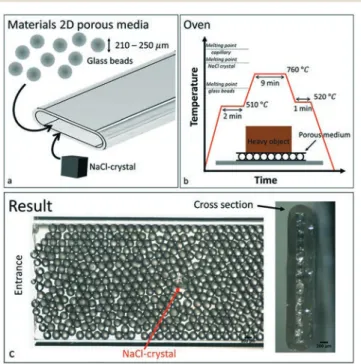
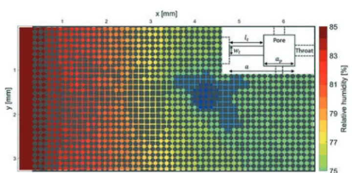
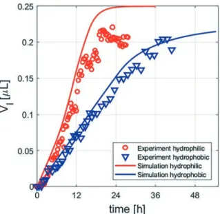
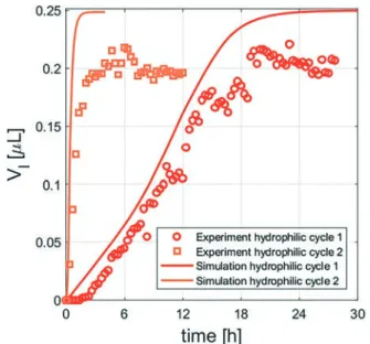
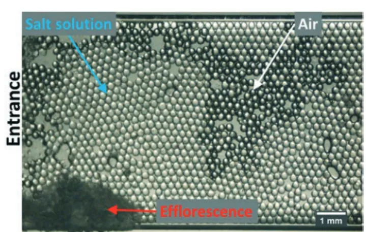
Figure
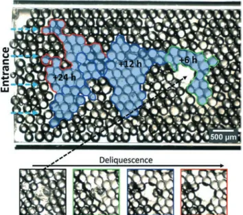
+6


Documents relatifs