been fully refereed, but have not been through the copy-editing and proof correction process. Wiley-Blackwell and the British Association of Dermatologists cannot be held responsible for errors or consequences arising from the use of information contained in these articles; nor do the views and opinions expressed necessarily reflect those of Wiley-Blackwell or the British Association of Dermatologists
This article is protected by copyright. All rights reserved. Article type : Original Article
Title: Assessment of the cutaneous immune response during Arthroderma benhamiae and Arthroderma vanbreuseghemii infection using an experimental mouse model
Running head: Mouse model for dermatophyte immune response study
Authors: L. Cambier 1†, A. Weatherspoon 2†, V. Defaweux 2, E.T. Bagut 1, 3,M.P. Heinen 1, N. Antoine 4, B. Mignon 1*
Affiliations: 1Veterinary Mycology Laboratory, Department of Infectious and Parasitic Diseases, Faculty of Veterinary Medicine, University of Liège, Liège, Belgium 2Human Histology Laboratory, Department of Preclinical Science, Faculty of Medicine, CHU Liège, Liège, Belgium 3Parasitology and Parasitic Diseases Unit, Faculty of Veterinary Medicine, University of Agricultural Sciences and Veterinary Medicine Cluj-Napoca, Cluj-Napoca, Romania 4Veterinary Histology Laboratory, Department of Morphology and Pathology, Faculty of Veterinary Medicine, University of Liège, Liège, Belgium
†
Both authors have contributed equally to this work
*Corresponding author: Bernard Mignon, Veterinary Mycology Laboratory, Department of Infectious and Parasitic Diseases, University of Liège, Liège, Belgium
Funding sources: This study was supported by grant 3.4558.10 from Fonds de la Recherche Scientifique Médicale (FRSM, Belgium) and 594/2012 from the Executive Unit for Higher Education, Research, Development and Innovation Funding (UEFISCDI, Romania). This publication was possible thanks to the agreement that binds Wallonia-Brussels (WBI) and Romania. L.C. is the recipient of a studentship from FRIA (Fonds pour la Formation à la Recherche dans l’Industrie et dans l’Agriculture, 1000 Brussels, Belgium). E.T.B. was the recipient of a research grant provided by the University of Liège.
Conflicts of interest: The authors report no conflicts of interest. The authors alone are responsible for the content and writing of the manuscript.
What’s already known about this topic?
• Pathophysiology and immunology of dermatophytoses are still poorly understood. • Most in vivo studies have been performed using guinea pig-based models.
• Several limitations, notably the lack of immunological tools for this animal species, render the development of a modern mouse model necessary for progress in
pathogenesis understanding. What does this study add?
• Using two peculiar fungal species isolated from men, Arthroderma benhamiae and Arthroderma vanbreuseghemii, a new mouse model of dermatophytosis was developed.
• Clinical, histopathological and immunohistological evaluations showed that this model is reproducible, fits with previous experimental infection models using guinea pigs and mimics superficial tinea in humans.
• For the first time, the cutaneous cytokine response was assessed during a
dermatophyte infection, showing that the role of the Th17 pathway should now be considered.
Abstract
Background Dermatophytoses are common but poorly understood skin infections. Most in vivo studies have been performed using the guinea pig as the experimental animal model, which has several limitations.
Objectives To develop a mouse model of dermatophytosis suitable for multiple purposes, including the investigation of immunity against dermatophytes.
Methods Two peculiar fungal species, Arthroderma benhamiae and
Arthroderma vanbreuseghemii, isolated from tinea in men having contact with rodents were used for epicutaneous inoculation. During the infection, clinical and histopathological follow-up were performed. The recruitment of immune cells was evaluated by immunofluorescence staining and the levels of cytokines mRNA were quantified by quantitative RT-PCR in skin of infected mice.
Results The skin symptoms and microscopic lesions, including the colonization of
keratinized epidermal and follicular structures by both dermatophytes, were highly similar to those observed in guinea pig infection models and in natural infections, mimicking acute superficial tinea in humans. The dermal inflammatory cellular infiltrate consisted of
macrophages, dendritic cells, and especially polymorphonuclear neutrophils, which are one of the histologic ‘clues’ to the diagnosis of dermatophytosis. The in situ cytokine profile was characterized by the overexpression of TGF-β, IL-1β, and IL-6 mRNAs during infection, suggesting a role of the Th17 pathway in the establishment of immunity.
Conclusions Our new reproducible and validated mouse model of dermatophytosis is a modern in vivo tool that allows a more in-depth understanding of the pathogenesis of human dermatophyte infections.
Introduction
Dermatophytes are pathogenic filamentous fungi responsible for superficial cutaneous infections called dermatophytoses.1 They specifically invade keratinized structures such as stratum corneum, hair, nails and claws. Based on their ecological features, three groups of dermatophyte species are recognized, namely anthropophilic, restricted to humans, zoophilic, found predominantly in animals and geophilic, recovered from the soil without or only sporadically causing disease.2
Despite their superficial localization in keratinized structures, dermatophytes can induce an adaptive immune response. The Th1 cellular response, associated with delayed type
hypersensitivity (DTH), appears to be correlated with clinical recovery and protection against reinfection.3-5 Nevertheless, the Th17 pathway could also be involved in the establishment of a protective immunity in dermatophytoses as has been shown for other fungi such as Candida albicans6-8 and Aspergillus fumigatus.7, 9 Little is known about the mechanisms involved in the establishment of a protective immune response during dermatophytosis.4, 5, 10 Some in vitro studies about immunity against dermatophytes have demonstrated that keratinocytes,11 neutrophils12 and dendritic cells13 are able to produce pro-inflammatory cytokines in response to infection. Nevertheless, in vivo studies are essential in furthering our understanding of immunity against dermatophytes. Therefore, the establishment of an animal model of dermatophytosis is required. Currently, the most frequently used animal model is the guinea pig14-17 but the inbred nature, and the genetic and immunologic tools available in the mouse
species, support the use of a model in the study of immune response against dermatophytes.18 Only few mouse models of dermatophytoses are available. A former model using
Trichophyton quinckeanum was successfully used for both histopathologic 19, 20 and immunologic precursory studies.21, 22 However, this model is questionable because T. quinckeanum is a rare agent of dermatophytosis in humans, moreover responsible for a particular clinical entity named mouse favus.23 Two other models using dermatophytes in mice were recently described but they are not representative of a superficial natural infection.24, 25
In this study, we focused on two zoophilic dermatophyte species that are frequently isolated from superficial tinea in men: Arthroderma benhamiae, which primarily affects guinea pigs and the closely related Arthroderma vanbreuseghemii which affects other animal species, notably cats and dogs but very rarely rodents including mice.26 Both species belong to the Trichophyton mentagrophytes complex26-28 and are readily manipulable at the genetic level.
29-31 These features urged us to test both A. benhamiae and A. vanbreuseghemii species by using
two isolates originating from humans in contact with either the guinea pig or mouse, respectively. Although some mycologists still use the nomenclature T. mentagrophytes complex, the sexual names A. benhamiae and A. vanbreuseghemii should always be used according to the rules proposed by the Amsterdam declaration on Fungal Nomenclature.32
The aim of this work was to develop a reproducible experimental mouse model of
dermatophytosis using A. benhamiae or A. vanbreuseghemii, and subsequently to assess the cutaneous immune response generated during infection. To this purpose, the immune cells recruited to the site of infection were defined and the cytokine response induced in skin of infected mice was assessed.
Materials and Methods Fungal strains
Arthroderma benhamiae IHEM 20163 (Institute of Hygiene and Epidemiology-Mycology [IHEM], Brussels, Belgium) was originally isolated from a patient suffering from
inflammatory tinea faciei and who had had previous contact with a guinea pig.33 This
A. benhamiae strain has been characterized at the molecular level27 and was successfully used to develop a guinea pig infection model for this species.29 Arthroderma vanbreuseghemii IHEM 22740 was chosen because of its peculiar origin, namely a patient in contact with mouse and having highly inflammatory tinea manum.27 This strain is closely related to A. vanbreuseghemii isolated from dogs and cats and causing various inflammatory tinea in humans.27 Both strains were grown for 15 days at 27 °C on Sabouraud’s plates (Sab; 2% glucose-1% peptone), and used for the experimental infection.
Animals and experimental infection
Eighteen specific-pathogen-free 2-month-old female mice of C57BL/6 strain (Charles River, Wilmington, MA, USA) were used and housed separately during the entire study period. Mice were divided into 3 groups. The A. benhamiae group (n=6) was inoculated with
4.2 × 10¹⁰ colony forming units (CFU), the A. vanbreuseghemii group (n=6) received 2 × 10¹¹ CFU while the mice from the control group (n=6) remained uninfected. Experimental
infection was performed under general anaesthesia (medetomidine [1 mg kg―1] and ketamine [40 mg kg―1]) administered by intraperitoneal injection. Backs of the animals were shaved and lightly abraded with a 25G needle. The inoculum (300 µl) consisting of mycelia and spores collected from cultures on Sab plates was suspended in 5% (w/w) poloxamer 407 and gently rubbed onto the skin with a sterile pipette tip. Mice from the control group received 300 µl of poloxamer only. For histology, immunoflorescence staining and quantification of
cytokines, 3 infected mice were randomly selected from each group and skin biopsies samples were collected. Animal experiments were approved by the local ethics committee (University of Liège, ethics protocol no. 943).
Clinical follow-up
Infected mice were monitored twice a week for 5 weeks by two independent examiners using clinical criteria i.e., alopecia and squamosis-crusting. Both investigators who remained the same throughout the experiment were blinded as to the status of mice (infected or not). For each clinical criterion, a score of 0, 1, 2 or 3 (absence, mild, moderate or severe skin lesion) was attributed. A clinical score was then calculated for each mouse by adding scores for both criteria. Finally, a median clinical score was calculated for each group of mice.
Histology
Skin samples collected at days 3, 7, 14 and 21 post-inoculation (PI) were fixed in 10%
neutralized buffered formalin and paraffin embedded for routine processing. Sections of 4 µm thick sections were stained with haematoxylin and eosin (H&E) for histopathologic
evaluation or with periodic acid-Schiff (counterstained with haematoxylin) to assess the fungal invasion in keratinized skin structures.
Immunofluorescence staining
At day 7 PI, skin samples collected from mice were immersed in tissue-TEK OCT
embedding medium (Sakura, Zouterwoude, The Netherlands) and quickly frozen at −80 °C. Sections (8-10 µm) were cut at −20 °C with a microtome (Microtom HM 500 OM, Thermo Fisher Scientific, Waltham, MA, USA), mounted on glass slides coated with poly-L-Lysine (Sigma, St. Louis, MO, USA), fixed in acetone for 10 min at 4 °C, and stored at −20 °C until
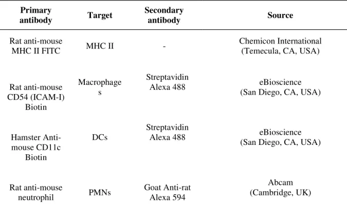
used. After rehydration in PBS, cryosections were incubated for 1 h at room temperature with appropriate primary antibodies diluted 1:100 in PBS. After washing in PBS, sections were incubated for 30 min at room temperature with Alexa Fluor488 streptavidin or species-specific secondary antibody (Molecular Probes, Leiden, The Netherlands). The characteristics of primary and secondary antibodies are listed in Table 1. A fluorescent dye for nuclear staining, the TOPRO-3-iodide (Molecular Probes) was added to cryosections labelled with anti-major histocompatibility complex (MHC) II and anti-CD54 antibodies. To test the general specificity of the antibodies used, samples were incubated with irrelevant antibodies. Negative controls were obtained by incubating samples with secondary antibodies only.
Quantification of cytokine mRNA levels in skin biopsies
At day 0, 7 and 21 PI, skin biopsy specimens were homogenized using the FastPrep® instrument (MP Biomedicals, Santa Ana, CA, USA). Total RNA was isolated using TRIzol reagent following the manufacturer's instructions (Invitrogen, Burlington, ON, Canada). The purified RNA was treated with DNase I (Invitrogen). Template cDNA was synthesized from RNA by reverse transcription, using iScript™ cDNA Synthesis kit (Bio-rad, Hercules, CA, USA). Reverse transcriptase was omitted in control reactions. The sequences of
oligonucleotide primers have already been published(12S rRNA, Tumor Necrosis Factor (TNF)-α, Interleukin (IL)-1β, IL-6 and IL-22)34-36 or were selected (IL-10 and Transforming Growth Factor (TGF)-β) using the Primer-BLAST program
(http://www.ncbi.nlm.nih.gov/tools/primer-blast/index.cgi?LINK_LOC=BlastHomeAd). The primers for mouse 12S rRNA (internal control) and cytokines were synthesized by Eurogentec (Liège, Belgium) (Table 2). The quantitative PCR (qPCR) reactions were assembled using the iQ™ SYBR® Green Supermix (Bio-rad) and subjected to the following protocol in a MiniOpticon System (Bio-rad): 10 min at 95 °C and 50 cycles of 45 s at 95 °C,
45 s at 60 °C and 45 s at 72 °C. The melting curve was performed from 45 °C to 95 °C in 1 °C/15 s increments. All assays were performed in duplicate. Results in terms of cycle thresholds were converted in folds 12S rRNA expression using the 2−ΔΔCt method. The level of cytokine mRNA in skin biopsies at day 7 or 21 PI was expressed relative to that in skin biopsies at day 0.
Statistical analysis
The two-way analysis of variance (ANOVA) test was used for the statistical comparison of median clinical scores between the infected and control mice. This test was performed with the Graph pad Prism 5.0 statistical software (GraphPad Software, San Diego, CA, USA). The levels of cytokine mRNA in skin biopsies, 7 and 21 days PI were compared with those determined in skin biopsies at day 0 using a general linear model (GLM procedure of SAS; SAS Institute Inc., Cary, NC, USA). P<0.05 was considered as statistically significant.
Results
Mice infected with A. benhamiae and A. vanbreuseghemii develop clinical lesions of dermatophytosis
All animals infected with A. vanbreuseghemii and 80% of mice infected with A. benhamiae showed consistent dermatophytosis lesions. This contrasts with previous experiments suggesting that the C57BL/6 strain mice are resistant to dermatophytosis.21 Clinical signs of dermatophytosis were obvious 4 days after infection and were maximal at day 19 PI for both dermatophyte species (Fig. 1). The lesions healed progressively until day 35 PI. The main clinical signs consisted of severe squamosis-crusting followed by transient alopecia. From day 11 PI until day 24 and 28 PI for A. benhamiae and A. vanbreuseghemii respectively, median clinical scores were significantly higher in both infected groups in comparison with
the controls (Fig. 2). The application of poloxamer 407 without fungus did not produce any lesions in the control group.
A. benhamiae and A. vanbreuseghemii colonize keratinized skin structures
To assess the presence and the localization of dermatophytes in keratinized skin structures of infected mice, skin biopsy sections were stained with PAS. In mice infected with both A. vanbreuseghemii and A. benhamiae, fungal elements consisting of both hyphae and arthroconidia were detectable from day 3 until day 14 PI in the stratum corneum and the keratinized part of the infundibular-isthmal hair follicles including the hair shafts. Subjectively, the most severe fungal colonization was observed at day 7 PI for both
dermatophyte species (Fig. 3). As expected, no fungus elements were found in skin of control mice.
A. benhamiae and A. vanbreuseghemii induce severe microscopic inflammatory skin lesions
To assess histologic lesions in infected mice, skin biopsy sections were stained with H&E. All infected animals showed extensive inflammatory lesions. At day 3 PI, both
dermatophytes induced a severe increase of epidermis thickness essentially due to both acanthosis (Figs. 4a, c) and hyperkeratosis, mainly orthokeratotic (Fig. 4a). These lesions were accompanied by moderate to severe multifocal to diffuse spongiosis (Fig. 4c) and by perivascular to diffuse dermal infiltration by immune cells (Fig. 4e). At 7 days PI, subcorneal pustules (Fig. 4d) were also observed. By day 14 PI, lesions had disappeared in mice infected with A. vanbreuseghemii, while acanthosis, spongiosis and dermal cellular infiltration
persisted beyond day 21 PI after infection with A. benhamiae. No lesions were observed in control mice (Fig. 4b).
Immune cells are recruited early after A. benhamiae and A. vanbreuseghemii infection To identify which immune cells were recruited to the infection site at day 7 PI,
immunofluorescence staining was performed on frozen skin biopsy sections (Figure 5). Large numbers of polymorphonuclear neutrophils (PMNs), MHC II expressing cells and to a lesser extent, dendritic cells (DCs) (CD11C+) and macrophages (CD54+) were recruited in the dermis of mice infected with both A. benhamiae and A. vanbreuseghemii. These cells were scarce in the skin of control mice.
Cytokine mRNA expression in infected skin
Cytokine mRNA levels were determined at days 7 and 21 PI and compared with those at day 0 (Fig. 6). Infection with both dermatophytes induced a statistically significant increase in the levels of TGF-β, IL-1β and IL-6 mRNA at day 7 PI. The level of IL-22 mRNA was also significantly increased in mice infected with A. benhamiae. At day 21 PI, the levels of TGF-β, IL-1β and IL-6 mRNA were significantly higher in mice infected with A. benhamiae, while infection with A. vanbreuseghemii induced a statistically significant increase in IL-1β mRNA level only. The levels of cytokine mRNA in control non-infected mice were not significantly different at both days 7 and 21 PI in comparison to day 0.
Discussion
We have developed a new and useful experimental mouse model of dermatophytosis using either A. benhamiae or A. vanbreuseghemii. Both species were isolated from naturally infected humans in contact with rodents. For both dermatophytes, clinical signs were identical from one animal to another, typical for a primary natural infection and disappeared spontaneously, indicating that this model is reproducible and appears to be suitable for in vivo studies on acute dermatophytoses.
The symptoms and microscopic skin lesions, including the colonization of keratinized epidermal and follicular structures by both dermatophytes, were highly comparable to those described in a recent guinea pig infection model for A. benhamiae,29 which was used to assess dermatophyte virulence factors in vivo.30 A high amount of fungus was observed at day 7 PI in keratinized structures of mice infected with both dermatophytes before the peak of skin lesions (at day 19 PI). This observation is consistent with the recruitment of immune cells at day 7 PI that are responsible for elimination of the fungus by inducing an
inflammatory response.
In contrast with guinea pig-based models,37 one of the major advantages of our mouse model lies in the homogeneity of skin symptoms and lesions between individuals, thanks to the inbred nature of animals. Additional benefits include lower operating costs and availability of many modern immunological tools, which render relevant mouse models of dermatophytoses desirable for furthering a comprehensive understanding of the pathogenesis of these
diseases.18
In addition to comparable clinical signs and microscopic inflammatory lesions, the nature of the immune cells recruited after the infection of mice with both A. benhamiae and
A. vanbreuseghemii was consistent with what most authors have observed using other models or during natural infections. The inflammatory response observed in skin of mice infected with both dermatophytes was characterized by an inflow of immune cells such as PMNs, macrophages and DCs. Similarly, the infiltration of skin by PMNs has been shown in experimentally infected mouse20 and is commonly described as one of the histologic ‘clues’ to the diagnosis of dermatophytosis in humans.38 Human PMNs and macrophages can exert cytotoxic activity against Trichophyton species including the most common dermatophyte
Trichophyton rubrum, via phagocytosis and production of superoxide anions39-41 and also via the formation of neutrophil extracellular traps (NETs).13
In addition to PMN and macrophages, our model also revealed infiltrating DCs, consistent with the role that these professional antigen presenting cells (APCs) are believed to play in the establishment of a protective immune response during dermatophytosis. Such a role is supported by in vitro experiments showing that both human DCs13 and several pattern recognition receptors on mouse DCs42, 43 can bind fungal components from both T. rubrum and Microsporum andouinii, and modulate immunity to dermatophytes. Additionally, cellular mediated immunity, which is known to be protective in dermatophytoses4, 5 is induced by the recognition of the MHC II at the APCs surface and the CD4+ T-cell receptor (TCR).44 In this work, the considerable infiltration of MHC II expressing cells in the skin of infected mice suggests that these cells could be involved in the immunity against A. benhamiae and
A. vanbreuseghemii. DCs and macrophages, both expressing MHC II on their surface45 were recruited in the skin of mice infected with both dermatophytes, which suggests that these cells could be important for the initiation of immunity against dermatophytosis.
In order to investigate the cutaneous immune response in our validated dermatophytosis mouse model, parallel to the characterization of the cells recruited to the site of infection, we assessed the cytokine response in situ, precisely because the set-up of a protective immune response is also correlated with the establishment of an appropriate cytokine response.46 The cytokine profile generated by A. benhamiae and A. vanbreuseghemii infection was a pro-inflammatory profile characterized by TGF-β, IL-1β and IL-6 production. These cytokines are involved in the establishment of the Th17 pathway47-49. In mice infected with A. benhamiae, an increase in IL-22 mRNA level was also observed. This cytokine, which is also produced
by Th17 cells,50 has a significant role in the establishment of skin and mucosa immunity.51 Altogether, these results indicate that the Th17 pathway could be involved in the
establishment of immunity against A. benhamiae and A. vanbreuseghemii. This hypothesis is strengthened by the weak IL-10 mRNA level produced in the skin of mice infected with both dermatophytes. This cytokine is known to reduce the inflammatory response by activating the Treg pathway.52 The potential role of the Th17 pathway in the immunity against
A. benhamiae and A. vanbreuseghemii infection is also supported by the recruitment of PMNs to the site of infection, as cross-talk exists between Th17 cells and PMNs.53-56 In conclusion, we have developed and validated a new model of dermatophytosis in mice, which is consistent with former in vivo models, most of which are based on the guinea pig, a commonly used animal species that is no very suitable for studying this disease. Thanks to the inter-individual homogeneity of clinical signs and skin lesions, our model will be particularly useful for the evaluation of antifungals and vaccines, and for identifying
dermatophyte virulence factors. Additionally, our model is consistent with the recent insights gained in vitro into the innate and specific immune response against dermatophytes. It appears to be, therefore, a promising modern animal model suitable to study, in depth, the host immune response in dermatophytosis. In the near future, the role of Th17 and the possible involvement of the Th1 or Th2 pathways in the initiation of a protective immunity against dermatophytes will be assessed by evaluating other pro-Th1, pro-Th2 and pro-Th17 cytokines in mice infected with A. benhamiae and A. vanbreuseghemii, as by using mice deficient for specific cytokines.
Acknowledgements
The authors thank Dr Vinciane Toppets, Jessica Collard and Joëlle Piret for technical assistance. They also thank Dr B. Evrard (Laboratory of Pharmaceutical Technology, University of Liège, Liège, Belgium) for providing the excipient.
References
1 Degreef H. Clinical forms of dermatophytosis (ringworm infection). Mycopathologia 2008; 166: 257-65.
2 Weitzman I, Summerbell RC. The dermatophytes. Clin Microbiol Rev 1995; 8: 240-59. 3 Calderon RA. Immunoregulation of dermatophytosis. Crit Rev Microbiol 1989; 16: 339-68. 4 Almeida SR. Immunology of dermatophytosis. Mycopathologia 2008; 166: 277-83.
5 Mignon B, Tabart J, Baldo et al. Immunization and dermatophytes. Curr Opin Infect Dis 2008, 21: 134-40.
6 Huang W, Na L, Fidel PL, Schwarzenberger P. Requirement of interleukin-17A for systemic anti-Candida albicans host defense in mice. J Infect Dis 2004; 190: 624-31. 7 Bozza S, Zelante T, Moretti S, et al. Lack of Toll IL-1R8 exacerbates Th17 cell responses in fungal infection. J Immunol 2008; 180: 4022-31.
8 Conti HR, Shen F, Nayyar N, et al. Th17 cells and IL-17 receptor signaling are essential for mucosal host defense against oral candidiasis. J Exp Med 2009; 206: 299-311.
9 Zelante T, Luca Ad, Bonifazi P, et al. IL-23 and the Th17 pathway promote inflammation and impair antifungal immune resistance. Eur J Immunol 2007; 37: 2695-706.
10 Vermout S, Tabart J, Baldo A, et al. Pathogenesis of dermatophytosis. Mycopathologia 2008; 166: 267-75.
11 Shiraki Y, Ishibashi Y, Hiruma M, et al. Cytokine secretion profiles of human
keratinocytes during Trichophyton tonsurans and Arthroderma benhamiae infections. J Med Microbiol 2006; 55: 1175-85.
12 Cambier L, Mathy A, Baldo A, et al. Feline polymorphonuclear neutrophils produce pro-inflammatory cytokines following exposure to Microsporum canis. Vet Microbiol 2013, 162: 800-5.
13 Heddergott C, Bruns S, Nietzsche S, et al. The Arthroderma benhamiae hydrophobin HypA mediates hydrophobicity and influences recognition by human immune effector cells. Eukaryot Cell 2012; 11: 673-82.
14 Mignon BR, Leclipteux T, Focant C, et al. Humoral and cellular immune response to a crude exo-antigen and purified keratinase of Microsporum canis in experimentally infected guinea pigs. Med Mycol 1999; 37: 123-9.
15 Brouta F, Descamps F, Vermout S, et al. Humoral and cellular immune response to a Microsporum canis recombinant keratinolytic metalloprotease (r-MEP3) in experimentally infected guinea pigs. Med Mycol 2003; 41: 495-501.
16 Saunte DM, Hasselby JP, Brillowska-Dabrowska A, et al. Experimental guinea pig model of dermatophytosis: A simple and useful tool for the evaluation of new diagnostics and antifungals. Med Mycol 2008; 46: 303-13.
17 Baldo A, Mathy A, Tabart J, et al. Secreted subtilisin Sub3 from Microsporum canis is required for adherence to but not for invasion of the epidermis. Brit J Dermatol 2010; 162: 990-7.
18 White TC, Oliver BG, Gräser Y, Henn MR. Generating and testing molecular hypotheses in the dermatophytes. Eukaryot Cell 2008; 7: 1238-45.
19 Hay RJ, Calderon RA, Collins MJ. Experimental dermatophytosis: the clinical and
histopathologic features of a mouse model using Trichophyton quinckeanum (mouse favus). J Invest Dermatol 1983; 81: 270-4.
20 Hay RJ, Calderon RA, Mackenzie CD. Experimental dermatophytosis in mice: correlation between light and electron microscopic changes in primary, secondary and chronic infections. Br J Exp Pathol 1988; 69: 703-16.
21 Calderon RA, Hay RJ. Cell-mediated immunity in experimental murine dermatophytosis. I. Temporal aspects of T-suppressor activity caused by Trichophyton quinckeanum.
Immunology. 1984; 53: 457-64.
22 Calderon RA, Hay RJ. Cell-mediated immunity in experimental murine dermatophytosis. II. Adoptive transfer of immunity to dermatophyte infection by lymphoid cells from donors with acute or chronic infections. Immunology. 1984; 53: 465-72.
23 Beguin H, Pyck N, Hendrickx M, et al. The taxonomic status of Trichophyton
quinckeanum and T. interdigitale revisited: a multigene phylogenetic approach. Med Mycol. 2012; 50: 871-82.
24 Nakamura T, Nishibu A, Yasoshima M, et al. Analysis of Trichophyton antigen-induced contact hypersensitivity in mouse. J Dermatol Sci 2012; 66: 144-53.
25 Venturini J, Álvares AM, Camargo MRd, et al. Dermatophyte-host relationship of a murine model of experimental invasive dermatophytosis. Microbes Infect 2012; 14: 1144-51. 26 Drouot S, Mignon B, Fratti M, et al. Pets as the main source of two zoonotic species of the Trichophyton mentagrophytes complex in Switzerland, Arthroderma vanbreuseghemii and Arthroderma benhamiae. Vet Dermatol 2009; 20: 13-8.
27 Symoens F, Jousson O, Planard C, et al. Molecular analysis and mating behaviour of the Trichophyton mentagrophytes species complex. Int J Med Microbiol 2011; 301: 260-6.
28 Symoens F, Jousson O, Packeu A, et al. The dermatophyte species Arthroderma benhamiae: Intraspecies variability and mating behaviour. J Med Microbiol 2013; 62: 377-85.
29 Staib P, Zaugg C, Mignon B, et al. Differential gene expression in the pathogenic dermatophyte Arthroderma benhamiae in vitro versus during infection. Microbiology 2010; 156: 884-95.
30 Grumbt M, Defaweux V, Mignon B, et al. Targeted gene deletion and in vivo analysis of putative virulence gene function in the pathogenic dermatophyte Arthroderma benhamiae. Eukaryot Cell 2011; 10: 842-53.
31 Alshahni MM, Yamada T, Takatori K, et al. Insights into a nonhomologous integration pathway in the dermatophyte Trichophyton mentagrophytes: efficient targeted gene disruption by use of mutants lacking ligase IV. Microbiol Immunol. 2011; 55: 34-43. 32 Hawksworth DL, Crous PW, Redhead SA, et al. The Amsterdam declaration on fungal nomenclature. IMA Fungus 2011; 2: 105-12.
33 Fumeaux J, Mock M, Ninet B, et al. First report of Arthroderma benhamiae in Switzerland. Dermatology. 2004; 208: 244-50.
34 Shire D. An invitation to an open exchange of reagents and information useful for the measurements of cytokine mRNA levels by PCR. Eur Cytokine Netw 1993; 4: 161-2. 35 Godinez I, Haneda T, Raffatellu M, et al.T cells help to amplify inflammatory responses induced by Salmonella enterica serotype typhimurium in the intestinal mucosa. Infect Immun 2008; 76: 2008-17.
36 Dewals B, Hoving JC, Horsnell WGC et al. Control of Schistosoma mansoni egg-induced inflammation by IL-4-responsive CD4+CD25-CD103+Foxp3- cells is IL-10-dependent. Eur J Immunol 2010; 40: 2837-47.
37 Vermout SM, Brouta FD., Descamps FF et al. Evaluation of immunogenicity and protective efficacy of a Microsporum canis metalloprotease subunit vaccine in guinea pigs. FEMS Immunol Med Microbiol 2004; 40: 75-80.
38 Ackermann AB. Dermatophytosis. In: Histology diagnosis of inflammatory skin diseases (Ackerman AB, Chongchitnant N, Sanchez J, Guo J, Bennin B, Reichel M, Randall MB, eds), Williams & Wilkins: Baltimore, 1997; 910.
39 Calderon RA, Hay RJ. Fungicidal activity of human neutrophils and monocytes on dermatophyte fungi, Trichophyton quinckeanum and Trichophyton rubrum. Immunology 1987; 61: 289-95.
40 Calderon RA, Shennan GI. Susceptibility of Trichophyton quinckeanum and Trichophyton rubrum to products of oxidative metabolism. Immunology 1987; 61: 283-8.
41 Dahl MV, Carpenter R. Polymorphonuclear leukocytes, complement, and Trichophyton rubrum. J Invest Dermatol 1986; 86: 138-41.
42 Chung JS, Yudate T, Tomihari M, et al. Binding of DC-HIL to dermatophytic fungi induces tyrosine phosphorylation and potentiates antigen presenting cell function. J Immunol 2009; 183: 5190-8.
43 Sato K, Yang XL, Yudate T, et al.. Dectin-2 is a pattern recognition receptor for fungi that couples with the Fc receptor gamma chain to induce innate immune responses. J Biol Chem 2006; 281: 38854-66.
44 Joffre O, Nolte MA, Sporri R, Sousa CRE. Inflammatory signals in dendritic cell activation and the induction of adaptive immunity. Immunol Rev 2009; 227: 234-47. 45 Bryant P, Ploegh H. Class II MHC peptide loading by the professionals. Curr Opin Immunol 2004; 16: 96-102.
46 Esche C, Stellato C, Beck LA. Chemokines: key players in innate and adaptive immunity. J Invest Dermatol 2005; 125: 615-28.
47 Bettelli E, Carrier YJ, Gao WD, et al. Reciprocal developmental pathways for the generation of pathogenic effector T(H)17 and regulatory T cells. Nature 2006; 441: 235-8. 48 Wilson NJ, Boniface K, Chan JR, et al. Development, cytokine profile and function of human interleukin 17-producing helper T cells. Nat Immunol 2007; 8: 950-7.
49 Volpe E, Servant N, Zollinger R, et al. A critical function for transforming growth factor-beta, interleukin 23 and proinflammatory cytokines in driving and modulating human T(H)-17 responses. Nat Immunol 2008; 9: 650-7.
50 Stockinger B, Veldhoen M. Differentiation and function of Th17 T cells. Curr Opin Immunol 2007; 19: 281-6.
51 Wolk K, Kunz S, Witte E, et al. IL-22 increases the innate immunity of tissues. Immunity 2004; 21: 241-54.
52 Sabat R, Grutz G, Warszawska K, et al. Biology of interleukin-10. Cytokine Growth Factor Rev 2010; 21: 331-44.
53 Pelletier M, Maggi L, Micheletti A, et al. Evidence for a cross-talk between human neutrophils and Th17 cells. Blood 2010; 115: 335-43.
54 Roussel L, Houle F, Chan C, et al. IL-17 promotes p38 MAPK-dependent endothelial activation enhancing neutrophil recruitment to sites of inflammation. J Immunol 2010; 184: 4531-7.
55 Griffin GK, Newton G, Tarrio ML, et al. IL-17 and TNF-alpha sustain neutrophil recruitment during inflammation through synergistic effects on endothelial activation. J Immunol 2012; 188: 6287-99.
56 Hsu SC, Wang LT, Yao CL, et al. Mesenchymal stem cells promote neutrophil activation by inducing IL-17 production in CD4+ CD45RO+ T cells. Immunobiology 2013; 218: 90-5.
Figure Legends
Figure 1: Comparative clinical follow-up of skin lesions in mice inoculated with Arthroderma benhamiae, Arthroderma vanbreuseghemii, and the excipient (control).
Both dermatophyte species induced comparable symptoms, which were obvious since day 4 post-infection (PI) and had spontaneously disappeared by day 32 PI. Macroscopic
inflammatory skin lesions consisted of scaling and crusting followed by transient but severe inflammatory alopecia. Control mice developed no skin lesions.
Figure 2: Kinetics of median clinical scores (± SD; n=6) reflecting the severity of
dermatophytic lesions in mice inoculated with Arthroderma benhamiae (a), Arthroderma vanbreuseghemii (b), and the excipient (control). The median clinical scores were calculated using the clinical scores attributed blindly to each mouse in a given group. For each mouse, a clinical score was determined by adding the score for alopecia (0, 1, 2 or 3) and the score for squmosis-crusting (0, 1, 2 or 3). Median clinical scores were significantly different between the control group and the A. benhamiae group from day 11 until day 24 post-infection (PI). Significant differences were also observed between the control group and the
A. vanbreuseghemii group from day 11 until day 28 PI. *P<0.05; **P<0.01; ***P<0.001.
Figure 3: Keratinized skin and follicular structures of infected mice are colonized after experimental inoculation with Arthroderma benhamiae and Arthroderma vanbreuseghemii. The fungal colonization in mice infected with A. vanbreuseghemii 7 days post-infection is shown. The stratum corneum (a) and hair follicles (b) were colonized by dermatophyte hyphae and arthroconidia (black arrows). Sections were stained with periodic acid-Schiff and counterstained with hematoxylin. Scale bars = 20 µm.
Figure 4: Mice infected with Arthroderma benhamiae (c) and Arthroderma vanbreuseghemii (a, d, e) develop comparable inflammatory epidermal and dermal lesions. Micrographs illustrate the main skin lesions consisting of severe acanthosis (a, c) and hyperkeratosis (a), accompanied by spongiosis (c), scattered subcorneal pustules (d), and perivascular-to-diffuse cellular infiltration in the dermis (e) at day 7 (a, b, d, e) and 21 post-infection (c). Control mice inoculated with the excipient (b) developed no skin lesions. Sections were stained with hematoxylin and eosin. †: hyperkeratosis, ‡: acanthosis, ↑: spongiosis. Scale bars = 20 µm.
Figure 5: Immune cells are recruited in the dermis of mice infected with both Arthroderma benhamiae and Arthroderma vanbreuseghemii. Micrographs show the connective tissue
around the infundibular and isthmal hair follicles of infected mice at day 7 post-infection or uninfected controls. Immunofluorescence staining revealed that polymorphonuclear
neutrophils (PMNs) (neutrophil+), class II major histocompatibility complex-expressing cells (MHC II+) and, to a lesser extent, dendritic cells (DCs) (CD11c+) and macrophages (CD54+) were recruited mainly around hair follicles. A fluorescent dye for nuclear staining, the
TOPRO-3-iodide, was added to cryosections immunostained with anti-MHCII and anti-CD54 antibodies. Scale bars = 75 µm.
Figure 6: Cytokine mRNA profile in the skin of mice at days 7 (a) and 21 (b) post-infection (PI) with Arthroderma benhamiae and Arthroderma vanbreuseghemii. In each group of mice, both uninfected control and infected, three animals were used to compare the levels of cytokine mRNAs before inoculation (day 0) and at days 7 and 21 PI. A statistically significant increase in TGF-β, IL-1β, IL-6, and IL-22 mRNA levels was observed in mice
infected with A. benhamiae at day 7 PI. Similar results were observed in mice infected with A. vanbreuseghemii, except for levels of IL-22 mRNA, which were not significantly higher than at day 0. At day 21 PI, a statistically significant increase in TGF-β, IL-1β, and IL-6 mRNA was observed in mice infected with A. benhamiae. In mice infected with
A. vanbreuseghemii, the increase in the levels of these cytokine mRNAs was statistically significant only for IL-1β. In uninfected control mice, the levels of cytokine mRNAs did not increase significantly through the study period. Data are representative of three independent experiments (mean ± SD). *P<0.05; **P<0.01; ***P<0.001.
Tables
Table 1. Antibodies used for immunofluorescence staining Primary antibody Target Secondary antibody Source Rat anti-mouse MHC II FITC MHC II - Chemicon International (Temecula, CA, USA)
Rat anti-mouse CD54 (ICAM-I) Biotin Macrophage s Streptavidin Alexa 488 eBioscience
(San Diego, CA, USA)
Hamster Anti-mouse CD11c Biotin DCs Streptavidin Alexa 488 eBioscience
(San Diego, CA, USA)
Rat anti-mouse neutrophil PMNs Goat Anti-rat Alexa 594 Abcam (Cambridge, UK)
MHC II: class II major histocompatibility complex DCs: dendritic cells
Table 2. Sequences of oligonucleotide primers Target gene Forward sense (5’-> 3’) Reverse sense (5’-> 3’) 12s
rRNA GGAAGGCATAGCTGCTGGAGGT CGATGACATCCTTGGCCTGA
TNFα TCTCATCAGTTCTATGGCCC GGGAGTAGACAAGGTACAAC
IL-1β TTGACGGACCCCAAAAGATG AGAAGGTGCTCATGTCCTCA
IL-6 GTTCTCTGGGAAATCGTGGA TGTACTCCAGGTAGCTAT GG
TGFβ GCTTTCGATTCAGCGCTCACT TCCAACCCAGGTCCTTCCTAA
IL-10 AGCCGGGAAGACAATAACTG CATTTCCGATAAGGCTTGG