HAL Id: hal-02109357
https://hal.archives-ouvertes.fr/hal-02109357
Submitted on 24 Apr 2019HAL is a multi-disciplinary open access archive for the deposit and dissemination of sci-entific research documents, whether they are pub-lished or not. The documents may come from teaching and research institutions in France or abroad, or from public or private research centers.
L’archive ouverte pluridisciplinaire HAL, est destinée au dépôt et à la diffusion de documents scientifiques de niveau recherche, publiés ou non, émanant des établissements d’enseignement et de recherche français ou étrangers, des laboratoires publics ou privés.
expressed in mammalian cells
M Karlova, N Voskoboynikova, G Gluhov, D Abramochkin, O Malak, A
Mulkidzhanyan, Gildas Loussouarn, H.-J Steinhoff, K Shaitan, O Sokolova
To cite this version:
M Karlova, N Voskoboynikova, G Gluhov, D Abramochkin, O Malak, et al.. Detergent-free solubiliza-tion of human Kv channels expressed in mammalian cells. Chemistry and Physics of Lipids, Elsevier, 2019, �10.1016/j.chemphyslip.2019.01.013�. �hal-02109357�
Detergent-free solubilization of human Kv channels expressed in mammalian cells
M.G. Karlova1, N. Voskoboynikova2, G.S. Gluhov1, D. Abramochkin1,3, O.A. Malak4, A.
Mulkidzhanyan2, G. Loussouarn4, H.-J. Steinhoff2, K.V. Shaitan1, O.S. Sokolova1
1
Department of Biology, Moscow Lomonosov State University, 119234, Moscow, Russia
2
Department of Physics, University of Osnabrück, 49069, Osnabrück, Germany
3
Laboratory of Cardiac Physiology, Institute of Physiology, Komi Science Center, Ural Branch, Russian Academy of Sciences, Syktyvkar, Russia
4
INSERM, CNRS, l'Institut du Thorax, Université de Nantes, 44007, Nantes, France
Address correspondence to: Olga S. Sokolova
sokolova@mail.bio.msu.ru
1 Leninskie Gory, bld 12, 119234, Moscow, Russia
Abstract
Styrene-maleic acid (SMA) copolymers are used to extract lipid-encased membrane
proteins from lipid bilayers in a detergent-free manner, yielding SMA lipid particles (SMALPs).
SMALPs can serve as stable water-soluble nanocontainers for structural and functional studies of
membrane proteins. Here, we used SMA copolymers to study full-length pore-forming
α-subunits hKCNH5 and hKCNQ1 of human neuronal and cardiac voltage-gated potassium (Kv)
channels, as well as the fusion construct comprising of an α-subunit hKCNQ1 and its regulatory transmembrane KCNE1 β-subunit (hKCNE1-hKCNQ1) with added affinity tags, expressed in mammalian COS-1 cells. All these recombinant proteins were shown to be functionally active.
Treatment with the SMA copolymer, followed by purification on the affinity column, enabled
extraction of all three channels. A DLS experiment demonstrated that Negative stain electron
microscopy and single particle image analysis revealed a four-fold symmetry within
channel-containing SMALPs, which indicates that purified hKCNH5 and hKCNQ1 channels, as well as
159 words
Key words
Human Kv channels; hKCNE1-hKCNQ1 complex; SMALP; electron microscopy; dynamic light
scattering; affinity purification
Highlights
● The SMA copolymer was used to extract full-length human cardiac and neuronal voltage-gated potassium (Kv) channels, overexpressed in mammalian cells, in the absence of
detergents.
● The SMA approach allowed to obtain water-soluble, monodisperse, concentrated preparations of SMALPs that contained human Kv channels and were suitable for
structural studies.
● Within SMALPs, the extracted channels retained their four-fold symmetry when studied by transmission electron microscopy (EM).
Introduction
Potassium (K+) channels control K+ uptake and efflux in cells (Yellen 2002; Kuang, Purhonen,
and Hebert 2015) and constitute one of the ubiquitous and most diverse classes of membrane
proteins (MPs). Voltage-gated K+ channels (Kv channels), found in all animal cells, compose
their largest group and are represented by twelve families (Kv1-Kv12) (Yu et al. 2005). Kv
channels are essential for the function of excitable cells (Yellen 2002), and, thus, for the
maintenance of cardiac activity (Wang and MacKinnon 2017). They are important for the
regulation of apoptosis (Pal et al. 2003), cell growth and differentiation (Deutsch and Chen
1993), and for the release of neurotransmitters (Singer-Lahat, Chikvashvili, and Lotan 2008) and
disorders (Wagner 2009) and pathological conditions such as neurological disorders (Watanabe
et al. 2000) and heart arrhythmias (Tester and Ackerman 2014). Kv channels are also involved in
the pathogenesis of multiple sclerosis (Judge et al. 2006) and in the development of tumors
(Camacho 2006). As far as activators and blockers can modulate the function of Kv channels
(Milescu et al. 2013), the latter represent promising drug targets (Thomas et al. 2004; Ikeda et al.
2010). Hence, unraveling the functional mechanism of Kv channels based on their structures is
an important task.
Methods of structural biology allow to preform detailed analyses of conformational
rearrangements during re-/deactivation of Kv channels using their three-dimensional (3D)
structure (Jensen et al. 2012). However, only a few 3D structures of Kv channels are known
from X-ray crystallography (Long et al. 2007; Jiang et al. 2003). Recently, single-particle
electron microscopy (EM) provided high-resolution structures of detergent-solubilized ion
channels, including the rat Kv10.1 (Eag1) (Whicher and MacKinnon 2016), the human Kv11.1
(herg) (Wang and MacKinnon 2017), and the mouse TRPC4 (Duan et al. 2018) channels.
Detergents, however, can negatively affect protein stability and the activity leading to
conformational modifications or even inactivation of MPs (De Zorzi et al. 2016).
A recently developed membrane mimetic system composed of so-called nanodiscs
(Bayburt, Grinkova, and Sligar 2002) was successfully applied to structural studies of
mammalian ion channels, such as the rat TRPV1 (Gao et al. 2016), the human TRPM4 (Autzen
et al. 2018), the mouse endolysosomal TRPML1 (Chen et al. 2017), the Kv1.2–2.1 paddle
chimera channels (Long et al. 2007; Matthies et al. 2018), and the full-length α-subunit of the
human Kv7.1 (hKCNQ1) (Shenkarev et al. 2018). Moreover, in vitro translation of small viral
(Kcv) and bacterial (KcsA and Fluc-Ec2) channels into nanodiscs, followed by their direct
reconstitution from these nanoparticles into standard bilayers, was suggested as a valuable tool
for functional studies (Winterstein et al, 2018). Detergents are not required in a novel alternative
integral MPs by direct extraction from natural membranes or artificial bilayers with given lipid
composition (reviewed, e.g., in (Dorr et al. 2016; Lee and Pollock 2016; Lee et al. 2016)). SMA
copolymers are non-selective in regard to the lipid type (Arenas et al. 2016; Dominguez Pardo et
al. 2017). Soluble SMA lipoprotein particles (SMALPs) consist of a lipid/protein core
surrounded by a stabilizing SMA copolymer belt with diameters of about 10 nm (Lee and
Pollock 2016). SMA copolymers were shown to extract both the α-helical bundle (Knowles et al.
2009; Orwick-Rydmark et al. 2012) and β-barrel proteins (Knowles et al. 2009), which maintain
protein integrity and activity. Moreover, SMA copolymers can solubilize entire membrane
protein complexes (Long et al. 2013; Swainsbury et al. 2014; Dorr et al. 2014; Bell, Frankel, and
Bricker 2015, Voskoboynikova et al. 2017). The small size and single-particle character of
SMALPs simplified their structural studies using EM techniques (Postis et al. 2015; Parmar et al.
2018).
In this study, we report the first application of the detergent-free SMA copolymer-based
approach to isolate the pore-forming α-subunits hKCNH5 and hKCNQ1 of human Kv channels
from mammalian cells. Most mammalian ion channels contain not only the pore-forming
subunits, but also the regulatory ones. Therefore, preparations for structural studies that contain
both types of subunits are highly desirable. In this work, we further isolated the fusion construct of the hKCNQ1 α-subunit and its auxiliary regulatory KCNE1 β-subunit through the direct solubilization of COS-1 cells by SMA, and, subsequently, analyzed three types of the resulting
SMALPs by EM.
Methods
Plasmid construction
pIRES2-EGFP/hKCNQ1-1D4. The sequence of the human potassium channel α-subunit
hKCNQ1 was amplified from the pCI/KCNQ1 plasmid by PCR. The forward primer was
sequence using the reverse primer
AATGGATCCTCATGCCGGAGCTACTTGTGAAGTTTCGGTGGACCCCTCATCGG. The
PCR mix contained 20% of betaine (Sigma-Aldrich) to overcome the excess of GC-pairs in the
matrix. The PCR product was ligated into the pIRES2-EGFP vector between EcoRI and BamHI
sites.
pcDNA6-V5-HisA/hKCNE1-hKCNQ1. The sequence of the fusion construct containing
human KCNE1 subunit fused directly to the N-terminus of the hKCNQ1 subunit was cloned
from the pcDNA3.1(-)/hKCNE1-hKCNQ1 plasmid (Wang, Xia, and Kass 1998) with the
forward primer CGCAAATGGGCGGTAGGCGTG and the reverse primer
CATCTATTCGAAGGACCCCTCATCGGG. The PCR product was ligated into the
pcDNA6/V5-His (A) vector (Invitrogen) between NheI and BstBI restriction sites, in frame with
the V5 epitope tag and a 6-histidine Ni-binding tag.
The pMT3-hKCNH5-1D4 vector was a gift from Dr. D. Wray from Leeds University,
UK.
Cell cultures and protein expression
The COS-1 cell line was maintained in Dulbecco’s modified Eagle’s medium (PanEco,
Russia), supplemented with 10% of fetal bovine serum (HyClone, USA). COS-7 cells (American Type Culture Collection) were cultured in Dulbecco’s modified Eagle’s medium (Invitrogen), supplemented with 10% fetal calf serum (Eurobio) and antibiotics (100 IU/ml penicillin and 100 μg/ml streptomycin; Gibco). Both cell lines were cultured at 5% CO2 and 37°C in a humidified
incubator.
Cells were transiently transfected with plasmids pIRES2-EGFP/hKCNQ1-1D4,
pcDNA6-V5-HisA/hKCNE1-hKCNQ1 and pMT3-hKCNH5 using the Metafectene PRO (Biontex,
Germany) for purification purposes and the Fugene 6 Transfection Reagent (Promega) for the
electrophysiological experiments. Cells were split 24 hr before transfection. Plasmid DNA was
up to 80% confluency. 48 hr after the transfection, cells were subjected to electrophysiological
experiments or harvested for further protein purification.
For protein purification, cells were washed twice with a cold PBS supplemented with
protease inhibitor cocktail (1 tablet per 50 ml) (Roche, Switzerland), harvested using a cell
scraper, frozen in liquid nitrogen and kept at -80°C until use. Protein expression was assessed
with 10% SDS-PAGE and immunoblotting using mouse monoclonal antibody against Rho-1D4
tag, rabbit polyclonal antibody against hKCNQ1 protein and rabbit polyclonal antibody against
hKCNE1 protein as primary antibodies (all from Abcam, UK). The secondary antibodies were
anti-rabbit (H+L) HRP-conjugated, anti-mouse (H+L) HRP-conjugated and anti-mouse (H+L)
AP-conjugated (all from BioRad, USA). Registration of the chemiluminescent or colorimetric
signal was performed on the ChemiDoc XRS+ imager using ImageLab software (BioRad).
Single-cell electrophysiology
In transfected COS cells, currents were recorded using the whole-cell configuration of the
patch-clamp technique. The COS-7 cells were continuously superfused with a HEPES-buffered
Tyrode solution containing (in mM): NaCl 145, KCl 4, MgCl2 1, CaCl2 1, HEPES 5, glucose 5,
pH adjusted to 7.4 with NaOH. The CHO cells were superfused with an external saline solution
containing (in mM): NaCl 150, KCl 5.4, CaCl2 1.8, MgCl2 1.2, glucose 10, HEPES 10, with pH
adjusted to 7.4. Currents were recorded at room temperature (24+/-1°C) The cells were visually
controlled using Nikon Ti-S inverted luminescent microscope (Tokyo, Japan).
The hKCNE1-hKCNQ1 current density was measured with patch pipettes (Kimble Chase; tip resistance: 1.8 to 2.5 MΩ) filled with an intracellular medium containing (in mM): KCl 100, K gluconate 45, MgCl2 1, EGTA 5, HEPES 10, pH adjusted to 7.2 with KOH. All
products were purchased from Sigma. Stimulation and data recording were performed with Axon
pClamp 10 through an A/D converter (Digidata 1440A), using an Axopatch 200B amplifier (all
Molecular Devices). The current density was measured using depolarizations from a holding
-40 mV for 1.2 sec where the tail current was measured (increment: 20 mV, stimulation
frequency: 0.125 Hz). The Boltzmann fit of the non-normalized activation curves was used to
estimate the full-activated hKCNE1-hKCNQ1 current density for each cell.
To measure hKCNQ1 current density, the patch pipettes of 1.5-2.5 MΩ resistance were
pulled from borosilicate glass (Sutter Instrument, Novato, CA, USA) and filled with K+-based
electrode solution containing (in mM): 140 KCl, 1 MgCl2, 5 EGTA, 4 MgATP, 0.3 Na2GTP and
10 HEPES with pHadjusted to 7.2 with KOH. Series resistance and capacitances of pipette and
cell were routinely compensated. Current amplitudes were normalized to the capacitive cell size
(pA/pF). The potassium current was elicited by a double-pulse protocol from the holding
potential of -40 mV by 5-s depolarizing pulses to -20 to +60 mV in 20-mV steps followed by 3-s
repolarization to -20 mV.
SMA solution preparation
The styrene maleic acid (SMA) copolymer with a styrene-to-maleic acid molar ratio of
3:1 (MW 9500 Da, supplied as an aqueous sodium salt solution SMA 3000 HNa) was kindly
provided as a gift by Cray Valley (Exton; PA; USA). The 5% (w/v) solution of SMA, which was
extensively dialyzed against 10 mM Tris-HCl, 150 mM NaCl, pH 8, was used for the preparation
of the SMALPs.
Preparation of protein-containing SMALPs
COS-1 cells expressing ion channel proteins were resuspended in the buffer A (10 mM
Tris-HCl, 150 mM NaCl, 2 mM DTT, 1 mM EDTA, protease inhibitor cocktail, pH 8) in the
presence of a 2.5% (w/v) SMA copolymer, incubated for 30 min at 4°C with shaking, sonicated
with an ultrasonic sonicator (Branson Ultrasonic Corporation, USA) for 15 sec on ice and
incubated for an additional 30 min at 4°C. Suspensions were centrifuged for 15 min at 200000g.
The pellet and supernatant were analysed by SDS-PAGE and immunoblotting. Supernatants
were subsequently purified on affinity resin.
The transfected COS-1 cells were resuspended in the buffer A containing 2.5% of the
CHAPS detergent and incubated for 1h at 4°C with gentle shaking. Unbroken cell nuclei were
pelleted using centrifugation for 5 min at 1500g, at 4°C. The supernatant was centrifuged for 15
min at 200000g.
Affinity chromatography
SMA-solubilized hKCNQ1 and hKCNH5 solutions were added to the NHS-activated
sepharose, (GE Healthcare, UK) conjugated with monoclonal anti Rho-1D4 antibody,
pre-equilibrated with buffer A, and incubated for 2 hrs at 4°C with gentle mixing. The suspension
was centrifuged at 3000g for 3 min at 4°C and the supernatant (column flow through) was
discarded. The resin was washed with 30 column volumes of buffer B (Buffer A, containing 330
mM NaCl, pH 8). The protein was eluted with the same buffer, supplemented with 0.2 mg/ml
Rho1D4 peptide (Almabion, Russia).
The SMA-solubilized hKCNE1-hKCNQ1 solution was added to the anti V5-tag pAb
agarose (MBL, Japan), pre-equilibrated with buffer A. The suspension was then incubated for 2
hrs at 4°C with gentle mixing. The resin was pelleted with brief centrifugation, and then washed
with 30 column volumes of buffer B and an additional 5 column volumes of PBS. The protein
was eluted with 2 mg/ml V5 peptide in PBS (MBL, Japan), supplemented with an additional 150
mM NaCl, 40 mM KCl, 2 mM DTT, 1 mM EDTA protease inhibitor cocktail.
Elution fractions were immediately applied to the glow-discharged EM grids and stained
with a 1% uranyl acetate solution. Simultaneously, they were analysed by SDS-PAGE and
immunoblotting with anti-1D4 and anti-hKCNQ1 antibodies. The effectiveness of solubilization
was estimated using ImageLab software (BioRad, USA).
Transmission electron microscopy
Copper grids (300 mesh formvar/carbon-coated) (Ted Pella, USA) were hydrophilized by
glow discharge (-20 mA, 45 sec) with Emitech K100X (Quorum Technologies, UK). A fresh
sample was removed with filter paper. Grids were then stained twice with a 1% aqueous uranyl
acetate solution for 30 sec at RT and air-dried.
Micrographs were acquired using an analytical transmission electron microscope
Jem-2100 (Jeol, Japan) equipped with a 2K x 2K CCD camera Ultrascan 1000XP (Gatan, USA). The
microscope was operated at 200 kV in a low dose mode, with a magnification of x40000 and a
defocus of 0.5-1.9 µm.
Image processing
To obtain the 2D projections of purified ion channels on carbon film, 11240 particles of
hKCNH5, 30000 particles of hKCNQ1 and 16531 particles of hKCNE1-hKCNQ1 were selected
from the corresponded EM images using Boxer and windowed into 100 x 100 pixel images.
These images were merged into stacks, filtered, normalized to a standard deviation of 1, and
subjected to the Multivariate statistical analysis (MSA) in IMAGIC5 (van Heel et al. 1996).
Final 2D classification was accomplished in RELION2.0.5 (Scheres et al. 2005).
Dynamic light scattering
Dynamic light scattering experiments were performed on a Brookhaven 90 Plus
instrument (Brookhaven Instruments Company, USA), in a thermostated cell at 20°C. The buffer
solution was filtered through 0.22 µm membrane filters. The scattered light was recorded at an
angle of θ = 90°, the accumulation time of the signal was 1 min. The measurements were
repeated 3-5 times and averaged. The mathematical processing of the experimentally recorded
autocorrelation functions of the scattered light was carried out using a package of programs
provided by the manufacturer.
Results
The full-length human ion channels, expressed in mammalian cells, are functional
We used transient expression in mammalian COS cells to express the full-length
hKCNQ1 subunits (Fig. 1). The α-subunit hKCNH5 had the 1D4 affinity tag, which resembles
the C-terminal 12 amino acids from rhodopsin (Oprian et al. 1987) for affinity purification
purposes. The α-subunit hKCNQ1 was also fused to the 1D4 tag at the C-terminus. To purify the
fusion protein hKCNE1-hKCNQ1, we modified it by adding a V5 (GKPIPNPLLGLDST)
affinity tag and a 6xHis tag to the hKCNQ1 subunit’s C-terminus.
Electrophysiological experiments on single cells, expressing the hKCNQ1 constructs,
confirm that the addition of an 1D4 affinity tag does not disturb the channel functioning (Fig. 2
A, B). In order to check whether the tags alter the fused channel activity and biophysical
properties, we performed electrophysiological experiments on single cells, expressing the
hKCNE1-hKCNQ1 fusion. The obtained current was typical of the hKCNE1-hKCNQ1 channel
lacking a tag (Fig. 2C). The averaged current density measured at -40 mV, after full activation of
the channel, amounted to 61.9 ± 14.0 pA/pF (n=8). Considering the slight difference in the
quantity of transfected plasmids, it was similar to current densities obtained with untagged
hKCNE1-hKCNQ1, amounting to 42.2 ± 13.1 pA/pF (n=15) (PubMed PMID 27590098).
Analyzing the channel activation also displayed similar voltage dependences of the two
constructs: the half-activation potential of the tagged hKCNE1-hKCNQ1 amounted to 24.7 ± 1.5
mV (n=8) vs. 24.6 ± 2.3 mV (n=12) for the untagged hKCNE1-hKCNQ1 (Fig. 2D). The slope of
the activation curve was also similar: 15.6 ± 0.5 mV (n=8) vs. 13.6 ± 0.7 mV (n=12).
The full-length human ion channels can be effectively solubilized using SMA
Whole cell membranes readily dissolved upon incubation with a 2.5% SMA solution for
30 min on ice. The suspension, which contained cell debris and DNA, was clarified after
sonication and successive centrifugations. We used western blotting on all stages of the
solubilization process to check the protein content in the whole cell membranes, solubilized by
2.5% SMA, compared to the detergent (2.5% CHAPS). Immunoblots showed the presence of
protein of interest in both the supernatant and the pellet, yet the supernatant solubilization yield
We then used affinity chromatography on 1D4 or V5 affinity resins to purify human ion
channels. The presence of protein of interest in the elution fraction was established by western
blot (Fig 3A, Fig. 4A, B) and electron microscopy (Fig. 3B, Fig. 4C, D). The purified protein
was further analyzed by dynamic light scattering (DLS) to assess the average size of assembled
protein-containing SMALPs (Fig. 3C). The intensity-weighted particle diameter was estimated to
be in the 15-nm range: consistent with the average size of the ion channel (15 nm is the diameter
of the cytoplasmic part of the EAG-1 channel, while the membrane-embedded part is about 10
nm (Whicher and MacKinnon 2016)) and previously reported data on SMA-solubilized
membranes (Dorr et al. 2016; Knowles et al. 2009; Orwick-Rydmark et al. 2012; Bagrov et al.
2016). The DLS data, thereby, indicate the monodisperse character of the SMALP preparations.
The amount of purified proteins was sufficient for their analyses by negative stain EM.
Yet, for high-resolution cryo-EM, much higher concentrations of protein are necessary, so we
concentrated one of our samples (hKCNE1-hKCNQ1) on Microcon concentrators (cut-off 30
kDa). It should be noted that, the detergent-solubilized protein tends to aggregate under the same
conditions (data not shown). For hKCNE1-hKCNQ1, we concentrated 500 µl of combined
elution fractions obtained to the final volume of 15 µl. Importantly, the concentrated protein
preparation was still monodispersed, according to our EM data.
Negative stain electron microscopy revealed tetrameric channel particles
Affinity purified ion channels solubilized in SMALPs were studied using a JEOL 2100
microscope at low-dose conditions. 197 images for hKCNQ1, 106 images for
hKCNE1-hKCNQ1 and 95 images for hKCNH5 were collected; each field of view contained a large
number of particles, 12-15 nm in the diameter, depending on the channel studied (Fig. 3B, Fig.
4C, D). About 37000 single particles from three data sets were collected semi-automatically
using EMAN Boxer; contrast transfer function correction was carried out in EMAN2.1 (Ludtke,
Baldwin, and Chiu 1999) and the particle sets were then subjected to reference-free classification
classification of hKCNQ1, 16531 particles were used for the 2D classification of
hKCNE1-hKCNQ1 and 11240 particles were used for the 2D classification of hKCNH5. The aligned
images were subjected to MSA, where each image was represented as a point in a
multidimensional space. MSA determines the new coordinate system where each aligned image
can be expressed as a linear combination of independent eigenimages. Eigenimages reflect
variations in particle densities, in relation to different symmetry. Thus, we could conclude that
the projections of the studied channels purified using SMA possess the four-fold symmetry (Fig.
3E, eigenimage #5, Fig. 4G, eigenimage #3, Fig. 4H, eigenimage #2), confirming that they are
present in SMALPs as tetramers. The resulting classes displayed projection structures of about
12-15 nm in diameter, depending on the channel (Fig. 3D, Fig. 4E, F). The orientation of the
particles on the grid was random, suggesting that the SMA did not affect the preferred
interactions of the purified protein with the carbon film, which occurs, e.g., with
detergent-solubilized Shaker channels (Sokolova et al. 2001).
Discussion
Recently, the use of polymer nanodiscs for protein purification became a hot topic. So
far, the characterizations in SMALPs were reported for small membrane proteins, including
bacterial KcsA (Dorr et al. 2014), ARC-B transporter (Postis et al. 2015) and the human KCNE1
transmembrane subunit (Sahu et al. 2013, Sahu et al. 2014, Sahu et al. 2017), expressed in E.
coli. Information on the use of SMALPs for purification of large eukaryotic channels is limited
to the human GPCR (Jamshad, 2015) and eukaryotic ABC transporters (Gulati, 2014), purified
from isolated membrane fractions. Here we report, for the first time, the application of SMALP
to the solubilization of full-length human Kv channels: pore-forming α-subunits hKCNQ1 and
hKCNH5, as well as the complex of the α-subunit hKCNQ1 with its auxiliary subunit hKCNE1.
All channels were purified from the COS-1 cell membrane using whole-cell solubilization. The
voltage-dependent potassium channels, while hKCNH5 is found in the central nervous system
(Bauer and Schwarz 2018).
The structures of homologous channels, KCNQ1 from Xenopus laevis (Sun and
MacKinnon 2017) and human Kv10.1 (Eag1) (Whicher and MacKinnon 2016), were studied
using cryo-EM. The flexible parts of both channels bearing Cys residues, including flexible
domains and loops, were truncated to prevent aggregation (Sun and MacKinnon 2017; Whicher
and MacKinnon 2016). Some cytoplasmic regions of the KCNQ1 channel were removed and the
final construct contained amino acids from position 67 to 610 (Sun and MacKinnon 2017).
Similarly, 114 amino acid residues at the C-terminus of the Kv10.1 channel were removed
(positions 773–886) (Whicher and MacKinnon 2016). As a result, the truncated channels had
slightly altered activation potentials. For correct functioning of the KCNQ channels,
phosphatidylinositol 4,5-bisphosphate (PIP2) is necessary (Loussouarn et al. 2003; Zaydman et
al. 2013). However, due to the use of detergent solubilization, followed by chromatography
purification, lipids were substituted by detergent and the KCNQ1 channel structure was solved in
the absence of PIP2 in the so-called ‘uncoupled’ state, with depolarized voltage sensor and a
closed pore (Sun and MacKinnon 2017).
Our first goal here was to express and purify those full-length human channels keeping
intact their large cytoplasmic domains and natural lipid environment. We used SMA copolymer
for the solubilization of whole mammalian cells to eliminate the use of detergents and to allow
one-step affinity purification. This approach allows preserving natural lipids from the
mammalian cell membrane in SMALPs, which is especially important when isolating the
KCNQ1 channel. Therefore, the presence of PIP2 lipids in channel-containing SMALPs could
likely facilitate the obtaining of the Kv7.1 channel structure with a pore in an opened state.
To our knowledge, some MPs are relatively difficult to solubilize from native sources,
which may be due to e.g., a low lipid/protein ratio (Dorr et al. 2016; Routledge et al. 2016).
concentration relatively high, because its drop below the critical micelle concentration (CMC)
may affect dispersity (Sokolova 2004) or the correct conformation of the purified channels (Lee
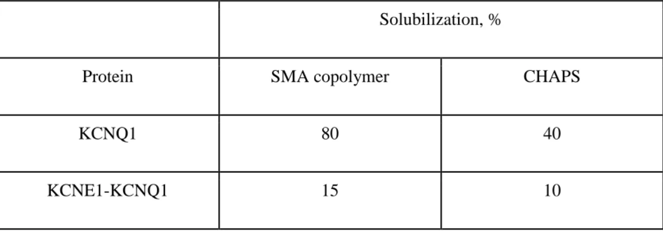
et al. 2005). We used transient transfection of the COS-1 cells to overexpress the Kv channels
and compared the effectiveness of their solubilization by SMA and detergent (Table 1). We
demonstrated that the SMA copolymer was more efficient at solubilization of the human
KCNQ1 channels, than CHAPS. Detergents have been used before in many structural studies
(for review see for example De Zorzi et al. 2016); high-quality preparations for EM could be
obtained by using the appropriate detergents and baculovirus expression system (Guo and
MacKinnon 2017). Yet, for channels expressed in mammalian cells, the solubilization in the
detergent often yielded rather low concentrations. The advantage of using SMALP is that the
solubilized membrane proteins can be easily concentrated on Microcon concentrators without
aggregation.
Our second goal was to develop the procedure for expression and purification of the complex of the α-subunit hKCNQ1 with its auxiliary subunit hKCNE1 for further cryo-EM experiments. To avoid the structural variability, due to various stoichiometry from one particle to
another that may further interfere with image processing, we used the fusion construct, which includes both α- and ß-subunits with the stoichiometry of subunits 4:4 (Choveau et al. 2011, Wang, Xia, and Kass 1998). Single-cell electrophysiological experiments confirmed that this
construct was fully active (Fig. 2C). We were able to isolate the complex from mammalian
COS-1 cells using SMALPs and to examine its 2D structure. Multivariate statistical analysis on the
aligned channel particles produced eigenimages (Fig. 4H), suggesting either two- or four-fold
symmetry, in agreement with current and previous (Shenkarev et al. 2018) data for the α-subunit.
Hence, incorporation into SMALPs did not affect the conformation of the purified fusion
channel. Moreover, in concordance with earlier reports (Routledge et al. 2016), we found that the
SMA-solubilized hKCNE1-hKCNQ1 construct was more stable, less prone to aggregation and
In summation, using SMA copolymers, we tested a method of detergent-free
solubilization of human ion channels, particularly, the cardiac and neuronal potassium
voltage-dependent channels. SMALPs appear to develop into convenient platform for studying the
structure of human ion channels and their complexes (which are hard to crystallize) using not
only cryo-EM, but also NMR methods, as well as other structural methods that require using the
single particle mode (including XFEL). The study of the structural and functional properties of
potassium voltage-dependent channels would help to clarify the mechanisms that cause
malfunction of these channels in case of point mutations. Understanding these mechanisms, in its
turn, would pave the way to methods of targeted correction of channel function.
Acknowledgements
Authors thank Dr. Irina Panova for her help with DLS experiments, Andrey Moiseenko
for his help with obtaining the electron microscopy images and Lisa Trifonova for proofreading
the manuscript. Electron microscopy was performed at the user facilities center ‘Electron microscopy in the life sciences’ of the Biology Department of Lomonosov Moscow State University.
The purification of hKCNQ1 and hKCNE1-hKCNQ1 by a SMA copolymer and their
investigations were financially supported by the Russian Foundation for Basic Research (Project
#18-504-12045 to K.V.S.); DLS experiments were supported by the German Research
Foundation (DFG) (Project #STE640/15 to H.-J.S.). Experiments on the purification of hKCNH5
ion channel and its EM studies were supported by the Russian Science Foundation young
investigators grant (Project #18-74-00087 to G.S.G.).
Solubilization, %
Protein SMA copolymer CHAPS
KCNQ1 80 40
KCNE1-KCNQ1 15 10
Figure legends
Fig. 1. Schematic representation of the channel expression constructs used in this
study.
Fig. 2. Expression of functional human ion channels in mammalian cells. (A)
Representative superimposed recordings of a CHO cell transfected with
pIRES2-EGFP/hKCNQ1-1D4. (B). Mean activation curves of tagged hKCNQ1 (n=9). (C) Representative
superimposed recordings of a COS-7 cell transfected with pCDNA6-V5-His/A-KCNE1-KCNQ1
and GFP as a reporter. Insert: voltage protocol, as detailed in the methods section. (D) Mean
activation curves of tagged hKCNE1-hKCNQ1 (n=8).
Fig. 3. The solubilization and purification of human ion channel (hKCNH5) by SMA
copolymer.
(A) Western blot of elution fraction containing hKCNH5; mouse monoclonal antibody
directed against 1D4 tag was used as a primary antibody: MW-protein ladder; E – elution with
1D4 peptide. (B) EM image of the elution fraction, stained with 1% UA. White arrows indicate
hKCNH5 particles. Bar size – 20 nm. (C) DLS curve of elution fraction of the hKCNH5 channel.
(D) Representative class-averages of hKCNH5. Bar size – 10 nm. Above each 2D average, the
placed for comparison. (E) Eigenimages generated upon classification of hKCNH5. Images
reflect variations in densities of particles related to the different symmetry. Eigenimage #5
demonstrates the presence of four-fold symmetry.
Fig. 4. The solubilization and purification of hKCNQ1 and hKCNE1-hKCNQ1 by
SMA copolymer. (A) hKCNQ1 protein expression, solubilization with SMA and purification on
anti-1D4 affinity resin. Line 1 - SDS-PAGE of KCNQ1 protein expression in COS-1 cells,
coomassie staining; line 2-6 - western blots, immunodetection with anti-1D4 Ab. 2 - COS-1 cells
extract; 3 - solubilization with 2,5% of SMA copolymer - supernatant; 4 - pellet; 5 - anti 1D4
column wash; 6 - elution fraction. (B) hKCNE1-hKCNQ1 fusion expression, solubilization with
SMA copolymer and purification on the anti-V5 affinity resin. Line 1 - SDS-PAGE of
hKCNE1-hKCNQ1 protein expression in COS-1 cells, coomassie staining; line 2-6 - western blots,
immunodetection with anti KCNQ1 Ab. 2 - COS-1 cells extract; 3 - solubilization with 2,5% of
SMA copolymer - supernatant; 4 - pellet; 5 - anti V5 column wash; 6 - elution fraction. EM
images of (C) purified hKCNQ1 and (D) hKCNE1-hKCNQ1, both stained with 1% UA. Arrows
indicate channel particles. Bar size – 20 nm; Representative 2D class-averages of (E) hKCNQ1
and (F) hKCNE1-hKCNQ1. Above each 2D average, the corresponded projection of the
available Xenopus laevis KCNQ1 channel structure (EMD-8712) is placed for comparison. Bar
size – 10 nm. Eigenimages, obtained after MSA of (H) hKCNQ1 particles and (G)
hKCNQ1-hKCNQ1 particles.
Literature
1. Arenas, R. C., J. Klingler, C. Vargas, and S. Keller. 2016. 'Influence of lipid bilayer
properties on nanodisc formation mediated by styrene/maleic acid copolymers',
2. Autzen, H. E., A. G. Myasnikov, M. G. Campbell, D. Asarnow, D. Julius, and Y. Cheng.
2018. 'Structure of the human TRPM4 ion channel in a lipid nanodisc', Science, 359:
228-32.
3. Bagrov, D.V., N. Voskoboynikova, G.A. Armeev, W. Mosslehy, G.S. Gluhov, T.T.
Ismagulova, A.Y. Mulkidjanian, M.P. Kirpichnikov, H.-J. Steinhoff, and K.V. Shaitan.
2016. 'Characterization of lipodisc nanoparticles containing sensory rhodopsin ii and its
cognate transducer from Natronomonas pharaonis', Biofizika (Biophysics) Moscow, 61:
942-49.
4. Bauer, C. K., and J. R. Schwarz. 2018. 'Ether-a-go-go K(+) channels: effective
modulators of neuronal excitability', The Journal of physiology, 596: 769-83.
5. Bayburt, T. H., Y. V. Grinkova, and S. G. Sligar. 2002. 'Self-assembly of discoidal
phospholipid bilayer nanoparticles with membrane scaffold proteins', Nano Letters, 2:
853-56.
6. Beekwilder, J. P., M. E. O'Leary, L. P. van den Broek, G. T. van Kempen, D. L. Ypey,
and R. J. van den Berg. 2003. 'Kv1.1 channels of dorsal root ganglion neurons are
inhibited by n-butyl-p-aminobenzoate, a promising anesthetic for the treatment of chronic
pain', The Journal of pharmacology and experimental therapeutics, 304: 531-8.
7. Bell, A. J., L. K. Frankel, and T. M. Bricker. 2015. 'High Yield Non-detergent Isolation
of Photosystem I-Lightharvesting Chlorophyll II Membranes from Spinach Thylakoids
IMPLICATIONS FOR THE ORGANIZATION OF THE PS I ANTENNAE IN
HIGHER PLANTS', Journal of Biological Chemistry, 290: 18429-37.
8. Camacho, J. 2006. 'Ether a go-go potassium channels and cancer', Cancer letters, 233:
1-9.
9. Chen, Q., J. She, W. Zeng, J. Guo, H. Xu, X. C. Bai, and Y. Jiang. 2017. 'Structure of
10. Choveau, F. S., N. Rodriguez, F. Abderemane Ali, A. J. Labro, T. Rose, S. Dahimene, H.
Boudin, C. Le Henaff, D. Escande, D. J. Snyders, F. Charpentier, J. Merot, I. Baro, and
G. Loussouarn. 2011. 'KCNQ1 channels voltage dependence through a
voltage-dependent binding of the S4-S5 linker to the pore domain', The Journal of biological
chemistry, 286: 707-16.
11. Cleverley, R. M., J. Kean, C. A. Shintre, C. Baldock, J. P. Derrick, R. C. Ford, and S. M.
Prince. 2015. 'The Cryo-EM structure of the CorA channel from Methanocaldococcus
jannaschii in low magnesium conditions', Biochimica et biophysica acta, 1848: 2206-15.
12. Craig, A. F., E. E. Clark, I. D. Sahu, R. Zhang, N. D. Frantz, M. S. Al-Abdul-Wahid, C.
Dabney-Smith, D. Konkolewicz, and G. A. Lorigan. 2016. 'Tuning the size of
styrene-maleic acid copolymer-lipid nanoparticles (SMALPs) using RAFT polymerization for
biophysical studies', Biochimica et biophysica acta, 1858: 2931-39.
13. De Zorzi, R., W. Mi, M. Liao, and T. Walz. 2016. 'Single-particle electron microscopy in
the study of membrane protein structure', Microscopy, 65: 81-96.
14. Deutsch, C., and L. Q. Chen. 1993. 'Heterologous expression of specific K+ channels in
T lymphocytes: functional consequences for volume regulation', Proceedings of the
National Academy of Sciences of the United States of America, 90: 10036-40.
15. Dorr, J. M., M. C. Koorengevel, M. Schafer, A. V. Prokofyev, S. Scheidelaar, E. A. van
der Cruijsen, T. R. Dafforn, M. Baldus, and J. A. Killian. 2014. 'Detergent-free isolation,
characterization, and functional reconstitution of a tetrameric K+ channel: the power of
native nanodiscs', Proceedings of the National Academy of Sciences of the United States
of America, 111: 18607-12.
16. Dorr, J. M., S. Scheidelaar, M. C. Koorengevel, J. J. Dominguez, M. Schafer, C. A. van
Walree, and J. A. Killian. 2016. 'The styrene-maleic acid copolymer: a versatile tool in
17. Duan, J., J. Li, B. Zeng, G. L. Chen, X. Peng, Y. Zhang, J. Wang, D. E. Clapham, Z. Li,
and J. Zhang. 2018. 'Structure of the mouse TRPC4 ion channel', Nature
communications, 9: 3102.
18. Gao, Y., E. Cao, D. Julius, and Y. Cheng. 2016. 'TRPV1 structures in nanodiscs reveal
mechanisms of ligand and lipid action', Nature, 534: 347-51.
19. Gulati, S., M. Jamshad, T. J. Knowles, K. A. Morrison, R. Downing, N. Cant, R. Collins,
J. B. Koenderink, R. C. Ford, M. Overduin, I. D. Kerr, T. R. Dafforn, and A. J. Rothnie.
2014. 'Detergent-free purification of ABC (ATP-binding-cassette) transporters',
Biochemical Journal, 461: 269-78.
20. Guo, Y. R., and R. MacKinnon. 2017. 'Structure-based membrane dome mechanism for
Piezo mechanosensitivity', Elife, 6.
21. Ikeda, M., Y. Tomita, A. Mouri, M. Koga, T. Okochi, R. Yoshimura, Y. Yamanouchi, Y.
Kinoshita, R. Hashimoto, H. J. Williams, M. Takeda, J. Nakamura, T. Nabeshima, M. J.
Owen, M. C. O'Donovan, H. Honda, T. Arinami, N. Ozaki, and N. Iwata. 2010.
'Identification of novel candidate genes for treatment response to risperidone and
susceptibility for schizophrenia: integrated analysis among pharmacogenomics, mouse
expression, and genetic case-control association approaches', Biological psychiatry, 67:
263-9.
22. Jamshad, M., J. Charlton, Y. P. Lin, S. J. Routledge, Z. Bawa, T. J. Knowles, M.
Overduin, N. Dekker, T. R. Dafforn, R. M. Bill, D. R. Poyner, and M. Wheatley. 2015.
'G-protein coupled receptor solubilization and purification for biophysical analysis and
functional studies, in the total absence of detergent', Biosci Rep, 35.
23. Jensen, M. O., V. Jogini, D. W. Borhani, A. E. Leffler, R. O. Dror, and D. E. Shaw.
2012. 'Mechanism of voltage gating in potassium channels', Science, 336: 229-33.
24. Jiang, Y., A. Lee, J. Chen, V. Ruta, M. Cadene, B. T. Chait, and R. MacKinnon. 2003.
25. Judge, S. I. V., J. M. Lee, C. T. Bever, and P. M. Hoffman. 2006. 'Voltage-gated
potassium channels in multiple sclerosis: Overview and new implications for treatment of
central nervous system inflammation and degeneration', Journal of Rehabilitation
Research and Development, 43: 111-21.
26. Knowles, T. J., R. Finka, C. Smith, Y. P. Lin, T. Dafforn, and M. Overduin. 2009.
'Membrane proteins solubilized intact in lipid containing nanoparticles bounded by
styrene maleic acid copolymer', Journal of the American Chemical Society, 131: 7484-5.
27. Kuang, Q., P. Purhonen, and H. Hebert. 2015. 'Structure of potassium channels', Cellular
and molecular life sciences : CMLS, 72: 3677-93.
28. Lee, S. C., Knowles, T. J., Postis, V. L. G., Jamshad, M., Parslow, R. A., Lin, Y.,
Goldman, A., Sridhar, P., Overduin, M., Muench, S. P., Dafforn, T. R. (2016). ‘A method for detergent-free isolation of membrane proteins in their local lipid environment’. Nature Protocols, 11(7), 1149–1162.
29. Lee, S. C., S. Khalid, N. L. Pollock, T. J. Knowles, K. Edler, A. J. Rothnie, R. T. Thomas
O, and T. R. Dafforn. 2016. 'Encapsulated membrane proteins: A simplified system for
molecular simulation', Biochimica et biophysica acta, 1858: 2549-57.
30. Lee, S. Y., A. Lee, J. Chen, and R. MacKinnon. 2005. 'Structure of the KvAP
voltage-dependent K+ channel and its dependence on the lipid membrane', Proceedings of the
National Academy of Sciences of the United States of America, 102: 15441-6.
31. Long, A. R., C. C. O'Brien, K. Malhotra, C. T. Schwall, A. D. Albert, A. Watts, and N.
N. Alder. 2013. 'A detergent-free strategy for the reconstitution of active enzyme
complexes from native biological membranes into nanoscale discs', Bmc Biotechnology,
13.
32. Long, S. B., X. Tao, E. B. Campbell, and R. MacKinnon. 2007. 'Atomic structure of a
voltage-dependent K+ channel in a lipid membrane-like environment', Nature, 450:
33. Loussouarn, G., K. H. Park, C. Bellocq, I. Baro, F. Charpentier, and D. Escande. 2003.
'Phosphatidylinositol-4,5-bisphosphate, PIP2, controls KCNQ1/KCNE1 voltage-gated
potassium channels: a functional homology between voltage-gated and inward rectifier
K+ channels', The EMBO journal, 22: 5412-21.
34. Ludtke, S. J., P. R. Baldwin, and W. Chiu. 1999. 'EMAN: semiautomated software for
high-resolution single-particle reconstructions', Journal of structural biology, 128: 82-97.
35. MacDonald, P. E., and M. B. Wheeler. 2003. 'Voltage-dependent K(+) channels in
pancreatic beta cells: role, regulation and potential as therapeutic targets', Diabetologia,
46: 1046-62.
36. Matthies, D., C. Bae, G. E. Toombes, T. Fox, A. Bartesaghi, S. Subramaniam, and K. J.
Swartz. 2018. 'Single-particle cryo-EM structure of a voltage-activated potassium
channel in lipid nanodiscs', eLife, 7.
37. Milescu, M., H. C. Lee, C. H. Bae, J. I. Kim, and K. J. Swartz. 2013. 'Opening the shaker
K+ channel with hanatoxin', The Journal of general physiology, 141: 203-16.
38. Oprian, D. D., R. S. Molday, R. J. Kaufman, and H. G. Khorana. 1987. 'Expression of a
synthetic bovine rhodopsin gene in monkey kidney cells', Proceedings of the National
Academy of Sciences of the United States of America, 84: 8874-8.
39. Orwick-Rydmark, M., J. E. Lovett, A. Graziadei, L. Lindholm, M. R. Hicks, and A.
Watts. 2012. 'Detergent-free incorporation of a seven-transmembrane receptor protein
into nanosized bilayer Lipodisq particles for functional and biophysical studies', Nano
Lett, 12: 4687-92.
40. Pal, S., K. A. Hartnett, J. M. Nerbonne, E. S. Levitan, and E. Aizenman. 2003. 'Mediation
of neuronal apoptosis by Kv2.1-encoded potassium channels', The Journal of
41. Pardo, Dominguez, J. J., J. M. Dorr, A. Iyer, R. C. Cox, S. Scheidelaar, M. C.
Koorengevel, V. Subramaniam, and J. A. Killian. 2017. 'Solubilization of lipids and lipid
phases by the styrene-maleic acid copolymer', Eur Biophys J, 46: 91-101.
42. Parmar, M., S. Rawson, C. A. Scarff, A. Goldman, T. R. Dafforn, S. P. Muench, and V.
L. G. Postis. 2018. 'Using a SMALP platform to determine a sub-nm single particle
cryo-EM membrane protein structure', Biochimica et biophysica acta, 1860: 378-83.
43. Postis, V., S. Rawson, J. K. Mitchell, S. C. Lee, R. A. Parslow, T. R. Dafforn, S. A.
Baldwin, and S. P. Muench. 2015. 'The use of SMALPs as a novel membrane protein
scaffold for structure study by negative stain electron microscopy', Biochimica et
biophysica acta, 1848: 496-501.
44. Routledge, S. J., L. Mikaliunaite, A. Patel, M. Clare, S. P. Cartwright, Z. Bawa, M. D.
Wilks, F. Low, D. Hardy, A. J. Rothnie, and R. M. Bill. 2016. 'The synthesis of
recombinant membrane proteins in yeast for structural studies', Methods, 95: 26-37.
45. Sahu, I. D., R. M. McCarrick, K. R. Troxel, R. F. Zhang, H. J. Smith, M. M. Dunagan,
M. S. Swartz, P. V. Rajan, B. M. Kroncke, C. R. Sanders, and G. A. Lorigan. 2013.
'DEER EPR Measurements for Membrane Protein Structures via Bifunctional Spin
Labels and Lipodisq Nanoparticles', Biochemistry, 52: 6627-32.
46. Scheres, S. H., M. Valle, R. Nunez, C. O. Sorzano, R. Marabini, G. T. Herman, and J. M.
Carazo. 2005. 'Maximum-likelihood multi-reference refinement for electron microscopy
images', Journal of molecular biology, 348: 139-49.
47. Shenkarev, Z. O., M. G. Karlova, D. S. Kulbatskii, M. P. Kirpichnikov, E. N.
Lyukmanova, and O. S. Sokolova. 2018. 'Recombinant Production, Reconstruction in
Lipid-Protein Nanodiscs, and Electron Microscopy of Full-Length alpha-Subunit of
48. Singer-Lahat, D., D. Chikvashvili, and I. Lotan. 2008. 'Direct interaction of endogenous
Kv channels with syntaxin enhances exocytosis by neuroendocrine cells', PloS one, 3:
e1381.
49. Sokolova, O. 2004. 'Structure of cation channels, revealed by single particle electron
microscopy', FEBS letters, 564: 251-6.
50. Sokolova, O. S., K. V. Shaitan, A. V. Grizel, A. V. Popinako, M. G. Karlova, and M. P.
Kirpichnikov. 2012. 'Three-dimensional structure of human Kv10.2 ion channel studied
by single particle electron microscopy and molecular modeling', Bioorg Khim, 38:
177-84.
51. Sokolova, O., L. Kolmakova-Partensky, and N. Grigorieff. 2001. 'Three-dimensional
structure of a voltage-gated potassium channel at 2.5 nm resolution', Structure, 9: 215-20.
52. Sun, J., and R. MacKinnon. 2017. 'Cryo-EM Structure of a KCNQ1/CaM Complex
Reveals Insights into Congenital Long QT Syndrome', Cell, 169: 1042-50 e9.
53. Swainsbury, D. J., S. Scheidelaar, R. van Grondelle, J. A. Killian, and M. R. Jones. 2014.
'Bacterial reaction centers purified with styrene maleic acid copolymer retain native
membrane functional properties and display enhanced stability', Angew Chem Int Ed
Engl, 53: 11803-7.
54. Tester, D. J., and M. J. Ackerman. 2014. 'Genetics of long QT syndrome', Methodist
DeBakey cardiovascular journal, 10: 29-33.
55. Thomas, D., A. B. Wimmer, K. Wu, B. C. Hammerling, E. K. Ficker, Y. A. Kuryshev, J.
Kiehn, H. A. Katus, W. Schoels, and C. A. Karle. 2004. 'Inhibition of human
ether-a-go-go-related gene potassium channels by alpha 1-adrenoceptor antagonists prazosin,
doxazosin, and terazosin', Naunyn-Schmiedeberg's archives of pharmacology, 369:
56. van Heel, M., G. Harauz, E. V. Orlova, R. Schmidt, and M. Schatz. 1996. 'A new
generation of the IMAGIC image processing system', Journal of structural biology, 116:
17-24.
57. Voskoboynikova, N., W. Mosslehy, A. Colbasevici, T. T. Ismagulova, D. V. Bagrov, A.
A. Akovantseva, P. S. Timashev, A. Y. Mulkidjanian, V. N. Bagratashvili, K. V. Shaitan,
M. P. Kirpichnikov, and H. J. Steinhoff. 2017. 'Characterization of an archaeal
photoreceptor/transducer complex from Natronomonas pharaonis assembled within
styrene-maleic acid lipid particles', Rsc Advances, 7: 51324-34.
58. Wagner, D.T. 2009. 'Palliative Hypofractionated Radiotherapy For Non-Small Cell Lung
Cancer (NSCLC) Patients Previously Treated By Induction Chemotherapy: Is It For
Many, Some, All, Or None?', Journal of thoracic disease, 1: 1-2.
59. Wang, W., and R. MacKinnon. 2017. 'Cryo-EM Structure of the Open Human
Ether-a-go-go-Related K(+) Channel hERG', Cell, 169: 422-30 e10.
60. Wang, W., J. Xia, and R. S. Kass. 1998. 'MinK-KvLQT1 fusion proteins, evidence for
multiple stoichiometries of the assembled IsK channel', The Journal of biological
chemistry, 273: 34069-74.
61. Watanabe, H., E. Nagata, A. Kosakai, M. Nakamura, M. Yokoyama, K. Tanaka, and H.
Sasai. 2000. 'Disruption of the epilepsy KCNQ2 gene results in neural hyperexcitability',
Journal of neurochemistry, 75: 28-33.
62. Whicher, J. R., and R. MacKinnon. 2016. 'Structure of the voltage-gated K(+) channel
Eag1 reveals an alternative voltage sensing mechanism', Science, 353: 664-9.
63. Winterstein, L. M. et al. Reconstitution and functional characterization of ion channels
from nanodiscs in lipid bilayers. J Gen Physiol 150: 637-646
64. Yellen, G. 2002. 'The voltage-gated potassium channels and their relatives', Nature, 419:
65. Yu, F. H., V. Yarov-Yarovoy, G. A. Gutman, and W. A. Catterall. 2005. 'Overview of
molecular relationships in the voltage-gated ion channel superfamily', Pharmacological
reviews, 57: 387-95.
66. Zaydman, M. A., J. R. Silva, K. Delaloye, Y. Li, H. Liang, H. P. Larsson, J. Shi, and J.
Cui. 2013. 'Kv7.1 ion channels require a lipid to couple voltage sensing to pore opening',
Proceedings of the National Academy of Sciences of the United States of America, 110: