HAL Id: tel-01237007
https://pastel.archives-ouvertes.fr/tel-01237007
Submitted on 2 Dec 2015
HAL is a multi-disciplinary open access
archive for the deposit and dissemination of sci-entific research documents, whether they are pub-lished or not. The documents may come from teaching and research institutions in France or abroad, or from public or private research centers.
L’archive ouverte pluridisciplinaire HAL, est destinée au dépôt et à la diffusion de documents scientifiques de niveau recherche, publiés ou non, émanant des établissements d’enseignement et de recherche français ou étrangers, des laboratoires publics ou privés.
Multiscale biomechanics of skin: experimental
investigation of the role of the collagen microstructure
Barbara Lynch
To cite this version:
Barbara Lynch. Multiscale biomechanics of skin: experimental investigation of the role of the collagen microstructure. Biomechanics [physics.med-ph]. Ecole Doctorale Polytechnique, 2015. English. �tel-01237007�
Th`
ese pr´
esent´
ee pour l’obtention du titre de
DOCTEUR DE L’´
ECOLE POLYTECHNIQUE
Sp´
ecialit´
e : M´
ecanique
par
Barbara Lynch
Sujet de th`
ese
Multiscale biomechanics of skin: experimental
investigation of the role of the collagen microstructure
Soutenue le 7 Septembre 2015 devant le jury compos´e de :
Karine BRUY`ERE-GARNIER IFSTTAR Rapporteure
Val´erie DEPLANO IRPHE, Aix-Marseille Universit´e Rapporteure
Edoardo MAZZA ETH Z¨urich Examinateur
Marie-Claire SCHANNE-KLEIN LOB, ´Ecole Polytechnique Examinatrice Claude VERDIER LIPhy, Universit´e Joseph Fournier Examinateur Jean-Marc ALLAIN LMS, ´Ecole Polytechnique Directeur de th`ese
Remerciements
Je tiens en premier lieu `a remercier les membres de mon jury, Karine Bruy`ere-Garnier, Val´erie Deplano, Edoardo Mazza et Claude Verdier, de m’avoir fait l’honneur d’´evaluer mon travail de th`ese.
J’aimerai ´egalement remercier Patrick Le Tallec de m’avoir accueillie au sein du Laboratoire de M´ecanique des Solides, ainsi que l’´Ecole Polytechnique pour le financement de cette th`ese.
Je remercie chaleureusement Jean-Marc Allain de m’avoir encadr´ee en qualit´e de directeur de th`ese durant ces trois ann´ees. Merci pour ta disponibilit´e, ta confiance et ton enthousiasme (mˆeme quand on avait “pas de peau”).
J’adresse ma sinc`ere gratitude `a Marie-Claire Schanne-Klein du Laboratoire Optique et Bios-ciences, qui m’a ´egalement beaucoup guid´ee pendant ce travail. Merci pour toutes nos discussions bilingues optique-m´ecanique, toujours tr`es enrichissantes.
Un grand merci `a ceux avec qui j’ai eu le plaisir de r´ealiser les manips tout au long de ces 3 ans : St´ephane Bancelin, Guillaume Ducourthial et Sotiris Psilodimitrakopoulos. Je remercie aussi Aur´elie Benoit qui a guid´e mes premiers pas sur les essais multi-´echelles et qui m’a initi´ee `a la corr´elation d’images.
Merci `a nos collaboratrices de l’Institut de G´enomique Fonctionnelle de Lyon, Florence Ruggiero, Christelle Bonod-Bidaud et Ruth Rubio Amador, qui nous ont fourni les peaux de souris. Je les remercie ´egalement pour leur d´etermination `a mettre la biologie `a la port´ee des physiciens et pour nos nombreux ´echanges fructueux.
Je souhaite une longue vie `a cette belle collaboration grˆace `a laquelle j’ai beaucoup appris, et j’adresse mes meilleurs vœux de succ`es `a Jean-S´ebastien, Guillaume et Florent, qui en pour-suivent les projets.
Pouvoir construire sa machine d’exp´erimentation sur mesure est un luxe, et je souhaite remercier ceux grˆace `a qui cela a ´et´e possible : Fran¸cois Lelong pour l’usinage des mors et Vincent de Greef pour la gestion du (capricieux) boˆıtier de commande.
Je tiens ´egalement `a remercier les personnes avec qui j’ai eu l’opportunit´e de collaborer sur divers projets annexes `a mon travail de th`ese principal, en particulier Gervaise Mosser et Aur´elien Tidu pour la m´ecanique des matrices de collag`ene ainsi que William Parnell, Tom Shearer et Riccardo de Pascalis pour la mod´elisation du tendon.
Entre mon bureau au bˆatiment 65, le LMS et la salle de manip au LOB, j’ai eu la chance de cˆotoyer pendant 3 ans de nombreuses personnes d’horizons tr`es vari´es : je les remercie d’avoir contribu´e chacune `a leur mani`ere `a rendre ces ann´ees sur le plateau tr`es agr´eables.
Je tiens `a remercier les doctorants, post-doctorants et stagiaires du LMS qui ont partag´e mon quotidien. Merci `a Jessica, David, Gwena¨el, Cl´ement, Christelle, Rapha¨el, Guilia, Kokou, Nicolas, Luka, Laurent... Merci, et bravo, `a mes chers co-th´esards : Armel, Dennis et Dimitri.
Je remercie ´egalement l’ensemble du secr´etariat du LMS : Alexandra, Anna, Christiane, Val´erie et Danielle, pour leur disponibilit´e et leur aide pr´ecieuse.
Je remercie l’ensemble du personnel du LOB de m’avoir toujours r´eserv´e un accueil chaleureux pendant nos si longues journ´ees de manip. Merci en particulier aux doctorants : Claire, Mouna, Nelly, Marianne, Kamel.
J’aimerai remercier enfin toutes les personnes avec qui j’ai eu l’occasion de partager le bˆatiment 65. Toute ma reconnaissance va en particulier `a mes co-bureaux successifs : Franz, C´ecile, Fran¸cois et Manon. Je remercie ici ´egalement Avin Babataheri qui nous permet d’effectuer nos exp´erimentations dans des conditions optimales en g´erant efficacement les stocks du laboratoire et le L2.
Merci `a mes amis pour leur oreille toujours attentive et leur bienveillance. Merci `a Olivier, MH, Cl´ement, Catherine, C´elia, Natacha, B´ea, Tristan, Manu, Charles & Charles.
A sincere thank you to Maggie and Kip for proofreading this document and for their continued support. Mes plus profonds remerciements `a mes parents, pour leur soutien sans faille et leurs encouragements au cours de ces (presque) 25 ann´ees d’´etude.
Table of Contents
General introduction 9
I Introduction 13
I.1 Microscopic scale: from collagen to skin . . . 13
I.1.1 Collagen . . . 13
I.1.1.a The collagen family . . . 14
I.1.1.b Collagen’s hierarchical structure . . . 15
I.1.1.c Soft collagenous tissues . . . 19
I.1.1.d Mechanical properties of collagen . . . 21
I.1.2 Skin . . . 22
I.1.2.a Properties and functions . . . 22
I.1.2.b Global structure . . . 23
I.1.2.c Microstructure of the dermis . . . 25
I.1.3 Imaging the architecture of soft collagenous tissues . . . 28
I.1.3.a Non-optical imaging . . . 28
I.1.3.b Optical imaging . . . 29
I.1.3.c Non-linear microscopy: 2PEF and SHG imaging . . . 32
I.2 Macroscopic scale: skin’s mechanical properties . . . 35
I.2.1 Mechanical characterisation . . . 35
I.2.1.a Ex vivo characterisation . . . 35
I.2.1.b In vivo characterisation . . . 37
I.2.2 Skin’s mechanical properties . . . 39
I.2.2.a Non-linear elasticity . . . 39
I.2.2.b Prestress and anisotropy . . . 40
I.2.2.c Viscoelasticity . . . 40
I.2.3 Soft tissues mechanical modelling . . . 43
I.2.3.a Phenomenological models . . . 43
I.3 Previous multiscale works . . . 45
I.4 Aim of the thesis . . . 47
II Experimental multiscale study of skin biomechanics 49 II.1 Mice skin samples . . . 49
II.1.1 Sample preparation . . . 50
II.1.2 Validation of preparation protocol with histological cuts . . . 51
II.1.3 Sample cutting for mechanical test . . . 52
II.2 Mechanical tensile tests . . . 53
II.2.1 Tensile test device . . . 53
II.2.2 In situ set-up . . . 54
II.2.3 Test procedure under the microscope . . . 57
II.2.4 Test procedure outside the microscope . . . 58
II.3 Mechanical analysis . . . 60
II.3.1 Stress/time and stress/stretch curve . . . 61
II.3.1.a Tangent modulus . . . 62
II.3.1.b Heel region length . . . 62
II.3.1.c Rupture . . . 63
II.3.2 Relaxation . . . 63
II.4 Optical analysis . . . 66
II.4.1 Local stretches . . . 68
II.4.2 Measure of porosity . . . 71
II.4.3 Fibre orientation . . . 72
III Evolution of micrometer-scale collagen organisation in mice skin upon mechanical strain 75 III.1 Multiscale biomechanics of mice skin . . . 76
III.1.1 Stress/stretch behaviour . . . 76
III.1.2 Relaxation analysis . . . 81
III.1.3 Evolution of fibre orientation . . . 83
III.1.4 Correlation of OI and stress . . . 86
III.1.5 Strain rate dependency . . . 87
III.1.6 Cycling experiments . . . 90
III.2 Discussion . . . 91
III.2.1 Incompatibility with conventional models . . . 91
III.2.2 Hypothesis on possible microstructural mechanisms . . . 94
IV Biomechanics of mice skin with genetic mutations affecting the microstructure 105 IV.1 Ehlers-Danlos Syndrome . . . 105
IV.1.1 Role of collagen V in collagen I fibrillogenesis . . . 106
IV.1.2 Microstructural modifications . . . 108
IV.1.3 Ehlers-Danlos Syndrome . . . 109
IV.1.4 Mice models for the investigation of EDS . . . 110
IV.2 peK mice: Results and Discussion . . . 113
IV.2.1 Genetic mutation and microstructural changes . . . 113
IV.2.1.a Literature review . . . 113
IV.2.1.b Measured characteristics . . . 115
IV.2.2 Mechanical behaviour under stretch . . . 116
IV.2.2.a Stress/stretch behaviour . . . 116
IV.2.2.b Relaxation analysis . . . 116
IV.2.2.c Strain rate dependency . . . 117
IV.2.3 Microstructural behaviour under stretch . . . 121
IV.2.4 Interpretation . . . 122
IV.3 pN mice: Results and Discussion . . . 124
IV.3.1 Genetic modification and microstructural changes . . . 124
IV.3.1.a Literature review . . . 124
IV.3.1.b Measured characteristics . . . 127
IV.3.2 Mechanical behaviour under stretch . . . 127
IV.3.2.a Stress/stretch behaviour . . . 127
IV.3.2.b Relaxation analysis . . . 129
IV.3.3 Microstructural behaviour under stretch . . . 129
IV.3.4 Interpretation . . . 131
V Biomechanics of aged mice skin 133 V.1 Effect of age on human skin . . . 133
V.1.1 Structural changes . . . 134
V.1.2 Mechanical changes . . . 139
V.2 Aged mice: Results . . . 141
V.2.1 Tissue morphology changes . . . 141
V.2.2 Mechanical behaviour under stretch . . . 142
V.2.2.a Stress/stretch behaviour . . . 142
V.2.2.b Relaxation analysis . . . 145
V.2.3 Microstructural behaviour under stretch . . . 146
V.2.4 Interpretation . . . 147
Conclusion and perspectives 151
A Statistical analysis 157
A.1 Normal distribution . . . 157
A.2 Mean, SEM and p-factor . . . 158
B Possible experimental bias 161 B.1 Tests under and outside the microscope . . . 161
B.2 WT mice and non-mutant peK and pN mice . . . 164
B.3 Thickness . . . 165
B.4 Hair density and relative porosity . . . 166
B.5 Male and female mice . . . 168
C Data tables 171
General introduction
At first sight, materials from biological origin, whether animals or plants, appear very diverse. What do wood, bone, skin and sponges have in common ? The diversity of biological materials illustrates admirably the variety of tasks they serve and the diverse mechanical environments they need to accommodate.
Most of these materials fulfil more than one function, amongst which one is generally intrinsically mechanical: to provide stiffness, elasticity, resilience, etc. Yet, natural materials are extremely efficient, in the sense that they are composed of a relatively small number of structural building blocks. Consequently, almost all natural materials are composites: adaptation of shape, geometry, size and orientation in a structural block arrangement is nature’s preferred strategy to optimise mechanical properties of biological materials to fulfil the complex requirements imposed by plants and animals.
Biomechanics is the application of methods and tools from mechanics to study biological sys-tems. Mechanics offers a complementary approach to understand complex biological systems at various scales, from environment probing at the scale of a single cell to whole body locomotion. It has already enabled significant advances in medicine in multiple fields, e.g. diagnosis, surgery, prosthetics, tissue engineering. When understanding the mechanical principles behind the normal function of cells and organs, it becomes possible to foresee outcomes of pathological modifications and conceive new ways to address them.
Material sciences find a particularly straightforward field of application in the investigation of tissue biomechanics. In particular, the application of an upscaling scheme, like the theory of ho-mogeneisation, allows for the deduction of macroscale laws and constitutive relationships from knowledge on microscale behaviours.
This PhD dissertation is the result of a project aimed at multiscale experimental characterisation of skin. Skin belongs to the category of connective tissues, one of the four principal types of animal tissue, together with nervous, epithelial and muscle tissues. Connective tissues are responsible for support and cohesion. Skin is the largest organ of the human body and serves several crucial tasks, such as protection against external aggressions (mechanical, chemical, bacteriological, radiation, etc.), temperature balance, hormonal excretions, sense of touch and appearance. The use of che-micals, the ageing process, wounds and some pathologies are some of the many factors that can
influence the properties of skin, including mechanical characteristics. Understanding the complex properties of skin, and in particular how microstructure impacts the macroscopic mechanical be-haviour, will be beneficial in a number of domains, ranging from tissue engineering to cosmetics, robotic surgery or car crash predictive assessment.
Skin is a multilayered composite structure: its thickest layer, and the most important from a mechanical point of view, is the dermis. It consists mostly of collagen fibres embedded in a host structure. A scaling up methodology requires precise information on the interplay between compo-nents at different scales during mechanical stimulation. Micromechanical experiments are however difficult to implement, as they require precise knowledge on multiple techniques and careful sample preparation.
We developed, optimised and validated a multiphysics and multiscale protocol to perform me-chanical experiments while simultaneously assessing microstructural reorganisation. Using Second Harmonic Generation imaging we observed the reorganisation of the collagen network inside the dermis while performing a uniaxial tensile test.
The first chapter introduces useful notions for the understanding of this work. We first present the family of soft connective tissues, skin in particular, from the microscopic scale. We are mostly interested in the structural components and microstructural organisation. We also briefly review in this section the imaging tools available for the investigation of tissues micro-architecture. Then we take a look at the macroscopic mechanical properties of skin: the characterisation methods are introduced, and skin’s complex mechanical properties (non-linear elasticity, anisotropy, viscoelas-ticity, etc.) are detailed. Finally we show the context of existent multiscale works within which this PhD work falls.
Chapter II develops the multiphysics experimental protocol we implemented for this study. The sample preparation and mechanical tensile test protocols are described. We then define the quanti-tative parameters that were developed for the needs of this work. Firstly we review the parameters used for the assessment of skin’s macroscopic mechanical properties. Secondly we introduce the information that were retrieved from the microscopic images obtained with Second Harmonic Ge-neration imaging, in terms of local stretch and fibre orientation assessment in particular.
Chapter III reviews the multiscale results on control mice skin. The experiments were first carried out on one-month-old mice from a control strain. The results yield information on the macroscopic mechanical properties and microscopic evolution of skin. On the one hand they allow us to validate our experimental protocol, while on the other hand the correlation between mechanical behaviour and collagen network reorientation gives unexpected results. These are remarkably different from those expected from the literature on microscopic models of soft collagenous tissues. This inade-quacy is discussed, and possible microstructural mechanisms are suggested.
Chapter IV reports the changes in multiscale properties observed in mice that have a mutation affecting skin’s microstructure. We studied two mice strains created in the context of studying a pathology that affects all collagenous tissues’ mechanical properties: Ehlers-Danlos syndrome. One of the main features of Ehlers-Danlos syndrome is hyperelastic skin. In both mice strains we tested, the microstructure of the dermis is modified, at different levels and various severities. Testing these mice strains with our protocol, we observed how these changes affect microstruc-tural reorganisation and ultimately macroscopic mechanical properties. This is an opportunity to test the conclusions we obtained on control mice skin to various levels of microstructural alteration. The last chapter addresses the impact of age on multiscale properties of skin. We carried out our multiscale experiments on mice skin aged 15 and 19 months old, both control and with a genetic mutation. The ageing process impacts deeply the microstructural composition and organisation of the dermis, which results in modifications of mechanical properties. We can characterise those al-terations with the quantitative parameters developed, at both macroscopic and microscopic scales. Once more, we also evaluated the adequacy of the hypothetic microstructural mechanisms sug-gested before on an actual perturbation of the dermis’ microstructure.
This work illustrates that the link between microstructure and mechanical properties in soft col-lagenous tissues may not be as simple as initially infered. Small changes in the microstructure can deeply impact the mechanical behaviour, while contradictory effects of more generalised modifi-cations lead to unexpected results. In the end, the aim is to get a better idea of how to approach the development of an effective multiscale model of skin.
Chapter I
Introduction
The purpose of this introductory chapter is to demonstrate the need for precise microstructural investigation of skin behaviour under mechanical stimulation.
The first section is dedicated to skin from a microscopic perspective, from its most fundamental component, collagen, to its structural organisation. The second part considers the tissue from the macroscopic scale, and focuses on the complex mechanical properties of skin. After reviewing the usual mechanical modelling of skin and in particular how it takes microstructure into account, we eventually present the rationale of this PhD work and its novelty in the field.
I.1
Microscopic scale: from collagen to skin
In this section we examine skin from a microscopic point of view. First we introduce collagen and the family of tissues called connective, or “collagen-rich”, tissues. Then we review the anatomy and structure of skin more precisely, as well as the experimental methods that can be used to investigate the architecture of collagen-rich tissues at the microscopic level.
I.1.1
Collagen
Proteins are macromolecules formed by one or more long chains of amino acids. They play a cru-cial role in many biological interactions in vivo, including response to stimuli, replication of DNA, transport of molecules or catalysis of metabolic reactions. The specific 3D structure of each protein is of the utmost importance in all of these tasks: if the right binding site is not well presented or if steric effect prevents contact, no interaction is possible.
The main structural proteins in animal life form are the collagens, forming the collagen family. Collagen is ubiquitous in animals, and represents around 30% of the total mass of proteins in vertebrates. It can be found in a number of different molecular forms, as well as presenting various microstructural organisations, including fibres. Collagen also exists in invertebrates, for example
in the byssus threads through which mussels attach themselves to rocks.
In vertebrates, collagen is one of the basic bricks composing what is called “collagen-rich” tissues, also called connective or collagenous tissues. Connective tissues are, in most animals, the most abundant and most widely distributed type of tissues, as opposed to the 3 other basic types of tissue, i.e. epithelial, muscle and nervous tissue. Connective tissues include fat tissues, dense fibrous tissue, cartilage, bone, blood, etc. Their common purpose is to provide support and protection. Connective tissues only encompass a few cells. The remaining material is called “extracellular matrix”, and is mainly composed of collagen.
I.1.1.a The collagen family
At least 28 different collagens are known in vertebrates [1]. The most important and abundant collagen type in collagen-rich tissues is collagen I: it is responsible for most of their structural properties. However, less abundant collagens can also affect the microstructure: it is the case for collagen type V for instance, which will be of particular relevance to our study.
The collagens are classified with roman numbers, I-XXVIII, some also have common names. The size, function, role and abundance in tissues vary considerably from one collagen to another. In terms of structure, some form fibrils, others form 2D networks, and some remain disorganised, for example in basement membranes, the specific layer of extracellular matrix that connects cellular and connective tissues in many organs.
There is no firm and commonly agreed upon definition of what qualifies as a collagen. In general, all proteins that assemble into triple helices (or present triple helical domains) that also fulfil support or assembly functions in tissues are regardered as collagen.
A triple helix is made by binding three amino acid chains, called α-chains, together, in what is called a tropocollagen. A tropocollagen triple helix is represented schematically in figure I.1. The α-chains are numbered with arabic numerals. The chain called α1 in one collagen type is not
identical to the α1 chain in another collagen type. A triple helix is called homotrimeric when all
three α-chains are identical and heterotrimeric when one chain differs from the other two or all three α-chains are different. Most collagens exist in more than one stable form, although one is generally much more abundant than the others.
Table I.1 summarises the 28 different types of collagen, with their preferred structural organisa-tion and distribuorganisa-tion in tissues in the human body. There also exists a larger group of so-called “collagen-like proteins”.
Collagen I is the most abundant collagen: 90% of collagen in vertebrates is type I collagen. While most collagen can present interruptions in their triple helical regions, collagen I triple helix shows no imperfection [1]. Collagen I forms fibrils, that combine into bigger entities to ensure the
struc-Figure I.1 – Tropocollagen triple helix binding 3 α-chains together. tural integrity of tissues.
I.1.1.b Collagen’s hierarchical structure
Like skin, many biological tissues are fibre composites with a multiscale hierarchical structure. This includes animal tissues, but also wood for instance, which is a composite of cellulose fibres. This structural organisation is one of nature’s most common strategy to customise mechanical properties adapted to mechanical stimulation while ensuring tissue integrity. For a larger view of nature’s hierarchical materials, see [2, 3].
In some tissues, such as bone or dentine, the stiffness can be tuned notably by the addition of mineralized elements. This is not suitable for soft tissues, such as skin, tendon, ligaments, cornea or arteries, that have to undergo large and multiaxial deformations. In these cases, the mechanical resistance is obtained mostly by a modification of the many levels of collagen hierarchical struc-ture.
This organisation is summarised in figure I.2: α-chains assemble in tropocollagens, that assemble into fibrils, that congregate to form fibres, which organise in turn into a microstructure specific to each tissue [4, 5]. It should be noted that while triple helices assemble inside cells, all other assembling processes take place outside the cells after excretion.
When describing the structure of a protein, four forms are generally distinguinshed. The primary form is the sequence of amino acids. The secondary and tertiary forms are respectively the local organisation and spatial arrangement of a single polypeptidic chain. In the case of collagen, the se-condary and tertiary forms are the same and correspond to the organisation in α-chains. α-chains are coiled upon themselves in an helix.
The quaternary form describes how different polypeptidic chains assemble with regards to one another to form the protein. In the case of collagen, it is the triple helical tropocollagen. The stability of the triple helix is ensured by hydrogen bonds between specific amino acids of different α-chains. In fibrillar collagens, the triple helix domain is continuous, while in non fibrillar colla-gens, the triple helix can be interrupted by non collagenous sequences [1].
Tropocollagens have a diameter of the ordre of 1.5 nm and are approximately 300 nm long for collagen type I.
struc-Collagen type Structural organisation Distribution in tissues
I Fibril-forming Non-cartilaginous connective tissues: tendon, ligament, cornea, bone, skin...
II Thin fibril-forming Cartilage, vitreous humour
III Fibril-forming Co-distributes with collagen I, especially in skin and vassal walls
IV Network-forming Basal membranes
V Fibril-forming Co-distributes with collagen I, in skin, cornea and embryonic tissues
VI Beaded-filament-forming Widespread, especially muscle, skin VII Anchroring fibrils Epidermis-dermis junction
VIII Network-forming Descement’s membrane in cornea
IX FACIT Co-distributes with collagen II, cartilage,
vitrous humour
X Network-forming Hypertrophic cartilage
XI Fibril-forming Co-distributes with collagen II
XII FACIT Found with collagen I
XIII Transmembrane Neuromuscular junctions, skin
XIV FACIT Found with collagen I
XV Endostatins Eye, muscles, microvessels
XVI FACIT Integrated into collagen fibrils
XVII Transmembrane Epidermis-dermis junction
XVIII Endostatins Basement membranes
XIX FACIT Basement membranes, muscles
XX FACIT Widespread distribution, corneal epithelium
XXI FACIT Widespread distribution
XXII FACIT Tissue junctions, e.g. hair follicle-dermis
XXIII Transmembrane Tissue junctions
XXIV Fibril-forming Developing cornea and bone
XXV Transmembrane Associated with type I
XXVI Beaded-filament-forming
XXVII Fibril-forming Embryonic cartilage, developing dermis, cornea, heart, cartilage
XXVIII Beaded-filament-forming Basement membranes
Table I.1 – The collagen family: 28 collagen types, with preferred structural organisation and distribution in tissues in the human body. FACIT stands for Fibril-Associated Collagen with Interrupted Triple helices. Endostatins stand for endostatins-producing collagens. Table adapted from [1].
ture of collagen fibrils has been investigated for quite a long time using X-rays and electronic microscopy [4].
The most remarkable characteristic of collagen fibrils is the arrangement of tropocollagens in overlapping and gap zones, as shown in figure I.3 a. This arrangement appears as a result of two tropocollagens bonding through a covalent link between specific amino acids, called lysines. The cross-links formation depends deeply on the specific alignment of amino acids in the tropocolla-gens, through the maximisation of apolar and ionic interactions [4]. This specific bonding leads to 20 nm long overlapping zones and 47 nm gap zones, and thus to a characteristic periodic ar-rangement with a period of 67 nm, commonly called D. This shifting period is considered the watermark of fibrillar collagens. In transmission electronic microscopy (TEM) it is revealed by a characteristic alternation of light and dark bands [1]. An example can be seen in a TEM image from lung tissue in figure I.4.
Collagen fibrils have diameters ranging from 12 nm to over 500 nm, and their length vary greatly depending on the tissue and stage of development considered, from several micrometers to milli-meters [6].
Figure I.2 – Structural organisation of fibrillar collagen from collagen α-chain to tropocollagen, to fibril, to fibre and finally to tissue-specific microstructure, with corresponding scale. Figure adapted from [7].
Fibrils assemble in turn to form collagen fibres. This process is not yet well understood, but seems to depend strongly on the tissue involved. In soft connective tissues such as tendon, cornea or skin, the fibrils are embedded in an environment called “ground substance”, composed mostly of water. Ground substance and non-collagenous elements of the extracellular matrix, such as elastin in skin for instance, appear to have a deep impact on the adherence of fibrils together [4].
Some studies demonstrated that the ground substance is not as randomly organised as was first believed. For instance, proteins called proteoglycans (described in section I.1.2.3) extend radially
Figure I.3 – Exemples of cohesive strategies between collagenous sub-units: a) Covalent bonds between tropocollagen hold the fibrils together. The longitudinal gap creates overlapping regions with a length of 67 nm, characteristic for fibrillar collagen. Image adapted from [7]. b) TEM image of a 10-week-old rat tail tendon stained with cupromeronic blue. Collagen fibrils can be seen, with proteoglycans filaments (arrowed) extending from one fibril to another. Scale bar is 500 nm. Image from [8].
from one collagen fibril to another and seem to contribute to hold the whole network together [8]. This is shown in figure I.3 b in the case of a rat tail tendon specifically stained to show proteo-glycans.
Collagen fibres can extend up to a few millimeters in length and a few micrometers in diameter. Since the initial investigations of Fung [9], many experimental studies have been aiming for an accurate description of the fibres arrangement in specific biological composite structures, tissues and plants. Some of these studies start from the fibrillogenesis process and focus on cross-links [10] or interactions between fibrils and the rest of the matrix [11]. The dynamic interplay of subunits during a mechanical assay (such as sliding) has been examined very little up to now [12], and will be discussed later in this chapter.
Figure I.4 – TEM image of lung tissue. Collagen type I fibrils can be seen, with characteristic per-iodic arrangement revealed through the alternance of dark and light bands. Image by L. Howard, Dartmouth College.
the particular functions of the tissue, such as resistance to the mechanical environment. I.1.1.c Soft collagenous tissues
Soft collagenous tissues such as tendons, ligaments, skin and cornea are all made of the same simple building blocks: fibrillar collagen, a tissue-dependent proportion of elastin fibrils, and a surrounding substance composed mainly of water, proteins, and a few cells. From this relatively simple recipe originate tissues that are very different in terms of aspect, function and properties. Soft collagenous tissues can be submitted to various mechanical environments. Tendons transmit displacement from muscle to bone: they need to be resilient, but do not sustain large displace-ments. Furthermore, most tendons are solicited in only one direction (with a few exceptions, such as shoulder tendons). Cornea on the other hand must sustain the inner pressure of the eye, but also respond to a very specific need: transparency. Finally, skin is solicited in various directions and its primary protective function requires a large extensibility.
To adapt to this wide range of mechanical environments while having simple building blocks com-mon to all tissues, the microstructural organisation has to be optimised.
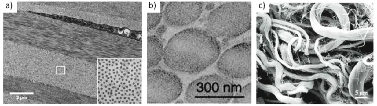
Figure I.5 shows images of cornea, tendon, and skin obtained with Transmission Electron Micro-scopy (TEM).
In the case of cornea the collagen fibrils organise in lamellae, stacked onto one another. They are also deposited in an hexagonal arrangement, as seen in the insert in figure I.5 a. This organisation is believed to be crucial for the transparency of the cornea [13].
Tendons also present a very organised microstructure, shown in figure I.5 b: fibres are unidirection-nal, parallel, aligned with the direction of mechanical stimulation. When observed with polarised light microscopy, the collagen fibres show a periodic waveform configuration, commonly called crimp, as shown in figure I.6 [14]. This crimp structure is believed to be intended as a safety
margin: the tendon can extend for a few percent, during which the crimp disappears, with no effect on fibres themselves [15].
Skin on the other hand, as seen in figure I.5 c, presents a very disorganised tangle of fibres. The fibres are often coiled or wavy. No preferred direction of alignment stands out at this scale. This organisation is ideal to allow the skin to sustain mechanical stimulation in any direction.
The fibre orientation is always closely related to the direction or directions in which the tissue is mechanically sollicited. When no direction is preferred, the fibres are randomly oriented, as in the vitreous humour for instance.
Figure I.5 – Variety of microstructure in functionally different tissues, TEM images from: a) Cornea (transversal cut). The fibrils are organised in lamellae stacked onto one another, in an hexagonal arrangement (insert). Image from Mich`ele Salvodelli, Hˆopital de l’Hˆotel-Dieu. b) Tendon (transversal cut). The fibres are unidirectionnal, parallel, aligned in the main direction. Image from [16]. c) Skin (horizontal cut). The fibres are coiled and form thick disorganised bundles. Image from [17].
Figure I.6 – Polarised light image of collagen fibre bundles from relaxed rat tendon. Crimps appear as triangles of different length and with variable width of the top angle. Scale bar is 200 µm. Image from [14].
The fibre orientation is not the only property that can be tuned to respond to different mechanical stimuli. For instance the collagen content, the cross-links between fibrils and the fibre diameter distribution can have crucial roles, as well as the properties of the rest of the extracellular matrix. It has been demonstrated for instance that the fibre diameter distribution alone could explain a
number of differences in the mechanical properties of collagenous tissues [18]. Indeed, numerous small fibrils, such as in the cornea for instance, present a large surface to the matrix, which pro-motes shear. On the other hand, large fibres, such as in tendons, sustain a larger number of lateral cross-links between fibres, which enhances the tensile properties.
I.1.1.d Mechanical properties of collagen
To understand how collagen-based networks confer elasticity and resilience to connective tissues, the investigation of the mechanical properties of each of the hierarchical sub-structures and their interplay can be valuable.
Many studies have been conducted in the past decade to determine the mechanical properties of isolated α-chains, collagen triple-helices, fibrils and fibres, either experimentally, theoretically or numerically [5, 18, 19, 20, 21, 22].
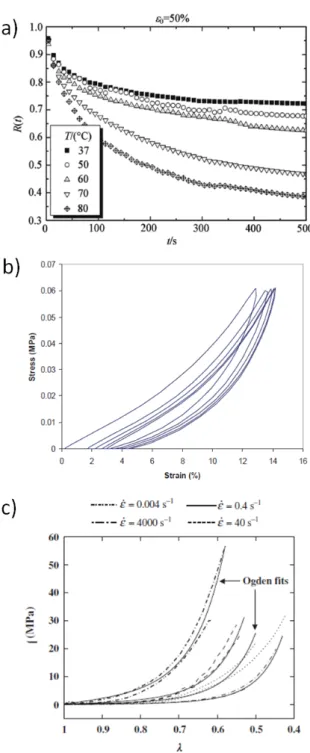
The main conclusion of these studies is that the average Young’s modulus is divided by approxi-mately 6 from the molecular level (≈ 6 GPa) to the fibrillar scale (≈ 0.9 GPa). The viscoelasticity on the contrary seems to be several orders of magnitude higher in fibrils (characteristic relaxation time from 10 to 100 s) than in collagen molecules (characteristic relaxation time ≈ 0.5 ns). This indicates that the viscous properties of the collagenous tissues cannot be attributed to collagen as a material alone, but also results from other mechanisms, such as sliding and slipping at different length scales [5] and from the properties of the surrounding matrix.
Isolated collagen fibrils have been investigated relatively extensively both experimentally and theo-retically. Collagen fibrils, as any slender structure, are anisotropic in the mechanical sense. While collagen fibrils have little strength in flexion or torsion, they exhibit a high tensile strength in their main direction, attributed to the intermolecular covalent cross-links. To test isolated collagen fi-brils mechanically, Atomic Force Microscopy (AFM) [20, 21], MicroElectroMechanical Systems (MEMS) [5, 19] and Optical Tweezers [22] are the most commonly used experimental devices. The results highlight the influence of hydration even at the fibrillar level, and show the intrinsic viscoelastic nature of collagen fibrils, illustrated in figure I.7 by the hysteresis that occurs during cyclic loading on a single fibril [21]. This viscoelasticity is however at a time scale an order of magnitude lower than the time scale of the whole tissue viscoelasticity.
Taking advantage of recent technical improvements in tissue engineering, some studies have been focusing on investigating the mechanical properties of specifically engineered collagen gels [23, 24]. These allow for customisation of the tissue morphological characteristics, and can help identify the microstructural parameters relevant to the macroscopic mechanical properties. They can however mimic the real organs only as well as we know the actual tissue’s microstructure.
Figure I.7 – Stress/strain curve for a cyclic loading (loading path shown in insert) on a single collagen fibril using AFM: the hysteresis reveals the viscoelasticity of collagen fibrils. Figure from [21].
collagenous sub-elements in a scaling-up process to get a comprehensive view of the mechanics of macroscopic structures. Adequate micromechanical experiments are necessary to comprehend the behaviour of the microstructure under mechanical stimulation.
I.1.2
Skin
Most studies of skin have been conducted on humans, and therefore all information from this chapter addresses the properties of human skin. It is generally accepted that murine skin is si-milar to human skin with only scale changes. The sisi-milarities between human and murine skin is evidenced for instance by the fact that it is possible to perform xenografts of human skin on mice, as illustrated in figure I.8. It is however possible that some differences exist between the two species, either microstructural, macroscopic or both.
Skin is the largest organ in the human body. It has an average surface of 1.5 - 2.0 m2 and weighs
3.5 - 4 kg in an average adult. It is a complex organ with multiple functions, reviewed in the first section below, and a variety of components, summarised in the second section. The third section focuses on the dermis, the most important layer for mechanical properties and the one we are most interested in.
I.1.2.a Properties and functions
The primary function of skin is one of protection and regulation. Skin plays an important role of structural support and protection from mechanical impacts and outer pressure. To this effect the mechanical strength of skin is crucial.
Figure I.8 – Histological section of skin after a xenograft (human skin transplanted onto immu-nodeficient mice) at junction site, coloured with hematoxylin and eosin (H&E) stain. H&E stain colours cell nuclei dark blue and proteins pink. Image from Fuhlbrigge Lab, Harvard.
factor of cancer development. When it has sustained a burn, the protective function of skin against UVs is impaired.
Skin is however not impermeable: it is an important exchange area. Through perspiration and muscle activation in goose bumps, skin contributes actively in the regulation of body tempera-ture. The precise control of body temperature is of the utmost importance: below 32˚C and above 41.6˚C body temperature a human being is considered in mortal danger. Perspiration also re-gulates body fluid balance. Some chemicals such as hormones can also be released through skin when needed.
Skin is our first barrier against micro-organisms and outer chemicals. In addition to a passive physical barrier, some elements of the immune systems, both innate and adaptative, are fully inte-grated in the skin. For example, Langherhans cells’ in the epidermis capture antigens and present them to the T-lymphocytes to engage the chain of immune reaction.
Finally, skin is our primary organ for the touch sense. An extensive network of nervous cells can detect and relay touch, heat, cold and pain. Alteration of this function is extremely imparing in everyday life and can lead to an increased risk of severe injuries.
I.1.2.b Global structure
Skin is a complex multilayered organ. It encompasses cells, fibres, complex molecules, sweat and sebaceous glands, hairs, nerves, blood vessels, etc. Figure I.9 presents a simplified representation of skin’s structure.
eye-Figure I.9 – Schematic representation of skin’s multilayered structure. Three layers are distin-guished, from top to bottom: epidermis, dermis and sub-cutaneous tissue. Image adapted from dermnetnz.org.
lids, to 1.5 mm on soles and palms. The epidermis is made of layers of cells, called keratinocytes, stacked onto each other. The epidermal cells can be seen coloured in dark blue in the histological section presented in figure I.10.
The cells originate from the junction between the epidermis and the dermis, called basal layer. This is where melanin is added to the keratinocytes by cells called melanocytes. No blood vessel goes through the epidermis. Some immune cells, such as the Langerhans’ cells, migrate through the epidermis to detect antigens.
The epidermal cells migrate from the basal membrane to the skin’s surface while filling with ke-ratin, a very resistant fibrous protein. The outermost layer of the epidermis is called the stratum corneum, and is entirely composed of dead cells glued together with lipidic cement. The cells from the stratum corneum detach progressively with friction and cleansing, leaving space for new cells. The mean turnover time of the epidermis, required to renew the cells completely, is roughly 39 days [25]. This time can be affected by various diseases, psoriasis for instance.
Under the epidermis lays the dermis. The epidermis-dermis junction presents finger-like waves, visible in figure I.10, to facilitate both adhesion and exchange of nutrients. The cohesion between the two layers is ensured by anchoring filaments of proteins: proteoglycans (called hemidesmo-somes) and type VII collagen fibrils.
The dermis is the thickest and most important layer of the skin for structure and mechanical resistance. A few cells, called fibroblasts, can be found in the dermis. Their role is to synthetise the components of the extracellular matrix which constitutes the greater part of the dermis. These components are collagen fibres of different types (I, III, V, VI, XII, XIV, XVI), elastin fibres and
Figure I.10 – Histological section of skin at the epidermis-dermis junction, coloured with H&E stain (transversal cut). H&E stain colours cell nuclei dark blue and proteins pink. E identifies the epidermis, D the dermis. Image adapted from mrcophth.com.
ground substance. The ground substance encompasses everything in the extracellular matrix that are not fibres, including, but not limited to, proteoglycans. The main component of the ground substance and of the dermis in general is water, which represents 64% of the whole skin. The exact microstructure of the dermis is detailed in the next section.
Sebaceous and sweat glands lay in the dermis. Small blood vessels run through the dermis and especially at the epidermis-dermis junction, to bring nutrients to the epidermal basal cells. Under the dermis lays the hypodermis, the deepest layer of the skin, also called “sub-cutaneous” tissue. It is composed of fat cells and is often interconnected with the fat layers underneath. The hair follicules initiate there, and blood vessels run through the hypodermis. The two main roles of the hypodermis are energy storage and shock absorption.
I.1.2.c Microstructure of the dermis
The dermis is a collagenous tissue in the general sense described earlier: it is a heterogenous structure made of a few cells and extracellular matrix. In our study, the cells of the dermis can generally be neglected, as their impact on the mechanical properties is considered negligible at the time scale we explore. It is however possible that dying cells, going through apoptosis, produce substances that can affect the extracellular network organisation.
The dermis is a composite structure in the mechanical sense of the term: a combination of collagen fibres, elastin fibrils and a host structure.
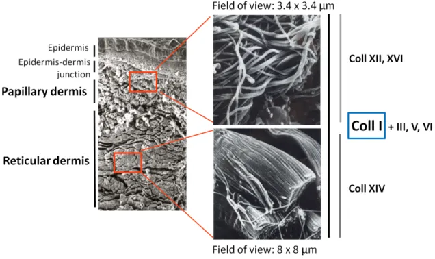
More than 70% of the dermis’ dry weight is attributable to collagen. The most abundant type is collagen I, that accounts of approximately 80% of total amount of collagen in the dermis. Collagen I forms long and large fibres, that are arranged in a tight network. The dermis is separated in two parts, based on morphology of the collagen network: the papillary dermis and the reticular dermis. Figure I.11 shows scanning electron microscope (SEM) images of the dermis and collagen types location, demonstrating the differences in structure and composition between papillary and reticular dermis.
The dermis layer that is closest to the epidermis is called papillary dermis, and owes that name to the finger-like structures of the epidermis-dermis junction, called papillae. The papillary dermis consists of fairly small fibres, curled and crimped, that are quite loosely arranged. It is commonly believed to be responsible for skin’s extensibility.
The deepest layer of the dermis is called reticular dermis. It consists of fibres that are arranged in thick bundles stacked densely onto one another. The fibres are straighter and thicker than in the papillary dermis. The reticular dermis is believed to be responsible for skin’s resilience. The papillary dermis is much thinner than the reticular dermis, with a ratio of approximately 0.2/0.8.
There are other types of collagen present in skin in lesser amounts. Collagen type III is the second most abundant collagen type in skin (approximately 20% of all collagen in adult skin). Other collagen types (type V, VI, XII, XIV, XVI) are much more scarce and represent only a few percent. Some of these collagen types (type III, type V) are fibrillar. The roles of these other collagens, not always well understood, are very diverse, from fibrillogenesis to adhesion. This is reflected in their uneven repartition in the two dermises, shown in figure I.11.
Figure I.11 – SEM images of the two dermises, papillary and reticular, and respective repartition of collagen types (type I, III, V, VI, XII, XIV, XVI). The papillary dermis consists of fairly small fibres, curled and crimped, loosely arranged, while the reticular dermis consists of thick bundles of fibres stacked densely onto one another. Image adapted from [26].
The host structure, which encompasses anything that is not collagen I fibre, is called in this work “extrafibrillar matrix”. The extrafibrillar matrix includes a variety of elements. One of its most important component are proteoglycans. These complex molecules make for only about 0.2% of
skin’s dry weight, but their primary function is of the utmost importance: they retain water, in amounts of up to 1000 times their own volume.
Proteoglycans are complex proteins that consists of a “core protein” with one or more covalently attached glycoaminoglycan chains (GAGs). Figure I.12 shows a schematic representation of a pro-teoglycan. GAGs consist of a long-chain, aminated polysaccharides. The most common GAGs in the dermis are hyaluronic acid and dermatan sulfate. Proteoglycans are categorised depending upon the nature of their GAGs chains. They also are usually categorised by size in atomic mass units, that can vary between 30 kDa (kiloDaltons) and 500 kDa.
Proteoglycans are visible when coloured with a specific stain, such as alcian blue. Using this technique, Scott and coworkers [27, 28] demonstrated that there is structure in the extrafibrillar matrix. Indeed, in most connective tissues, such as skin, the proteoglycans attach to collagen fibres at regular intervals, and extend perpendicularly from fibre to fibre. They are thus believed to be important structural components of the dermis, that contribute to force transmission.
Figure I.12 – Schematic representation of a proteoglycan: a core protein with covalently attached glycoaminoglycan chains (GAGs).
Another major component of the matrix is elastic fibres, the most important of which being elas-tin. Elastin is a protein that forms small fibres, much thinner than collagen fibres, as shown in figure I.13. Elastin is often presented as providing “elasticity” to skin [29]. In skin, elastin fibres are believed to be wrapped around collagenous fibres, but their exact role and mechanism of action remains unclear [30, 31].
The embryogenesis of the dermis is a complex process, during which even collagen types that are present in small amounts can play an important role. Pathologies of collagen fibrillogenesis have very serious effects on skin and other collagenous tissues, which remain generally poorly unders-tood. One of these pathologies, Ehlers-Danlos syndrome, is particularly relevant to our study and is described in Chapter IV.
The next section focuses on imaging methods enabling the investigation of the microstructural architecture of soft collagenous tissues.
Figure I.13 – Histological section of skin (transversal cut) coloured with van Gieson’s stain. Van Gieson’s stain colours collagen pink and elastic fibres violet. Image from the School of Anatomy and Human Biology, University of Western Australia.
I.1.3
Imaging the architecture of soft collagenous tissues
When describing a tissue, whether to characterise it (biologically or mechanically speaking) or to diagnose a pathology, the examination of inner structure is valuable. This section will briefly introduce some imaging techniques available to investigate the architecture of soft collagenous tissues. Some allow in vivo imaging, whereas other necessitate to take a cut of the tissue, some are destructive, some not.
We will primarily consider optical imaging techniques: they are particularly appropriate for dy-namic imaging of soft tissues during a mechanical test, for reasons discussed later. Nevertheless, other imaging techniques, briefly presented in the following section, can be used to unravel the microscopic architecture of soft collagenous tissues.
I.1.3.a Non-optical imaging
A prime example is electron microscopy, Transmission Electron Microscopy (TEM) or Scanning Electron Microscopy (SEM), mostly. Electron microscopy gives access to very small scales with a very good resolution, of about a nanometer or less, over a small field of view. These imaging techniques require a careful preparation of the sample in thin cuts, that need to be resistant both to vacuum and electronic beams. A SEM image of the dermis is shown in figure I.14 a. SEM has been applied to the investigation of fibre reorganisation during a mechanical tensile test on skin by Brown [32] and later by Belkoff and Haut [33]. However, as it is a destructive method, a different sample had to be observed at each deformation step.
Figure I.14 – Imaging skin with non-optical imaging: a) SEM image of rat skin dermis. Scale bar is 100 µm. Image from [33]. b) MRI image of in vivo human skin. H indicates the hypodermis, D the dermis and E the epidermis. Image from [34].
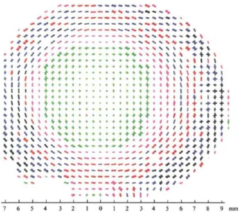
X-ray diffraction techniques, such as Small-Angle X-ray Scattering (SAXS), allow specific imaging of organised tissues, such as the tendon [35] or the cornea [36]. Figure I.15 shows an example of fibre bundle orientation map obtained from a human cornea with X-ray diffraction. The resolution of X-ray imaging ranges between 0.1 and 0.5 mm. This technique is intrinsically limited to a few types of tissue, and damage has been observed when imaging tissues for a long time.
Finally, other imaging techniques are commonly used in in vivo imaging for diagnosis purposes, such as ultrasounds (using sound waves) and Magnetic Resonance Imaging (MRI, using magnetic fields and radio waves). They are non invasive and fairly easy to use, although requiring specific equipment. The images, such as the one seen in figure I.14 b for human skin, cover a large spatial field of view, but have a low resolution (≈ 0.1 mm at best) and are sometimes difficult to interpret due to non-specific imaging.
I.1.3.b Optical imaging
Optical imaging techniques are valuable for imaging soft collagenous tissues with relatively large fields of view, good resolutions, light sample preparation and reasonably short imaging times. De-pending on the technique used, the lateral resolution can reach 0.2 to 0.5 µm. When 3D imaging is possible, the axial resolution ranges from 1 to 5 µm. These advantages makes optical imaging ideal for microscopic observations of tissues during a mechanical test.
Optical imaging techniques can be classified based on the sources of contrast they depend upon, relying on various types of interaction between light and matter.
One of the most common ways to examine the microstructure of a tissue is to do an histological section. A biopsy of the tissue is embedded in paraffin, and very thin slices of the tissue are cut (a few micrometers thick). The sections are then deparaffinised, coloured with a chosen stain spe-cific to the components sought and observed under an optical microscope (widefield imaging with
Figure I.15 – Map of fibre bundle orientation in human cornea obtained with X-ray diffraction. Image from [36].
white light). One of the most classical stain for skin is H&E, hematoxylin and eosin, in which cell nuclei are stained purple and proteins (such as collagen) are stained pink. Figure I.16 a shows an histological section of skin stained with H&E.
It is also possible to stain proteins, carbohydrates or lipids with specific antibodies: this technique is called immunohistochemistry. Antibodies can, theoretically, be obtained for any antigen: it is then possible to stain only a specific type of collagen for example.
Histological and immunohistochemical analyses allow for a precise and relatively immediate iden-tification of a sample’s components, as they are coloured with respects to their nature. Although time consuming, a 3D observation of the tissue is possible with successive sections. These analyses are however very invasive and inherently destructive. They only allow for a local characterisation of a whole organ. Furthermore, it remains mostly a qualitative analysis of the microstructure. With a similar principle, it is possible to use polarised light instead of or in combination with specific staining to observe microstructures in the sample. An example of polarised light imaging of tendon without stain was shown earlier in this chapter, see figure I.6.
Reflectance can also be used as a source of contrast in soft collagenous tissues. Examples of imaging techniques making use of reflected light are Optical Coherence Tomography (OCT) or reflectance confocal microscopy.
Optical Coherence Tomography is based on low-coherence interferometry [39], typically employing near-infrared light to enhance penetration in biological tissues, mostly made of water. Depending on the system used, the micrometer resolution can be obtained, and the images, such as the one seen in figure I.16 b, can scan large regions of the tissue.
The advantages of OCT imaging are numerous: no sample preparation, 3D imaging is possible and the imaging time is very short (up to a few images per second). This imaging technique requires
Figure I.16 – Optical imaging of soft collagenous tissues: a) Histological vertical section of skin stained with H&E. Scale bar is 1 mm. b) OCT image of in vivo skin. Image size is 1.5 × 0.6 mm2.
E identifies the epidermis, D the dermis and sc the stratum corneum. Image from [37]. c) Sagittal section of porcine skin stained with picrosirius red and observed with confocal microscopy. Scale bar is 300 µm. Image from [38]. d) Tendon observed in confocal microscopy, cell nuclei are stained using acridine orange. Scale bar is 50 µm. Image from [12].
however a tissue with optical interfaces (different refractive indices), and its main drawback is the complete lack of imaging specificity.
Finally, fluorescence has been used more and more extensively in the past decades as a source of contrast to image soft collagenous tissues. Most of the time, imaging techniques based on fluo-rescence require the introduction of exogenous fluorophores to induce contrast, but they can also rely on endogenous signals. The tissues are then imaged either with widefield microscopy, confocal microscopy or multiphoton microscopy.
Figure I.16 c shows an example of porcine skin imaged with confocal microscopy. This imaging technique was also used for micromechanical experiments by Screen and colleagues: they moni-tored the displacements of stained cell nuclei in tendons submitted to uniaxial tensile tests, to compare local and global strains [12]. A typical image with a group of nuclei identified is shown in figure I.16 d. Their results will be further discussed in relation with our results in Chapter III. For these techniques, no sample cutting is required, which means that delicate or deep tissues can be examined, sometimes even in vivo. The resolution is good (usually sub-micrometric), with a relatively large field of view (up to 1 mm). These techniques are however limited by nature by the small penetration of light in tissues: except for the cornea (which is transparent), the scattering of light greatly limits the penetration. Furthermore, especially when using exogenous signals, the
outcoming signal decreases with exposure time, which limits the possibility of observing a long-time effect such as microstructural reorganisation in tissues. This effect is called photobleaching. Notably, photobleaching can also be used to create patterns in the depth of the tissue, which can be monitored during mechanical loading to compute local stretches [40, 41].
In skin, there is a strong endogenous fluorescence signal from cells, epithelial cells for example, and from keratin in the hair. Melanin absorbs but does not re-emit light.
Confocal microscopy remains the most commonly used optical imaging method to image biological tissues. It allows for 3D imaging, with light sample preparation, a large field of view and good resolution. Collagen fibres in particular can be observed using their intrinsic fluorescence, which is however extremely weak, non specific and difficult to distinguish from fluorescence arising from other components.
In the following section we present fairly recent optical imaging techniques: non-linear micro-scopy, and in particular 2-photon excitation fluorescence and Second Harmonic Generation. These techniques are more difficult to implement than confocal microscopy, but can solve two major drawbacks of this technique: the small tissue penetration (maximum 30 µm) and the lack of specifity in the absence of exogenous signal (which require sample preparation).
I.1.3.c Non-linear microscopy: 2PEF and SHG imaging
Multiphoton microscopy regroups imaging techniques using non-linear optical effects as a source of contrast in biological samples. Multiphoton microscopy techniques, such as 2-photon excitation fluorescence (2PEF), Second Harmonic Generation (SHG), Third Harmonic Generation (THG) or non-linear Raman imaging, rely on the recombination of two or more photons into one photon by the tissue, hence the term “non-linear”.
Figure I.17 a shows simplified electronic states diagrams (Jablonski diagrams) for conventional (linear) fluorescence (i) and non-linear imaging: 2-photon excitation fluorescence and Second Har-monic Generation (ii). In the case of 2-photon excitation fluorescence the principle is the following: a molecule absorbs two photons and access an excitated state, then reemits one photon while going back to its original energy level.
The probability of occurance of non-linear processes is greater in areas with more photons, that is to say the focal volume (the probability is in N2 for 2PEF or SHG). In linear imaging techniques, the probability of emission is proportional to the number of photons. This spatial specificity in photon emission is essential in non-linear microscopy: a non-linear excitation will lead to an out-come signal intrinsically localised. This property is illustrated in figure I.17 b. When a fluorescein solution is excited with an excitation wavelength corresponding to linear fluorescence, the signal is extended to an excitation cone (i), whereas with a non-linear effect the signal is intrinsically localised to a point in the focal plane (ii). To image the whole sample in 3D, one just needs to scan the sample by moving the focal point. A thick sample can be imaged by optical sectioning,
i.e. by changing the focal plane.
Overall, multiphoton microscopy and confocal microscopy have comparable resolutions. However, the unique property of intrinsic signal localisation in multiphoton microscopy optimises the signal-to-noise ratio, and enhances the tissue depth available to imaging in highly scattering tissues, such as soft collagenous tissues.
Figure I.17 – Non-linear imaging: a) simplified Jablonski diagrams for i) 1-photon and ii) 2-photon excitation signals (2PEF and SHG): two photons are recombined into a single photon, b) spatial resolution for i) linear and ii) non-linear fluorescence of a fluorescein solution. Non-linear fluo-rescence is intrinsically localised, whereas in linear fluofluo-rescence the excitation is generalised to a cone. Images adapted from [7].
Non-linear imaging techniques can be combined, together or with other imaging techniques, as long as one can distinguish the different signals from their wavelengths.
Multiphoton microscopy has a lot of applications in neurosciences and developmental biology. They have indeed the intrinsic advantages of small invasiveness, deep penetration and ability to rely on endogenous signals. They require however a large laser power to exploit the rare non-linear effects in tissues. This can be obtained with pulsed lasers (femtosecond lasers), that allow for a
large peak power with a small mean power, and do not deteriorate the tissue.
Second Harmonic Generation (SHG) imaging is a non-linear imaging technique: the signal results from the combination of two photons of the same wavelength into a photon of halved wavelength. SHG is a coherent effect, meaning that the phase of the emitted photon is strictly related to the phase of the excitation photons. This means that the signal is amplified by constructive inter-ferences if the local environment is organised. In a disorganised, random structure, destructive interferences annihilate the signal. This particularity is summarised schematically in figure I.18 a. Thus, for the same basic SHG-emitting material, the signal can only be seen in organised enough structures, such as fibres. This specificity is illustrated by examples of materials that can be ob-served using SHG, e.g. fibrillar collagens, cellulose fibres, some crystalline polymers.
In all proteins, the SHG signal comes from peptidic chains in amino acids. All collagen molecules that present a triple helical domain emit a small SHG signal. However, only fibrillar collagens emit significant SHG signals, i.e. detectable with a microscope. In figure I.18 b, we can see a combined SHG/2PEF image of a kidney with fibrosis. SHG signal, in green, emerges from type I collagen, which is a fibrillar collagen. No SHG signal can be observed in the glomerulus, where immunochemical labelling reveal however the presence of non fibrillar collagen IV.
Further details on the non-linear optical microscopy techniques used in this study can be found for example in [7].
Figure I.18 – Conditions on microstructural organisation for SHG observation: a) generation of SHG signal by parallel molecules leads to constructive interferences, while anti-parallel molecules produce destructive interferences, b) SHG/2PEF image of a human kidney with fibrosis. SHG (green) reveals fibrillar collagen (type I). No SHG signal can be seen in the glomerulus, where non fibrillar collagen IV is known to be present. Images from [7].
I.2
Macroscopic scale: skin’s mechanical properties
Skin’s mechanical properties are of great importance in many clinical and cosmetical applications. They have hence been studied experimentally both ex vivo and in vivo for a long time [42]. This section first reviews the techniques allowing for skin’s mechanical characterisation, ex vivo and in vivo. We then detail the complex mechanical properties of skin.
I.2.1
Mechanical characterisation
I.2.1.a Ex vivo characterisation
Ex vivo tests have the benefit of allowing for the exploration of a wide range of parameters. Skin can be isolated from fat, muscles and bone to characterise only the properties of skin, and not of an ensemble of tissues. While with in vivo analysis, only the top layers of the skin are accessible,
ex vivo analysis allow the whole depth of the tissue to be tested. Layers of skin can also be isola-ted to test independently. The failure mechanisms can be evaluaisola-ted through destructive tests not available in vivo.
Finally, another key advantage of ex vivo testing is that samples can be modified using chemicals to evaluate separately the contribution of each component, for instance collagenase breaks the peptide bonds in collagen. This method has been used to vary the proportions of collagen [43], elastin [44] and proteoglycans [45].
The ex vivo results can also serve as database to compare real tissues and their tissue-engineered imitations.
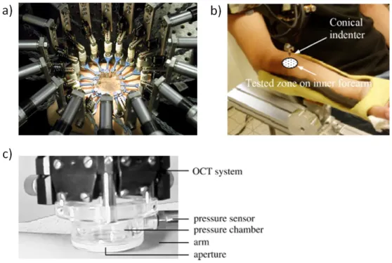
The tensile properties of skin have been extensively studied. Uniaxial, biaxial and multiaxial tests have been performed. For reviews, see [30, 46, 47]. Figure I.19 shows some examples of set-ups for these tests.
Figure I.19 – Tensile tests on ex vivo skin: a) uniaxial test (1970, image from [48]), b) biaxial test (1974, image from [49]), c) multiaxial test with 12 automated motor axes and camera mounted above the testing region for Digital Image Correlation (2011, image from [38]).
Inflation tests can also be used to test for skin tensile multiaxial properties [50]. Compression, torsion and shear tests have also been carried out. Finally, some studies test the epidermis-dermis cohesion with delamination tests. Examples of set-ups for these methods are shown in figure I.20. The great variety of experimental protocols should however encourage us to be careful when comparing two studies or matching model to experiment. Indeed, many variables can influence the results, such as:
– Animal tested: e.g. human [53], rat [43, 45], dog [54], pig [55, 56], cat [48]. – Age of the animal: e.g. neonatal, adult, aged.
– Site and orientation: e.g. rib, rump, forehead, abdomen, back.
– Sample preparation: e.g. tested right after excision, frozen, with layers and/or hairs removed. – Hydration method: e.g. immerged, sprayed, covered in gel, no hydration.
Figure I.20 – Examples of other possible mechanical set-ups on ex vivo skin: a) Set-up for pure shear test. Image from [51]. b) Delamination test between epidermis and dermis. Image from [52].
– Loading path: e.g. preconditioning or not, strain rate, cycling, maximum stretch ratio.
One of the main differences between mechanical test protocols is the method used for displacement measurements. In recent years, contact extensometers have been supplemented by non-contact me-thods, such as Digital Image Correlation (DIC), which will be used in the present work.
Digital Image Correlation is a post-processing technique now frequently used in mechanics. The principle is to identify in a deformed image the displacements of a pattern from the reference image. The matching of the two images makes use of a similarity criterium, called correlation coefficient, which relies on the hypothesis of conservation of local contrast in the immediate sur-roundings of the point sought.
This technique gives a point-by-point displacement field at the local scale, that can be compared with the imposed displacement to correct for slippage or unfolding of the tissue. Digital Image Correlation is generally used in 2D only, but 3D DIC is also available. Details on DIC and its applications to biological materials can be found in [57, 58, 59].
However, ex vivo tests have two main drawbacks. As mentioned before, they isolate the tissue from its natural environment. This eases the identification of skin properties, but removes the natural prestress and hydration control. Furthermore, the preparation of the tissue is an inevitable source of damage: from extraction to potential preparation (such as layer separation), the tissue can become deteriorated. This inevitably results in mechanical characterics different from those measured with in vivo analysis.
I.2.1.b In vivo characterisation
In vivo tests evaluate skin’s properties in natural prestress and hydration state. Experiments have mostly been implemented on humans. Obviously, only low loads can be applied with in
vivo analysis. Testing can either be carried out in plane or perpendicular to the surface. Four main categories of in vivo tests can be identified: torsion, tension, suction and indentation. For reviews, see [60, 61]. Some set-ups for in vivo assessment of skin mechanics are shown in figure I.21.
Figure I.21 – In vivo tests of skin biomechanical properties: a) Multiaxial tensile test on a sub-ject’s arm. Image from [62]. b) Indentation test. Image from [63]. c) Suction device set-up combi-ning suction experiments and OCT imaging. Image from [64].
To test the skin in its surface plane, torsion tests and tensile tests can be carried out. These tests rely mostly on the properties of epidermis and upper dermis [61]. The torsion test consists in the application of a disc to the skin’s surface, which is rotated with a prescribed torque. Tensile tests consist in attaching two (for uniaxial tests) or more (for multiaxial tests) tabs to the skin and imposing a displacement.
Suction and indentation measurements evaluate skin’s biomechanical properties perpendicularly to the surface. In suction tests a vacuum is applied to the skin’s surface, which deforms due to the decrease in pressure. The deformation of the tissue can be related to its mechanical properties. Indentation is a commonly used technique in materials science: a rigid indenter is pressed to the skin’s surface. The measured loads and displacements can be linked to the material properties. In these techniques, the lower layers of the skin contribute to the mechanical properties measured as well as the upper layers [61].