Université de Montréal
The Role of Protein Convertases in Bigdynorphin and
Dynorphin A Metabolic Pathway
par
ALBERTO RUIZ ORDUNA
Département de biomédecine vétérinaire Faculté de médecine vétérinaire
Mémoire présenté à la Faculté de médecine vétérinaire en vue de l’obtention du grade de maître ès sciences (M.Sc.)
en sciences vétérinaires option pharmacologie
Décembre, 2015
i
Résumé
Les dynorphines sont des neuropeptides importants avec un rôle central dans la nociception et l’atténuation de la douleur. De nombreux mécanismes régulent les concentrations de dynorphine endogènes, y compris la protéolyse. Les Proprotéines convertases (PC) sont largement exprimées dans le système nerveux central et clivent spécifiquement le C-terminale de couple acides aminés basiques, ou un résidu basique unique. Le contrôle protéolytique des concentrations endogènes de Big Dynorphine (BDyn) et dynorphine A (Dyn A) a un effet important sur la perception de la douleur et le rôle de PC reste à être déterminée. L'objectif de cette étude était de décrypter le rôle de PC1 et PC2 dans le contrôle protéolytique de BDyn et Dyn A avec l'aide de fractions cellulaires de la moelle épinière de type sauvage (WT), PC1 -/+ et PC2 -/+ de souris et par la spectrométrie de masse. Nos résultats démontrent clairement que PC1 et PC2 sont impliquées dans la protéolyse de BDyn et Dyn A avec un rôle plus significatif pour PC1. Le traitement en C-terminal de BDyn génère des fragments peptidiques spécifiques incluant dynorphine 1-19, dynorphine 1-13, dynorphine 1-11 et dynorphine 1-7 et Dyn A génère les fragments dynorphine 1-13, dynorphine 1-11 et dynorphine 1-7. Ils sont tous des fragments de peptides associés à PC1 ou PC2. En plus, la protéolyse de BDyn conduit à la formation de Dyn A et Leu-Enk, deux peptides opioïdes importants. La vitesse de formation des deux est réduite de manière significative dans les fractions cellulaires de la moelle épinière de souris mutantes. En conséquence, l'inhibition même partielle de PC1 ou PC2 peut altérer le système opioïde endogène.
Mots-clés: Dynorphines, Dynorphine A, Proprotéines convertases, Protéolyse, Peptides
ii
Abstract
Dynorphins are important neuropeptides with a central role in nociception and pain alleviation. Many mechanisms regulate endogenous dynorphin concentrations, including proteolysis. Proprotein convertases (PCs) are widely expressed in the central nervous system and specifically cleave at C-terminal of either a pair of basic amino acids, or a single basic residue. The proteolysis control of endogenous Big Dynorphin (BDyn) and Dynorphin A (Dyn A) levels has a profound impact on pain perception and the role of PCs remain unclear. The objective of this study was to decipher the role of PC1 and PC2 in the proteolysis control of BDyn and Dyn A levels using cellular fractions of spinal cords from wild type (WT), PC1 -/+ and PC2-/+ animals and mass spectrometry. Our results clearly demonstrate that both PC1 and PC2 are involved in the proteolysis regulation of BDyn and Dyn A with a more important role for PC1. C-terminal processing of BDyn generates specific peptide fragments Dynorphin 1-19, Dynorphin 1-13, Dynorphin 1-11 and Dynorphin 1-7 and C-terminal processing of Dyn A generates Dynorphin 1-13, Dynorphin 1-11 and Dynorphin 1-7, all these peptide fragments are associated with PC1 or PC2 processing. Moreover, proteolysis of BDyn leads to the formation of Dyn A and Leu-Enk, two important opioid peptides. The rate of formation of both is significantly reduced in cellular fractions of spinal cord mutant mice. As a consequence, even partial inhibition of PC1 or PC2 may impair the endogenous opioid system.
Keywords: Dynorphins, Dynorphin A, Proprotein convertases, Proteolysis, Opioid
iii
Table of contents
Résumé………...i Abstract………...………ii Table of contents………...……….iii Tables list………...………vi Figures list………...…..vii Abbreviations list………...……….x Acknowledgements……….……….…xiii Introduction……….……….1 LITERATURE REVIEW…...……….………4Chapter I -Endogenous mechanisms of pain………...………5
I.1-Pain generalities………...……….5 I.2-Pain pathologies………...……….5 I.2.1-Neuropathic pain……….……….…….5 I.2.2-Nociceptive pain……….………...……6 I.2.3-Inflamatory pain……….…...…..6 I.3-Pain physiology....………...….…..7 I.3.1-Nociceptive process….………...….…….………...…..7
I.3.1.1-Primary sensory neurons…...………...7
I.3.1.2-Ion channels………..….…8
I.3.1.2.1-Sodium channels……….….…..9
I.3.1.2.2-Calcium channels……….………….….9
I.3.1.2.3-Potassium channels……….…….……..9
I.3.1.2.4-Chloride channels……….………...…..10
I.3.1.3-Pain receptors, nociceptors………..………10
I.3.1.3.1-Heat sensitive receptors….………....……….…..11
iv
I.3.1.3.3-Mechano-transducers……….………..12
I.3.1.3.4-Cold sensitive receptors………….……….………....…….12
I.3.2-Ascending pathway……… ….13
I.3.2.1-First synapse……….………14
I.3.2.1.1-Principal synaptic neurotransmitters and receptors……….15
I.3.3-Descending pathway
…
………...16I.3.3.1-Opioid receptors and endogenous opioid peptides…….……….16
I.3.3.1.1-Opioid receptors….…...………….……….….17 I.3.3.1.2-Endorphins….………...…….…..18 I.3.3.1.3-Enkephalins….……….………...………...……..19 I.3.3.1.4-Endomorphins….……….19 I.3.3.1.5-Dynorphins…….……….…….19 Chapter II-Dynorphins…....….………..21 II.1-Dynorphins generalities….………..……….………....………..21 II.2- Prodynorphin….………….……….………..21
II.3-Endogenous dynorphin peptides………..…….………....…….23
II.3.1-Bigdynorphin……….………24
II.3.2-Dynorphin A………...……….………..25
II.3.2.1-Physiological role of Dynorphin A ….………..……….25
II.3.3-Dynorphin B……….…...………..26
II.4-Protein convertases……….………...…..………..27
II.4.1-Tissue and cellular expression of PCs………...…………28
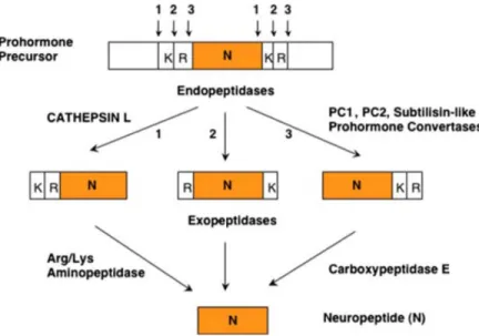
II.4.2-Proneuropeptides processing……….………29
II.4.3-Prodynorphin processing by PC1 and PC2.……….…..31
II.4.3.1-PC1(-/-) and PC2(-/-) phenotypes……..…………..………….……….33
Chapter III-Mass spectrometry...……….……….35
v III.1.1-Sample separation……….………36 III.1.1.1-HPLC system………36 III.1.2-Sample ionization………...………..38 III.1.2.1-Electrospray ionization
…
………...………..38 III.1.3-Mass analyzer……….………..40 III.1.3.1-Quadrupole……….……….………..41 III.1.3.2-Time of flight………..……….….………42 III.1.3.3-Ion trap……….………….…………43 III.1.3.4-Orbitrap……….……….………….………..44 III.1.4-Ion detector………....…….….……….45III.1.5-Nomenclature for peptide fragmentation….……….….……….…..45
III.1.6-MS data acquisition modes……….……….…….47
III.1.6.1-Full scan MS mode……….……….……….…….47
III.1.6.2-SRM and MRM modes……….……....…………47
III.1.7-Absolute quantification by isotope dilution….…….………...….………48
HYPOTHESIS AND OBJECTIVES....……….…………...………..50
ARTICLE………...….….………....……….……52
GENERAL DISCUSSION……….…..……….…………...….……82
1-Summary of obtained results….……….………84
2-Relevance of the observations….……….……….………..…..85
3-Implication of the results………….………….….……….………...87
4-Limitations……….……….……….……..……...….88
CONCLUSIONS……….……….……….……….…………....…89
vi
Tables list
Table 1: Most representative opioid peptide derived from the processing of their respective
precursors. Affinity observed for opioid receptors………...……… .17
Table 2: Prodynorphin-derived peptides amino acid sequences………23 Table 3: PCs tissue distribution and subcellular location……….………29
vii
Figures list
Figure 1: Primary afferent axons. Peripheral nerves include large-diameter (Aβ),
medium-diameter (Aδ) and small-medium-diameter unmyelinated (C) afferent fibers……….…..8
Figure 2: Thermal, mechanical and chemical noxious stimuli are perceived by nociceptors
located at the terminal of afferent neurons. The activation of nociceptors leads to the generation of action potentials which rely noxious perceptions to the brain…….…11
Figure 3: Schematic of the pain pathway. (1) The PNS is responsible for noxious stimulus
perception and the transmission of noxious information to the spinal cord though primary afferent neurons. (2) Signaling between afferent and secondary neurons take place at the first synapse. (3) These secondary order neurons transmit the information to the thalamus which engages the somatosensory cortex, providing information about the location and intensity of the painful stimulus. Other projections engage the thalamus with cingulate cortex and amygdala, contributing to the emotive component of the pain experience. (4) Noxious stimulus inputs activate the endogenous modulating system, stimulating the synthesis of endogenous opioids and their release into the first synapse, resulting in a modulation of the synaptic activity………..…..……...13
Figure 4: Opioid-derived peptides are generated from the endoproteolytic processing of
proopioid precursors POMC, PDyn and PEnk. With the exception of dynorphins, the enzymatic formation of opioid peptides is regulated by cleavage at dibasic amino acid positions………18
Figure 5: Mouse, rat and human Prodynorphin proteomic alignment. High homology is
observed in multiple regions between species. The region encoding Bdyn is completely homologue for the three species. This homology shows high conservation of this gene through evolution………...……….22
Figure 6: Involvement of Dyn A in pain modulation. (1) Release of Dyn A enhances the
viii
inhibits the release of excitatory neurotransmitters and (3) decreases the excitability of postsynaptic receptors in addition to (4) regulate the release of Dyn A...…..26
Figure 7: Pathways for proneuropeptides processing………30 Figure 8: Schematic representation of PDyn precursor shows possible paired and single basic
cleavage sites (KR and R). Various processing intermediates, such as 10-, 8- and 4-(BDyn) kDa peptides were identified, as well as some final opioid peptide products such as Dyn A, Dyn B and Dyn 1-8. The shaded portions represent Leu-Enk………....32
Figure 9: Potential BDyn and Dyn A-derived peptides from PC1 and PC2 cleavage….…33 Figure 10: Schematic representation of the different sections of a MS system…………...35 Figure 11: (A) HPLC operating mode. The sample is injected into the system, the mobile
phase is composed by a mixture of organic and aqueous solvents in a specific proportion and carry the sample into the column. The type of interaction between the molecule with the stationary and mobile phases makes each analyte to leave the column at different retention times (chromacademy.com). (B) Principle of RPLC with
gradient elution………..…37
Figure 12: Schematic of a typical electrospray source……….39 Figure 13: (A) HPLC eluent containing the sample analysis is sprayed into small droplets
and converted into gas phase ions. The ions carried by the electric field and the high vacuum are then introduced through a transfer capillary into the MS. (B) The potential applied into the needle tip makes that the droplets of the same polarity are repelled from the needle towards the cone generating the so-called Taylor cone. As the droplets traverse the space between the needle tip and the cone, solvent evaporates until it reaches the point that the surface tension can no longer sustain the charge (the Rayleigh limit) at which point a "Coulombic explosion" occurs and the droplet is dissociated producing charged analyte molecules………...…..39
ix
Figure 15: Schematic of a TOF mass analyzer………..…42 Figure 16: Schematic representation of an ion trap mass analyzer. Ions of a specified m/z are
trap in the analyzer. Non-selected ions are ejected from the trap………43
Figure 17: Orbitrap operating basis. Ions are injected into the orbitrap, where, as a
consequence of the high voltage applied on the central electrode, ions oscillate around the electrode driven by the strong electric field inside the orbitrap. Ions oscillation frequency is then measured and converted into a useful signal………..45
Figure 18: Peptide fragmentation nomenclature………...46 Figure 19: (A) Full scan acquisition mode. All ions generated at the ions source are injected
into the detector. The total ion chromatogram (TIC) shows all the ions detected on the basis of their retention time. (B) SRM and MRM mode. Specific m/z ions are selected and fragmented. Extract ion chromatogram (XIC) is extracted by monitoring a specific transition. A specific ion (precursor ion) is selected and fragmented. Then, one (SRM) or more (MRM) of its fragment ions generated, are isolated and injected into the detector. This operating mode also allows to obtain structural information from the MS2 spectra by studying the fragmentation pattern………....48
Figure 20: Leu-Enk MS2 spectra show how the peaks corresponding to the fragment ions from the unlabeled standard and the labeled peptide can be differentiated owing to the differences between their masses. XICs are extracted for each peptide by monitoring the same specific MRM transition for both peptides………..49
x
Abbreviations list
(-/+) Knockdown (-/-) Knockout
A Aspartic acid
ACTH Adrenocorticotropic hormone
AMPA α-amino-3-hydroxy-5-methyl-4-isoxazolepropionic acid APCI Atmospheric pressure chemical ionization
ASIC Acid-sensing ion channel ATP Adenosine triphosphate BDyn Big dynorphin
C Cysteine
Cav calcium voltage-gated channels
cDNA Complementary deoxyribonucleic acid CGRP Calcitonin gene related peptide
CNS Central nervous system D Aspartic acid
DEG/ENaC Degenerin/epithelial Na+ channel
DOR δ-opioid receptor DRG Dorsal root ganglion
Dyn Dynorphin E Glutamic acid EM Endomorphin End Endorphin
ER Endoplasmic reticulum ESI Electrospray ionization
ESI-MS Electrospray ionization coupled mass spectrometer F Phenylalanine
g Gravitational constant G Glycine
GABA γ-Aminobutyric acid H Histidine
xi
HPLC High performance liquid chromatography HRAM High resolution accurate mass
I Histidine K Lysine
KCNK K+ channel subfamily K
KOR κ-opioid receptor L Leucine
Leu-Enk Leu-Enkephalin M Metionine
m/z Mass to charge ratio Met-Enk Met-Enkephalin
min Minute
MOR µ-opioid receptor
MRM Multiple reaction monitoring MS Mass spectrometer
MSH Melanocyte-stimulating hormone N Asparagine
Nav Voltage-gated sodium channels NMDA N-methyl-D-aspartate
NK1/2 Neurokinin-1/2 receptor NKA Neurokinin A
NKB Neurokinin B
NPLC Normal-phase liquid chromatography
p Significance level (in statistics) P Proline
PAG Periaqueductal PC Proprotein convertase PDyn Prodynorphin
PEnk Proenkephalin
pH power of hydrogen (acidity measurement) PNS Peripheral nervous system
xii
Q Glutamine
Q-orbitrap Quadrupole coupled orbitrap mass spectrometer QqQ Triple quadrupole mass spectrometer
Q-TOF Quadrupole coupled TOF mass spectrometer R Arginine
RPLC Reverse phase liquid chromatography RVM Rostral ventromedial medulla
S Serine SP Substance P
SRM Selected reaction monitoring T Threonine
TGN trans-Golgi network
TIC Total ion current chromatogram TOF Time of flight mass analyzer
TRPA Transient receptor potential ankyrin TRPM Transient receptor potential melastatin
TRPV Transient receptor potential vanilloid V Valine
W Tryptophan WT Wild type
XIC Extract ion chromatogram Y Tyrosine
xiii
Acknowledgements
Foremost, I would like to express my gratitude to my supervisor Dr. Francis Beaudry, for trusting me for this master’s project, guiding and helping me in all the time of research and writing of this thesis. His encouragement, patience and understanding are the reasons of my success.
To my committee members, Dr. Christopher Price and Dr. Younes Chorfi for their assistance and suggestions throughout my project.
To my lab mates for those good moments we had during these two years, I have really enjoyed all this time we have been working together.
Finally, my most special gratitude to my family for their unceasing support on my decision of moving far from home being always behind encouraging me, without them this thesis would have not been possible.
Introduction
Patients suffering from chronic or neuropathic pain have a highly compromised quality of life and account for approximately 20-25% of the population worldwide. Given the importance of managing pain in medicine, the complex mechanisms that encompass pain perception, transduction and modulations are currently subject of intense research.
Pain is usually caused by a noxious stimulation of the peripheral nervous system (PNS). The PNS is responsible for perceiving those stimuli and to project that information through afferent neurons to the spinal cord. The synaptic transmission takes place at the external laminae of the dorsal root between afferent neurons and secondary order neurons. This process is called first synapse and regulates the intracellular signaling between the PNS and the central nervous system (CNS), relaying nociceptive information to the brain where it is perceived as pain. The communication between neurons is mediated by the release of excitatory neurotransmitters from the terminal of the afferent neurons. The neurotransmitters, like glutamate and substance P (SP), activate secondary neurons by their interaction with post-synaptic receptors such as N-methyl-D-aspartate (NMDA) and neurokinin-1 (NK1) receptors. When a noxious stimulus is perceived, the release of those excitatory neurotransmitters is enhanced. As a response to that perception, the brain is able to modulate the activity at first synapses through different modulatory pathways. The release of endogenous opioid peptides into the first synapse is the principal endogenous mechanism for the alleviation of pain.
Four families of endogenous opioid peptides have been described to date. They include endorphins, endomorphins, enkephalins and dynorphins. These peptides are synthetized as large and inactive proneuropeptides and requires endoproteolytic processing to generate the bioactive peptides, which play an essential role in the endogenous modulation of pain. Several studies have shown that protein convertases (PCs), specifically PC1 and PC2, are involved into C-terminal endoproteolytic processing of proneuropeptides through their cleavage at basic residues of proteins and peptides. In neuronal cells, proneuropeptides and PCs are synthetized and packed into dense-core vesicles. During the axonal transport of these vesicles, proneuropeptides are processed by PCs prior their release by exocytosis at the first
synapse. As a consequence of a noxious stimulation, the production and the release of vesicles containing endogenous opioid peptides are significantly enhanced.
Dynorphins have been identified as an important family of endogenous opioid peptides with potent analgesic effects. Prodynorphin is the proneuropeptide precursor of dynorphins. Early studies have partially described the endoproteolytic processing of prodynorphin, including a fundamental role for PC1 and PC2. Those preliminary studies established that the action of PC1 and PC2 is needed for the formation of different high molecular weight dynorphin-peptides, Bigdynorphin (BDyn), Dynorphin A (Dyn A) and Dynorphin B (Dyn B). However, the different contribution of each endoprotease, including PC1 and PC2, in the regulation of endogenous BDyn and Dyn A levels still remains unclear.
The presence of paired and single basic residues on the primary sequence of BDyn and Dyn A suggest further C-terminal processing catalyzed by PC1 and PC2 leading to the formation of several important N-terminal metabolites. Further processing of BDyn and Dyn A can lead to the formation of bioactive peptides including Dynorphin 1-19 (Dyn 1-19), Dynorphin A (Dyn A), Dynorphin 1-13 (Dyn 1-3), Dynorphin 1-11 (Dyn 1-11), Dynorphin 1-10 (Dyn 1-10), Dynorphin 1-7 (Dyn 1-7) and Dynorphin 1-6 (Dyn 1-6). Interestingly all these prodynorphin-derived peptides encode a copy of Leu-Enkephalin (Leu-Enk), another important opioid peptide, at their N-terminal. Thus, Leu-Enk might be an important metabolic product of BDyn and Dyn A. The objective of this project is to study the metabolism of BDyn and Dyn A, identify and quantify the rate of formation of the metabolites, as well as clarify the role of PC1 and PC2 in regulation of the concentration of both neuropeptides.
This study was designed to develop an in vitro experimental procedure to show the enzymatic degradation of BDyn and Dyn A and to elucidate the roles of PC1 and PC2 in the proteolytic control of endogenous dynorphins levels. Mice lumbar spinal cord S9 fractions were isolated from 3 different mice genotype, wild type (WT), PC1-knockdown (PC1-/+) and PC2-knockdown (PC2-/+), and the cellular homogenates containing among other enzymes PC1 and PC2, were used as a source of endogenous enzymes for the in vitro digestion. High performance liquid chromatography (HPLC) separation coupled with an electrospray
ionization mass spectrometer (ESI-MS) was used for the identification and quantification of BDyn and Dyn A metabolites. Moreover, an isotopic dilution method was employed for peptide quantification.
The study was designed to provide a better understanding of the mechanisms involved in the endogenous control of peptide levels and their impact on pain modulation pathways. Since opioid drugs are widely used in pain treatment with serious side-effects, a better mechanistic understanding of endogenous opioid metabolic pathways could lead to the development of innovative strategies in the treatment of pain.
CHAPTER I-ENDOGENOUS MECHANISMS OF PAIN
I.1-Pain generalitiesPain has been defined by the International Association for the Study of Pain as an unpleasant sensory and emotional experience associated with actual or potential tissue damage, or described in terms of such damage. This experience is essential for human survival since it protects body tissues from damage and alerts of stimulation in any part of the body by activating the surveillance mechanism and evoking the so-called muscular reflex arc.
Pain can be caused by diverse methods including physical injuries, infections or other forms of noxious stimulation. Understanding the nature of pain as well as its complex molecular mechanisms associated with pain perception and transduction is a challenge and many of these mechanisms are still not well understood.
Depending on its nature, pain can be subdivided into neuropathic, nociceptive and inflammatory pain. They are each generated for different reasons and need to be treated accordingly.
I.2-Pain pathologies I.2.1-Neuropathic pain
The central nervous system (CNS) is responsible for pain perception. Any alteration on the CNS can lead to an inadequate perception making pain persist long after the initiating cause has ceased (Gold and Gebhart, 2010). Neuropathic pain is provoked from disorders, damage or injuries on peripheral nerves, dorsal root ganglion (DRG) or the CNS. Patients suffering neuropathic pain exhibit persistent pain independent of a stimulus (Devor, 2006). Depending its influence in the sympathetic nervous system, pain perception ranges from a persistent burning sensation evoked for any alteration on C-sensory neurons, to acute pain perception resulting by large myelinated A fibers. Alterations on the CNS also evoke two
characteristic pain sensitizations; hyperalgesia and allodynia (Ossipov and Porreca, 2005). Hyperalgesia is characterized as an enhanced and more prolonged pain response to noxious stimuli as a result of abnormal processing of nociceptor input. This is a pain hypersensitivity that usually accompanies inflammation (Schaible, 2006; Woolf and Salter, 2000). On the other hand, allodynia is a state of pain characterized by a pain sensation caused by a normally innocuous stimulus. It can be evoked by the action of low threshold myelinated Aβ fibers on an altered CNS and by the reduction of the activation threshold of nociceptors present in the PNS (Woolf and Mannion, 1999).
I.2.2-Nociceptive pain
Nociceptive pain, also described as physiological pain, is caused by injuries like cuts, burns and other excessive stimulation of nociceptors. Its purpose is to protect tissues from further damage activating withdraw reflexes (Schaible, 2006). It is characterized by aching, sharp, or throbbing sensations. Well localized, constant and time limited, this kind of pain typically resolves once the tissue damage heals (Woolf and Salter, 2000). Treatments with opioids-like drugs such as morphine or codeine tends to respond well to nociceptive pain (Matthes, et al., 1996; Mogil et al., 1996).
I.2.3-Inflammatory pain
After injury, primary sensory neurons and other non-neural cells respond producing and releasing chemical mediators at the site of tissue injury. This ensemble of chemicals, known as inflammatory soup, includes peptides (bradykinins, prostaglandins, interleukins), lipids and neurotransmitters (serotonin, ATP) among others chemical compounds. The interaction of those compounds with nociceptors leads to an increase of nociceptors sensitivity altering neuronal excitability (Julius and Basbaum, 2001). That results in the generation of pain in the absence of any stimulation. Moreover, the release of neurotransmitters, such as Substance P (SP) and Calcitonin Gene Related Peptide (CGRP), from the terminal fibers induces vasodilation and activation of many non-neuronal cells, including mast cells and neutrophils, which will contribute to the release of additional molecules into the inflammatory soup (Chiu et al., 2012). Inflammatory pain is treated using
anti-inflammatory drugs which reduce the release of essential molecules involved in neurogenic inflammation (Piomelli et al., 2014; Murata et al., 1997).
I.3-Pain physiology
I.3.1-Nociceptive process
The nociception process includes the detection of noxious stimuli at cutaneous and deep somatic tissues innervated by primary afferent neurons, and the subsequent transmission of that information to the brain. The afferent neurons present nociceptors on their terminals that are responsible for noxious or damaging stimuli transduction being activated when the stimuli are sufficient to initiate an action potential (Kidd and Urban, 2001). The information encoded as an action potential is then transmitted through afferent neurons located in the dorsal horn of the spinal cord where, within specific laminae, a synaptic transmission will occur with second order neurons. Sensory information is then carried through these secondary order neurons to supraspinal structures where it is transduced and finally perceived as pain. (Woolf and Salter, 2000).
I.3.1.1-Primary sensory neurons
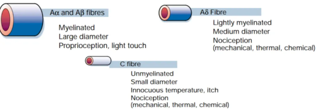
Nociceptors are commonly associated with primary afferent neurons (Aδ and C fibers) relaying information about noxious perceptions from the periphery to the CNS, making up the so-called nociceptive system. Primary sensory neurons are activated when a harmful or damaging stimuli are perceived by the nociceptors. C-fibers and Aδ-fibers are considered as the afferent fibers signaling nociceptive perceptions whereas Aβ-fibers are not involved on pain transmission and they predominantly carry information concerning innocuous perceptions such light touch or pressure (Figure 2).
Figure 1: Primary afferent neurons include large-diameter (Aβ), medium-diameter (Aδ) and
small-diameter unmyelinated (C) afferent fibers (Adapted from Julius and Basbaum, 2001)
Aβ fibers have the largest diameter and are myelinated. The layer of myelin, a dielectric compound which prevents the electric current from leaving the axon, confers a high speed of propagation of impulses along these fibers (Russell, 1992). Aβ fibers are not involved in pain perception but allow the integration of innocuous stimuli such as cold or warm feeling, sense of touch, vibration and light pressure (Julius and Basbaum, 2001).
Aδ fibers are medium-diameter fibers and are also myelinated. They are polymodal and more importantly, play a central role in the transmission of intense heat, mechanical and chemical noxious perception. The high speed of propagation of noxious stimuli though these fibers, conferred by their myelination and diameter, link Aδ fibers on the perception of acute and fast pain called first pain(Adriaensen et al., 1983).
C-fibers, are the smallest in diameter and are unmyelinated. Their small diameter and the lack of myelination results in a slow conduction velocity (Bouhassira, 2009). These fibers mediate the so-called second or slow pain. C-fibers are also polymodal responding to thermal, mechanical and chemical stimuli (Basbaum et al., 2009). Both Aδ and C-fibers have elevated activation threshold and are involved in noxious stimulus perceptions (Schaible, 2006).
I.3.1.2-Ion channels
Harmful or damaging stimuli perception is mediated in the first instance by voltage-gated ion channels composed of complex transmembrane proteins. The activation of ion channels leads the efflux or influx of specific ions through the channel resulting in a polarization or depolarization of the cell membrane modulating the electrical excitability of
neurons. A vast majority of nociceptors are ion channels or are associated with them allowing rapid membrane depolarization (Takayama et al., 2015). Ion channels permeable to different ions have been identified along the CNS. They play a fundamental role in the generation of action potentials and their propagation through afferent fibers.
I.3.1.2.1-Sodium channels
Voltage-gated sodium channels (Nav) are present in all sensory neurons. They initiate action potentials in neuronal cells though the influx of Na+, producing a fast depolarization of the membrane. Many Nav have been identified and they have similar functions (Goldin et al., 2000). Nav1.7, Nav1.8 and Nav1.9 have been demonstrated to be critical for pain perception. Genetically engineered mice lacking those channels shown a clear insensibility to pain (Cummins et al., 2007).
I.3.1.2.2-Calcium channels
In neuronal cells, calcium voltage-gated channels (Cav) are the principal ionic channels involved in regulating the release of neurotransmitters during synaptic transmission. Cav2.2, located on nerve terminals, has been described to be essential for initiating presynaptic neurotransmitter release by neuronal cells as a response to the influx of Ca2+ (Catterall, 2000; Catterall and Few, 2008). Moreover, the influx of Ca2+ through Cav into cell cytosol is also crucial to regulate the activity of cytosolic enzymes and other biochemical processes (Flavell and Greenberg, 2008). In addition, as shown below, several nociceptors, such as TRPV receptors are in fact Cav, the activation of which leads to membrane depolarization and the generation of action potentials(Fernández-Carvajal et al., 2011).
I.3.1.2.3-Potassium channels
Potassium channels are primarily involved in repolarizing the membrane. The influx of K+ though these channels modulates the formation of action potentials on sensory neurons. The activity of potassium channels is essential to control the length and frequency of the action potential (Brady et al., 2005). In addition, potassium channels have been demonstrated to be associated with various opioid receptors, which open specific potassium channels and prevent the excitation and propagation of action potentials (Ocaña et al., 2004).
I.3.1.2.4-Chloride channels
The concentration of Cl- ions in neurons is low, therefore an influx or efflux of Cl -through chloride channels drastically leads to a rapid membrane polarization or depolarization respectively, making these channels crucial for the control of neuronal excitability (De Koninck, 2007). Chloride channels such as anoctamin 1 have been found associated with nociceptors providing a rapid depolarization mediated by the efflux of Cl -(Takayama et al., 2015). In contrast, the activation of the chloride channels γ-aminobutyric acid (GABA) receptors, one of the most important synaptic receptors, leads to a hyperpolarization of the membrane by the influx of Cl-, resulting in a reduction in the probability of action potential initiation and causing neuronal inhibition (Brady et al., 2005; Duran et al., 2010).
I.3.1.3-Pain receptors, nociceptors
Pain usually starts with the activation of sensory receptors known as nociceptors (Woolf, 2011; Scholz and Woolf, 2002). Nociceptors are located at terminal axons of peripheral sensory neurons that innervate skin, organs, joints and viscera. Nociceptors are able to respond selectively to different tissue stimulations (Gold and Gebhart, 2010). They respond to specific noxious threshold stimuli but do not respond to innocuous stimuli. Their activation can result from noxious heat and cold perception (heat/cold sensitive receptors), from chemical compounds (sensitive to chemical compounds receptors) or from mechanical stimuli (mechano-transducers) (Figure 1) (Schaible, 2006). The activation threshold of those different nociceptors depends on the tissue or organ innervated. As an example, activation threshold in tissues such as the cornea is low compared to other tissues such as the skin (Gold and Gebhart, 2010). Moreover, the sensitivity of nociceptors can be altered after tissue injury owing to the release of inflammatory chemical compounds which reduce nociceptor activation threshold (Chiu et al., 2012). The excitation of nociceptors by mechanical, thermal and chemical stimuli evokes membrane depolarization, leading to the generation of an action potential. (Gold and Gebhart, 2010)
Figure 2: Thermal, mechanical and chemical noxious stimuli are perceived by nociceptors located
at the terminal of afferent neurons. The activation of nociceptors leads to the generation of action potentials which rely noxious perceptions to the brain (Adapted from Scholz and Woolf, 2002).
I.3.1.3.1-Heat sensitive receptors
Transient receptor potential vanilloids (TRPV) are responsible for heat stimuli perception. Their thermal stimulation open the channels and allow the influx of Ca2+, resulting in cell depolarization (Schaible, 2006). Heat-sensitive TRPV receptor family comprises 3 essential receptors TRPV1, TRPV2 and TRPV3. TRPV1 and TRPV2 are activated by noxious heat (>42° C) (Fernández-Carvajal et al., 2011), whereas TRPV3 is activated by innocuous warmth (30°C–40°C), maintaining its activation at noxious temperatures. Those receptors and expressed at high levels along the PNS and CNS in small diameter C and Aδ fibers. Its activity is enhanced under inflammatory pain conditions (Julius and Basbaum, 2001).
I.3.1.3.2-Chemical sensitive receptors
Acid-sensing ion channel receptors (ASICs) are the most important and studied chemical sensitive receptor. ASICs are Na+ channels opening at acidic pH (<5) (Sazanavets and Warwicker, 2015; Babinski et al., 1999). They serve among other things as a receptor
for extracellular proton release following a tissue injury (Sutherland et al., 2001). In addition, some thermo-sensitive nociceptors such as TRPV1 and TRPM8 are also considered chemically sensitive receptors. TRPM8 can be activated, in addition to thermal stimulus, by menthol and TRPV1 by capsaicin and acids (Karashima et al., 2007; Bandell et al. 2004). The importance of the polyvalence of some nociceptors is reflected, for example, by the involvement of TRPV1 receptors on the burning pain perceived during inflammation, which is a result of the interaction of protons contained in the inflammatory soup with TRPV1 heat-sensitive receptors (Reid and Flonta, 2001). Chemical heat-sensitive receptors have been reported to be present along PNS and CNS in C and Aδ fibers (Iida et al., 2003).
I.3.1.3.3-Mechano-transducers
Although the molecular mechanisms of mechano-transducers are not fully understood, recent studies suggested a major role for degenerin/epithelial Na+ channel (DEG/ENaC) and K+ channel subfamily K (KCNK) for the transduction of mechanical stimulus (Mano and Driscoll, 1999; Bautista et al., 2008). Likewise, the TRPV2 channels can respond to osmotic stretch in addition to noxious heat, denoting their role in mechano-transduction (Basbaum, et al., 2009). A variety mechano-transducer ranging from high threshold activation mechano-receptors are found on C and Aδ fibers and low threshold mechano-transducers which are found on Aβ fibers capable of detecting light pressure, vibration or texture.
I.3.1.3.4-Cold sensitive receptors
Transient receptor potential melastatin 8 (TRPM8) and transient receptor potential ankyrin 1 (TRPA1) ion channels have been proposed as the most essential cold sensitive receptors, whose activation allows the entry of Na+ and Ca2+ ions to the cell. TRPM8 is the best known cold sensitive receptor. This receptor is responsible for cold perceptions within the range of innocuous temperatures below 30°C. (Fernández-Carvajal et al., 2010). On the other hand, TRPA1 receptor is responsible for the perception of cold stimuli within noxious range (<15ºC) (Basbaum, et al., 2009). Both TRPM8 and TRPA1 are also sensitive cooling agents such as menthol and eucalyptol (Karashima et al., 2007; Bandell et al. 2004). These
nociceptors were identified on C and Aδ fibers, into the CNS and in a variety of tissues (Dhaka et al. 2007; Simone and Kajander 1996).
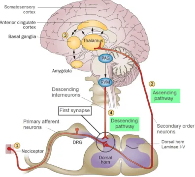
I.3.2-Ascending pathway
Sensory information perceived by the nociceptors is carried through the primary afferent neurons and reach the spinal cord. The ascending pathway comprises the process of signaling between primary afferent neurons and secondary order neurons at the spinal cord, as well as the ascension of the information through the secondary order neurons to supraspinal structures (Figure 3). In the brain noxious information is translated and then perceived as a noxious stimuli. The signaling process between primary afferent neurons and secondary order neurons is known as the first synapse. This process takes place at the spinal cord and allows the communication between neurons via the release of neurotransmitters.
Figure 3: Schematic of the pain pathway. (1) The PNS is responsible of noxious stimuli perception
and the transmission of noxious information to the spinal cord though primary afferent neurons. (2) Signaling between afferent and secondary neurons take place at the first synapse. (3) These secondary order neurons transmit the information to the thalamus which engage the somatosensory
cortex, providing information about the location and intensity of the painful stimulus. Other projections engage the thalamus with cingulate cortex and amygdala, contributing to the emotive
component of the pain experience. (4) Noxious stimulus inputs activates the endogenous modulating system, stimulating the synthesis of endogenous opioids and their release into the first synapse, resulting in a modulation of the synaptic activity (Adapted from de Lalouvière et al. 2014).
I-3.2.1-First Synapse
In the late 19th century the first neuronal connections were discovered. The connections were found to be discontinuous and the communication between neurons was observed to be carried out by the so-called synaptic transmission (López-Muñoz et al., 2006). Nowadays the communication between neurons is known to be mediated by the release of excitatory neurotransmitters from the terminal of primary neurons. Through the interaction of these released neurotransmitters with their receptors located on secondary neurons, the secondary neurons are activated and the information is transferred. The first synapse is the process through which the PNS and the CNS are connected. It takes place at the dorsal horn of the spinal cord within laminae I to V and allows the signaling between afferent and secondary order neurons which relay information to supra-spinal structures (Basbaum et al., 2009).
The synaptic process is a dynamic mechanism of neurotransmitters release which keeps a basal activity of sensory neurons. However, noxious stimulations of the PNS and CNS enhances the activity at the first synapse. Action potentials generated as a consequence of a harmful perception by the nociceptors, are propagated through primary sensory neurons stimulating the synthesis of specific peptidergic neurotransmitters at the DRG along with the opening the Ca2+ channels at the terminal of the neuron. These peptides involved in the signaling of noxious information between neurons are packed into secretory vesicles and are carried through the axons to the presynaptic terminal (Zhang et al., 1995). The high concentrations of Ca2+ at the terminal, caused by the Ca2+ influx through the Ca2+ channels activated by the action potential, facilitate the release of the vesicle content from the presynaptic terminal into the synaptic gap by exocytosis (Margeta et al., 2008; Südhof, 2004). The interaction between the released neurotransmitters with specific post-synaptic receptors stimulates the postsynaptic terminal and generates a new action potential at the secondary order neuron (Brady et al., 2005).
I-3.2.1.1-Principal synaptic neurotransmitters and receptors
Several neurotransmitters are involved in the neuronal synaptic communication. Glutamate and ϒ-aminobutyric acid (GABA) are the major chemical neurotransmitters involved, and it is believed that approximately 80-90% of the synapses in the CNS are glutamatergic (Brady et al, 2005). Glutamine is synthesized in neurons and metabolized to glutamate by the mitochondrial enzyme glutaminase (Olsen and DeLorey, 1999). Glutamate binds to N-methyl-D-aspartate (NMDA) and α-amino-3-hydroxy-5-methyl-4-isoxazole-propionic acid (AMPA) postsynaptic receptors producing excitatory postsynaptic response (Purves et al., 2001). The NMDA receptor is an ionotropic receptor which, following activation, produces an influx of cations mainly Na+ and K+ but also Ca2+ into the cell. On the other hand, AMPA receptor is also an ionotropic transmembrane receptor, and which gates mainly Na+ and K+, but not Ca+2 ions. Their activation and the consequent influx of cations produces a membrane depolarization at the postsynaptic terminal, resulting in the generation of an action potential (Purves et al., 2001). Glutamate is the precursor for GABA, a major inhibitory neurotransmitter. GABA activates GABA receptors, which are ionotropic channels permeable to Cl-. The transmembrane influx of Cl- through GABA receptors leads to membrane hyperpolarization reducing the presynaptic release of neurotransmitters as well as the excitability of postsynaptic receptors (Petroff et al., 2002; Schousboe et al., 2007).
The perception of intense stimulus is directly associated with the release of peptidergic excitatory neurotransmitters including tachykinins and calcitonin gene-related peptide (CGRP). Thus, perceptions on the noxious range promote the production of tachykinins and CGRP at the DRG and their subsequent release into the synaptic gap. Tachykinins are a family of neurotransmitters that includes peptides such as Substance P (SP), Neurokinin A (NKA) and Neurokinin B (NKB). These tachykinin-related peptides act on neurokinin receptors (i.e. NK1, NK2 and NK3). The activation of neurokinin receptors, particularly the activation of NK1 by SP, generates a greater post-synaptic response and enhances NMDA receptors activity (Gao and Peet, 1999; Teodoro et al., 2013). CGRP also plays an important role in nociception, it is a potent vasodilator and the release of CGRP potentiates the action of SP by inhibiting its enzymatic degradation and by enhancing its release (Gangula et al., 2000; Bennett et al., 2000; Biella et al., 1991).
I.3.3-Descending pathway
The perception of well-being is the result of the equilibrium between an incessant release of excitatory neurotransmitters at spinal levels, which allow the brain to perceive noxious information, and the suppressive influences of the same importance that descends from the brain. When the ascending signal is more intense than the suppressing action, pain appears (Beaulieu 2005).
By the time the brain receives noxious stimuli inputs this perception is projected to the periaqueductal gray (PAG) and the rostral ventromedial medulla (RVM) activating the descending pathway (see Figure 3) and leading to a nociceptive modulation (Lovick, 1991; Helmstetter et al., 1998). This modulating process is principally mediated by the release of endogenous opioid peptides from descending interneurons into the first synapse. Through the interaction of these opioid peptides with their respective pre- and post-synaptic opioid receptors the synaptic activity is modulated, reducing the intensity of noxious perceptions.
I.3.3.1-Opioid receptors and endogenous opioid peptides
Since their discovery in 1970s, research related to opioid peptides revealed fundamental CNS functions (Brownstein, 1993). Endogenous opioid peptides participate in pain modulation producing analgesia and a sense of well-being (Froehlich, 1997). Synthetized as large and biologically inactive precursors at the DRG, pituitary and adrenal gland, they require enzymatic processing to generate active opioid peptides. Similar to other neuropeptides, opioid peptides are synthetized and packed into dense core vesicles (Hook, 2006). Following noxious stimuli, the synthesis and release of these vesicles are enhanced, leading to measurable modulatory effects on pain perception (Alberts et al., 2002).
Opioid peptides are classified into four major families; endorphins, endomorphins, enkephalins and dynorphins. Each family is derived from a distinct precursor (Figure 4) and has a characteristic anatomical distribution and physiological activities (McDonald and Lambert, 2014). Moreover, each family of endogenous opioid peptides has specific affinity for the opioid receptors present into the CNS. These receptors include µ-opioid receptors (MOR), κ-opioid receptors (KOR) and δ-opioid receptors (DOR) (Table 1).
Precursor Endogenous peptide Amino acid sequence
Affinity for Opioid receptors
MOR DOR KOR
Pro-Opiomelanocortin β-endorphin YGGFTMTSEKSQTPLVYLFKNAIIKNAYKKGE +++ ++ -
Unknown Endomorphin-1 YPWF-NH2 +++ - -
Endomorphin-2 YPFF-NH2 +++ - -
Pro-Enkephalin Met-Enkephalin YGGFM ++ +++ -
Leu-Enkephalin YGGFL + +++ -
Pro-Dynorphin Dynorphin A YGGFLRRIRPKLK + - +++
Dynorphin B YGGFLRRQFKVVT + + +++
Table 1: Most representative opioid peptide derived from the processing of their respective
precursors. Affinity observed for opioid receptors (Stein et al. 2009; Merg et al., 2006; Beaulieu, 2005; Mansour et al., 1995; Raynor et al., 1994).
I.3.3.1.1-Opioid receptors
Opioid receptors are G protein-coupled receptors characterized by 7 transmembrane domains. Pharmacological, behavioral, and receptor binding studies have suggested the existence of at least three types of opioid receptors including MOR, KOR and DOR (Snyder and Pasternak, 2003). These receptors are highly abundant in the brain and the spinal cord. Opioid receptors are mainly located in the superficial dorsal horn (Zöllner and Stein, 2007). These receptors induce specific pharmacological response and they differ in their binding characteristics, even though a specific opioid peptide can interact with more than one type of opioid receptor (Lutz and Pfister, 1992; Ji et al., 1995; Mcnally and Akil, 2002).
The binding of opioid peptides to opioid receptors initiates a series of biochemical events that usually culminates in the stimulation of potassium efflux though the potassium ion channels, leading to a repolarization that results in various effects, including analgesia and euphoria (Ikeda et al., 2002; Maldonado et al., 2001; North et al., 1987). KORs are located presynaptically on primary afferent neurons in the dorsal horn of the spinal cord where they participate in the inhibition of the release of excitatory neurotransmitters such as SP, CGRP and glutamate. DORs are present on postsynaptic terminals of secondary order
neurons, and they decrease the excitability provoked by the activation of other postsynaptic receptors such as NK1 and NMDA. MORs are located either at presynaptic or postsynaptic terminals, therefore, MORs can either modulate the release of excitatory neurotransmitters or decrease the excitability of postsynaptic receptors (McDonald and Lambert, 2014).
I.3.3.1.2-Endorphins
Endorphins are endogenous opioid peptides produced during arduous exercise, excitement or pain perception, specifically inducing analgesia and well-being feeling (Sprouse-Blum et al., 2010). They are found widely distributed in the PNS and CNS (Marvizón et al., 2009). Endorphins are derived from the precursor proopiomelanocortin (POMC) a 241 amino acids protein (Smith et al., 1988). Its endoproteolytic processing by protein convertases (PCs) generate various bioactive peptides, such as β-endorphin (β-End), in addition to several non-opioid neuropeptides including adrenocorticotropic hormone (ACTH) and α-, β- and γ- melanocyte-stimulating hormone (α-MSH, β-MSH and γ-MSH) (Mousa et al., 2004). Binding affinity experiments of β-End for opioid receptor have shown a primary affinity for MOR, even though a high affinity for DOR was also reported (Mansour et al., 1995).
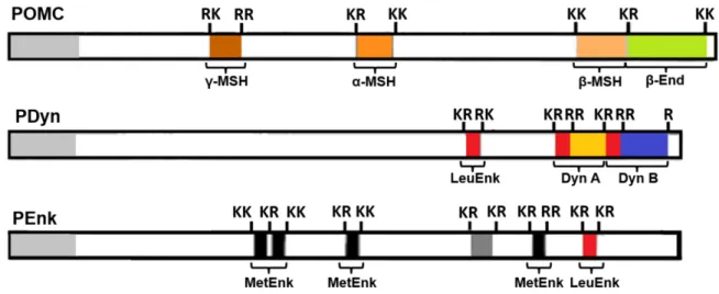
Figure 4: Opioid-derived peptides are generated from the endoproteolytic processing of proopioid
precursors POMC, PDyn and PEnk. With the exception of dynorphins, the enzymatic formation of opioids peptide are regulated by cleavages at dibasic amino acid positions.
I.3.3.1.3-Enkephalins
Enkephalins are pentapeptides found in many different regions in the CNS, suggesting that these peptides are involved in many physiological functions. Among other functions enkephalins are involved in pain perception, mood and behavior by altering emotional responses as well as by acting on cardiovascular and respiratory functions (Przewłocki and Przewłocka, 2001; Mediavilla, 1977). In pain modulation, enkephalins have a potent action but with a short duration due to their rapid degradation by metallopeptidases (Mosnaim et al., 2008). Proenkephalin (PEnk), a protein constituted of 267 amino acids, is proteolytically cleaved into enkephalin peptides. PEnk processing by PCs results in the generation of 4 copies of Met-enkephalin (Met-Enk) and one copy of Leu-Enkephalin (Leu-Enk) (Loh et al., 2002). Both enkephalins have high affinity for DOR and moderate affinity, approximately tenfold lower, for MOR (Roques et al., 2012; Zöllner and Stein, 2007).
I.3.3.1.4-Endomorphins
The most recent family of endogenous opioid peptides discovered is endomorphins. They have a key role in pain perception, responses related to stress, and complex functions such as reward, arousal, and vigilance (Fichna et al., 2007). Although the endomorphin precursor still remains unidentified, two endomorphin peptides have been identified; endomorphin-1 (EM-1) and endomorphin-2 (EM-2). Both endomorphins differ just in one amino acid and are quite distinct from the other opioid peptides (endorphins, enkephalins and dynorphins), which all share the YGGF amino acid sequence at the N-terminus. EM-1 and EM-2 are widely distributed throughout the CNS and bind selectively to the MOR. Both endomorphin peptides have high affinity and similar potency for MOR (Zadina et al., 1997;
Hackler et al., 1997; Martin‐Schild et al., 1999). I.3.3.1.5-Dynorphins
Endogenous dynorphin peptides are generated from the endoproteolytic processing of their precursor prodynorphin by PCs. This family of dynorphin peptides includes big dynorphin, dynorphin A and dynorphin B. They have primary affinity for KOR and their biological
functions comprise, among several others, a key role in pain modulation. The functions of the principal dynorphin peptides will be thoroughly described in the next chapter.
CHAPTER II-DYNORPHINS
II.1-Dynorphins generalitiesThe modulation of sensory information has been shown to take place in the dorsal horn of the spinal cord, more specifically at the first synapse (Honore et al., 2000; Levine, 1993). Dynorphins are a family of endogenous opioid peptides that have potent analgesic effects and have been identified as neuropeptides involved in endogenous pain inhibition (Kuner, 2010; Mika et al., 2011). Dynorphin peptides are widely distributed in the mammalian CNS and have a primary affinity for KOR (Stein et al. 2009; Schaible, 2006). This class of opioid peptides is known to be involved in a wide range of functions, including mood, motor activity and homeostatic response to injury and contributes to perceptual distortion in schizophrenia, dementia and bipolar disorders. An altered expression of dynorphins is also observed in drug abusers and psychiatric patients (Solbrig and Koob, 2004; Hurd, 2001; Hurd et al., 1996). Like other endogenous opioid peptides, bioactive dynorphins result from the processing of its large and inactive precursor, Prodynorphin (PDyn). PCs, specifically PC1 and PC2, found in neuronal and endocrine cells, were reported to be involved into PDyn proteolytic processing by cleaving its basic amino acid residues (Berman et al., 2000; Dupuy et al., 1994; Day et al., 1998).
II.2- Prodynorphin
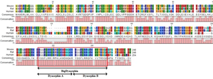
Dynorphin peptides are derived from PDyn, a 254 amino acid biologically inactive protein which undergoes enzymatic degradation by PCs generating dynorphin peptides. PDyn was first characterized in 1982 from porcine neuronal tissues by using cDNA hybridization (Kakidani et al., 1982). As shown in figure 5, mouse, rat and human PDyn present a high homology at the amino acid level, sharing an identical sequence for the region encoding dynorphin A (Dyn A) and dynorphin B (Dyn B) (Civelli et al., 1985).
PDyn, similarly to other proneuropeptides, is synthesized in the endoplasmic reticulum (ER), of neuronal cells, and is transferred to the Golgi apparatus where it is packed in dense core vesicles together with the endoproteases PC1 and PC2 (Hook et al., 2008).
Dense core vesicles are then transported along the axons to the nerve terminal. Classical models postulated that PDyn processing by endoproteases starts at the trans-Golgi network (TGN) and continues during the axonal transport of the secretory vesicles (Hökfelt et al., 2000; Alberts et al., 2002). Potassium-induced depolarization produced by neuronal activity, stimulates the production, the migration and the exocytotic release of dynorphin-containing vesicles into the first synapse (Seidah and Chrétien, 1999; Zhou et al., 1999; Arvan and Castle, 1998). Others have proposed that vesicles containing unprocessed PDyn are stored at the nerve terminal waiting for depolarization to induce precursor processing and its release into the synaptic space (Yakovleva et al., 2006).
The highest concentrations of unprocessed PDyn are found in the hypothalamus, striatum, and hippocampus. Less important amounts are also found in the midbrain, nucleus tractus, brainstem, and cerebral cortex. In non-brain tissues, PDyn is found in the adrenal gland, spinal cord, testis, and anterior pituitary (Civelli et al., 1985).
Figure 5: Mouse, rat and human Prodynorphin proteomic alignment. High homology is observed in
multiple regions between species. The region encoding BDyn is completely homologue for the three species. This homology shows high conservation of this gene through evolution.
II.3-Endogenous Dynorphin peptides
Several active dynorphin peptides have been identified in mammalian brain and spinal cord as potential products from PDyn proteolytic processing. These PDyn-derived peptides include BDyn, Dyn 1-19, Dyn A, Dyn B, Dyn 1-13, Dyn 1-11, Dyn 1-10, Dyn 1-9, Dyn 1-8, Dyn 1-7 and Dyn 1-6 (Lu et al., 2001; Mansour et al., 1995; Reed et al., 2003; Prokai et al., 1998; Chou et al., 1994) (Table 2). So far, BDyn, Dyn A and Dyn B have been the dynorphin peptides which were extensively studied with a strong emphasis on Dyn A physiological activities.
Peptide Amino acids Sequence
Bigdynorphin 1-32 YGGFLRRIRPKLKWDNQKRYGGFLRRDFKVVT Dynorphin 1-19 1-19 YGGFLRRIRPKLKWDNQKR Dynorphin A 1-17 YGGFLRRIRPKLKWDNQ Dynorphin B 20-32 YGGFLRRDFKVVT Dynorphin 1-13 1-13 YGGFLRRIRPKLK Dynorphin 1-11 1-11 YGGFLRRIRPK Dynorphin 1-10 1-10 YGGFLRRIRP Dynorphin 1-9 1-9 YGGFLRRIR Dynorphin 1-8 1-8 YGGFLRRI Dynorphin 1-7 1-7 YGGFLRR Dynorphin 1-6 1-6 YGGFLR
Table 2: Prodynorphin-derived peptides amino acid sequences
Dyn 1-19, in spite of being the major product from Bigdyn endoproteolytic processing (Berman et al., 2000), owing to its rapid conversion into Dyn A by carboxipeptidases is not a highly abundant peptide in the CNS and its role is still quite unclear (Berman et al. 2001).
Dyn 1-13 has been presented as an extraordinarily potent opioid peptide with a long duration of action (Goldstein et al., 1979), acting on KOR (Oka et al., 1982). Its administration was reported to induce catalepsy and analgesia in rats (Herman et al., 1980). Studies also shown that the expression of opiate withdrawal symptoms after the administration of Dyn 1-13 were suppressed in mice (Takemori et al., 1992 and 1993; Khazan
et al., 1983; Aceto et al., 1982; Hooke et al., 1995), suggesting that, in vivo, Dyn 1-13 does not behave as a typical KOR agonist, but its role still needs to be clarified.
On the other hand, the effects of Dyn 1-11, Dyn 1-10, Dyn 1-9, Dyn 1-8, Dyn 1-7 and Dyn 1-6 remains relatively unexplored. Even though they have been identified as proteolytic products of PDyn, BDyn or Dyn A, just few studies have been published corroborating their antinociceptive effects, while their specific physiological role still remains to be determined (Lu et al., 2001; Mansour et al., 1995; Reed et al., 2003; Prokai et al., 1998; Chou et al., 1994;
Herman et al., 1980).
II.3.1-BigDynorphin
BDyn is the largest and bioactive PDyn-derived peptide, consisting on Dyn A and Dyn B bound by arginine-lysine (KR) paired basic amino acids. In addition to serving as a precursor for Dyn A and Dyn B, this peptide may also have its own function. Found at substantial levels in the pituitary gland, brain and spinal cord (Xie and Goldstein, 1987; Day and Akil, 1989), the effects of BDyn differs from those of the other dynorphin peptides. The intrathecal and intracerebral administration of BDyn to mice were reported to produce a nociceptive behavioral response of the animal (Tan-No et al. 2002). These nociceptive responses were associated with the interaction of BDyn with NMDA receptors (Merg et al., 2006; Chen et al., 1995). In addition, the binding affinities and potency of BDyn for opioid receptors were also studied, showing that BDyn also has a strong affinity for KOR. Although BDyn affinity for KOR was comparable to Dyn A, its potency was significantly greater compared to other dynorphins such as Dyn A or Dyn B (Merg et al., 2006; Kuzmin et al., 2006).
The selectivity and potency of BDyn in activating KOR along with the behavioral effects observed in mice, mediated by the activation of NMDA receptors, suggest that a deficient processing of PDyn, resulting in high levels of BDyn, could lead to an enhancement on nociceptive perception, while a normal processing of PDyn leads generally to a predominant activation of KOR by BDyn and other dynorphin peptides, resulting in antinociceptive effects.
II.3.2-Dynorphin A
Dyn A was the first dynorphin peptide to be identified (Cox et al., 1975). The observation of potent analgesic effects when administered intrathecally to mice raised the interest in this opioid peptide for further studies in pain research (Hayes et al., 1983). Dyn A is widely found throughout the CNS, being more abundant especially in the brain, in areas like hypothalamus, substantia nigra and periaqueductal gray. High concentrations are also found in the dorsal horn of the spinal cord and in the pituitary gland. (Tan-No et al., 1997).
Studies revealed that Dyn A participates in pain modulation pathways, mediated in part by its release in the brain and spinal cord (Mizoguchi et al., 2006). Its participation in the first synapse was corroborated a posteriori when high levels of Dyn A were found in intrinsic neurons projecting into laminae I, II and V of the spinal cord dorsal horn (Draisci et al., 1991; Przewlocki et al., 1983), where neurons responding to noxious input are contained (Mika et al., 2011). Subsequent studies based on the injection of Dyn A into the subarachnoid space of the spinal cord of rats showed a strong and long-lasting analgesic effect, with a greater potency than morphine (Merg et al., 2006). That effect was completely reversed by the administration of naxolone, a potent opioid receptor antagonist, demonstrating that Dyn A acts on KOR (Han and Xie, 1982; Nakazawa et al., 1985). The affinity and potency of Dyn A for KOR has been reported to be substantially higher than for other dynorphin peptides (Merg et al. 2006). However, intrathecal administration of Dyn A at high doses was observed to induce long-lasting mechanical and thermal allodynia in rats, a response that was not blocked by naloxone, suggesting that when Dyn A is administered at high doses, it is able to interact with other receptors such as NMDA in addition to KOR (Laughlin et al. 1997; Vandera et al. 1996; Shukla et al. 1994). Those studies suggested that Dyn A has inhibitory or excitatory effects, depending on its concentration (Caudle et al. 1994).
II.3.2.1-Physiological role of Dynorphin A
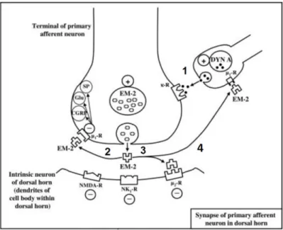
EM-2 has been proposed to have an important involvement in antinociception mediated by Dyn A at spinal cord levels (Draisci et al., 1991). The mechanism proposed by Mizoguchi and workers (Mizoguchi et al. 2006) and later supported by Fichna and co-workers (Fichna et al. 2007) establish that the release of Dyn A from descending interneurons
and its subsequent interaction with presynaptic KOR, promote the production and release of EM-2 from the terminal of primary afferent neurons. The released EM-2 preferentially stimulates MOR, a presynaptic and postsynaptic receptor, leading to the inhibition of the release of excitatory neurotransmitters such as SP, glutamate and CRGP, as well as causing a decrease of the excitability of postsynaptic receptors such as NMDA or NK1 receptors (Figure 5). Moreover the presence of MOR in the descending dynorphin-containing neurons also make EM-2 having an important regulator of the release of Dyn A (Ohsawa et al., 2001; Sakurada et al., 2001; Mizoguchi et al., 2006; Fichna et al., 2007).
Figure 6: Involvement of Dyn A in pain modulation. (1) The release of Dyn A enhances the
production and release of EM-2. Thus through its interaction with MOR, EM-2 (2) inhibits the release of excitatory neurotransmitters and (3) decreases the excitability of postsynaptic receptors
in addition to (4) regulate the release of Dyn A. (Fichna et al., 2007)
II.3.3-Dynorphin B
As a primary product from Bigdyn, Dyn B is present in the same neuronal fibers and tissues that contains Dyn A (Zamir, 1984). Dyn B has a primary affinity for KOR, however its affinity for the receptor and its potency is lower compared to Dyn A (Merg et al., 2006). Intrathecal injections of Dyn B in rats produced potent and long-lasting analgesic effects (Han and Xie, 1982). Nevertheless, the physiological role of Dyn B is still far from clear.
Mizoguchi and colleges reported that the physiological role of Dyn B completely differs from Dyn A. They observed that Dyn B activated KOR induced the release of another potent antinociceptive peptide, analogue to dermorphin (Mizoguchi et al. 2006a). This mechanism of action was similar to the role observed for Dyn A involving the release of EM-2. These interesting findings strongly suggest that the dynorphin peptides act through separate pathway to bring about pain relief.
II.4-Proprotein convertases
Several neuropeptides and hormones as well as a variety of other endogenous peptides are derived from large and inactive proteins which require endoproteolytic processing for the biosynthesis of the active peptides. Protein convertases (PCs) are a family of enzymes catalyzing protein cleavage at monobasic amino acid residues such as single lysine (K) or arginine (R) as well as at paired basic residues such as RR, KR, RK and KK (Hook and Brennand, 2014; Rouillé et al., 1995). As an exception, when a basic position is flanked by a proline at the C-terminal, the conformational restrictions imposed by this amino acid on the peptide avoids any possible cleavage by PCs (Vanhoof et al. 1995).
Seven PCs were identified in mammalian tissues including PC1, PC2, PC4, PC5 , PACE4, PC7 and furin, officially named as proprotein convertase subtilisin/kexin type 1, 2, 4, 5, 6,7 and furin respectively (Seidah et al. 1998). Like most secreted proteins, PCs are synthesized in the ER as immature PCs which require post-translational modifications to become fully active (Seidah et al. 2008). Following their synthesis, PCs are transported to downstream compartments of the secretory pathway where they are N-glycosylated at various sites and folded into an active conformation within the ER (Benjannet et al. 1993; Steiner, 1998). As an exception, PC2 does not follow this process, instead, immature PC2 is transported with a binding protein (7B2) to acidic immature secretory granules, where it is autocatalytically activated (Mousa et al., 2004; Mbikay et al., 2001).
PCs and other secreted proproteins are then packed in secretory vesicles as they leave the ER. During the transport of those vesicles, active PCs process proproteins within the vesicles (Alberts et al., 2002). PCs require Ca2+ as a cofactor to cleave their substrates (Linard et al., 1995). In addition, PC1 and PC2 are maximally active in the acidic pH
environment (5.5-6.5) of immature secretory granules (Shennan, 1995), being less effective outside the vesicles (Seidah and Prat, 2012). Since all PCs possess overlapping functions, substrate specificity is dictated by different tissue and cellular distribution (Steiner et al., 1992).
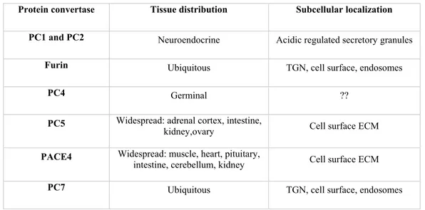
II.4.1-Tissue and cellular expression of PCs
The tissue distribution and intracellular localization of PCs is varied as presented in Table 3. Furin and PC7 are widely distributed in the lymphatic system, in the liver and in the kidney, and are localized predominantly in endosomes, on the cell surface and in the trans-Golgi Network (TGN) (Hatsuzawa et al., 1990; Schalken et al., 1987). PC5 and PACE4 are expressed in both endocrine and non-endocrine tissues, primarily in the brain, the digestive system and the adrenal cortex. They can be found intracellularly in the cell surface and in the extracellular matrix (ECM) (Lusson et al., 1993; Nakagawa et al., 1993). The expression of PC4 differs from other PCs since this enzyme is predominantly synthesized in testicular germ cells, the placenta and the ovary. PC4 is important in fertility, but its intracellular location is still not well characterized (Seidah and Prat, 2012; Torii et al., 1993). Neuronal and endocrine cells are rich in PC1 and PC2. They are found inside the cells in the TGN and are stored within the acidic regulated secretory vesicles (Seidah et al., 2008 and 1999). PC2 has been described to be the major protein convertase within the CNS (Seidah et al., 1998).
Tissue and cellular location of PCs are fundamental in the determination of their role and target substrate. The rich expression of PC1 and PC2 in neuroendocrine cells confers a key role of these enzymes in the processing of several proneuropeptides and prohormones (Hook et al., 2008).