HAL Id: inserm-00110160
https://www.hal.inserm.fr/inserm-00110160
Submitted on 8 Jan 2007HAL is a multi-disciplinary open access archive for the deposit and dissemination of sci-entific research documents, whether they are pub-lished or not. The documents may come from teaching and research institutions in France or
L’archive ouverte pluridisciplinaire HAL, est destinée au dépôt et à la diffusion de documents scientifiques de niveau recherche, publiés ou non, émanant des établissements d’enseignement et de recherche français ou étrangers, des laboratoires
Endothelial differentiation gene-2 receptor is involved in
lysophosphatidic acid-dependent control of 3T3F442A
preadipocyte proliferation and spreading.
Céline Pagès, Danièle Daviaud, Songzhu An, Stéphane Krief, Max Lafontan,
Philippe Valet, Jean Sébastien Saulnier-Blache
To cite this version:
Céline Pagès, Danièle Daviaud, Songzhu An, Stéphane Krief, Max Lafontan, et al.. Endothelial differentiation gene-2 receptor is involved in lysophosphatidic acid-dependent control of 3T3F442A preadipocyte proliferation and spreading.. Journal of Biological Chemistry, American Society for Bio-chemistry and Molecular Biology, 2001, 276 (15), pp.11599-605. �10.1074/jbc.M010111200�. �inserm-00110160�
1 Endothelial Differentiation Gene-2 receptor is involved in lysophosphatidic
acid-dependent control of 3T3F442A preadipocyte proliferation and spreading
Céline Pagès, Danièle Daviaud, Songzhu An#, Stéphane Krief@, Max Lafontan, Philippe Valet and Jean Sébastien Saulnier-Blache*.
I.N.S.E.R.M U317, Institut Louis Bugnard, Université Paul Sabatier, CHU Rangueil, Batiment L3, 31403, Toulouse cedex 04, France.
# Department of Medicine, University of California, San Francisco, CA 94143-0711 @ Smithkline Beecham Laboratoires Pharamceutiques, 35762 Saint-Grégoire, France.
Running Title: EDG-2 receptor in preadipocytes Corresponding author
Phone: (33) 0562172956 Fax: (33) 0561331721
Email: [email protected]
JBC Papers in Press. Published on January 4, 2001 as Manuscript M010111200
Hal author manuscript inserm-00110160, version 1
Hal author manuscript
ABSTRACT
EDG-2, EDG-4, EDG-7 and PSP24 genes encode distinct lysophosphatidic acid (LPA) receptors. The aim of the present study was to determine which receptor subtype is involved in the biological responses generated by LPA in preadipocytes. Growing 3T3F442A preadipocytes express EDG-2 and EDG-4 mRNAs, with no expression of EDG-7 nor PSP24 mRNAs. Quantitative RT-PCR revealed that EDG-2 transcripts were 10 fold more abundant than that of EDG-4. In order to determine the involvement of EDG-2 receptor in the responses of growing preadipocytes to LPA, stable transfection of antisense EDG-2 cDNA was performed in growing 3T3F442A preadipocytes. This procedure, led to a significant and specific reduction in EDG-2 mRNA and protein. This was associated with a significant alteration in the effect of LPA on both cell proliferation and cell spreading. Finally, the differentiation of growing preadipocytes into quiescent adipocytes led to a strong reduction in the level of EDG-2 transcripts. Results demonstrate the significant contribution of EDG-2 receptor in the biological responses generated by LPA in 3T3F442A preadipocytes.
Introduction
Lysophosphatidic acid (LPA: 1-acyl-2-hydroxy-sn-glycero-3-phosphate), is a bioactive phospholipid, present in serum and other biological fluids (1,2). LPA controls a wide variety of cellular responses (mitogenesis, cytoskeletal rearrangements, cell adhesion, ion transport, apoptosis) through the activation of specific G-protein coupled receptors (3,4). A first potential LPA receptor, called vzg1 (5), was cloned in mouse and found homologous to the Endothelial Differentiation Gene-1 (EDG-1) (6), a high affinity receptor for another bioactive phospholipid: sphingosine-1-phosphate (7). A human gene exhibiting 97% homology with vzg1 was then identified and called EDG-2 (8). Two other human genes, called EDG-4 and EDG-7, have also been cloned and proposed to be LPA receptors (9,10). At the beginning of the present work the cDNA sequences of EDG-4 and EDG-7 mouse orthologues were not yet available. Since then, EDG-4 mouse orthologue was cloned and sequenced (11). A fourth receptor, called PSP24, was cloned in xenopus (12) and mouse (13) and initially proposed to be a LPA responsive receptor. However, PSP24-receptor gene exhibits poor homology with EDG-receptor gene family, and is actually related to the Platelet Activating Factor receptor.
Most evidences that EDG-2, EDG-4, EDG-7 and PSP24 are LPA-receptors are deduced from their ability to increase or restore LPA activity following their overexpression in cells (5,9,10 ,12). However, the relative contribution of endogenously expressed LPA-receptors in the biological activities of LPA, remains poorly defined. Because pharmacological tools to study LPA-receptors are very limited (no available antagonists, difficulties to perform receptor binding studies) one way to address the question, is to alter LPA-receptor expression by using antisense strategies or gene invalidation methods.
We recently observed that conditioned media prepared from adipocytes exposed to an α2-adrenergic stimulation, increased proliferation and spreading (reflecting a reorganization of actin cytoskeleton) on an α2-adrenergic-insensitive murine preadipose cell line: 3T3F442A. Analysis based on the use of a lysophospholipid-specific phospholipase (phospholipase B) and [32P] phospholipid labeling, revealed the involvement of LPA in the trophic activities of adipocyte conditioned media (14). Because of the intimate coexistence of adipocytes and preadipocytes within adipose tissue, LPA released by adipocytes could play an important role in paracrine/autocrine control of preadipocyte growth, a key event involved in adipose tissue development. Therefore, a better understanding of the cellular mechanisms of LPA action in preadipocytes could help to develop pharmacological and/or genetic strategies to control adipogenesis.
The trophic action of LPA in growing 3T3F442A preadipocytes can be specifically desensitized by chronic exposure to high concentration of LPA (14). Exposure of growing 3T3F442A preadipocytes to LPA leads to rapid and pertussis toxin sensitive
activation of the Mitogen Activated kinases ERK1 and ERK2 (15). Whereas those observations suggest the involvement of a G-protein coupled receptor(s) in the action of LPA in 3T3F442A preadipocytes, the identity of the discrete LPA receptor(s) responsible for these biological effects remains to be determined.
In the present work, we studied the expression of EDG-2, EDG-4, EDG-7 and PSP24 receptors, and attempted to determine their relative contribution in the biological responses of 3T3F442A preadipocytes to LPA (proliferation and spreading). Based upon quantification of gene expression, and antisense cDNA transfection, EDG-2 receptor was found to be predominantly involved in LPA-dependent control of 3T3F442A preadipocyte proliferation and spreading.
Material and Methods Cells
3T3F442A preadipocytes were grown in 10% donor calf serum supplemented DMEM as previously reported (16). The medium was changed every two days. Conversion of 3T3F442A preadipocytes into adipocytes was obtained by cultivating confluent cells in DMEM supplemented with 10% fetal calf serum plus 50 nM insulin as previously described (14).
Non quantitative RT-PCR analysis
Total RNAs were extracted using RNeasy mini kit (Qiagen). One microgram of total RNA was treated with 1U RNAse-free Dnase I (Gibco BRL) for 15 min at room temperature followed by further inactivation with 1µl EDTA (25 mM) 10 min at 65°C. Then, RNA was reverse transcribed for 60 min at 37°C using Superscript II (Life Technologies) RNase H- Reverse Transcriptase (RT) and subjected to amplification. A minus RT reaction was performed in parallel to ensure the absence of genomic DNA contamination. Polymerase chain reaction (PCR) was carried out in a final volume of 50
µl containing 3 µl of cDNA, 1 µl dNTP (10mM), 5 µl 10x PCR buffer (10 mM Tris-HCl, pH 9, 50 mM Kcl and 0,1% Triton X-100), 3 µl MgCl2 (25 mM), 1.5 µl sense and antisense specific oligonucleotide primers (10µM) and 1.25 unit of Taq DNA polymerase (Promega). Conditions for the PCR reaction were: initial denaturation step at 94°C for 2 min, followed by 35 cycles consisting in 1 min at 94°C, 1 min at 54°C (EDG-2), 57°C (EDG-4), 49°C (EDG-7), 57°C (PSP24) or 58°C (EDG-1), 72°C for 90 s. After a final extension at 72°C for 6 min, PCR products were separated on 1.5% agarose gel and amplification products were visualized with ethidium bromide. In some experiments we analysed the influence of the number of PCR cycles on the intensity of the amplification products.
Oligonucleotide primers for non quantitative RT-PCR
Detection of sense mRNAs:
EDG-2 primers were designed from mouse EDG-2 cDNA (vzg-1) (5): sense, 5’-ATCTTTGGCTATGTTCGCCA-3’ and antisense, 5’-TTGCTGTGAACTCCAGCCA-3’. Those primers are 100% identical between mouse and human (8).
EDG-4 primers were designed from the 570 bp fragment of mouse EDG-4 cDNA cloned in the present study (see below): sense, TGGCCTACCTCTTCCTCATGTTCCA-3’ and antisense, 5’-GGGTCCAGCACACCACAAATGCC-3’. Those primers are specific to mouse.
EDG-7 primers were designed from a GenBank Mouse EST, (Clone ID 2192692, GenBank # AW107032) which exhibits 82% identity on nucleotide level and that we
assumed to correspond to mouse EDG-7. This was confirmed later on after the identification of the mouse EDG-7 (GenBank # NM_012152): sense, 5’-AGTGTCACTATGACAAGC-3’ and antisense, 5’-GAGATGTTGCAGAGGC-3’. Those primers are 100% identical between mouse and human.
PSP24 primers were designed such as these are homologous to xenopus (12), mouse (13) and human (GenBank accession number : HSU92642 ) : sense, GGCCATCCTGCTCATCATTAGCG-3’ and antisense, 5’-GGTGGTGAAGGCCTTGGTTTTGAA-3’.
EDG-1 primers were designed from mouse EDG-1 gene (17): sense, 5’-GTCCGGCATTACAACTACAC-3’ and antisense, 5’-TATAGTGCTTGTGGTAGAGC-3’. Those primers are 100% identical between mouse and human ** (6).
Detection of antisense mRNAs:
Detection of EDG-2 antisense mRNAs was performed with an antisense primer designed from the sequence of stabilization of mRNA in pcDNA3.1 vector (5’-CAACAGATGGCTGGCAACTA-3’), and a specific sense primers designed from human EDG-2 (5’-CTGTGAAATTACAGGGATGGA-3’).
Quantitative RT-PCR analysis
cDNA was synthesized from 2 µg of total RNA in 20 µl using random hexamers and murine Moloney leukemia virus reverse transcriptase (Life technologies). A minus RT reaction was performed in parallel to ensure the absence of genomic DNA contamination. Design of primers was done using the Primer Express software (Applied Biosystems). Real-time quantitative RT-PCR analyses were performed starting with 50 ng of reverse transcribed total RNA with 200 nM of both sense and antisense primers in a final volume of 25 µl using the Sybr Green PCR core reagents in a ABI PRISM 7700 Sequence Detection System instrument (Applied Biosystems). Standard curves were determined after amplification of 5x10² to 5x10⁶ copies of purified amplicons generated from 3T3-F442A cDNA by non quantitative RT-PCR. Quantification of EDG2 and EDG4 mRNA steady state copy numbers were performed using internal primers located within the amplicons' sequence. Internal sense and antisense primers and size of products were for EDG-2 and EDG-4: CTGTGGTCATTGTGCTTGGTG-3', CATTAGGGTTCTCGTTGCGC-3' and 231 bp; GGCTGCACTGGGTCTGGG-3', 5'-GCTGACGTGCTCCGCCAT-3' and 214 bp.
Northern blot analysis 32
P-labelled probes were obtained by Nick-translation of cDNA fragments purified from the coding region of mouse EDG-2 (1.1 kbp), mouse EDG-4 (0.57 kbp), mouse EDG-1 (1.3 kbp) and mouse aP2 (0.6 kbp) genes. Twenty µg of total RNAs were separated by electrophoresis in 1% agarose gel containing 2.2 M formaldehyde,
transferred onto a nylon membrane (Schleicher and Schuell, Dassell, Germany) and UV-crosslinked. The blot was incubated overnight at 68°C in hybridization buffer containing 0.5 M Na2HPO4-12H2O, 1 mM EDTA, 7% SDS, 1% BSA, 32P-labelled cDNA probes pH 7. The blot was finally washed at a final stringency of 0,5X SSC; 0,1% SDS and autoradiographied.
Cloning of a mouse EDG-4 cDNA fragment
Mouse cDNAs were synthesized from total RNAs isolated from NIH3T3 cells with random primers and reverse transcriptase. PCR was done with degenerate primers 5'-CTiGCCiATCGCCGTiGAGCGiCA-3' and 5'-ACiACCTGiCCiGGiGTCCAGCA-3' corresponding to the third and the sixth transmembrane domains of the human EDG-4, respectively (9). The PCR conditions were 35 cycles of 94 °C for 1 min, 60 °C for 1 min, and 72 °C for 2 min. A product of approximately 570 bp was obtained and cloned into pCR2.1-TOPO vector (Invitrogene). The sequence of this mouse cDNA fragment was 85% and 93% identical to human EDG-4 at the nucleotide and amino acid levels, respectively. Therefore, this cDNA fragment was assumed to correspond to the mouse EDG-4 gene. During the time of the present study a full length cDNA encoding mouse EDG-4 cDNA was cloned (11) (GenBank accession number : AF218844). Our sequence was 100% homologue with this cDNA.
Stable transfection with antisense vector
The coding region of human EDG-2 cDNAs (8) was subcloned in the antisense direction in pcDNA3.1 vector (Invitrogene). Construct was verified by restriction mapping and sequencing. Antisense EDG-2 cDNA vector or empty pcDNA3.1 vectors were transfected in exponentially growing 3T3F442A preadipocytes by calcium phosphate precipitation followed by G418 (neomycin) selection as previously described (16). Gene expression and functional analysis were performed on individual G418-resistant cell clones.
Western blot analysis
Preadipocyte proteins were solubilized in RIPA buffer, and 50 µg protein were separated on 11% SDS-PAGE and transferred on nitrocellulose as previously described (18). The blot was preincubated for 2 h at room temperature in TBST buffer containing 5% dry-milk (TBST-DM) and then overnight at 4°C in TBST-DM containing 1/5000e anti-vzg1 receptor antibody (5). After extensive washing with TBST-DM, the blot was incubated with peroxidase conjugate secondary anti-rabbit antibody 1/5000e (Sigma) for 1 h and washed again. Immunostained proteins were visualized using the enhanced chemiluminescence detection system (ECL, Amersham Pharmacia Biotech).
Cell proliferation and spreading
Cell proliferation was determined as previously described (14). After 48 h culture in 10% donor calf serum supplemented DMEM, cells were serum-deprived and exposed for additional 48h to various growth factors such as foetal calf serum, 1-oleoyl-LPA. Cell number was determined using Coulter counter. In some experiments LPA present in foetal calf serum was suppressed by overnight treatment at 37°C with 0,1U/ml phospholipase B (EC 3.1.1.5 ; Sigma Chemical Co.).
Cell spreading was used as an index of actin cytoskeleton reorganization and was quantified as previously described (19). Briefly, preconfluent cells were washed with PBS and placed in serum free DMEM for 30-60 min to induce cell retraction characterised by a reduced cell area. Cell spreading was measured by the increase in cell area generated after 20 min. exposure to 1-oleoyl-LPA, or sphingosine-1-phosphate. Cell area was measured under a microscope connected to video camera and image analysis program (Visiolab).
Results
A/ Expression of EDG-receptor mRNA in growing 3T3F442A preadipocytes. The presence of EDG-2, EDG-4, EDG-7 and PSP24 mRNAs in total RNA prepared from 3T3F442A preadipocytes was first tested by non-quantitative RT-PCR. Total RNA prepared from different tissues were used in parallel as positive control: brain (for EDG-2 and EDG-1 (5,8,17)), spleen (for EDG-4 (9)), heart (for EDG-7 (10)). As shown in figure 1, EDG-2 and EDG-4 mRNAs, but not EDG-7 nor PSP24 mRNAs were detected in 3T3F442A preadipocytes. In parallel, transcripts for the sphingosine-1-phosphate receptor EDG-1 were also detected in 3T3F442A preadipocytes.
The relative proportion of EDG-2 versus EDG-4 mRNAs was quantified using real time RT-PCR. As shown in Table 1, EDG-2 mRNAs were found to be 10 fold more abundant than EDG-4 mRNAs (Table 1). By using Northern blot analysis on total RNA, EDG-2 (as well as EDG-1) mRNAs were easily detectable after 24 h autoradiography (Figure 2) whereas EDG-4 mRNAs remained undetectable after 7 days exposure (not shown). Results showed that although EDG-2 and EDG-4 transcripts could be detected in growing 3T3F442A preadipocytes, EDG-2 transcripts were predominantly expressed. B/ Stable transfection of antisense EDG-2 cDNA in growing 3T3F442A preadipocytes.
In order to determine the contribution of EDG-2 receptor in the bioactivity of LPA, an EDG-2 antisense cDNA was stably transfected into 3T3F442A preadipocytes. Thirteen G418-resistant cell clones were isolated and the presence of antisense mRNAs was determined by RT-PCR. For that, specific primers designed for antisense EDG-2 mRNA detection (see Material and Methods). Six cell clones were found to express antisense EDG-2 mRNA (Figure 3A). Based upon Northern blot analysis, the six clones exhibited a lower expression of endogenous EDG-2 mRNAs as compared to 3T3F442A preadipocytes transfected with the empty vector (Figure 3B). Clone 7 and clone 24 were those expressing the lowest amount of endogenous EDG-2 mRNA (Figure 3B). It was then tested whether down-regulation of endogenous EDG-2 mRNAs observed in clone 7 and clone 24 was accompanied by a reduction in EDG-2 protein level. Thus, Western blot analysis was performed with a polyclonal antibody raised against mouse-EDG-2 receptor (Vzg-1) (5). As previously described (5), the receptor was detected as a protein with a molecular weight between 31 and 45 kDa protein (Figure 3C). In clone 7 and clone 24, the amount of this protein was lower when compared to 3T3F442A preadipocytes transfected with the empty vector (Figure 3C). No detectable reduction in the amount of EDG-2 receptor protein was observed in clone 9, 12 and 22 (not shown). Results showed that stable transfection with EDG-2 antisense cDNA allowed to isolate
3T3F442A preadipocyte clones with decreased expression of endogenous EDG-2 receptor.
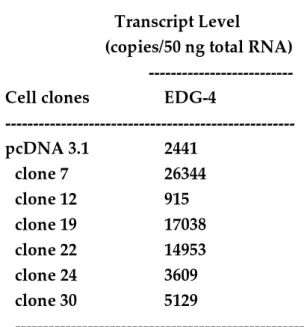
In parallel, we tested whether stable expression of EDG-2 antisense cDNA would affect EDG-4 receptor expression. EDG-4 transcripts were thus quantified in the six clones depicted in figure 3B. Because EDG-4 mRNAs could not be detected by Northern blot, real time RT-PCR was used for their quantification. When compared to 3T3F442A preadipocytes transfected with the empty vector, EDG-4 mRNA level was found to be increased in clone 7, clone 19 and clone 22, and significantly decreased in clone 12 (Table 2). Therefore, down-regulation of EDG-2 transcripts was not systematically accompanied by a compensatory up-regulation of EDG-4 transcripts.
C/ Influence of EDG-2 antisense cDNA transfection on LPA-dependent proliferation.
We previously demonstrated that 1-oleoyl-LPA, the most active LPA species, increases proliferation in growing 3T3F442A preadipocyte (14). In 3T3F442A preadipocytes transfected with empty vector, 1 µM 1-oleoyl-LPA by itself induced a significant increase (150% of the control) in cell number (Figure 4). Among the six G418-resistant cell clones of the Figure 3B, only clone 7 and clone 24 exhibited a significant reduction in the proliferative response induced by 1 µM 1-oleoyl-LPA (Figure 4). Clone 7 and clone 24 were those harboring detectable alteration of EDG-2 receptor protein expression (see Figure 3C). It was also noticeable that clone 7 exhibited an alteration of preadipocyte responsiveness to LPA despite the existence of the high level of expression of EDG-4 transcripts (Table 2). Clone 12, which exhibited no alteration of EDG-2 transcripts but a significant reduction of EDG-4 transcripts (Table 2), revealed no significant alteration in the proliferative response to LPA. Results showed that reduction of EDG-2 receptor expression was accompanied by an alteration in the proliferative response of 3T3F442A preadipocytes to LPA. In parallel, a poor contribution of EDG-4 receptor in this response was suggested.
LPA is abundant (0.5 to 2.5 µM) in serum (20), and contributes to its biological activity (21-24). In 3T3F442A preadipocytes transfected with the empty vector, 10% serum led to a large increase in cell number, which was significantly reduced (about 30%) by pre-treatment of the serum with phospholipase B (Figure 5). Phospholipase B is a lysophospholipase previously shown to hydrolyze LPA and suppress its bioactivity (14,21,25). Therefore, LPA significantly contributed to the proliferative response of 3T3F442A preadipocytes to serum. In clone 7, the response to serum was significantly lower as compared to 3T3F442A preadipocytes transfected with the empty vector. In addition, it was not significantly modified by phospholipase B treatment (Figure 5). Similar results were obtained with clone 24 (not shown). Results showed that clone 7 exhibited a strong reduction in its proliferative response to the LPA present in serum.
Finally, it was noticeable that the phospholipase B-insensitive proliferative response to serum was not significantly different between clone 7 and empty vector transfected cells (Figure 5). This showed that clone 7 exhibited no alteration in the proliferative response to phospholipase B-insensitive growth factors.
D/ Influence of EDG-2 antisense cDNA transfection on LPA-dependent spreading
In several cell types, including 3T3F442A preadipocytes (14,25), 1-oleoyl-LPA (LPA) and sphingosine-1-phosphate (S1P) (another bioactive phospholipid acting via a distinct receptor than LPA), induce a rapid and powerfull reorganization of actin cytoskeleton, leading to a rapid spreading of the cells previously retracted by serum-deprivation (Cont in Figure 6C). In 3T3F442A preadipocytes transfected with empty vector (pcDNA3.1 in Figure 6C), LPA (left panel in Figure 6C) and S1P (right panel in Figure 6C) induced a dose-dependent increase in cell spreading. This effect was quantified by measurement of cell surface (figure 6AB). Detectable spreading effect was observed with 10 nM of both LPA and S1P (Figure 6ABC). In clone 7, the dose-response curve generated by LPA was significantly shifted to the right, with a detectable spreading effect observed only at 100 nM (Figure 6A and C left panel). On the contrary, the dose response curve generated by S1P was not significantly different between 3T3F442A preadipocytes transfected with empty vector and clone 7 (Figure 6B and C right panel). A weak but not significant reduction in maximal response of sphingosine-1-phosphate was observed in clone 7. Results showed that clone 7 exhibited a significant and specific reduction in its spreading response to LPA.
E/ Expression of EDG-2 mRNAs during the conversion of growing 3T3F442A preadipocytes into growth arrested adipocytes .
When cultured in fetal calf serum and insulin (see Material and Methods), confluent 3T3F442A preadipocytes can be converted into growth arrested adipocytes (26). Conversion into adipocytes is also characterized by a increased expression of mRNAs encoding adipocyte-specific proteins. Among them is the adipocyte-lipid-binding protein (ALBP) encoded by aP2 gene (27). The influence of adipose conversion of 3T3F442A preadipocytes was tested on the expression of EDG-2 mRNAs. By using Northern blot analysis, it was observed that adipocyte conversion (characterized by a rapid increase in aP2 mRNA level) was accompanied by a coordinate and strong reduction in EDG-2 mRNA level (Figure 7). Results suggested that EDG-2 receptor likely played a more important role in growing preadipocytes than in growth arrested and differentiated adipocytes.
Discussion
The results of the present study bring several evidences for a predominant contribution of EDG-2 receptor in the responses of 3T3F442A preadipocytes to LPA. Among the four potential LPA-receptors (2, 4, 7 and PSP24), only EDG-2 and EDG-4 transcripts were found in 3T3F44EDG-2A preadipocytes. Quantitative analysis of transcript abundance, revealed predominance of EDG-2 receptor over EDG-4 receptor. This predominance was also found in another preadipose cell line: 3T3L1 (Krief et al, personal data). Therefore EDG-2 receptor is likely primarily involved in the action of LPA in preadipocytes. This assessment is supported by experiments showing that antisense-directed reduction in EDG-2 receptor expression significantly altered the responses of 3T3F442A preadipocytes to LPA. It was indeed possible to isolate 3T3F442A-derived cell clones exhibiting significant reduction of endogenous EDG-2 mRNA and protein level, associated with significant reduction of the cellular responses to LPA: proliferation and cytoskeleton reorganization (Figures 4 to 6). This reduction was specific to LPA since, in parallel, responses to sphingosine-1-phosphate (Figure 6) or to other phospholipase B-insensitive growth factors (Figure 5) were not significantly altered.
Nevertheless, this antisense strategy did not completely block the action of LPA. This very likely resulted from a partial blockade of EDG-2 receptor expression following antisense cDNA transfection since a substantial decrease, but not total disappearance, of EDG-2 expression is elicited by EDG-2 antisense stable expression. In addition, one cannot completely exclude the possible contribution of another receptor in the residual responses generated by LPA. Beside EDG-2 receptors, 3T3F442A preadipocytes also express EDG-4 receptor. However, the data of the Table 2 reveal that variations of EDG-4 transcript expression in EDG-2 antisense expressing clones cannot be correlated with modifications of the proliferative response to LPA. Although we are aware that these data should be confirmed at the protein level, they strongly suggest the poor contribution of EDG-4 receptor in the responses of 3T3F442A preadipocytes to LPA.
Finally, EDG-2 transcripts were predominantly expressed in growing preadipocytes and were strongly reduced in growth-arrested and differentiated adipocytes. The precise mechanisms involved in down-regulation remain unclear, and are currently under investigation. Whatsoever, this observation strongly supports the role of EDG-2 receptors in the proliferative response to LPA in growing preadipocytes.
Although several studies have shown that overexpression of EDG-2 cDNA restores or increases LPA-sensitivity in mammalian cells (5,8), the specific contribution of endogenously expressed EDG-2 receptor remained poorly documented. Goetzl et al. (28) have shown that the antiapoptotic response of a human T-lymphocyte cell line to LPA could significantly be reduced by transfection with EDG-2 plus EDG-4 antisense
cDNA. Specific contribution of EDG-2 remained to be determined. The present study brings evidences for a specific contribution of EDG-2 receptor endogenously expressed in preadipocytes.
Previous work from our laboratory (14) revealed that LPA can be produced by adipocytes and play an important role in paracrine/autocrine control of proximal preadipocytes. Because of the involvement of EDG-2 receptor in the control of the proliferation of preadipocytes, this receptor appears to be an interesting target to control preadipocyte proliferation, one of the key events of adipose tissue development.
Acknowledgments: We thank Dr. Jerold Chun for providing vzg-1 antibody, Dr. Gabor Tigyi for providing xenopus PSP24 vector and mouse PSP24 sequence, and Isabelle Lefrère for expert technical assistance. This work was supported by grants from the "Institut National de la Santé et de la Recherche Médicale" (APEX #4X405D), the "Association pour la Recherche sur le Cancer" (#5381), the "Laboratoires Clarins" and the "Institut de Recherche Servier".
References
1. Gaits, F., Fourcade, O., F, L. B., Gueguen, G., Gaigé, B., Gassama-Diagne, A., Fauvel, J., Salles, J.-P., Mauco, G., Simon, M.-F., and Chap, H. (1997) FEBS Lett. 410, 54-58
2. Jalink, K., Hordijk, P. L., and Moolenaar, W. H. (1994) Biochim. Biophys. Acta. 1198, 185-196
3. Goetzl, E., and An, S. (1998) FASEB J. 12, 1589-1598
4. Chun, J., Contos, J. J., and Munroe, D. (1999) Cell Biochemistry and Biophysics 30, 213-242
5. Hecht, J., Weiner, J., Post, S., and Chun, J. (1996) J. Cell Biol. 135(4), 1071-1083
6. Hla, T., and Maciag, T. (1990) J. Biol. Chem. 265, 9308-9313
7. Lee, M., Brocklyn, J. V., Thangada, S., Liu, C., Hand, A., Menzeleev, R., Spiegel, S., and Hla, T. (1998) Science 279(5356), 1552-1555
8. An, S., Dickens, A., Bleu, T., Hallmark, O., and Goetzl, E. (1997) Biochem. Biophys. Res. Com. 231, 619-622
9. An, S., Bleu, T., Hallmark, O., and Goetzl, E. (1998) J. Biol. Chem. 273(14), 7906-7910
10. Bandoh, K., Aoki, J., Hosono, H., Kobayashi, S., Kobayashi, T., Murakami-Murofushi, K., Tsujimoto, M., Arai, H., and Inoue, K. (1999) J Biol Chem 274(39), 27776-85
11. Contos, J., and Chun, J. (2000) Genomics 64, 155-169
12. Guo, Z., Liliom, K., Fischer, D. J., Bathurst, I. C., Tomei, L. D., Kiefer, M. C., and Tigyi, G. (1996) Proc Natl Acad Sci USA 93, 14367-14372
13. Kawasawa, Y., Kume, K., and Shimizu, T. (1998) FASEB J. 18(869)
14. Valet, P., Pagès, C., Jeanneton, O., Daviaud, D., Barbe, P., Record, M., Saulnier-Blache, J., and Lafontan, M. (1998) J. Clin. Invest. 101(7), 1431-1438
15. Pagès, C., Girard, A., Jeanneton, O., Barbe, P., Wolf, C., Lafontan, M., Valet, P., and Saulnier-Blache, J. (2000) NY Acad Sci 905, 159-164
16. Bouloumié, A., Planat, V., Devedjian, J.-C., Valet, P., Saulnier-Blache, J.-S., Record, M., and Lafontan, M. (1994) J. Biol. Chem. 269, 30254-30259
17. Liu, C. H., and Hla, T. (1997) Genomics 43(1), 15-24
18. Bétuing, S., Daviaud, D., Valet, P., Bouloumié, A., Lafontan, M., and Saulnier-Blache, J. (1996) Endocrinol. 137(12), 5220-5229
19. Pagès, C., Rey, A., Lafontan, M., Valet, P., and Saulnier-Blache, J. (1999) Biochem Biophys Res Com 265, 572-576
20. Saulnier-Blache, J. S., A., Girard, M.F., Simon, M., Lafontan, and P., Valet (2000) J. Lipid Res. 41, 1947-1951
21. Tigyi, G., and Miledi, R. (1992) J. Biol. Chem. 267, 21360-21367
22. Eichholtz, T., Jalink, K., Fahrenfort, I., and Moolenaar, W. H. (1993) Biochem. J. 291, 677-680
23. Tokumura, A., Iimori, M., Nishioka, Y., Kitahara, M., Sakashita, M., and Tanaka, S. (1994) Am. J. Physiol. 267, C204-C210
24. Sasagawa, T., Suzuki, K., Shiota, T., Kondo, T., and Okita, M. (1998) J Nutr Sci Vitaminol 44, 809-18
25. Ridley, A. J., and Hall, A. (1992) Cell 70, 389-399
26. Bétuing, S., Valet, P., Lapalu, S., Peyroulan, D., Hickson, G., Daviaud, D., Lafontan, M., and Saulnier-Blache, J. S. (1997) Biochem. Biophys. Res. Com. 235, 765-773
27. Bernlohr, D. A., Angus, C. W., Lane, M. D., Bolanowski, M. A., and Kelly, T. J. (1984) Proc. Natl. Acad. Sci. USA 81, 5468-5472
28. Goetzl, E. J., Shames, R. S., Yang, J., Birke, F. W., Liu, Y. F., Albert, P. R., and An, S. (1994) J. Biol. Chem. 269, 809-812
Table 1: Expression of EDG-2 and EDG-4 transcripts in growing 3T3F442A preadipocytes.
---
Receptor Transcript Level
---
EDG-2 15500 ± 1944
EDG-4 1550 ± 597
_________________________________
EDG-2 and EDG-4 mRNAs were quantified from growing 3T3F442A preadipocytes using real time RT-PCR (see Materials and Methods). In each experiment EDG-2 and EDG-4 transcripts were quantified from the same cDNAs preparation. Results were obtained from 4 experiments and expressed in copies of mRNA per 50 ng total RNA.
Table 2: Expression of EDG-4 transcripts in 3T3F442A preadipocytes transfected with EDG-2 cDNA antisense.
---
Transcript Level
(copies/50 ng total RNA) ---
Cell clones EDG-4
--- pcDNA 3.1 2441 clone 7 26344 clone 12 915 clone 19 17038 clone 22 14953 clone 24 3609 clone 30 5129 ---
EDG-4 mRNAs were quantified in antisense EDG-2 cDNA transfected 3T3F442A preadipocytes using real time RT-PCR (see Materials and Methods). Results corresponded to one experiment performed in triplicate.
Legend of Figures.
Figure 1: RT-PCR detection of EDG-2, EDG-4 and EDG-1 mRNA in 3T3F442A
preadipocytes. RT-PCR was performed from 1 µg of DNAse-I treated total RNA (see
Material and Methods) extracted from growing 3T3F442A preadipocytes (3T3), mouse brain (brain), mouse spleen (spleen), human heart (heart). Thirty-five PCR cycles were performed from the same RT with specific primers for EDG-2, EDG-4, EDG-7, PSP24 and EDG-1 (see Material and Methods). Representative result of at least 3 separate experiments.
Figure 2: Northern blot detection of EDG-2, EDG-1 mRNA in 3T3F442A
preadipocytes. Twenty µg of total RNAs extracted from growing 3T3F442A
preadipocytes or mouse brain were analyzed by Northern blot using specific [32P]labeled probes directed against EDG-2 (A) and EDG-1 mRNAs (B). A 18S ribosomal RNA probe was used to insure equal well loading. Data presented are representative of at least 3 separate experiments.
Figure 3: Expression of EDG-2 antisense and sense mRNAs in G418-resistant cell clones transfected with EDG-2 antisense cDNA. (A) The presence of antisense EDG-2 mRNAs was examined by RT-PCR analysis (see Material and Methods) in G418-resistant cell clones (7 to 31) transfected with antisense EDG-2 cDNA and in empty pcDNA3.1 transfected cells. (B) EDG-2 mRNA level in G418-resistant cell clones analyzed by Northern blot as described in Figure 2. (C) EDG-2 receptor protein level analyzed by Western blot (see Material and Methods) in clone 7, clone 24 and in empty pcDNA3.1 transfected cells: representative of two separate experiments.
Figure 4: Influence of LPA on the proliferation of G418-resistant cell clones transfected with EDG-2 antisense cDNA. Each cell clone were seeded and grown in 10% donor calf serum supplemented DMEM. After 48 h, serum was removed and each cell clone was grown for an additional 48 h in the presence or absence of 1 µM 1-oleoyl-LPA. Cell number obtained in each clone was determined as described in Material and Methods and compared to that obtained with empty pcDNA3.1 transfected cells. Each column represents the mean ± SE of 3 to 5 independent experiments depending on cell clone. Statistical analysis was performed using the Student's t test: * P<0.05 when comparing LPA activity in each clone to that measured in empty pcDNA3.1 transfected cells.
Figure 5: Influence of antisense EDG-2 cDNA transfection on the proliferative response to serum. Clone 7 and empty pcDNA3.1 transfected 3T3F442A preadipocytes
were seeded and grown in 10% donor calf serum supplemented DMEM. After 48 h, the medium was changed with fresh 10% fetal calf serum supplemented DMEM pre-treated (+) or not (-) with 0.1 U/ml of phospholipase B overnight. Cell number was determined after 48 h as described in Material and Methods. Each column represents the mean ± SE of 5 independent experiments. Statistical analysis was performed using the Student's t test: P<0.05 when comparing the effect of serum with that of phospholipase B-treated serum (*), and P<0.05 when comparing pcDNA3.1 with clone 7 in the absence of phospholipase B (#). NS: non statistically different.
Figure 6: Influence of EDG-2 antisense cDNA expression on the cell spreading response to LPA. Growing clone 7 (black boxes) or empty pcDNA3.1 transfected cells (white boxes) were retracted by serum deprivation as described in Material and Methods, and exposed to increased concentrations of 1-oleoyl-LPA (A) or sphingosine-1-phosphate (B). After 15 min cell surface was measured as the intensity of cell spreading. Each value represents the mean ± SE of 3 independent experiments. Statistical analysis was performed using the Student's t test: P<0.05 when comparing clone 7 (black boxes) and empty pcDNA3.1 transfected cells (white boxes) (*). (C) Photo of one representative experiment of cell spreading.
Figure 7: Expression of EDG-2 and aP2 mRNAs during conversion of 3T3F442A preadipocytes into adipocytes. 3T3F442A preadipocytes were grown in donor calf serum (SVD) supplemented DMEM until confluence. At confluence, the medium was replaced by fetal calf serum supplemented DMEM plus insulin (SVF+insulin) in order to induce adipose conversion (see Material and Methods). Total RNAs were extracted at different time during the course of preadipocytes conversion into adipocytes, and mRNAs were detected by Northern blot. Data are representative of at least 3 separate experiments.