HAL Id: hal-02188401
https://hal.archives-ouvertes.fr/hal-02188401
Submitted on 19 Jul 2019HAL is a multi-disciplinary open access archive for the deposit and dissemination of sci-entific research documents, whether they are pub-lished or not. The documents may come from teaching and research institutions in France or abroad, or from public or private research centers.
L’archive ouverte pluridisciplinaire HAL, est destinée au dépôt et à la diffusion de documents scientifiques de niveau recherche, publiés ou non, émanant des établissements d’enseignement et de recherche français ou étrangers, des laboratoires publics ou privés.
Easy formation of functional liposomes in water using a
pH- responsive microbial glycolipid: encapsulation of
magnetic and up-converting nanoparticles
Lisa Van renterghem, Fabrizio Guzzetta, Patrick Le griel, Mohamed Selmane,
Ghazi Ben Messaoud, Tabitha Tan su teng, Sierin Lim, Wim Soetaert, Sophie
Roelants, Beatriz Julian-Lopez, et al.
To cite this version:
Lisa Van renterghem, Fabrizio Guzzetta, Patrick Le griel, Mohamed Selmane, Ghazi Ben Messaoud, et al.. Easy formation of functional liposomes in water using a pH- responsive microbial glycol-ipid: encapsulation of magnetic and up-converting nanoparticles. ChemNanoMat, Wiley, 2019, 5 (9), pp.1188-1201. �10.1002/cnma.201900318�. �hal-02188401�
Easy formation of functional liposomes in water using a
11
pH-responsive microbial glycolipid: encapsulation of
12
magnetic and up-converting nanoparticles
13
Dr. Lisa Van Renterghem,a Dr. Fabrizio Guzzetta,b Patrick Le Griel,c Mohamed
14
Selmane,c Dr. Ghazi Ben Messaoud,c Tabitha Tan Su Teng,d Prof. Sierin Lim,d Prof. Wim
15
Soetaert,a,e Dr. Sophie Roelants,a,e Dr. Beatriz Julián-López,b Dr. Niki Baccilec,*
16 17
a - InBio – Center for Industrial Biotechnology and Biocatalysis, Department of Biotechnology 18
Faculty of Bioscience Engineering, Ghent University 19
b - Institute of Advanced Materials (INAM), Universitat Jaume I, Avda. Sos Baynat s/n, 12071 20
Castellón, Spain 21
c - Sorbonne Universités, CNRS, Collège de France, Chimie de la Matière Condensée de Paris 22
UMR 7574, 4, Place Jussieu, 75005 Paris, France 23
d - School of Chemical and Biomedical Engineering, Nanyang Technological University, 70 24
Nanyang Dr., Singapore 637457, Singapore 25
e - Bio Base Europe Pilot Plant, Rodenhuizekaai 1, 9042 Gent, Belgium 26
27
Corresponding author: niki.baccile@upmc.fr 28
29
Abstract 30
The compartmentalization of colloids into topologically closed, vesicular, microphases offers 31
an attractive mean to concentrate a functional cargo in aqueous solutions for a range of 32
biomedical, cosmetic, and biotechnological applications. In this paper, we develop a simple, 33
phospholipid-free, phase change method employing a pH-responsive glycolipid. The method is 34
applied to the encapsulation of a sonicated, metastable, aqueous dispersion of functional 35
colloids in the lumen of lipid vesicles: uncoated magnetic maghemite -Fe2O3 and oleic-acid
36
coated upconverting NaYF4:Yb/Ln (Ln= Er or Tm) nanoparticles (NPs). We find a stable
37
liposomal dispersion containing a sub-population of crowded liposomes with high 38
concentrations of NPs. The encapsulated NPs, formed at nearly neutral pH and room 39
temperature, are stable over time and towards extrusion. The vesicular microphase is entirely 40
composed of pH-responsive glycolipids, which undergo a pH-mediated mesoscopic structural 41
transition from an open lamellar (2 < pH < 4) to topologically closed vesicular state (pH > 4). 42
We also show that encapsulation successfully works with a stable colloidal aqueous dispersion 43
of iron clusters stabilized in ferritin cages. This compartmentalization approach combining self-44
assembly with an orthogonal nonequilibrium dispersion of nanoparticles shows untapped 45
potential for synthesizing unusual classes of mixed matter. 46
47
Introduction 48
Encapsulation of nanoparticles into topologically-closed liposomes represents one of 49
the most valuable advances for therapeutics purposes, because it enables an integration of the 50
properties (e.g., optical, magnetic, and luminescent) of nanoparticles (NPs) with those of 51
liposomes (protection, stability, biocompatibility).1 Since the early 80s, nanoparticle-loaded
52
liposomes have been explored to probe cell-liposome interactions on gold2 and magnetite.3,4
53
More recently, there has been more focus on the the diversity of not only the nature of the 54
encapsulated NPs, like silica and quantum dots,5 but also to achieve better encapsulation
55
methods.6 The latter allows, for instance, high NPs load uptakes by the liposomes7 or the
56
development of stimuli-responsive vectors based on lipids8,9 or polymers.10,11,12 In the last
57
decade, liposomal encapsulation of iron-loaded ferritin cages has even been associated with the 58
fundamental questions underlying the origins of life.13
59
The case of magnetoliposomes, an idea being at least three decades old,3,4,14 is
60
particularly interesting. Magnetoliposomes refer to liposomes that contain magnetic NPs 61
(mainly Fe3O4 and -Fe2O3) in their lumen, lipid bilayer or at the vesicle surface,1,9,15 and which
62
are particularly interesting for the well-known applications in hyperthermia or as contrast agent 63
in magnetic resonance imaging.16 Either alone or in combination with drug loading, liposomal
64
protection increases the blood circulating time, bioavailability and delivery.1,8,9 The first
65
reported preparation approach to insert NPs into the liposome lumen employed a classical 66
precipitation method:4 simple mixing of pre-formed DMPG17 and DPPG18 phospholipid
67
liposomes with lauric acid coated Fe3O4 particles was shown to precipate NP-loaded vesicles.
68
Other methods have been employed, such as reverse phase evaporation,14,19,20,21 or in-situ
69
liposomal precipitation of the NPs.6 Thin film hydration, double emulsion methods, or ethanolic
70
injection can also be used.13,21,22,23 Recent review papers address the synthesis of
71
magnetoliposomes more extensively.1,9,16 Similar encapsulation strategies are employed for
72
other materials such as metal,24 oxides5 or luminescent nanoparticles including quantum dots5
73
and upconverting lantanide-doped fluorides.25
74
Although these methods are valuable, some of them use toxic halogenated solvents (e.g., 75
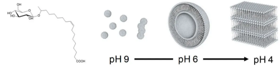
chloroform), require elaborate methodologies (e.g., evaporation under vacuum) and in general 76
the use of stable colloids. The latter is an important point. Most, if not all, of these methods 77
require the use of stable colloids in general. In most cases, many nanoparticle systems are 78
passivated by a hydrophobic layer,25 which easily guarantees encapsulation in the vesicle
79
bilayers.9,15,16 However, ligand exchange, commonly regarded at as tedious unavoidable step,
80
use of surfactants or hydrophilic coatings, become then necessary for encapsulation in the 81
lumen in water-based media.16,21 Moreover, the interaction between the (coated or uncoated)
82
NPs with the phosphate groups of phospholipids may destabilize the bilayer membrane, thereby 83
affecting the vesicle formation mechanism and reducing their stability.1,9 It is then interesting 84
to develop an encapsulation water-based method applicable to a broad range of colloids out of 85
thermal equilibrium. Non-equilibrium encapsulation is generally obtained in food and material 86
science through dynamic processes such as spray drying,26,27 while recent theoretical work
87
could explore its possibilities in the compartimentalization within elastic geometries like 88
carboxysomes.28
89
Lipid switches29,30 were recently combined with classical phospholipid liposomes to
90
deliver a cargo upon application of an external stimulus (pH, ion). In this paper, we take 91
inspiration of this concept to develop a fully water-based (at nearly neutral pH) bulk phase-92
change method to encapsulate, rather than release, a cargo composed of metastable colloids into 93
vesicles. The latter are compartments entirely free of phspholipids and only composed of pH-94
responsive lipid switches. Vesicle formation is dynamic, reversible, rapid and entirely 95
controlled through pH. We apply this method to the formation of magnetoliposomes, chosen as 96
model system abundantely described in the literature,4,9 from an unstable dispersion of bare
97
(uncoated) maghemite -Fe2O3 nanoparticle aggregates. We then extend it to the encapsulation
98
of hydrophobic, oleic acid coated, NaYF4:Yb/Ln (Ln= Er or Tm) upconverting nanocrystals.31
99
Both systems, otherwise sedimenting/segregating in/from water, are driven out of equilibrium 100
by applying strong sonication before encapsulation through a pH-jump process. 101
The method proposed here involves a lamellar-to-vesicle phase transition, conceptually 102
analogous to the mechanism occurring in thin film hydration or in the preparation of 103
vesosomes,32 but driven by a simple pH jump (generally ~4 to ~6) in bulk water. Instead of
104
common complex formulations combining phospholipids with pH-responsive lipids8,33,34 or
105
block-copolymers,35 we use a single pH-sensitive microbial glycolipid (GL) (Figure 1).36
106
Microbial glycolipids are biobased compounds known for their stimuli-responsive 107
properties,37,38,39 their biodegradability and low toxicity,40-45 and for these reasons particularly
108
interesting for biomedical applications. GL are known to form a set of closed vesicular 109
Using a combination of cryo-TEM, light and X-ray scattering, we find that high loads 111
of nanoparticles tend to occupy a limited number of vesicular compartments. This feature is 112
also observed upon loading of ferritin nanocages,47 a result which agrees with the finding of
113
Luisi et al.,13 who proposed that the massive occupation of a small fraction of vesicles,
114
coexisting with a large majority of empty vesicles, could explain the compartimentalization of 115
heregenous genomic material in the early stages of life formation. 116
117
Figure 1 - Acidic form of the microbial glucolipid GC18:1 (GL) and its corresponding pH-dependent phase
118
behaviour at room temperature: micelles, vesicle, lamellar.
119 120
Material and Methods 121
Synthesis of glycolipid GC18:1 (GL). GLs were produced in a scaled-up bioreactor 122
experiment (100 L) by the S. bombicola strain ∆ugtB1 described by Saerens et al.36 The detailed
123
production, purification and hydrolysis process to obtain the compound in Figure 1 has been 124
reported elsewhere.46 GLs contain about 90 % and 7 % of the subterminal (glycosidic bond
125
between glucose and the C17 of the fatty acid chain, Figure 1) and terminal (glycosidic bond 126
between glucose and the C18 of the fatty acid chain) congener, respectively. The remaining 3 127
% is constituted by di-unsaturated C18:2 and saturated C18:0 impurities. 128
Preparation of iron oxide nanoparticles (NPs). Iron oxide NPs were synthesized 129
using the co-precipitation method by adapting the standard protocol48,49 to obtain the inverse
130
spinel structure typically observed in magnetite, where [Fe2+]/[Fe3+]= 0.5. Specifically, 0.177 g
131
of FeCl3 •6H2O was mixed with 0.108 g of FeCl2 •4H2O in a round-bottom flask containing 20
132
mL of Milli-Q water. To this solution, 2.7 mL of a 28 % ammonium hydroxide solution was 133
gradually added under continuous mechanical stirring. The system was maintained in an argon-134
rich environment to limit oxidation. The resulting black precipitate was extracted by 135
magnetically-assisted sedimentation and washed with MilliQ water. This operation was 136
repeated three times to remove any residual salts. 137
Preparation of upconverting nanoparticles (UCNP). Upconverting oleic acid capped 138
NaYF4:Yb/Er and NaYF4:Yb/Tm nanoparticles (20%-Yb and 0.5%-Ln, Ln= Er, Tm) molar
ratio replacing yttrium ions in the lattice, labelled as UCNPs) of size between 20 nm and 40 nm 140
were prepared according to ref. 50. 141
Iron-loaded ferritin cages preparations and characterization. The iron-loaded 142
ferritin cages (AfFtn) are prepared as previously described.47,51 Briefly, the ferritin is derived
143
from the archaeon Archaeoglobus fulgidus and was produced recombinantly in E. coli strain 144
BL21(DE3)CodonPlus-RIL (Stratagene) with IPTG induction. The harvested cells were 145
sonicated and heat-treated at 85°C for 10 minutes. The insoluble fraction and denatured proteins 146
were removed using ultracentrifugation and the supernatant was passed through 0.22 µm filter. 147
No further purification was performed for this report. Iron was loaded by adding Fe2SO4
148
dropwise until 4800 Fe/cage had been loaded. To remove the unloaded iron, the sample was 149
desalted, concentrated using 100 kDA MWCO Amicon filter, and sterile filtered using 0.22 µm 150
syringe filter. The iron-loaded ferritin (Fe4800)AfFtn preparation was characterized by 151
dynamic light scattering (DLS) technique to confirm that the hydrodynamic size was ~13 nm 152
and the protein concentration was estimated using Bradford assay.47,51
153
NPs-containing vesicle preparation. Ten mL of the NPs solution were set to pH 9 and 154
bath-sonicated during 30 min to one hour. After sonication, the NPs solution was mixed at room 155
temperature to 10 mL of a micellar GL solution (0.29 g GL in 10 mL) at pH 9. The NPs GL 156
mixture at pH 9 (referred to NPs GL, pH9, sample S2) was kept under stirring for few minutes 157
before use. Note: the NPs solution is not sonicated in the presence of the GL solution. According 158
to the data in Baccile et al.,46 GL self-assembles into vesicles at pH values below 6.2. Thus, to
159
prepare vesicles, two aliquots of the NPs GL solution at pH 9 were acidified using 1 M HCl 160
solution: in aliquot 1, the pH is lowered to 6, and this sample is referred to as NPs GL, pH6 161
(sample S3) and in aliquot 2, the pH is lowered below 4 and then increased again to pH 6 and 162
named NPs GL, pH2→pH6 (sample S4). The latter will also be referred to as the manual pH-163
jump approach 2, where approach 1 corresponds to sample S1, that is a control experiment 164
only containing NPs and free of GL. In a second control experiment (sample S5), the pH of the 165
NPs (GL-free) solution was lowered to 6 with 1 M NaOH, and 0.45 g of non-acetylated acidic 166
sophorolipids were subsequently added, similarly to the two-step procedure described in 167
Baccile et al.55 Sophorolipids are microbial glycolipids having a disaccharide headgroup and
168
known to self-assemble into micelles, instead of vesicles, under the acidic pH conditions.46 The
169
detailed sample compositions are given in Table 1. 170
Table 1 – Composition table of the samples studied in this work. GL: acidic glucolipid GC18:1 (Figure 1),
171
NPs: -Fe2O3 nanoparicles,55 SL: sophorolipids.55 The concentration of NPs mother solution is estimated by
and NPs (4.2 mg/mL) solutions, both at pH 9. pHi, pHm and pHf respectively indicate the initial, middle and
174
final pH. If no pH change has occurred, pHi = pHf. All pH variations are performed by hand using 1 M HCl
175
and 1 M NaOH solutions.
176
Sample name Sample code mg/mL CGL / mg/mL CSL / mg/mL CNPs / pHi pHm pHf
GL - 10 - - 9 - 6 NPs S1 - - 4.2 9 - 9 NPs GL pH 9 S2 10 - 3.2 9 - 9 NPs GL pH 6 S3 10 - 3.2 9 - 6 NPs GL pH 2 → pH 6 S4 10 - 3.2 9 2 6 NPs SL pH 6 S5 - 10 3.2 9 - 6 177
The manual pH-jump (approach 2) described above has been performed manually and 178
for this reason we have tested a series of additional methods to evaluate the robustness of the 179
encapsulation process and to control the vesicle size distribution. These experiments have been 180
performed on both a NPs-free solution constituted of vesicles only (GL in Table 1) and a mixed 181
NP and GL (sample S4 in Table 1). Extrusion (approach 3): after the manual pH-jump 182
(approach 2) the solution is extruded (10 cycles) at 1 mL/min through a 0.45 μm filter. 183
Extrusion is commonly used to control vesicle size in the hundred nanometer range.52,53,54
184
Sonication (approach 4): after the manual pH-jump (approach 2), the solution is sonicated for 185
20 s using an immersive ultrasound probe. Sonication is commonly used to prepare vesicle of 186
diameter below 100 nm.52 Controlled pH-jump (approach 5): after the pH is lowered to 2, pH
187
is increased to 6 using a 1 M NaOH solution injected at 0.5 μL/min under stirring. Dialysis pH-188
jump (approach 6): after the pH is lowered to 2, pH is increased to 6 by dialyzing the solution 189
against a water reservoir at pH 6 using a 3500 MWCO (Spectra/Por®) membrane. Finally, to
190
guarantee homogeneous dispersion of the vesicles and NPs, solution are kept under stirring 191
during the manual pH-jump and controlled pH-jump processes. 192
Preparation of iron-loaded ferritin-containing vesicles. The procedure was adapted 193
from the NPs-containing vesicle process. The stock (Fe4800)AfFtn solution (0.88 mg/mL, pH 194
7.4) was mixed with the a micellar GL solution (40 mg/mL, pH 7.4) to achieve respective final 195
concentrations of 0.66 mg/mL and 10 mg/mL. The pH was then lowered to about 3.9 (using ~ 196
6 μL HCl 5 M) to avoid (Fe4800)AfFtn aggregation. The pH is then increased to 6 using three 197
selected approaches described above. Manual pH jump (approach 2): pH is manually increased 198
by manual addition of 1 M NaOH solution. Controlled pH-jump (approach 5): a 1 M NaOH 199
solution is injected at 0.5 μL/min under stirring until pH 6 was achieved. Extrusion (approach 200
3): the (manual) pH jump solution is extruded (10 cycles) at 1 mL/min through a 0.45 μm filter. 201
Note: (Fe4800)AfFtn is water-dispersible, therefore it is never sonicated. 202
Preparation of UCNP-containing vesicles. To a 10 mg/mL micellar GL solution in 203
water at pH 9, we add the dried oleic-acid-capped NaYF4:Yb/Er and NaYF4:Yb/Tm UCNP
204
powder at concentration of 5 mg/mL. The UCNP sample is water-insoluble and for this reason 205
sonication is necessary to help dispersion. For the encapsulation, we employ the manual pH 206
jump (approach 2): pH is reduced to 4 (manual dropwise addition of μL amounts of 0.5 M HCl) 207
and then increased to ~6 (manual dropwise addition of μL amounts of 1 M NaOH). The same 208
protocol applies to a control solution only containing the UCNP sample, without adding GL. 209
Despite the hydrophobic character of the oleic acid-coated UCNP, sonication is enough to 210
disperse them in water, although sedimentation is fast for the control, GL-free, experiment. 211
Sedimentation experiments using photoluminescence spectroscopy. Free 212
sedimentation: the UCNP dispersion is placed in a quartz cuvette and irradiated with a CW laser 213
source (λ= 980 nm) at a power density of 105 W/cm2. Emission spectra were recorded after 0,
214
10, 20, 30, 60, 120, 300, 600, 1200, 1800, 3600, 5400 s. Forced sedimentation: The setup and 215
conditions for photoluminescence measurements were identical to the free sedimentation. 216
However, the encapsulated UCNP dispersion was centrifuged at 2000 rpm in a time scale 217
comprised between 0 and 300 seconds, and emission spectra were taken after 10, 20, 30, 60, 218
120 and 300 seconds of centrifugation. 219
Light scattering (LS) experiments. Light scattering experiments were performed on a 220
Malvern Zetasizer Nano ZS instrument (λ= 633 nm) at constant shutter opening and same 221
sample-to-detector distance. The diffused light was expressed in terms of the derived count rate 222
in kilocounts-per-seconds (kcps). All solutions were diluted 100 times prior to analysis. Two 223
measurements were performed, the first one on the solution itself and the second one on the 224
supernatant of the same solution, after magnetically-assisted sedimentation of the suspended 225
NPs using a neodymium magnet. 226
Transmission Electron Microscopy (TEM). TEM was performed on a FEI Tecnai 120 227
Twin microscope operating at 120 kV and equipped with a high resolution Gatan Orius CCD 228
4k x 4k numeric camera. DigitalMicrograph™ software was used for image acquisition. TEM 229
images acquired in cryogenic mode were obtained from the same instrument. The sample holder 230
was a Gatan Cryoholder (Gatan 626DH, Gatan). DigitalMicrograph™ software was used for 231
image acquisition. Cryo-fixation was done on a homemade device. The solutions were 232
deposited on glow discharged holey carbon coated TEM copper grids (Quantifoil R2/2, 233
Germany). Excess solution was removed and the grid was immediately plunged into liquid 234
experimentation. 236
Small Angle X-ray Scattering (SAXS). SAXS experiments were recorded at the high 237
brilliance ID02 beamline at the ESRF synchrotron (Grenoble, France) using a 1 m detector-to-238
sample distance and a flow-through polycarbonate capillary. Aquisition time was 239
1 s per pH value. pH was changed in-situ in the experimental hutch and followed live. More 240
details on the experimental setup have been provided in Baccile et al.46 Data were acquired
241
using a CCD camera and integrated azimuthally to obtain a typical I(q) spectrum. Contribution 242
of the solvent (water at pH 11.6) and capillary were measured prior to the experiment and 243
accordingly substracted during the data treatment. All data were corrected for the transmission 244
of the direct beam. 245
Additional SAXS experiments were performed on a laboratory three-pinhole type S-246
MAX 3000 RIGAKU Nanoviewer instrument using a monochromatic Cu-Kα radiation 247
produced by a microfocus (20 microns x 20 microns) sealed tube X-ray source (MicroMax 002+ 248
RIGAKU working at 40 W) and equipped with a two-dimensional multi-wire proportional gas 249
detector. The sample-to-detector distance was set to 1469 mm. The applied voltage and filament 250
current were 40 kV and 50 mA respectively. The q-range calibration was made using a silver 251
behenate standard sample (dref = 58.38 Å). The measured intensity was always divided by the 252
sample transmission and appropriate masking was done to eliminate the beam-stop shadow 253
contribution and imperfection of the 2D detector. Quartz sample holders of 1 mm diameter were 254
used in a flow-through mode using a home-made device, thus assuring that the background 255
signal was constant for all samples. The subtracted background was constituted by the water-256
containing quartz capillary. The acquisition time per sample is 1 h. 257
X-ray Didffraction (XRD). XRD data were recorded on the dried powders using a 258
Bragg-Brentano θ-2θ (λ= 1.54 Å) goniometer Bruker D8 Discover instrument. 259
Differential thermal analysis (DTA). DTA was performed in a Mettler-Toledo 260
TGA/SDTA851e/LF/1600 instrument. The analysis of the powders was carried out in Pt 261
crucibles at a heating rate of 5 ºC/min under air atmosphere over the range of 20-900 ºC and 262
using alumina as reference. 263
Photoluminescence spectroscopy. Upconversion photoluminescence was recorded 264
using an infrared laser diode RLTMDL-980-2W module (980 nm ± 5 nm, 2 W cw) from 265
Roithner LaserTechnik, and a Black Comet CXR (StellarNet) optical fiber as detector. The 266
emission was measured using dispersions of the UCNPs (10 mg in 1mL of ethanol for bare 267
nanoparticles, or in 1 mL of water for the samples encapsulated in glycolipids) and focusing 268
the laser on the samples at a pump density of 105 W/cm2.
270
Results and discussion 271
Encapsulation of sonicated iron oxide nanoparticles. The XRD of the bare iron oxide 272
NPs is represented by the diffractogram N°1 on Figure 2, which shows the d-values: 2.967, 273
2.527, 2.092, 1.714, 1.614, 1.478 Å, corresponding, to the (220), (311), (400), (422), (511), 274
(440) Bragg diffraction planes of the iron oxide spinel cubic structure (JCPDS file, N° 19-275
0629), respectively. This is attributed to a maghemite structure, previously identified using 276
Mössbauer spectroscopy.55 The typical size (below 10 nm) and aggregation state of this sample
277
are shown by TEM in Figure S 1 (sample referred to as NPs).55 The magnetic nature of the NPs
278
(Figure 3) shows the attraction of the NP sample to the magnetic field of a neodymium magnet 279
leaving the supernatant solution completely clear. The observation is also confirmed by the 280
light scattering (LS) data in Figure 3 (sample S1) before (white bars, strong scattering) and after 281
(grey bars, no scattering) exposure to the magnet. This is explained by the strong aggregation 282
of the uncoated NPs (Figure S 1, NPs sample) in water. On the other hand, GL mainly assembles 283
into micelles in water at pH 9, as shown by the characteristic SAXS scattering signal, shown in 284
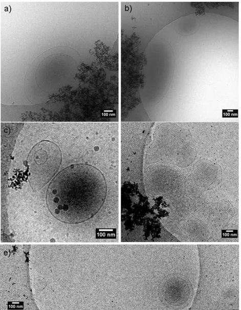
Figure 2b. The same compound is also known to form vesicles when the pH is lowered from 9 285
to 6, as previously demonstrated by us37,46 and shown here by the typical SAXS scattering signal
286
recorded at pH 6.07 in Figure 2b. The SAXS pattern is characterized by a broad oscillation at 287
q above 0.1 Å-1, attributed to the form factor of a lipid bilayer, and an intense scattered intensity
288
at q below 0.05 Å-1, the slope of which in log-log scale is close to -2, a typical value found for
289
planar surfaces. A detailed analysis of the form factor through model-dependent fitting of SAXS 290
data presented in Figure 2b can be found in ref. 37. 291
Addition of GLs to the bare nanoparticle solution at pH 9 does not modify the NPs 292
crystal structure (XRD pattern N°2, Figure 2a), nor the overall aggregation state (sample NPs 293
GL, pH9 in Figure S 1). However, upon exposure to the magnet most NPs are removed from 294
the solution, but the latter still has a light brown colour, indicating a small dispersion of NPs in 295
solution. This is confirmed by the slightly higher scattering signal in LS data (sample S2) in 296
Figure 3. Given the strong molecular similarity between GLs and sophorolipids (SLs), GLs can 297
certainly stabilize a small fraction of the iron oxide NPs by a simple adsorption at their surface 298
via the carboxylate group, a phenomenon observed in a previous study.55
300
Figure 2 – a) X-ray diffraction patterns of 1) iron oxide NPs, 2) NPs GL at pH 9 and 3) NPs GL at pH 6.
301
Reported values relate to the Miller (hkl) indices of the Bragg diffraction planes. b) SAXS curves were
302
recorded on the ID02 line of ESRF synchrotron for a pure GL solution at pH values between 3 and 9. The
303
solutions at pH 6.07 and pH 3.19 have been directly obtained from the solution at pH 9.00 by adding 1 M
304
HCl solution.
305 306
When the pH of the NPs GL solution is lowered from 9 to 6, one can confirm that the 307
nanoparticle crystal structure is intact (XRD pattern N° 3, Figure 2a). Upon approaching an 308
external magnet to the vial, most NPs are attracted, thus leaving the solution slightly yellowish 309
(Figure 3b, sample S3) due to the spurious presence of GL-coated NPs in solution. Light 310
scattering of sample S3 (diluted, Figure 3a) qualitatively confirms the loss in the scattered 311
intensity of the supernatant upon magnetically-assisted sedimentation. Nevertheless, Figure 3b 312
shows that sample S3 (undiluted) supernatant is slightly turbid, if compared to samples S1 or 313
S2; this is explained, and actually expected,46 by the presence of vesicles, confirmed by SAXS
314 20 30 40 50 60 70 2 theta 1) (220) (311) (400) (422) (511) (440) 2) 3) 0.01 0.1 1 pH 3.19 pH 6.07 q (Å-1) pH 9.00 0.18 0.36
a)
b)
I / a .u .(Figure 2b) and the image in Figure 3b (GL, pH 6), both corresponding to a NP-free GL solution 315
at pH 6. The TEM image of sample 3 (NPs GL pH6, Figure S 2a) confirms that nanoparticle 316
size, morphology, structure and aggregation are not affected by the presence of the GL. 317
However, GL can stabilize the nanoparticle surface, as commented above and as expected from 318
the previous work performed on sophorolipids-stabilized iron oxide nanoparticles.55 Light
319
scattering experiments performed on sample S3 (Figure 3b), as well as its dim yellowish colour 320
and aggregation state (Figure S 2a) are comparable to a control system composed of 321
sophorolipid-stabilized iron oxide nanoparticles (sample S5, Figure 3a, Figure S 1, Figure 3b). 322
323
Figure 3 – Light scattering data (a) and magnetically-assisted sedimentation images (b) for bare NPs
324
(sample S1), GL-containing NPs at pH 9 (NPs GL, pH9, sample S2), GL-containing NPs at pH 6 (NPs GL,
325
pH6, sample S3), GL-containing NPs at pH 6 after a transition at pH 2 (NPs GL, pH 2 → pH 6, sample S4),
326
NPs-free GL solution at pH 6 (GL, pH6) and SLs-containing nanoparticle solution at pH 6 (NPs SL pH6,
327
sample S5. Sample S5: control sample composed of sophorolipids-stabilized iron oxide NPs.55
328 329
GLs are known to precipitate into a lamellar phase when the pH is lowered below 4,46
330
also illustrated by the SAXS pattern recorded on a nanoparticle-free GL solution at pH 3.19 and 331
shown in Figure 2b. In the last figure, two sharp diffraction peaks at q = 0.18 Å-1 and 0.36 Å-1
332
overlaps the typical vesicle form factor profile; they respectively correspond to the first and 333
second order interplanar distances lamellar stacking (d= 34.8 Å and d= 17.4 Å). The cryo-TEM 334
analysis of a GL nanoparticle solution at pH below 4 displays the coexistence of nanoparticle 335
clusters and flat lamellae (Figure S 3). Under these conditions, NPs can be easily removed by 336
magnetically-assisted sedimentation, leaving a solid white precipitate in the vial. However, 337
when pH is increased again to 6, the nanoparticle solution becomes extremely stable towards 338
magnetically-assisted sedimentation: light scattering of sample S4 (Figure 3) indicates strong 339
scattering before and after sedimentation. This is confirmed by the images showing the 340
magnetically-sedimented undiluted sample (Figure 3b, sample S4): the dark solution indicates 341
that most NPs are now stabilized. We anticipate that stability is observed over several hours of 342
continuous exposure to the neodymium magnet. 343
Figure S 2b-d show that particle size (~10 nm) is unchanged compared to the GL-free 344
NP solution and high magnification (Figure S 2b) and Fourier transform (Figure S 2c) indicate 345
the crystallinity (Figure S 2d) of the particle with a typical distance of 2.9 Å, attributed to the 346
(220) plane of maghemite. 347
348
Figure 4 – a-d) Cryo-TEM images of NPs GL samples obtained after a pH jump (pH 9 →) pH 2 → pH 6
349
(NPs GL pH2→pH6, sample S4). e-g) Laboratory SAXS data recorded on e) bare NPs and NPs-free GLs at
350
pH 6 (GL pH6), f) NPs GL samples at pH 6 directly adjusted from pH 9 (NPs GL pH6, sample S3) before
351
(black curve) and after (grey curve) magnetically-assisted sedimentation, g) NPs GL samples at pH 6
352
adjusted from pH 9 after passing through pH 2 (NPs GL pH2→pH6, sample S4) before (black curve) and
353
after (grey curve) magnetically-assisted sedimentation
354 355
Although GL adsorption is not excluded, the only surface passivating effect of GL 356
cannot explain the colloidal stability at pH 6, because only a fraction of the NPs are in fact 357
concerned, as already shown for sample S3 and control sample S5.55 The remarkable stability
of the nanoparticle suspension is rather explained by the encapsulation of nanoparticle clusters 359
within (mainly) multilamellar vesicles, as shown by cryo TEM experiments in Figure 4a-d. 360
These data are statistically-confirmed by complementary SAXS experiments presented in 361
Figure 4e-f: panel a) shows the typical signatures of bare NPs (black curve) and GL in solution 362
at pH 6 (grey curve). The difference in the scattering intensity (although not in absolute scale) 363
reveals the strong difference between NPs and GL in terms of contrast with the solvent (water). 364
Additionally, the high-q region above 0.1 Å-1 in the GL sample, although very noisy, shows the
365
beginning of the form factor oscillation, which is very clear in the synchrotron-collected data 366
on a larger q-scale (Figure 2b, pH 6.07). The highly stable pH 2 → pH 6 system (Sample S4, 367
Figure 4g) shows that the signal is dominated by the NPs scattering before and after 368
magnetically-assisted sedimentation, thus confirming that vesicles prevent their removal. 369
Finally, we have specifically verified the orthogonality between vesicle-formation and 370
NPs dispersion: the pH 9 → pH 2 → pH 6 method is applied to a NPs-free GL solution and the 371
corresponding cryo-TEM images in Figure S 4 confirm the presence of vesicles, and in 372
particular of multilamellar vesicles. One can compare these results to the sample S3 (NPs GL 373
pH 9 → pH 6), of which the cryo-TEM (Figure 5) and SAXS (Figure 4f) before (black curve) 374
and after (grey curve) magnetically-assisted sedimentation indicate that the NPs are not 375
preferentially encapsulated. Cryo-TEM shows that the NPs mostly aggregate around vesicles, 376
while SAXS shows the typical signal of the NPs before and a pattern typical of the vesicles 377
after magnetically-assisted sedimentation. 378
379
Figure 5 – Cryo-TEM images of NPs GL samples prepared at pH 6 directly adjusted from pH 9 (NPs GL
380
pH 6, sample S3)
381 382
Stability, control of the size distribution and quantification. Time-dependent stability and 383
size control of the encapsulated NPs are two important parameters for potential applications in 384
nanomedicine and they are tested hereafter. Figure 6 shows the time stability of a GL solution 385
mixed with NPs in the presence and absence of a magnet employing different approaches of 386
encapsulation, but all performed using the pH jump (pH 9 → pH 2 → pH 6) process (sample 387
4). Approach 1 and approach 2 are, respectively, the vesicle-free NPs (sample S1) control and 388
the manually-controlled pH jump (sample S4). Among the others, approach 3 and approach 4 389
respectively use extrusion and sonication after encapsulation, both being classical methods to 390
control vesicle size in the 100 nm range (extrusion)52,53,54 and below 50 nm (sonication).52
391
Extrusion is also commonly employed to obtain unilamellar vesicles from multilamellar 392
vesicles. Finally, approach 5 and approach 6 are meant to perform the pH 2 → pH 6 jump 393
either through a controlled NaOH injection (1 M NaOH at 0.5 μL/min, approach 5) or through 394
a very slow modification in pH (approach 6, using dialysis). Without vesicles (filled squares, 395
Figure 6), the NPs sediment after several minutes in the absence of magnet, as expected from 396
the Stokes law, while sedimentation is immediate upon use of a magnet. Whichever the 397
approach of encapsulation, all vesicles-stabilized NPs solutions are similarly stable in time 398
against sedimentation (Figure 6, left-hand): after 15 h, the transmittance is almost constant, 399
while the vesicle-free control is clear. In the presence of a magnet (Figure 6, right-hand), none 400
of the solution shows the same immediate collapse as found in the vesicle-free control (black 401
squares), thus indicating that encapsulation is always successful. However, after 2 h of exposure 402
to the magnet, the transmission is not equivalent among the tested solutions, probably indicating 403
a disparity in terms of the encapsulation efficiency. Extruded, sonicated and controlled pH jump 404
(respectively, approach 3, 4 and 5) seem to be the most efficient approaches. 405
406
407
Figure 6 – Time-dependent stability of NPs GL pH 2 → pH 6 (sample S4) using approaches 1 through 6
(refer to figure and materials and methods section). Data on the left-hand side refer to simple decantation
409
(no magnet) while data on the right-hand side are acquire using magnetically-assisted sedimentation of the
410
solution (vials sit on a neodymium, as shown on figure)
411 412
Extrusion is known to be a severe process that can destabilize the most stable 413
nanoparticle systems.56 Vesicles-stabilized NPs prepared by pH jump provide an ultra-stable
414
dispersion of NPs upon extrusion (approach 3). Video 1 shows that a vesicles-stabilized NP 415
solution can be extruded repeatedly (we tested up to ten cycles) without aggregation of the NP 416
onto the filter, while a vesicle-free NP solution cannot be extruded once (Video 2). The 417
advantage of employing various sample preparation approaches is the possibility to control the 418
vesicles size distribution, as formulated theoretically in the development of nonequilibrium 419
compartments.28 Figure 7 shows the autocorrelation function and the corresponding number
420
distribution of the (vesicle-free) iron oxide NPs solution (in red), the (NPs-free) GL vesicle 421
solution obtained through the pH jump (in green) and the vesicles-stabilized NPs solution (in 422
black) obtained with approaches 2 through 6. Both manual and dialysis pH jump approaches 423
provide a heterogenous dispersion of vesicles and NPs with a broad size distribution, in 424
agreement with the cryo-TEM images of sample 4 in Figure 4. On the contrary, extrusion, 425
sonication and controlled pH jump provide a homogeneous set of NPs dispersion, of which the 426
size distribution can be tuned between 50 nm (sonication) and 500 nm (controlled pH jump). In 427
particular, the comparison between the autocorrelation function of the NPs-free vesicles and 428
the vesicles-stabilized NPs systems in the controlled pH jump approach clearly shows that the 429
size distribution of the latter is controlled by the process of vesicle formation. Selected cryo-430
TEM images for the sonication and extrusion processes are also shown in Figure 7 (right-hand 431
side) to support the DLS data. Cryo-TEM shows that sonication produces vesicles between 20 432
nm and 50 nm, while extrusion produces vesicles of size contained between about 100 nm and 433
300 nm. In both cases the average size and size distribution are in good agreement with the DLs 434
data. White arrow in the cryo-TEM image recorded on the extruded vesicles-stabilized NPs 435
system also shows a typical vesicle highly loaded with iron oxide NPs. 436
437
Figure 7 – Autocorrelation functions (left-hand panels) and number size distribution (right-hand panels) of
438
NPs GL pH 2 → pH 6 solutions (sample 4) prepared using approaches 2 through 5, respectively referring
439
to (manual) pH jump, extrusion, sonication, controlled pH jump and dialysis pH jump. The experimental
440
detail of each approach is provided in the materials and methods section. To support the DLS data on size
441
control, cryo-TEM images are provided on the right-hand side for the sonication and extrusion methods.
442
White arrow point at a typical vesicle highly loaded with iron oxide NPs.
443 444
Vesicles-stabilized NPs can be reproducibly prepared by the pH jump approach, which 445
controls a lamellar-to-vesicle phase transition when passing from highly acidic pH (it works 446
between 2 and 4) to pH ~6. Stability and size control of the NP-encapsulated vesicles depends 447
on the method the lamellar-to-vesicle phase transition line is crossed. This behaviour identifies 448
a kinetically controlled process, where the non-equilibrium NPs dispersion is randomly 449
encapsulated during the process of bilayer folding into vesicles. To estimate the repeatability 450
of the encapsulation process and the loading efficiency, the manual pH jump experiment 451
(sample S4 in Figure 3, approach 2 in Figure 6) was repeated twenty times. By a simple 452
gravimetric method, we find an encapsulation efficiency of 59.2 ± 16.0 %. These values are 453
obtained by weighting the mass of the NPs collected through the magnetically-assisted 454
sedimentation with respect to the total mass of the initial NPs load. Figure S 5 qualitatively 455
shows two systems, where the NPs retain rate in solution is in the order of 50 % (Figure S 5b) 456
and above 50 % (Figure S 5c), and where Figure S 5a shows the control image of a typical NPs-457
free GL vesicle solution. Considering the broad variation across experiments in the 458
encapsulation efficiency (± 16.0 %), we did not estimate useful to perform more precise 459
measurements. However, loading efficiency values above 50 % can be compared with the 460
highest values reported in the literature for magnetoliposome systems, with encapsulation 461
occurring both in the lumen and bilayer.57,58,59,60
462 463
Understanding the uneven encapsulation profile. At the moment, the only hypothesis 464
supporting the encapsulation of magnetic iron oxide NPs (magnetoliposomes) is the lamellar-465
to-vesicle transition occurring during the pH 2 → pH 6 jump process, a similar process 466
described for vesosomes from cochleate cylinders,32 or in more standard thin film hydration.13
467
In this work, such a mechanism is strongly suggested by the fact that both unilamellar and 468
multilamellar vesicles are produced during the micelles-to-vesicle (pH 9 → pH 6)46 and
469
lamellar-to-vesicle (pH 9 → pH 2 → pH 6, Figure S 4) transition. The folding process of the 470
bilayer membrane into closed objects during the pH 2 → pH 6 jump, and which was already 471
shown to be driven by temperature for GL,46 internalizes the NPs in solution. Nanoparticles are
472
locally aggregated but their volumetric distribution is homogeneous at a macroscopic scale due 473
to previous sonication (in the absence of GL vesicles) and stirring (in the presence of GL 474
vesicles). Nonetheless, the variability in loading efficiency is quite high (± 16 %), a fact which 475
can be explained by several factors. Figure 6 and Figure 7 show the impact of the encapsulation 476
process on the stability and size distribution, where the finest control provides the most stable 477
systems. Manual variation, although quite robust and easy to perform, obviously suffers from 478
an intrinsic variability, especially in a kinetically controlled process. Locally, cryo-TEM shows 479
a coexistence of heavily-loaded vesicles and empty vesicles, rather than a homogeneous 480
encapsulation event. Interestingly, a similar behaviour was reported by Bothun59 and by Luisi,13
481
respectively on water-dispersible magnetite and iron-loaded ferritin, the latter using a statistical 482
approach employing cryo-TEM. Unfortunately, any attempt to improve the homogeneity of the 483
encapsulation among the vesicles, for instance using different molarities (e.g., 1 M and 0.1 M) 484
of NaOH, was vain, suggesting other mechanisms behind the encapsulation phenomenon. 485
To better understand whether or not the uneven encapsulation profile is specific to this 486
system, we have decided to repeat the pH-driven encapsulation experiment using ferritin 487
nanocages containing an iron core. Luisi et al.13 have demonstrated that encapsulation of ferritin
488
(performed through thin film hydration and ethanol injection) in the lumen of 1-palmitoyl-2-489
oleoyl-sn-glycero-3-phosphatidylcholine vesicles is not described by a classical Poisson-490
Boltzmann distribution function, but rather by a small fraction of heavily-loaded vesicles 491
material similar to the one employed by Luisi et al.;13 it is characterized by an iron core (4800
493
iron atoms) of 8 nm enclosed in a proteic cage of 13 nm in diameter. 494
(Fe4800)AfFtn is encapsulated in GL vesicles using three main approaches: manual pH 495
jump (approach 2), controlled pH jump (approach 5) and extrusion (approach 3). These 496
experiments are all analyzed by cryo-TEM presented in Figure 8. Typical (Fe4800)AfFtn is 497
indicated by the white arrows in Figure 8a1,b2. Irrespective of the encapsulation approach, we 498
systematically observe empty, or close to empty, vesicles coexisting with loaded vesicles 499
(Figure 8a1,b1,c1), and in few cases, highly loaded vesicles (a thorough statistical analysis is 500
however out of the scope of this work). In particular, Figure 8b3,b4,c2,c3 show the side-to-side 501
coexistence of extremely crowded vesicles next to empty ones, as concluded by Luisi et al.13
502
Whether there may be an influence of the encapsulation approach or not, we do not have enough 503
statistically-meaningful data in our possession to conclude on this point, but this is not excluded, 504
as shown by cryo-TEM images corresponding to the extrusion approach. Figure 8c1, for 505
instance, shows a typical example of a large burst vesicle (arrow 1), from which (Fe4800)AfFtn 506
particles are free of diffusing from inside to outside and vice versa. One can also observe small 507
portions of a smaller vesicle being heavily loaded (arrow 2). We did not observe similar events 508
occurring in the manual and controlled pH jump. This can be explained by the reduction in 509
vesicle size polydispersity (Figure 7) after extrusion, a fact that can involve vesicle bursting 510
and intervesicular fusion. 511
These experiments show that the phase change encapsulation approach developed in 512
this work using the pH-responsive GL provides the same uneven distribution profile of the 513
ferritin nanocages within the lumen of the vesicular compartments, similarly to what was 514
described in ref. 13. To this regard, the data shown in Figure 4 are neither specific to the 515
encapsulation of iron oxide NPs nor to the pH-driven encapsulation method, but they seem to 516
follow an accepted, although not fully understood, distribution profile, which cannot be 517
described by a standard Poisson-Boltzmann distribution function. On another level, the 518
encapsulation of (Fe4800)AfFtn also suggest that the method developed in this work could be 519
extended to the encapsulation of a broader range of water-stable nanocolloids. 520
522
Figure 8 - Cryo-TEM images of GL samples containing Ferritin-4800 (average diameter of 13 nm) obtained
523
after a pH jump (pH 7.4 →) pH 3.9 → pH 6. The following approaches (please refer to the method section
524
for more information) are employed: a1-a2) controlled pH jump; b1-b4) manual pH jump; c1-c3) extrusion
525
after manual pH jump.
526 527 528
Extending the pH-jump encapsulation to aqueous dispersions of hydrophobic oleic-529
stabilized upconverting nanoparticles 530
UCNP are interesting colloids with a wide variety of applications for their ability to 531
absorb energy in the near infrared (NIR) and convert it to visible light. The most important 532
concerns the field of bioimaging because living tissues are transparent in the NIR.31 The actual
533
synthesis procedure occurs in organic media, from which the UCNP result in poor water 534
solubility due to their hydrophobic coating. Strategies to make UCNP hydrosoluble, which 535
generally consists in tedious ligand exchange procedures, are crucial for their application in the 536
biomedical field. In this sense, it could be very interesting to avoid the ligand exchange step 537
and to directly encapsulate the UCNP bearing their original hydrophobic coating. The 538
sonication-assisted phase change method developed in the previous section for bare iron oxide 539
NPs could be an interesting alternative to more classical encapsulation approaches,25 which
540
would require surface engineering steps to make UCNP water-dispersible. Furthermore, as 541
shown in many magnetoliposome systems, stable NPs bearing a hydrophobic coating 542
classically partition in the lipid bilayer of the vesicles,1,9,15,16,56,57 and in general only the
543
employment of surfactants, which guarantee oil-water phase transfer, forces them to aggregates 544
in the lumen.21 Otherwise, standard hydrophilic stabilizers (e.g., citric acid, polymers) are used
545
to surface stabilizers that help them aggregate in the lumen.16,57,59,60 In this section, we make
546
the hypothesis that the sonication-assisted phase-change method employing the pH-responsive 547
GL can also be used to encapsulate hydrophobic colloids under the same conditions employed 548
to form magnetoliposomes. We employ UCNP used with an average diameter between 20 nm 549
and 40 nm; they are composed of NaYF4:Yb/Er and NaYF4:Yb/Tm, both coated with oleic acid.
550
Due to their oleic coating, they aggregate in water, either sedimenting or immediately adsorbing 551
on the glass vial. 552
NaYF4:Yb/Er and NaYF4:Yb/Tm UCNP are prepared according to a standard
553
procedure;50 their typical XRD pattern (Figure S 6a) depicts a classical mixture of hexagonal
554
and cubic crystal structures, while the corresponding TEM micrographs (Figure S 6b) show a 555
polydisperse set of sub-50 nm nanoparticles (size distribution is given in Figure S 6c). The oleic 556
hydrophobic coating is shown by DTA analysis (red profile in Figure S 6e). Given their 557
hydrophobic coating, they are hardly water-dispersible, unless sonication is applied. In this 558
case, the UCNP powder mix with water but it precipitates within the order of minutes, as shown 559
by both light scattering (Figure 9a) and photoluminescence spectroscopy (Figure 9b,c). When 560
the UCNP are added to a GL solution and are submitted to a manual pH jump (approach 2), we 561
find a prompt dispersibility in water and an enhanced colloidal stability. Combination of light 562
scattering (Figure 9a, sensitive to both nanoparticles and vesicles), with photoluminescence 563
spectroscopy (Figure 9b,d, only sensitive to UCNP), show stability of the encapsulated UCNP 564
in solution up to one hour. The stability is also confirmed by photoluminescence spectroscopy 565
data recorded on a series of centrifuged samples: encapsulated UCNP emit significant amount 566
of light up to 2000 rpm for as long as 1 min of centrifugation time (Figure 9e). These results 567
are independent of the nature of doping element (Er or Tm). These data are also completed by 568
XRD and DTA results (Figure S 6d,e) showing that encapsulation does not affect the crystalline 569
structure of the UCNP and, most importantly, that the passivating hydrophobic layer is not 570
exchanged with GL. The typical DTA signature of GL (black curve in Figure S 6e) is different 571
than the signature of the hydrophobic coating in both as-synthesized and encapsulated UNCP 572
samples (Figure S 6e). 573
These experiments show that hydrophobic, aggregated, UCNPs can easily be dispersed 574
in an aqueous environment assisted by sonication followed by exposure to GL and employing 575
the phase change method. Absence of oleic-GL ligand exchange shown by DTA indicates that 576
the stabilization process mainly occurs by their encapsulation in the GL vesicles, as shown by 577
cryo-TEM (arrows 1 and 2 point at encapsulated NaYF4:Yb/Er particles in Figure 9f,g). The
578
colloidal stability, tested here up to one hour, is impressive if compared to the control system 579
composed of oleic acid coated UCNP in water, although less long-lasting than what we have 580
found for iron oxide NPs. At the moment, we do not have a clear explanation for this, but the 581
hydrophobic coating should certainly favour phase separation more than the bare surface of 582
iron oxide NPs, and cryo-TEM data (arrow 3 in Figure 9g) also shows that it is not uncommon 583
to find aggregated UCNP clusters outside of the vesicles. 584
585
Figure 9 – Sedimentation of static (0 rpm) GL-free and GL-encapsulated NaYF4:Yb/Ln (Ln= Er or Tm)
586
UCNP samples measured by a) light scattering (λ= 633 nm, θ= 90°, constant shutter opening, two replica
587
are presented) and b) photoluminescence spectroscopy (λexc= 980 nm), of which the time evolution of the
emission spectra for free sedimentation of NaYF4:Yb/Er are given in c) (nanoparticles in water) and d)
589
(encapsulated nanoparticles). Pictures in a) correspond to irradiated (λexc= 980 nm) NaYF4:Yb/Er UCNP
590
dispersed in GL vesicles (top picture) and cyclohexane (bottom image). e) Time evolution of the
591
photoluminescence emission (λexc= 980 nm) of encapsulated NaYF4:Yb/Er UCNP with forced sedimentation
592
(centrifugation at 2000 rpm). f-g) Typical cryo-TEM image of an encapsulated (arrows 1, 2) and
non-593
encapsulated (arrow 3) NaYF4:Yb/Er sample
594 595
The results presented in this work are summarized in Figure 10. Non-equilibrium 596
entrapment of metastable functional nanoparticles can occur via the same lamellar-to-vesicle 597
phase change process assisted by sonication. In this work, we successfully tested the 598
encapsulation of bare (uncoated) iron oxide NPs and oleic acid coated UCNP. Iron-loaded 599
ferritin nanocages were also encapsulated with the goal of understanding the origin of the 600
uneven distribution of the colloids within the vesicular compartments, in comparison to the 601
Luisi’s encapsulation experiments.13 However, these preliminary data suggest that stable
602
colloids could also be encapsulated through the same method. As a general remark, it seems 603
that this approach could be potentially extended to a broad class of nanocolloids both stable and 604
unstable in water, including those bearing a hydrophobic coating. This is an interesting point 605
compared to the literature, where efficient encapsulation in the vesicle lumen commonly 606
requires surface stabilization of the nanoparticles via hydrophilic coatings, while hydrophobic 607
coatings tend to partition the nanoparticles in the lipid bilayer.16,57 In terms of encapsulation
608
efficiency, the highest standards in the literature settle around 50%, but with the clear distinction 609
between hydrophilic and hydrophobic colloids, respectively encapsulated in the lumen and 610
bilayer.16,57 An interesting work by Amstad et al.56 shows that iron oxide nanoparticles bearing
611
a stable catechol-based hydrophobic coating partition in the bilayer membrane with an 612
encapsulation efficiency of about 30%, while labile carboxylic hydrophobic induce 613
uncontrolled NPs aggregation outside the vesicles and stable catechol-based hydrophilic 614
coatings induce encapsulation in the lumen. Uncoated NPs aggregates can actually be 615
encapsulated in the lumen, but the efficiency is very low (< 10%) compared to the use of stable 616
colloids bearing a hydrophilic coating and for which efficiency can reach 50%.60
617
Our data collected with the phase-change method using GL show that the encapsulation 618
efficiencies in the lumen can reach values as high as 70% on uncoated iron oxide NPs 619
aggregates (estimated through gravimetric methods), or about 50% (after 1 h) using 620
hydrophobic oleic acid coated UCNP (estimated with emission luminescence spectroscopy). 621
The values settle among the highest found in the literature and they also show that the 622
sonication-assisted phase change method of encapsulation in the lumen can be easily applied to 623
a broad range of as-prepared colloids, for which the classical steps involving the tedious ligand 624
exchange process could be possibly avoided. Nonetheless, despite the fact that our data do not 625
suggest that surface chemistry has a major impact on the vesicle-formation process from the 626
lamellar phase above pH 4, we cannot exclude an impact on the other features, like the 627
encapsulation efficiency, the NPs distribution within the vesicles or effects on the vesicle size 628
and stability, as found by others.56 Furthermore, the average encapsulation efficiency found
629
here settles around 60% with very broad variability (± 16%). It is also not excluded that the 630
latter could be influenced by the surface chemistry of the nanocolloids but also by the process 631
of bringing the NPs out of equilibrium through sonication, a parameter of which the role should 632
be better explored in future work. Finally, for realistic applications, the stability of the GL 633
vesicles in a broader pH range, especially above pH 6-7, and in cell culture media must be 634
studied in more details. 635
636
637
Figure 10 – NPs encapsulation into vesicles: the pH jump method used in this work start from a glucolipid
638
solution above pH 9 (micellar) and then lowered to pH < 4 (lamellar). Sonication assists dispersion of NPs,
639
which coexist with the micellar and lamellar phase, according to the pH value. The cartoon at pH< 4 shows
640
that clusters of NPs coexist with the lamellar phase, although exact positioning of the clusters (intra or
641
interlamellar) is still unclear. The lamellar-to-vesicle transition occurs above pH 4 and it is responsible for
642
the encapsulation process, resulting a small fraction of heavily-loaded vesicles.
643 644
Conclusion 645
This paper shows the possibility to form magnetoliposomes from a biobased lipid source 646
simply using pH as a physico-chemical trigger in water at room temperature. The vesicle-647
forming properties of a new family of microbial glycolipids is exploited to encapsulate 648
magnetite (-Fe2O3) NPs within glucolipid (GLs) vesicles. More precisely, acidic C18:1 GLs,
649
produced by S. bombicola strain ΔugtB1 are compounds constituted by a -D-glucose 650
headgroup linked through a glycosidic bond to the subterminal carbon (C17) of hydroxylated 651
oleic acid, thus leaving a free-standing COOH group. As shown by SAXS and cryo-TEM, this 652
compound mainly forms micelles at neutral to basic pH, vesicles between pH 4 and pH 6 and a 653
lamellar phase below pH 4. Direct exploitation of the lamellar-mediated vesicle-forming 654
mechanism when the pH is increased back from below 4 to pH 6 leads to an encapsulation of 655
NPs simultaneously dispersed in the GL solution. Cryo-TEM shows the presence of few but 656
densely-crowded magnetoliposomes. We have repeated the encapsulation experiment using 657
iron-loaded ferritin (Fe4800)AfFtn and we do obtain analogous results: very few heavily loaded 658
vesicles coexisting with empty ones. A similar distribution profile was reported for ferritin 659
nanocages encapsulated within the lumen of liposomes obtained from standard phospholipids 660
using more classical embedding approaches (ethanol injection, thin film hydration). The 661
analogy between our data and the literature for the specific ferritin system suggests that the 662
uneven distribution of NPs within vesicles found in this work does not depend on our conditions 663
of work, but it seems to be a more general, yet misunderstood, phenomenon. 664
In our experiments, we evaluate an encapsulation efficiency in the lumen of about 60%, 665
a value which settles among the highest in the literature, especially if compared to the use of 666
either uncoated or hydrophobic nanoparticles, whereas the former have shown to be 667
encapsulated in the lumen but with efficiencies below 10% and the latter are generally 668
encapsulated in the lipid bilayer. Nonetheless, variability is quite high (± 16%), a fact which 669
can depend both on the surface chemistry of the NPs and, above all, on the sonication step 670
required to bring the NPs out of equilibrium. Although at the moment it is not clear how to 671
achieve a precise control of the encapsulation efficiency, we were able to control the average 672
diameter between tens and several hundred of nanometers by varying the encapsulation process, 673
still keeping the same pH change approach. Sonication was found to provide the smallest 674
vesicle size while controlled pH change provides the largest vesicles. The extrusion 675
experiments, besides stabilizing the average vesicle diameter around 100 nm, nicely 676
demonstrate the high stability of the magnetoliposomes towards multiple extrusion cycles. 677
Besides unprotected iron oxide nanoparticles, using the simple phase change 678
encapsulation process in the presence of GL, we were able to encapsulate and stabilize two 679
upconverting sub-50 nm nanoparticles systems composed of oleic-acid coated NaYF4:Yb/Ln
680
(Ln= Er or Tm). These water-insoluble materials could be dispersed in water and stabilized up 681
sedimentation conditions, and in the order of minutes under centrifugation at 2000 rpm. These 683
results suggest that the present method of encapsulation in the lumen may be applied to a broad 684
range of colloids, for which typical, but tedious, ligand exchange steps could be potentially 685 avoided. 686 687 Acknowledgements 688
The research leading to these results has received funding from the European Community’s 689
Seventh Framework Programme (FP7/2007-2013) under Grant Agreement 690
n° Biosurfing/289219 and a national IWT innovation mandate grant with project number 691
140917. This work was also supported by Spanish Government (MINECO, project MAT2015– 692
64139-C4-2-R) and Universitat Jaume I (UJI-B2018-71 project). The mobility of LVR was 693
financially supported by The Research Foundation - Flanders (FWO) through an international 694
mobility scholarship. The mobility of F. Guzzetta was supported by COST Actions of the 695
European Commission (COST-STSM-ECOST-STSM-MP1202-010916-079377) and Balaguer 696
Gonel Foundation. Generalitat Valenciana is acknowledged for the PhD fellowship of FG. 697
NTU-Northwestern Institute for Nanomedicine (Singapore) is kindly acknowledged for funding 698
SL at School of Chemical and Biomedical Engineering. Serveis Centrals d’Instrumentació 699
Científica from UJI is also acknowledged for instrumental facilities. The SAXS experiments 700
were performed on beamline ID02 at the European Synchrotron Radiation Facility (ESRF), 701
Grenoble, France. We are grateful to Dr. Sylvain Prévost, our local contact at the ESRF for 702
providing assistance in using beamline ID02. 703
704
Keywords 705
Biosurfactants; Encapsulation; Glycolipids; Magnetoliposomes; Vesicle 706
707 708
1 M. R Preiss, G. D. Bothun, Stimuli-responsive liposome nanoparticle Assemblies, Expert Opin. Drug Deliv., 2011, 8, 1025-1040
2 K. Hong, D. S. Friend, C. G. Glabe, D. Papahadjopoulos Liposomes Containing Colloidal Gold Are A Useful Probe Of Liposome-Cell Interactions, Biochim. Biophys. Acta, 1983, 732, 320-323
3 H. Kiwada, J. Sato, S. Yamada, Y. Kato, Feasibility of Magnetic Liposomes as a Targeting Device for Drugs, Chem. Pharm. Bull., 1986, 34, 4253-4258
4 M. De Cuyper, M. Joniau Magnetoliposomes Formation and structural characterization, Eur. Biophys. J., 1988, 15, 311-319
5 C.-S. Chen, J. Yao, R. A. Durst, Liposome encapsulation of fluorescent nanoparticles: Quantum dots and silica nanoparticles, J. Nanopar. Res., 2006, 8, 1033–1038
6 C. Sangregorio, J. K. Wiemann, C. J. O’Connor, Z. Rosenzweig, A new method for the synthesis of magnetoliposomes, J. Appl. Phys., 1999, 85, 5699
7 A. Wijaya, K. Hamad-Schifferli, High-Density Encapsulation of Fe
3O4 Nanoparticles in Lipid Vesicles, Langmuir, 2007, 23, 9546-9550
8 R. R. Sawant, V. P. Torchilin, Liposomes as ‘smart’ pharmaceutical nanocarriers, Soft Matter, 2010, 6, 4026– 4044
9 E. Reimhult, Nanoparticle-triggered release from lipid membrane vesicles, New Biotechnol., 2015, 32, 665-672 10 R. T. Pearson, M. Avila-Olias, A. S. Joseph, S. Nyberg, G. Battaglia, Smart Polymersomes: Formation, Characterisation and Applications, in Smart Materials for Drug Delivery: Vol. 1, 2013, 179-207, RSC Smart Materials No. 2, Ed. C. Alvarez-Lorenzo and A. Concheiro, The Royal Society of Chemistry
11 R. Chandrawati, F. Caruso, Biomimetic Liposome- and Polymersome-Based Multicompartmentalized Assemblies, Langmuir, 2012, 28, 13798−13807
12 G. Fuks, R. Mayap Talom, F. Gauffre, Biohybrid block copolymers: towards functional micelles and vesicles, Chem. Soc. Rev., 2011, 40, 2475–2493
13 P. L. Luisi, M. Allegretti, T. P. De Souza, F. Steiniger, A. Fahr, P. Stano, Spontaneous protein crowding in liposomes: a new vista for the origin of cellular metabolism, ChemBioChem, 2010, 11, 1989–1992
14 S. Mann, J. P. Hannington, Formation of iron-oxides in unilamellar vesicles. J. Colloid Interface Sci., 1988, 122, 326–335
15 C. Bonnaud, C. A. Monnier, D. Demurtas, C. Jud, D. Vanhecke, X. Montet, R. Hovius, M. Lattuada, B. Rothen-Rutishauser, A. Petri-Fink, Insertion of nanoparticle clusters into vesicle bilayers. ACS Nano 2014, 8, 3451-3460 16 C. A. Monnier, D. Burnand, B. Rothen-Rutishauser, M. Lattuada, A. Petri-Fink, Magnetoliposomes: opportunities and challenges, Eur. J. Nanomed., 2014, 6, 201–215
17 1,2-dimyristoyl-sn-glycero-3-phosphoglycerol 18 1,2-dipalmitoyl-sn-glycero-3-phosphoglycerol
19 E. Viroonchatapan, M. Ueno, H. Sato, I. Adachi, H. Nagai, K. Tazawa, I. Horikoshi, Preparation and Characterization of Dextran Magnetite-Incorporated Thermosensitive Liposomes: An on-line Flow System for Quantifying Magnetic Responsiveness, I. Pharm. Res., 1995, 12, 1176-1183
20 F. Szoka, Jr., D. Papahadjopoulos, Procedure for preparation of liposomes with large internal aqueous space and high capture by reverse-phase evaporation, Proc. Natl. Acad. Sci. U.S.A., 1978, 75, 4194-4198