Biochem. J.(1988) 250,313-324 (Printedin Great Britain)
The
active-site-serine
penicillin-recognizing
enzymes as
members
of the
Streptomyces R61 DD-peptidase family
Bernard JORIS,* Jean-Marie GHUYSEN,*II Georges DIVE,* Andre RENARD,t Otto
DIDEBERG,j
Paulette
CHARLIER,4
Jean-Marie FRERE,* Judith A. KELLY,§ Jeffrey C.BOYINGTON,§ Paul C. MOEWS§ and James R. KNOX§*Servicede Microbiologie, Universite de Liege, Institut de Chimie, B6, B-4000 SartTilman(Liege 1), Belgium,
tEurogentec S.A., Campusdu SartTilman, B6Bldg, B-4000Liege, Belgium,
IService
deCristallographie, Universite de Liege, Institut dePhysique, B5, B-4000 Sart Tilman (Liege 1), Belgium, and §Institute of Materials Science,Department of Molecular and Cell Biology, UniversityofConnecticut, Storrs, CN06268, U.S.A.
Homology searches and amino acid alignments, using the Streptomyces R61
DD-peptidase/penicillin-binding protein as reference, have been applied to the
fl-lactamases
of classes A and C, the Oxa-2,1-lactamase
(considered as the first known member of an additional class D), thelOW-Mr
DD-peptidases/ penicillin-binding proteins (protein no. 5 of Escherichia coli and Bacillus subtilis) and penicillin-bindingdomains of the high-Mrpenicillin-binding proteins (PBP1A, PBP1B, PBP2 and PBP3 of E. coli). Though theevolutionarydistance may varyconsiderably,all thesepenicillin-interactiveproteins and domains appear to be members of a single superfamily ofactive-site-serine enzymes distinct from the classical trypsin or subtilisinfamilies. The aminoacidalignmentsrevealseveral conserved boxes that consist of strictidentities
orhomologous amino acids. The significance ofthese boxes is highlighted by the known results of X-ray
crystallography, chemical derivatization and site-directed-mutagenesis experiments.
INTRODUCTION
The active-site-serine DD-peptidases, involved in
bacterial cell-wallmetabolism, catalyse the attack ofthe C-terminal D-alanyl-D-alanine peptide bond in peptido-glycanprecursors. Theyare inactivated by the
,f-lactam
antibiotics (penicillins, cephalosporins andmono-bactams), whoseendocyclic amide
linkage
is equivalentto the scissile peptide bond in the
peptidoglycan
precursors. In turn, the active-site-serine fl-lactamases
are defensive enzymes; they hydrolyse the ,f-lactam
antibiotics into biologically inactive metabolites. These two groups of enzymes not only bind similar ligands, theyalsooperatebyacommonacyl-enzymemechanism. Central to this mechanism is the transfer of the
0
11
electrophilic group R-C of the scissile (peptide, amide) bond to the
hydroxy
group of the active-site serineresidue.Theester-linkedacyl-(penicilloyl-,cephalo-sporoyl-) enzymes formed by reaction between the ,-lactam antibioticsand the,-lactamases areusuallyvery
short-lived. In contrast, those formed by reaction with the DD-peptidases are usually very long-lived.
Conse-quently, the
,J-lactam
antibioticsaresubstrates ofthe ,-lactamases andmechanism-based inactivatorsof the DD-peptidases, which thus behave as penicillin-binding proteins (PBPs) (for reviews, see Cartwright & Waley,1983; Ghuysen etal., 1984; Frere &Joris, 1985).
Genesequencinghas
yielded
the amino acid sequences of fifteen ,-lactamases andDD-peptidases/PBPs (for
references, see Table 1). In parallel with this, X-ray crystallography has revealed details on the three-dimensional structure and active-site environment ofsome ofthese proteins (forreferences, see Table 1). On thebasis of theseadvances, therelationshipbetween the
penicillin-recognizing enzymes has been analysed and assessed.
MATERIALS ANDMETHODS
Enzymes (Table 1)
The 8-lactamases and the low-Mr DD-peptidase/PBP of Streptomyces R61 are water-soluble (periplasmic or
extracellular) proteins. Theprecursorof theStreptomyces
DD-peptidase, however, possesses in addition to a peptide signal, a cleavable 26-amino-acid C-terminal extension. Should it not be removed during maturation, this C-terminal extension might function as a stop-transfer
sequence through which the enzyme would become
membrane-bound (Duez et al., 1987).
The low-Mr DD-peptidases/PBPs of Escherichia coli and Bacillus subtilis are inserted into the plasma membrane by a non-cleaved C-terminal signal-like peptide segment, whereas the bulk of the polypeptide chain is on the periplasmic side of the membrane.
Replacement, by genetic engineering, ofthe C-terminal 21-amino-acid region of the E. coli PBP5by a shorter 9-amino-acid sequence (possessing two arginine and one lysine residues) causes excretion of the PBP in the periplasm in the form of a water-soluble derivative (Ferreiraetal., 1988).
The
high-M,
PBPs ofE.coli arebifunctional proteins. They possess a C-terminal penicillin-binding domain thatcatalyses the penicillin-sensitivepeptidoglycantrans-peptidase reaction and an N-terminal domain that is
assumed to catalyse the penicillin-insensitive
peptido-Abbreviations used:PBP(s),penicillin-binding protein(s);S.D.U.,standard-deviation unit.
11 Towhomcorrespondence andreprintrequestsshould besent.
Vol. 250
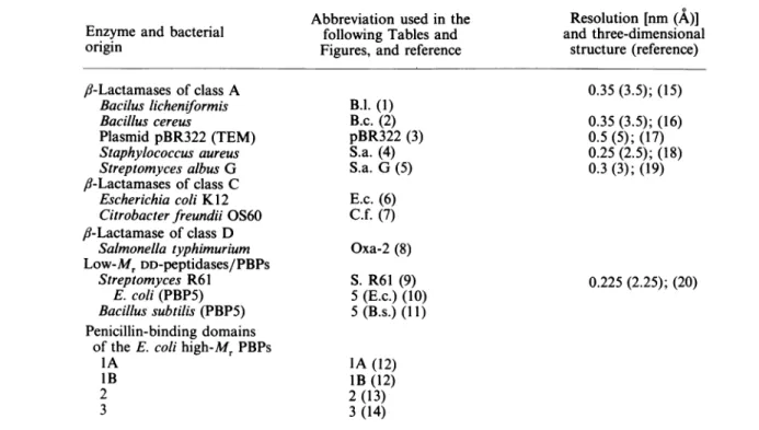
Table 1. Origin and main characteristics (primaryandtertiary structures) of theenzymesstudied
Key to reference numbers cited inthe Table below: (1)Neugebaueretal. (1981); (2)Sloma & Gross(1983); Madonnaetal. (1987); (3) Sutcliffe(1978);(4) Wang & Novick(1987); (5)Dehottayetal.(1987); (6) Jaurin & Grundstrom (1981); (7) Lindberg &Normark (1986); (8) Dale etal. (1985); (9) Duezet al. (1987); (10) Broome-Smith et al. (1983); (11) Toddet al. (1986); (12)Broome-Smith et al. (1985a); (13) Asohetal.(1986);(14) Nakamuraetal.(1983); (15)Kellyetal. (1986);(16)Samraoui etal. (1986); (17) Knoxetal. (1976); (18) Herzberg &Moult (1987); (19)Didebergetal.(1987); (20) Kelly etal. (1987).
Abbreviation used inthe Resolution [nm (A)]
Enzyme and bacterial following Tables and and three-dimensional
ongin Figures, and reference structure (reference)
fl-Lactamases of class A Baciluslicheniformis Bacillus cereus Plasmid pBR322 (TEM) Staphylococcus aureus Streptomyces albus G fl-Lactamasesof class C Escherichia coli K12 Citrobacterfteundii OS60
,8-Lactamase of class D Salmonella typhimurium Low-MrDD-peptidases/PBPs Streptomyces R61 E. coli (PBP5) Bacillus subtilis (PBP5) Penicillin-binding domains
of theE. colihigh-Mr PBPs IA lB 2 3 B.l. (1) B.c.(2) pBR322 (3) S.a. (4) S.a. G(5) 0.35(3.5); (15) 0.35(3.5); (16) 0.5(5); (17) 0.25(2.5); (18) 0.3 (3); (19) E.c.(6) C.f. (7) Oxa-2 (8) S. R61 (9) 5 (E.c.)(10) 5 (B.s.) (11) 0.225(2.25); (20) IA(12) lB(12) 2(13) 3 (14)
glycan transglycosylase reaction. A gene fusion that removes the N-terminal 240-amino-acid region of PBP3 and links the C-terminal 349-amino-acidregiontothe
N-terminal of the
fl-galactosidase
results in a truncatedpolypeptide that still binds penicillin (Hedge & Spratt, 1984).PBP1Band PBP3areheld in theplasmamembrane
attheirN-terminus, withessentiallyall theproteinin the
periplasm (Spratt et al., 1987). It is assumed that the
same organization applies to PBP1A and PBP2.
Amino acid alignments and homology searches
Alignments ofpairs ofproteins were made by using
the Goad & Kanehisa procedure (1982), itself -an
extension of the Needleman-Wunsch algorithm (1970).
In this procedure, comparisons are made from the smallest unit of significance, i.e. two amino
acids,
onefrom eachprotein.Eachpairof amino acids isassigneda
score, the value of which is basedon the relative amino-acid-substitution
frequencies
found among families ofhomologous proteins (the score varies from 0 to -17; the more negative the score, the better the homology)
(Dayhoff, 1972). All the pair combinations of amino acids are then introduced in atwo-dimensional arrayin
which all possible pathways are signified by lines
connecting cells of the array. From
this,
theoptimum-match pathway is derived by connecting those
partial
pathways that maximize the final score (SEQHP pro-gram; Kanehisa, 1982). This score is the sum of the individual scores of the connected cells,including
agappenalty factor every time a deletion is made. The
significance of the comparison between
pairs
ofaligned
sequencesis assessed usingthe SEQDP program (Kane-hisa, 1982). This program gives the score of the best
alignment of two entire sequences according to the original algorithm of Needleman-Wunsch (1970). The significance of the score is expressed by the standard-deviation unit(S.D.U.)of thescoresofagivennumber of random sequences (20 in the present study) of the same
composition (Dayhoff, 1978). An S.D.U. value of 5 or
higher indicates a statistically significant homology. A negative value is obtained when themeanrandom score
is better than the score obtained for the two sequences undercomparison.
The McLachlan procedure (Staden, 1982) was also usedtopresent allpossiblecomparisonsbetweenportions
ofpairsofproteinson amatrix inagraphical form,thus
givingarough,butimmediate,estimateof thesignificance
of any correlation. For this purpose, the two sequences
tobecomparedaredivided into allpossiblesegmentsof
a given length and each segment of one protein is
comparedwitheach segment of the otherprotein. Given thateachpair of amino acids isassigneda score(ranging
inthiscasefrom 2 to27; thehigherthescore, the better thehomology), onlythose segments havingatotalscore
above a certain threshold value are graphically repre-sented. The threshold valuewassuch that theprobability
that similarity occurred by chance was lower than 1 in
1000.
Prediction ofsecondary structures
The computerized Robson's empirical approach
described
by
Gamieretal.(1978)
served to estimate theEvolution of penicillin-interactive proteins
+H
~N---
-
O
PBPs/domainsC-N
XK
-HT
-
C-NH-CO-nN
~~~~~~~~EKTG
---C
ProteisIKSG
ProteinsI
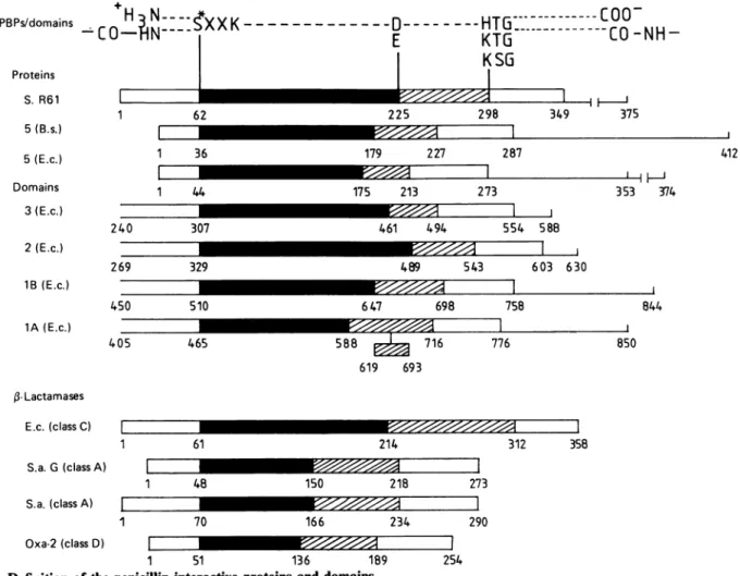
62 225 29883I
349 I 3737S lIiI
I 1 36 179 227 287 412 I I I 1 4 240 307 269 329 450 510 175 213 273-
H/mm 1 461 494 554 588-_
489 543 603 630 647 698 758 465 588 716 619 693 E.c. (class C) 1 61 214 776 3 3 353 374 844 J B50 8 iIZjI 312 358 S.a. G (classA) IZZ1 48 S.a. (classA) E I I 1 70 150 218 273 166 234 290 Oxa-2(class D) I _ 1 51 136 11
Fig. 1.Definition of thepenicillin-interactive proteinsand domains
I
1
89 254
Theposition oftheconserved boxesSer*-Xaa-Xaa-Lys, where Ser* istheactive-site serine residue (box II), AsporGlu(box V) and His-Thr-Gly,Lys-Thr-GlyorLys-Ser-Gly (box VII) alongtheaminoacid sequencesareshown. Forboxes,seealsoFig.
4and, forabbreviations, seeTable 1. C-Terminal extensions assumedto bedispensableforactivity ( )andpeptide segments involvedinplasmamembrane insertion(-I I-)arealso shown. The 619-693insertionin theE. coliPBP1Awasproposed by Broome-Smith etal.(1985a). Allthesequences aredrawnat thesame scale. Theblackandhatched areasdefine thesegments extendingbetween box IIand box V,and between box Vand boxVII, respectively.
a-helix and f-strandpotentials along thesequences. The
selected average decision constants, -100 for the
/3-strands and -80 for thea-helices, werethose proposed
for ana/fl-type structure.
The above computations were carried outon aVAX
11/780 computer.
RESULTS
Definition of the penicillin-interactive proteins and domains
The active-site serine residue(Ser*)inall theenzymes
listed inTable 1 isflankedbyalysine residueatthe third
position on its carbonyl side (conserved sequence
Ser*-Xaa-Xaa-Lys). This serine residue is close to the N-terminus ofthe
,-lactamases,
thelOW-MrDD-peptidases/PBPsand thepenicillin-binding(transpeptidase) domain ofthe E. coliPBP3 (atposition 67,instead of 307 in the intactPBP). Moreover, alltheenzymeslisted in Table 1
possessa conserved triad His-Thr-Gly, Lys-Thr-Gly or
Lys-Ser-Gly (the substitutions His/Lys or Thr/Ser are
known to occur with high frequencies in homologous
proteins). Inthewater-soluble,3-lactamasesand
Strepto-mycesR61 DD-peptidase/PBP,this triadoccursabout 60
residues upstream of the C-terminal end of the protein.
On the basis of these facts, the penicillin-binding (DD-peptidase) domain was assumed to start 60 residues upstream of theactive-site serine residue in the
high-M,
membrane-bound PBPs and to terminate 60 residues
downstream of theaforementioned triadin the low-and
high-Mr
membrane-bound PBPs. Fig. 1 defines thepenicillin-interactive proteins and domains and shows the relative positions of the conserved tetrad
Ser-Xaa-Xaa-Lys and triad His-Thr-Gly, Lys-Thr-Gly or
Lys-Ser-Gly along the aminoacidsequences. Theamino acid
numbering used for thisFigureandthroughoutthetextis
that of Ambler (1980) for the class A ,-lactamases of
B. licheniformis, B. cereus, pBR322 and Staphylococcus
aureus,and ofLindberg& Normark(1986)forthe class-C ,3-lactamases. In other cases, the numbering is that
givenintheoriginalpapers(seeTable1).Residue 1 refers
to the mature proteins.
Correlation between amino acidsequences
The amino acidsequences of thepenicillin-interactive proteinsand domains were compared pairwiseby using
1Il S.R61 5(B.s.) 5(E.c.) Domains 3(E.c.) 2(E.c.) 1B(E.c.) 1A(E.c.) 405 -Lactamases I 7 I 315 I I
H-%ON 00 * en t Co N O W 00 00 00 l,t ONm 00 t - 0I) cn asm - 11. r- cnt 0 00 o o N o o o o o b~~~~~~~~~~~Ci CN l" V) d4* N 0 0% ( 00 o- N) ON 0n o '1% 1.0 0n n _ 1 ll ON _ (N 't\t O0% en cN Rt Rt %%0 Br)_~C. . . 0 0% O N 00 B) .Ct BO) B 0 r-NR en It (IR en IN ~%c 00 (I') e; -; en -4 0 Br) II00 (10 0 w~C o0 6; '- N o II -00 eln a-, 00 Br _ N \0 N 0% '1 00 Br)N
N
qg r 00 o / 0t- Br/ t 'Tr / 0 - eNI Fc ./ e I 0F 0F /41 /~4
0% Cf) B) C'% / / r4 (4 N / o6 i / / ^ 00 en / t 6 / //)
(I
00 C1 "t tn 00 / --I Ci6 ci 0 3 V en 00 00 (-c 00 0 oo _ W VI cn 40n
VI ri en en oN 1%O N 00 e t It t en 0 wO en t I'l N en oot "C C1 W W1 o-rI I. Br '10r N Itr4en 'I- "C Br) VI) BI)
N r% C N t r IT it It oo 0% r4 Br 4% o N N W% 0 NI "t NI t N 0 t l ' ( 00 00 0 N t% _ 0% ( 'IO en en 00 lt en 'r N 0 coi co 0
a
< m= Ci VA Sdgd Sdgd .-1 St°V 'S-mso-0en 00 6 m I.) 0 U,Ce 0n. 1)11 ._ _ ._ CO-o r_ Q*= 7-U, Cd Ce C) Cd 04-Ci Ce x 0 C.) m 'Ce C. I-0' 0Ce Ce. _, D 0 0 0 .Ce C3e CeU 00 o oo ct m 0o, 0; .0 ,d CeC o0 CDe 0 CeN I)Ce - o .dI) E cD ,0 u5 ._ ,, U,; C.) -._ D ,~ 0 _ I-01) Uq U8
O CeCe oCeI
PC 1) c Ps * -Ps s 0 0 .0 .C. 0a *Co0
0Y 0 2 4) .0 Q 1988 U; 1:6 C) SoSL'WUj:)U-j-#~~~317 Evolution ofpenicillin-interactive
proteins
Lil -CD en
L9H
S/(33)s 19H S/StO9H
*S/Is
I~ ~ ~ U) H\*
L9IHS/'33
CD C-cic CD I 2 _-4 t-riA w * LUas ri~ 04-e4
C Os CD .r I-H *~~~~~~U CDiK
('33) s CDcoi
\ \ I_0\
t~~~~ ~~~~z ~~U) \ I \. I -I H~~~~~~* C I CD _ 1= en 0 I H * \) \(Duz
4I.. hen ' II~~~~.
1-S
0 -0~ " H U)-\) en Vol.High-Mr PBPsofE. coli
3- 2 1A 1B
Low-Mr PBPs
Fig. 3. Tentativefamily tree of the penicillin-recognizing enzymes
Notethat the Oxa-2
,-lactamase
of classDis notrepresented. Significances (inS.D.U.)are encircled. Fororganism abbreviations, seeTable 1.the Goad-Kanehisa (1982) algorithm and a uniform gap penalty of + 8. The significance of the pair combinationswasassessedby usingtheSEQDP program. The homology indexes, S.D.U., thus generated (Table 2) revealed that several groups of enzymes matched wellor at least significantly through the amino acid sequences (S.D.U. >5). However, when the pairs or groups thus defined were compared with each other, the overall
similarity became marginally significant or completely
vanished. Figs. 2a-2d illustrate the significance ofthe
comparisons in the form of McLachlan graphs.
Reference structure and calibration marks
The above procedures are known to give consistent results only with closely related proteins. In particular,
theyfailed torevealhomologybetween theStreptomyces
R61 DD-peptidase ontheonehand and the,B-lactamases
ofclass A(fromB.licheniformis,B.cereus,Staphylococcus
aureus, and Streptomyces albusG) ontheother,in spite
of the fact that, on the basis ofX-ray-crystallographic studies, these proteins are very similar in terms of the
spatialarrangement ofsecondarystructures(Kellyetal.,
1986; Samraoui et al., 1986; Herzberg& Moult, 1987;
Didebergetal., 1987). Moreover,asshown inFig. 3, the groupsofhomologous enzymes highlighted bythe data of Table 2 (i.e. the penicillin-binding domains of the
high-Mr PBPs, the low-Mr PBPs, the
f,-lactamases
ofclass A and class
,J-lactamases
of classC)could be linkedto each other through particular pairs of enzymes characterized by S.D.U. values ranging from 4.3to 8. In
this family tree, from which the Oxa-2
,-lactamase
was excluded,the Streptomyces R61 DD-peptidase servedas a bridge between the ,-lactamases of class Aand C.Consequently, any possible correlation between the
penicillin-interactive proteins and domains was
re-examined. For this purpose, the conserved tetrad Ser*-Xaa-Xaa-Lys and triadHis-Thr-Gly (intheStreptomyces R61 DD-peptidase/PBP), Lys-Thr-Gly or Lys-Ser-Gly
(in the otherpenicillin-interactiveproteins anddomains)
wereused ascalibration marks. When this research was
initiated, thesetwogroupsof amino acidswereknownto
occupycritical positions in thethree-dimensional
struc-ture of the Streptomyces R61 DD-peptidase/PBP (Kelly
etal., 1987). Ser*-62 was at the N-terminal end of one of the helices of the'all-a'region, sothat, afterone turnof the helix, the side chains of Lys-65 was brought back within the active-sitearea.Inturn,the triad
His298-Thr299-Gly300 was on the other side of the pocket on the
innermost strand ofthe five-stranded ,-sheet, with the imidazole ring also pointing to the active site.
Amino-acid-alignment editing
The Streptomyces R61 DD-peptidase (used as a template) and each of the other penicillin-interactive
proteins and domainswerealignedpairwise by selecting,
among the possible partial pathways of the Goad-Kanehisa (1982) comparison matrices, those having a score ofatleast -30(includingtheuniform gappenalty
of +8). From thisstarting point,adjustmentsweremade
such that (i) the two calibration marks defined above wereeffectivelyaligned; (ii) the deletions/insertions were restricted to stretches possessing residues known to
favourloop or turn formation (Pro, Gly, Asp); (iii) the helix and ,-strand potentials [as predicted by the Robson-Garnier (Gamier et al., 1978)procedure] were
not, or only slightly, affected; and (iv) the alignments previously proposed forpairs orgroups of homologous proteins (the class-A,-lactamases, class-C 8-lactamases and high-Mr PBPs)were not, oronly slightly, modified. Finally, for each pair combination, the sequence of the Streptomyces R61 DD-peptidase, used as reference, was
keptunbroken,and thedeletions and insertionsrequired
for an optimal match were introduced in each of the sequencesundercomparison.The results showninFig.4
led to the following observations. (1) The alignments highlighted seven conserved regions or boxes (marked
I-VII in Fig. 4) consisting of strict identities or
homologous residues. (2) The 'cost' of the editing, in 1988
Evolution ofpenicillin-interactive proteins 319 Table3. Search for homology between thealigned amino-acid-sequence portions (Fig. 4) of thepenicillin-recognizing enzymes
Thecostofeditingisexpressed asapercentageofresidueseliminatedfromthe original sequences. Forabbreviations, seeTable 1. Forfurtherexplanation,see thetext.
Numberof residues in the:
Comparison Significance Aligned Original Cost of
Enzyme score (S.D.U.) sequences sequences editing(%)
EA C.) C) E.c. -313 B. 1. B.c. < pBR322 S.a. G S.a. c Oxa-2 1. ZZZ ::. C-i 0. L. to M ..- 0. .r. IA IB 2 3 -228 -143 -153 -123 -127 -86 - 106 -156 -119 -197 40.38 20.81 9.19 8.14 11.0 10.92 10.30 9.62 11.35 9.25 11.45 309 250 243 244 254 239 207 232 235 282 248 358 273 257 259 273 254 254 372 309 335 308 X. 5
(E.c.)
-77 4.13 218 273 3 0. 5 (B.s.) -181 14.27 234 287* Ifthelarge insertion occurring between positions 619 and 693 (see Fig. 4) is not included in the calculation.
13 8 6 6 7 6 18.5 37 (or23*) 24 16 20 20 18
terms of the percentage of amino acids eliminated from
theoriginal sequences(Table 3), ranged from 6to 13%
for the
,f-lactamases
of class Aand C, 18% for theOxa-2
,3-lactamase
and did not exceed 24% for the low-Mr and high-MrPBPs. InthecaseofPBP1A, the 74-residuestretch 619-693 was excluded from the calculation, as
previously proposedbyBroome-Smithetal. (1985a). (3) The portions of the original sequences that were effectively aligned generated S.D.U. values (Table 3) and gaveriseto McLachlangraphs(Figs. 2e-2h) indicatinga significant homologybetween the Streptomyces R61 DD-peptidase and each of the other penicillin-recognizing
enzymes, including the Oxa-2
fl-lactamase.
Comparison of primary and tertiarystructures
Structural data(thatwere notavailablewhen the work decribed here was carried out) on the
,-lactamases
ofS. aureus
(Herzberg
&Moult,
1987)
andStreptomyces
albus G(Dideberget
al., 1987)
allowedonetoposition (i)
the
secondary
structuresalong
the aminoacidalignments
of Fig. 4, and
(ii)
the conserved 'boxes' I-VII in theknown three-dimensional structures. The
polypeptide
'scaffolding'andactive-site
configuration
of thesetwo,8-lactamases are virtually identical. However, too small one-turn helices, a3 and a7 in the
staphylococcal
/8-lactamase were not numbered in theStreptomyces
/3-lactamase.
Consequently,
helices al, 2,X49
c5,
c6,c68,
Cc9,
Cci
andac1inthestaphylococcal protein
areequivalent
to helices H1, H2,
H3,
H4,H5,
H6,
H7, H8 and H9 respectively in the Streptomycesprotein.
As shown in Fig. 4, the alignmentsmade
by
referenceto the Streptomyces R61
DD-peptidases/PBP
did notintroduce any gap in the
secondary
structures of theStreptomyces and staphylococcal ,-lactamases except in helix H3 (a4). Essentially, the deletions that were introduced in the sequences did affect the loops between helices H2 and H3 (a3 and a4) and between helices
H4
and H5 (cx5 and cc6). Obviously, the Streptomyces DD-peptidase/PBP and the class-A ,-lactamases have thesame pattern of secondary structures, except that helix H3 (a4) in the Streptomyces DD-peptidases/PBP might beabout two turnslonger that the corresponding helix in
the ,-lactamases.
In turn, Fig. 5 shows the positions of boxes 1-Vil in the three-dimensional structure of the Streptomyces albus G
,f-lactamase
(Didebergetal., 1987). BoxIdefines strandSl,
and box VII is on strand S3. StrandS,
is adjacent to thefl-meander
structure formed by strandsS3, S4and
S.,
and strandS3 forms one side of the active-sitearea(withthe e-amino group oflysinepointing to theactive-siteserineresidue). BoxesIIand VI are on helices H2(a2)and H7 (cx9)respectively. H2, with the active-site serine residue at the N-terminal end, forms the back of the active-site area, and
H7
is atthe surface of the 'all-ac' region. Finally, box III, onaloop connectinghelices H2 andH3 (a2and a4),and box V, on aloop connectinghelices H5 and H6(a6 and
a,8),
are atthe entrance of thecavity. Box IV occurs afew residuesonthecarbonylside of boxIII.
DISCUSSION
The algorithms presently available for the search of correlation between amino acid sequencesgiveconsistent
results only with closely related proteins. Yet, when
applied to a large number of active-site-serine
fi-lactamases and DD-peptidases (PBPs), these procedures
~~~~~~~~~~~ 0~~~~~~~~~~~~~~~~~~~~~~~~~~~ ai a% vwN V 14 In in -V~~~~~~~~~~~ ,-~~~~~~~~~~U) .0) ~ ~ ~ ~ ~ ~ ~ ~ ~ ~ ~ ~ ~ ~ ~ ~ ~ O 1~~~~~*.ou*.L~~~~~~~~~~~~~~~~~~~~Uc
of~~~~~~~~~~~~~~.~I
cx E;~~~co.;
.
U))"~ " )" 0" *U) *Of
CI
..I~~I
o~~~~~ *.~ ~ ~ ~~~) l :l U . P.- Il ofI 4 Ofm4 I 01 cn * ..*.*nM
M M U ) -, .C ~~~~~~~~~~~~~~~~~~~~~~~~~~~~~~g - I ~~~~~~-~~~~ ~~~ -~~~ -~~~~:~~~~~0 C4i lj :fIl
~~~~~~~~~~~~~ .1:~~~~~~10
CU)~~~~~U. c tn cm gn~~~~ go ~~~~~ ~~~ oz xC, ~ ~ ~ ~ ~ ~ ~.4~O~~ U3) a O '~~ ~0)0)1W ~ ~ 0 K I.) U)l O U) U3~~~~~~~~~~~~~~~~18Evolution ofpenicillin-interactive proteins 321 - .14 in eq 0 0 LO at 01 e-. GG 4) OD 41) 41 q q e eq eN 0 LA eq e - e- 1. ID U) -~~~~~~~~~~~~~~~~~~~~~~~~~~~~~i o ** * 4k ** ~~~~~~~~~~~~~~~~~~~~~ uU)~~~~c CA) 5-44
(1~~~~~~~~~~~~~~~~~~4(
z ~~~~~~~~~~~~0 uOl of O~~~~~~tn ol0113~~~~~~~~~~1
~~U)**U) ~~~~~~~t U z ~V4~~~~~~~~~' ~ ~g (2 .. -n 0 0Z U) tn C U) cn U)C H4(2 HC4 H H *r~ t4 ~ .~ U) U) V I >:_
V
-~~~~~~~~~~~~~~~~~~
cc~~~~~~~~~~~~~~~~~~~~~~~~~~~~~~~~~c ::o U)enU)ul rn PI cn~ ~ ~ ~~~~~~~.~U)~~I H z U) -cn (2 **) u cn4~~~~~~~~~~~~~~~~~~~~ ocn E-4~~~~~~~i a% ~ * ~ *W - - 2UC)-/ cm~~~~~~ 0~~~~~~~~~~~~~~~~~~~~~~-ci2I-I~~~~~~~~~~~~~~~~~~~~~~~~~t cc .1C~~~~~~~~~~~~
a eq C(C)$ .~~~~~~5 eq C,~~~~~~~~~~~~~~u -i,6 14 u m 4 99 1- R - W IX 6 u 44 C4 tiD 0 6 m r-i pi m Si " 14 fn wo iEn - (d L) fn co im (I in to 0 Ir -4 1-4 tn I in LAFig. 5. Positions of boxesI-VIHinthe Streptomyces albus Gj'-lactamase molecule
I, Gly24-Asp29; II,Ser*48-Lys5l; III, Gln82; IV, Lys92; V, Glu'50; VI, Trp194;VII, Lys218-Gly220.
suggestthat these enzymes, except the Oxa-2,3-lactamase,
behave as members of a single family tree (Fig. 3).
Moreover, when aligned with reference to the
Strepto-myces R61 DD-peptidase, the /,-lactamases, including
the Oxa-2
,f-lactamase.
the low-MrDD-peptidases/PBPsand the penicillin-binding domains of the E. coli high-Mr PBPs all show significant homology with the Streptomyces R61 DD-peptidase
through
major portionsof the amino acid sequences (Table 3). In all likelihood these penicillin-interactive proteins and domains are
related inanevolutionary senseand form asuperfamily
ofactive-site-serine enzymes. Depending on the
evolu-tionarydistance, theymayhave very different sequences and distinct functionalities and specificities. Yet they
would share the same type of polypeptide scaffolding (distinct from that of the classical trypsin and subtilisin
families). Predictional (thepresent paper) and structural studies support this view. Thus the 349-amino-acid DD-peptidase/PBP of Streptomyces R61 and the 280-amino-acid
,J-lactamases
of class A, though lacking, at firstsight,relatedness inprimarystructure, areverysimilar in
the extent and distribution ofthe
regions
ofsecondary structures (Kellyetal., 1986; Samraoui, 1986; Herzberg&Moult, 1987;Didebergetal.,1987).Inaddition,all the
penicillin-interactive proteins and domains possess
sev-eral conserved boxes that consist of strict identities or
homologousamino acids. Five of these boxes(II, III,IV,
VandVIIinFig. 5)occupycriticalpositionsin the three-dimensional structure of the Staphylococcus aureus and Streptomycesalbus G
fl-lactamases.
Inparallelwiththis,
andasdiscussed below,amino acidreplacementsineach of these boxes affect or abolish the activity of several
,f-lactamases
and low-Mr PBP5 andhigh-M,
PBP3 ofE. coli.
Box II
Theimportance of box II is, of course,
well-established,
since it contains the active-site serine residue. With the pBR322,3-lactamaseit has been shown that (i) inversion ofthe Ser*-Thr dyad to Thr-Ser provides E. coli with an
ampicillin-sensitive phenotype (Dalbadie-McFarland
etal., 1982); (ii) replacement of Ser* by Cys generates a thiol ,-lactamase whose substrate specificity is distinct from that of the wild-type enzyme (Sigal et al., 1982, 1984); (iii) alteration of Thr to many other residues has little effect, yet cells with
fl-lactamase
mutants having Tyr, Trp, Asp, Lys or Arg at this position have no observable resistanceto ampicillin (Schultz & Richards, 1986).Inturn,studies carried out with E. coli PBP3 have shown that(i)replacement of Ser* by Ala or Thr results in aprotein that does not bind penicillin (Houba-Herinetal., 1985) and (ii) alteration of Thr to Pro produces an
E. coli mutant that has high level of resistance to
cephalexin (Hedge & Spratt, 1985). Notethat alteration ofSer* to Cys has also been examined, but has yielded
conflicting results (Houba-Herin et al., 1985; Broome-Smithetal., 1985b). Finally, achimaeric mutant protein
containing a 30-amino-acid insert which comprises
box II of E. coli PBP5 in place of the equivalent
29-amino-acid region of thepBR322,6-lactamase,does not
confer an antibiotic-resistance phenotype. This mutant
has acquired detectable DD-peptidase activity towards the substrate analogue Ac2-L-Lys-Ala-D-Ala (Richards,
1986).
Box III
Nitration of the staphylococcal
,J-lactamase
shows that Tyr-105 of box III is readily derivatized and mustoccupyaparticular site withasteric hindrance such that
Evolution of penicillin-interactive proteins 323
it is prevented fromparticipating in intermolecular
cross-linking (Bristow &Virden, 1978). Box IV
Alteration of Val-Ala-Arg of box IV to Gly-Ala-Arg results in another type of modified PBP3 with high-level resistance to cephalexin (Hedge & Spratt, 1985). Simi-larly, alteration ofGly,followingimmediatelyboxIV, to Asp in the E. coli PBP5 produces a mutantprotein that still binds penicillin but cannot undergo deacylation
(Broome-Smith & Spratt, 1984). Box V
Glu-168, which occurs two positions downstream of box V, is the main site of derivatization of the B. cereus
/,-lactamase
by1-(3-dimethylaminopropyl)-3-ethylcar-bodi-imide. Though Glu- 168 is not conserved (and probably not essential), it has been proposed that its chemical conversion might destroy catalytic activity by influencing the nearby conserved Glu-166 of box V (Little et al., 1986). The fact that, as shown by
X-ray-crystallographicstudies(Herzberg&Moult, 1987;
Dide-bergetal., 1987), the Glu of box V in the Staphylococcus aureus and Streptomyces albus G 8-lactamases has its side chain pointing to the enzyme's active site, strongly supports the view that thiscarboxylate may be important. Box VII
Theincreased cephalosporinase activityof the mutant HI of the pBR322 ,-lactamase, obtained by directed selective pressure on the host cells (Hall & Knowles, 1976), can now be understood. An independent isolate
having the same substrate activity spectrum as mutant HI carries a change just after box VII, whereby Ala is replaced by Thr (W. Blattler & J. R. Knowles, personal
communication). Also, alterations to Ile of either one of the two Thrresidues of box VII or immediately following box VII give rise to a physiologically non-functional E. coli PBP3 which still, however, binds penicillin
(Hedge, 1985).
Theactive-site-serine penicillin-interactive enzymes of the Streptomyces R61 DD-peptidase family differ from the peptidases of the trypsin and subtilisin families in
having carboxypeptidase as against endopeptidase activityand inpreferringDasopposedtoLconfiguration
intheirligands. TheproposedalignmentsofFig.4show that histidine is not a conserved residue in the fifteen
penicillin-recognizingenzymesstudied.Moreover,the /J-lactamase of Streptomyces albus G, when cloned in
Streptomyces lividans,is excreted
by
the hostcells in theform of mutiple molecular species (due to
multiple
cleavage sites of thesignal
peptide),
and one ofthesespecies is histidine-free (Dehottay et al., 1987). These
observations also contrast the
penicillin-interactive
en-zymes with the
peptidases
of thetrypsin
and subtilisinfamilies, where histidine is an invariant element of the
catalytic machinery. The fact remains,
however,
that whatever the family to which an active-site peptidase belongs, the mechanism of thecatalysed
rupture of the scissile peptide(amide)
bond in asusceptible carbonyl
donor is basically identical. In support of this
view,
cephalosporins, which for a long time were consideredexclusively as antibacterial agents targeted against the DD-peptidases/PBPs, can be remodelled into mechanism-based inactivators of the (LL-)endopeptidases of the trypsin family (Doherty et al., 1986).
The work inLiegewassupported bythe Fonds de Recherche
de la Faculte de Medecine, the Fonds de la Recherche
Scientifique Medicale, Brussels (contract n°. 3.4507.83), the
Region wallonne (contract n°. C2/C16/246/20428), the
Gouvernement belge (action concertee n°. 81/86) and the Commission of the European Communities (contract BAP-0197-B). B.J. isChargede recherches and G. D. is Chercheur qualifie of the Fonds National de la Recherche Scientifique (FNRS,Brussels). The work in Storrswassupportedbygrant GM-37742toJ. R. K. and grant RR-01955toJ. A. K. from the National Institutes of Health.
REFERENCES
Ambler,R. P.(1980) Philos.Trans. R.Soc.London Ser.B289, 321-331
Asoh, S., Matsuzawa, H., Ishino, F. Strominger, J.,
Matsuhashi, M. & Ohta, T. (1986) Eur. J. Biochem. 160, 231-238
Bristow, A.F.&Virden, R. (1978) Biochem. J. 169, 381-388 Broome-Smith, J. K. & Spratt, B. G. (1984) FEBS Lett. 165,
185-189
Broome-Smith, J. K., Edelman, A. & Spratt, B. G. (1983) in the Target of Penicillin (Hakenbeck, R., H6ltje, J. V. & Labischinski, H., eds.), pp. 403-408, Walder de Gruyter, Berlin
Broome-Smith,J.K.,Edelman,A.,Youssif, S. & Spratt, B. G. (1985a) Eur. J. Biochem. 147, 437-446
Broome-Smith, J. K., Hedge, P. J. & Spratt, B. G. (1985b) EMBO J. 4,231-235
Cartwright, S. J. & Waley, S. G. (1983) Med. Res. Rev. 3, 341-382
Dalbadie-McFarland, G.,Cohen, L. W., Riggs, A. D., Morin, C., Itakura, K. &Richards, J. H. (1982) Proc. Natl. Acad. Sci. U.S.A. 79, 6409-6413
Dale,J. W.,Godwin,D., Mossakonska, D., Stephenson, P. & Wall, S.(1985) FEBS Lett. 191, 39-42
Dayhoff, M.0. (1972) in Atlas of Protein Sequence and
Structure, vol. 5 (Dayhoff, M.O., ed.), p. 17, National Biomedical ResearchFoundation, Silver Spring, MD Dehottay, P., Dusart, J., De Meester, F., Joris, B., Van
Beeumen, J., Erpicum, T., Frere, J. M. & Ghuysen, J. M. (1987) Eur. J. Biochem. 166, 345-350
Dideberg, O.,Charlier, P., Wery, J. P., Dehottay, P., Dusart, J., Erpicum, T., Frere, J. M. & Ghuysen, J. M. (1987) Biochem. J.245, 911-913
Doherty,J. B.,Ashe, B. M.,Argenbright, L. W.,Barker, P.L., Bonney, R.J., Chandler, G. O., Dahlgren, M. E., Dorn, C.P.,Finke,P.E.,Firestone, R.A.,Fletcher,D.,Hagmann, W.K.,Mumford, R.,O'Grady,L.,Maycock, A. L., Pisano, J.M., Shah, S. K. & Thompson, K. R. (1986) Nature (London) 322, 192-194
Duez, C., Piron-Fraipont, C., Joris, B., Dusart, J., Urdea, M. S., Martial, J.A., Frere, J. M. & Ghuysen, J. M. (1987) Eur. J. Biochem. 162, 509-518
Ferreira, L.C. S., Schwarz, U., Keck, W., Charlier, P., Dideberg, 0.&Ghuysen, J. M. (1988) Eur. J.Biochem., in thepress
Frere, J. M. &Joris, B. (1985) CRC Crit. Rev. Microbiol. 11,
299-396
Gamier, J., Osguthorpe, D.J. & Rodson, B. (1978) J. Mol. Biol. 120, 97-120
Ghuysen, J.M., Frere, J.M., Leyh-Bouille, M.,
Nguyen-Disteche, M., Coyette, J., Dusart, J., Joris, B., Duez, C.,
Dideberg, O.,Charlier,P., Dive, G. &Lamotte-Brasseur,J.
(1984) Scand. J. Infect. Dis. Suppl.42, 17-37
Goad, W. B.&Kanehisa, M. T. (1982)Nucleic Acids Res. 10, 247-263
Hall, A. & Knowles, J. R. (1976) Nature (London) 264, 803-804 Hedge, P. J. (1985) Ph.D. Thesis, University of Sussex
Hedge, P. J. & Spratt, B.G. (1984) FEBS Lett. 176, 179-184 Hedge, P. J. & Spratt, B.G. (1985) Eur. J. Biochem. 151,
111-121
Herzberg, 0. &Moult, J. (1987) Science236, 694-701 Houba-Herin, N., Hara, H., Inouye, M. &Hirota, Y. (1985)
Mol. Gen. Genet. 201, 499-504
Jaurin, B. & Grundstrom, T. (1981) Proc. Nat. Acad. Sci. U.S.A. 78, 4897-4901
Kanehisa, M.I. (1982) Nucleic Acids Res. 10, 183-195 Kelly, J. A., Dideberg, O., Charlier, P., Wery, J. P.,Libert, M.,
Moews, P.C., Knox, J. R., Duez,C., Fraipont, C., Joris, B., Dusart,J.,Frere, J. M.&Ghuysen, J. M. (1986) Science 231, 1429-1437
Kelly, J. A., Boyington, J. C., Moews, P. C., Knox, J. R., Dideberg, O., Charlier, P.,Libert, M., Wery, J. P., Duez, C., Joris, B., Dusart, J., Frere, J. M. &Ghuysen,J. M.(1987) in Frontiers of Antibiotic Research (Umezawa, H., ed.) (Proceedings of the Takeda Science Foundation Symposium on Bioscience, 1987), Academic Press, Tokyo,in the press Knox, J. R.,Kelly, J. A., Moews, P. C. & Murthy, N. S. (1976)
J. Mol. Biol. 164, 865-875
Lindberg, F. & Normark, S. (1986) Eur. J. Biochem. 156, 441-445
Little, C., Emanuel, E. L., Gagnon, J. & Waley, S. G. (1986) Biochem. J. 240, 215-219
Madonna, M.J.,Zhu, Y. F. & Lampen, J. 0. (1987) Nucleic Acids Res. 15, 1877
McLachlan, A. D. (1971)J. Mol. Biol. 61, 409-424
Nakamura, M., Maruyama, I. N., Soma, M., Kato, J., Suzuki, H. & Hirota, Y. (1983) Mol. Gen. Genet. 191, 1-9 Needleman, S. B. & Wunsch, C. D. (1970) J. Mol. Biol. 48,
443-453
Neugebauer, K., Sprengl, R. & Schaller, H. (1981) Nucleic Acids Res. 9, 2577-2588
Richards, J. H. (1986) Nature (London) 323, 187
Samraoui, B., Sutton, B. J., Todd, R. J., Artymiuk, J. J., Waley, S. G. & Phillips, D. C. (1986) Nature (London) 320, 378-380
Schultz, S. C. & Richards, J. H.(1986) Proc. Natl. Acad. Sci. U.S.A. 83, 1588-1592
Sigal, I. S.,Harwood, B.G. &Arentzen, R.(1982)Proc.Natl. Acad. Sci. U.S.A. 79, 7157-7160
Sigal, I.S., DeGrado, W.T., Thomas, B.J. &Petteway, S. R. (1984) J. Biol. Chem. 259, 5327-5332
Sloma, A. &Gross,M.(1983)Nucleic Acids Res. 11,4997-5004 Spratt, B. G., Bowler,L.,Edelman, A. & Broome-Smith, J. K. (1987) Proc. Am. Soc. Microbiol. Conf. Antibiot. Inhibition Bact. Cell Surf. Assem. Funct., Philadelphia, 17-20 May
1987, in the press
Staden, R. (1982)Nucleic Acids Res. 10, 2951-2961
Sutcliffe, J.G. (1978) Proc. Natl. Acad. Sci. U.S.A. 75, 3737-3741
Todd, J. A., Roberts, A.N., Johnstone, K., Piggot, P.J., Winter, G. &Ellar, D. (1986) J. Bacteriol. 167, 257-264 Wang, P.Z. & Novick, R. P. (1987) J. Bacteriol. 169,
1763-1766
Received 18May 1987/28 August 1987; accepted 9October 1987