Université de Montréal
The effect of exercise training on cholesterol and bile acid
metabolism in ovariectomized rats
par Zahra Farahnak
Département de kinésiologie
Thèse présentée à la Faculté des études supérieures en vue de l’obtention du grade de Philosophiae Doctor (Ph.D.)
en Sciences de l’activité physique
Novembre, 2016
Université de Montréal
Faculté des études supérieures et postdoctorales
Cette thèse intitulée:
The effect of exercise training on cholesterol and bile acid
metabolism in ovariectomized rats
Présentée par: Zahra Farahnak
a été évaluée par un jury composé des personnes suivantes:
Dave Ellemberg, président-rapporteur Jean-Marc Lavoie, directeur de recherche
Natalie Chapados, codirectrice Julie Lavoie, membre du jury David St-Pierre, examinateur externe Dave Ellemberg, représentant du doyen de la FES
i
Résumé
Il existe un nombre grandissant de preuves au cours des dernières années que la diminution de la sécrétion des œstrogènes chez les animaux ovariectomisés (Ovx) et chez les femmes ménopausées conduit à une accumulation importante de triglycérides (TG) dans le foie. Cependant, les évidences de perturbations dans le métabolisme du cholestérol, en lien avec la diminution des œstrogènes, sont limitées à des observations de niveaux élevés de cholestérol total dans le plasma trouvés chez la femme ainsi que chez les animaux. En fait, l'impact de la suppression des œstrogènes sur le métabolisme du cholestérol dans le foie a reçu peu d'attention et montre quelques controverses. Par conséquent, les trois études présentées dans cette thèse ont été réalisées chez des rats Ovx, comme modèle animal de femmes post-ménopausées, afin de documenter les effets du retrait des œstrogènes sur les marqueurs moléculaires clés du métabolisme du cholestérol et des acides biliaires dans le foie et dans l'intestin et des effets potentiels de l’entraînement physique. Il a été en effet démontré que l'entraînement physique peut réduire le niveau plasmatique de cholestérol. Une amélioration du transport du cholestérol en périphérie vers le foie pour sa sécrétion subséquente dans la bile et pour son l'excrétion de l'organisme a été suggérée, bien que les mécanismes sous-jacents ne soient pas entièrement compris.
Dans la première étude, nous avons démontré que les rattes Ovx nourris avec une diète standard et une diète standard + cholestérol avait un taux de cholestérol total dans le foie plus élevé (P <0,05) que les rattes avec une ovariectomie simulée (Sham) nourris avec ces deux derniers types de diète, tandis que la teneur en triglycérides du foie était plus élevée chez les rattes Ovx que chez les rattes Sham nourris avec une diète standard, une diète standard + cholestérol et aussi une diète riche en grasses + cholestérol. Étonnement, la diète standard + cholestérol a été associée à un niveau plasmatique plus faible (P <0,001) de cholestérol total et de triglycérides chez les rats Ovx que les rats Sham, ce qui suggère une diminution de la sécrétion de lipoprotéines à très basses densités (VLDL). Par conséquent, la transcription de plusieurs marqueurs clés de la synthèse des VLDL, y compris la microsomal triglyceride transfer Protein (MTP) et apoB-100, ont été réduites (P <0,05) chez les rattes Ovx par rapport aux rattes Sham nourris avec tous les trois types diètes et cette diminution de MTP et
apoB-ii
100 était plus prononcée chez les rats nourris avec la diète standard + cholestérol. Pour aller un peu plus loin, dans la deuxième étude, nous avons déterminé les effets de l'entraînement physique sur les marqueurs clés hépatiques de la voiefarnesoid X receptor (FXR) - small heterodimer partner (SHP) - de cholestérol 7 alpha-hydroxylase (CYP7A1) (FXR-SHP-CYP7A1) impliquée dans la conversion de cholestérol en acides biliaires et de leur excrétion chez les rat Ovx nourris avec une diète standard + cholestérol.
Notre groupe expérimental principal comprenait des rats Ovx nourris avec une diète riche en cholestérol (Ovx-Chol). Ce groupe a été comparé à un groupe de rats Ovx nourris avec une diète standard (Ovx-SD) et un groupe de rats Sham nourris avec une diète riche en cholestérol (Sham-Chol) pour observer, respectivement, l'effet de l'alimentation et l’effet du retrait de l'œstrogène. Les résultats de cette étude ont démontré que les niveaux de cholestérol total dans le plasma et dans le foie ne sont pas affectés par l'entraînement physique dans aucune des conditions expérimentales. L'alimentation en cholestérol a induit une accumulation plus importante chez les rats Sham et Ovx a mené à une accumulation du cholestérol dans le foie significativement plus élevée (P <0,001) que chez les rats Ovx-SD. Un effet principal d'entraînement physique (P <0,05) a été trouvée dans l’expression génique du SHP et de CYP7A1. Ce dernier gène est reconnu pour son implication majeure sur le contrôle de la biosynthèse des acides biliaires à partir du cholestérol. De plus, cette étude a montré que le récepteurs des LDL (LDL-R) et proprotein convertase subtilisin/kexin type 9 (PSCK9) au foie, qui sont impliqués dans l'absorption du cholestérol de la circulation, ne sont pas influencés par l’entraînement physique. Ces résultats suggèrent que l'entraînement physique module le métabolisme du cholestérol chez les animaux Ovx par un réglage positif de la formation des acides biliaires. Un nombre croissant de preuves récentes suggèrent que le transport inverse du cholestérol (RCT) peut également passer par une voie non-biliaire connue sous le nom « transintestinal cholesterol excretion » (TICE). En effet, le foie et l'intestin sont impliqués dans l'excrétion du cholestérol excédentaire du corps. Dans cette optique, dans la troisième étude, nous avons élargi nos recherches afin de déterminer si l'entraînement physique module l’expression des récepteurs de cholestérol de la membrane intestinale qui sont impliqués dans TICE chez les rats intacts et Ovx nourris avec une diète standard et une diète riche en cholestérol. Les résultats de cette étude ont montré que l'entraînement physique a augmenté (P
iii
<0,01) l’expression génique intestinale de LDL-R et de PCSK9 impliquées dans la captation du cholestérol intestinal de la circulation et de leur récepteur nucléaire, « sterol regulatory element-binding protein 2 » (SREBP2) (P <0,05) chez les rats Sham et Ovx par rapport aux rats sédentaires (Sed). D'autre part, l’expression des gènes hépatiques de LDL-R et de PCSK9 ont été supprimées (P <0,01) par l’alimentation riche en cholestérol, mais pas affectée par l'entraînement physique. L'expression du gène « flavin monooxygénase 3 » (FMO3), en tant que régulateur de l'équilibre du cholestérol dans le foie, a été diminuée de façon significative (P <0,01) par le cholestérol alimentaire chez les rats Sham et Ovx par rapport aux rats nourris avec la diète standard, mais demeure inchangée suite à l'entraînement physique et le retrait des œstrogènes. Un réglage positif de l'expression de gènes du LDL-R et PCSK9 intestinale par l'entraînement physique chez les rats intacts et Ovx suggère que l'entraînement physique peut contribuer à l’accroissement de l'élimination de cholestérol par la voie TICE.
Dans l'ensemble, nos résultats indiquent qu'une combinaison d’une diète riche en cholestérol et un retrait des œstrogènes a mené à une diminution de l'expression des gènes des marqueurs essentiels de la synthèse de VLDL, ce qui implique une réduction de l'excrétion du cholestérol du foie. Il semble que la réduction de LDL-R hépatique pourrait être due à l'accumulation du cholestérol dans le foie. De plus, nos résultats ont présenté l’entraînement physique comme une intervention non pharmacologique appropriée pour stimuler l'excrétion du cholestérol excédentaire de l'organisme par le réglage positif des gènes impliqués dans la biosynthèse des acides biliaires dans le foie et les récepteurs intestinaux de cholestérol dans la voie TICE.
Mots-clés : Ovariectomie, Rat, Diète riche en cholestérol, l'assemblage VLDL, LDL-R, PCSK9, Accumulation de cholestérol dans le foie, Entraînement physique, CYP7A1, la voie TICE.
iv
Abstract
There has been accumulating evidence in recent years that the estrogen deficient state in ovariectomized (Ovx) animals and in postmenopausal women results in substantial liver triglyceride (TG) accumulation. However, evidence of disturbances in cholesterol metabolism in link with estrogen deficiency is limited to observations of higher plasma total cholesterol levels found in human as well as in animals. In fact, the impact of estrogen withdrawal on liver cholesterol metabolism has received little attention and shows some controversies. Therefore, the three studies presented in this thesis have been conducted in Ovx rats, as an animal model of post-menopausal women, to investigate the effects of estrogen withdrawal on key molecular markers of cholesterol and bile acid metabolism in liver and in transintestinal cholesterol excretion (TICE), and also to determine the potential role of exercise training as a positive alternative intervention. It has been shown that exercise training can improve plasma cholesterol levels. An enhanced transport of peripheral cholesterol toward the liver for subsequent secretion into bile and excretion from the body has been suggested; however, the underlying mechanism for this action is not fully understood.
In the first study, we showed that estrogen withdrawal was associated with higher (P < 0.05) liver total cholesterol under the standard diet and the standard diet + cholesterol diet, while liver triglyceride (TG) content was higher in Ovx than in Sham rats in all three dietary conditions which are the standard diet, the standard diet + cholesterol and the high fat diet + cholesterol. Surprisingly, the standard diet + cholesterol was associated with lower (P < 0.001) plasma total cholesterol and TG levels in Ovx than in Sham rats, suggesting a decrease in very low-density lipoprotein (VLDL) secretion. Accordingly, several transcripts of key markers of VLDL synthesis including microsomal triglyceride transfer protein (MTP) and apoB-100 were decreased (P < 0.05) in Ovx compared to Sham rats under the three dietary conditions and even more so for MTP and apoB-100 when rats were fed the standard diet + cholesterol. To go one step further, in the second study we determined the effects of exercise training on hepatic key markers of farnesoid X receptor (FXR)-small heterodimer partner (SHP)-cholesterol 7 alpha-hydroxylase (CYP7A1) (FXR-SHP-CYP7A1) pathway, involved in cholesterol conversion into bile acid and excretion from the body, in Ovx cholesterol fed rats. Our main
v
experimental group was Ovx rats fed a high cholesterol diet (Ovx-Chol) that was compared, on one hand, to a group of Ovx rats fed a standard diet (Ovx-SD) to observe the effects of the diet and, on the other hand, compared to a group of Sham operated rats fed the cholesterol diet (Sham-Chol) to observe the effect of estrogen withdrawal. Results of this study showed that plasma and liver total cholesterol levels were not affected by exercise training in any of the experimental conditions. Cholesterol feeding in both Sham and Ovx rats resulted in significantly (P<0.001) higher hepatic cholesterol accumulation than in Ovx-SD rats. A main effect of training (P< 0.05) was, however, found for transcripts of SHP and CYP7A1. The SHP and CYP7A1 transcripts were increased by training. These results suggest that exercise training through up-regulation of genes involved in bile acid formation may modulate cholesterol metabolism in Ovx animals. Finally, a recent growing body of evidence suggests that reverse cholesterol transport (RCT) can also proceed through a non-biliary pathway known as transintestinal cholesterol excretion (TICE). Indeed, both liver and intestine are involved in excretion of the excess cholesterol from the body. Based on this concept, we expanded our research to determine whether exercise training has an effect on intestinal membrane cholesterol receptors involved in TICE pathway in intact and Ovx rats fed a normal and a high cholesterol diet. Results of the third study showed that exercise training increased (P< 0.01) transcripts of intestinal LDL-R and PCSK9, which are involved in intestinal cholesterol uptake from circulation, and their nuclear transcription factor, intestinal sterol regulatory element-binding protein 2 (SREBP2) (P< 0.05) in both Sham and Ovx rats compared to rats remaining sedentary (Sed). On the other hand, hepatic LDL-R and PCSK9 gene expression was suppressed (P< 0.01) by cholesterol feeding but not affected by exercise training. Flavin monooxygenase 3 (FMO3) gene expression, as a cholesterol balance regulator in liver, was significantly decreased (P<0.01) by cholesterol feeding in both Sham and Ovx rats compared to rats were fed the SD diet but unchanged following exercise training and estrogen withdrawal. An up-regulation of intestinal gene expression of LDL-R and PCSK9 following voluntary wheel running in intact and Ovx rats suggests that exercise training may contribute to increased cholesterol elimination through the TICE pathway.
Overall, our results indicate that a high cholesterol diet and ovariectomy combine to decrease the gene expression of key markers of VLDL synthesis suggesting a reduction in
vi
cholesterol excretion from the liver. Alternatively, it seems that reduced hepatic LDL-R transcript found in Ovx animals might be due to hepatic cholesterol accumulation. Moreover, our findings introduced exercise training as an appropriate non-pharmacological intervention to stimulate the excretion of the excess cholesterol from the body through upregulation of genes involved in bile acid biosynthesis in liver and intestinal basolateral cholesterol transporters in TICE.
Keywords: Ovariectomy, Rat, High cholesterol diet, VLDL assembly, LDL-R, PCSK9, Hepatic cholesterol accumulation, Exercise training, CYP7A1, TICE pathway.
vii
Table of contents
Résumé ... i
Abstract ... iv
Table of contents ... vii
Liste of figures ... ix
Abbreviations ... x
Acknowledgements ... xiv
Introduction ... 1
Chapter 1: Review of Literature ... 4
1.1 Reverse Cholesterol Transport (RCT) ... 4
1.1.1 Hepatobiliary pathway ... 6
1.1.1.1 Cholesterol influx into liver ... 6
1.1.1.2 Hepatic cholesterol uptake from circulation ... 8
1.1.1.3 Cholesterol excretion from the liver ... 13
1.1.1.3.1 Bile acids formation and the enterohepatic circulation... 14
1.1.1.3.2 VLDL assembly ... 19
1.1.2 Non-biliary TICE pathway ... 21
1.1.2.1 Step1: The role of lipoproteins in the TICE pathway ... 22
1.1.2.2 Step 2: Cholesterol receptors at intestinal basolateral membrane ... 24
1.1.2.3 Step 3: Cholesterol trafficking from basolateral to the apical membrane of enterocyte ... 25
1.1.2.4 Step 4: Cholesterol efflux via intestinal apical transporters into the lumen ... 26
1.2 The effects of high cholesterol diet, estrogen withdrawal and exercise training on reverse cholesterol transport (RCT) ... 30
1.2.1 Hepatobiliary pathway ... 30
1.2.1.1 Hepatic cholesterol accumulation ... 30
1.2.1.2 Hepatic cholesterol uptake from circulation ... 32
1.2.1.3 Cholesterol excretion from the liver ... 36
viii
1.2.1.3.2 Bile acid biosynthesis ... 38
1.2.2 Non-biliary TICE pathway ... 42
1.3 General objective of thesis and presentation of manuscripts ... 44
Chapter 2: Original research articles ... 46
2.1 Article 1 ... 46
2.2 Article 2 ... 70
2.3 Article 3 ... 98
Chapter 3: General discussion and conclusion ... 129
3.1 General discussion ... 129
3.2 Conclusion ... 133
ix
Liste of figures
Figure 1. Schematic representation of the main pathways of cholesterol excretion. Adapted from (Brufau et al. 2011). ... 5 Figure 2. HDL formation and cholesterol influx into liver. Taken from (Wiener et al. 2012) ... 7 Figure 3. Cholesterol uptake by the liver. Taken from (Rader 2006) ... 8 Figure 4. LDL-cholesterol metabolism in the presence (a) or absence of PCSK9 (b). Taken from (Dadu et al. 2014). ... 11 Figure 5. Cholesterol excretion from the liver. Adapted from (Jonker et al. 2009) ... 13 Figure 6. Transporters involved in bile acid reabsorption in the ileum. Adapted from (Schaap et al. 2014) ... 15 Figure 7. The molecular mechanisms of FXR pathway in bile acid synthesis in liver and intestine. Adapted from (Ory 2004; Inagaki et al. 2005) ... 18 Figure 8. VLDL assembly in liver. Taken from (Bartosch et al. 2010) ... 20 Figure 9. A model of non-biliary transintestinal cholesterol excretion (TICE). Adapted from (Le May et al. 2013)... 27 Figure 10. Dietary cholesterol absorption. Adapted from (Wang et al. 2013). ... 29
x
Abbreviations
ABCA1: Adenosine triphosphate binding cassette transporter A1 ABCB1 a/b: ATP-binding cassette transporter B1 a and b
ABCG5/G8: Adenosine triphosphate binding cassette transporters G5/G8 ACAT-2: Acyl-CoA:cholesterol acyltransferase
apoA-1: Apolipoproteins A-1 apoB: Apolipoprotein B
apoER2: Apolipoprotein E receptor 2
ASBT: Apical sodium-dependent bile acid transporter BSEP: Bile salt export pump
CE: Cholesteryl ester
CEPT: Cholesteryl ester transfer protein
Cideb: Cell death-inducing DNA fragmentation factor alpha (DFFA)-like effector B Chol: Cholesteryl
CPF: CYP7A1 promoter binding factor CYP7A1: Cholesterol 7 alpha-hydroxylase DGAT2: Diacylglycerol acyltransferase 2 E2: 17β-estradiol
ER: Endoplasmic reticulum ERα: Estrogen receptor α EX: Exercise
FGF15/19: Fibroblast growth factor 15/19 FGFR4: Fibroblast growth factor receptor 4 FMO3: Flavin monooxygenase 3
FC: Free cholesterol
FXR: Farnesoid X receptor HC: High cholesterol
HDL: High density lipoprotein HF: High fat
xi
HMGCoA-r: 3-hydroxy 3-methylglutaryl coenzyme A reductase LPL: Lipoprotein lipase
iBABP: Ileal bile acid binding protein IBAT: Ileal bile acid transporter IDL: Intermediate-density lipoprotein KO: Knockout
LCAT: Lecithin:cholesterol acyltransferase LDL: Low density lipoprotein
LDL-C: Low density lipoprotein cholesterol LDL-R: Low-density lipoprotein receptor
LIMP2: Lysosomal integral membrane protein-2 LRP1: LDL receptor-related protein
LRH-1: Liver related homologue-1 LXR: Liver X receptor
MDR2: Multidrug resistance protein 2
MTP: Microsomal triglyceride transfer protein NAFLD: Non-alcoholic fatty liver disease NASH: Nonalcoholic steatohepatitis NPC1: Niemann-Pick type C1 NPC2: Niemann-Pick type C1 NPC1L1: Niemann-Pick C1-Like 1
NTCP: Na+- dependent taurocholate cotransport peptide Ostα-Ostβ: Organic solute transporter α and β
Ovx: Ovariectomized
PCSK9: Proprotein convertase subtilisin/kexin type 9 PLTP: Phospholipid transfer protein
PXR: Pregnane X receptor
RCT: Reverse cholesterol transport Sar1a: Small GTPase a
Sed: Sedentary SD: Standard diet
xii Sham: Sham operated
SHP: Small heterodimer partner
SR-BI: Scavenger receptor class B class I
SREBP-2: Sterol regulatory element binding protein-2 TC: Total cholesterol
TG: Triglyceride
TICE: Transintestinal cholesterol excretion VLDL: Very low density lipoprotein
VLDLR: Very low-density lipoprotein receptor WT: Wild type
xiii
xiv
Acknowledgements
I would like to express my thanks to my supervisor, Dr. Jean-Marc Lavoie for his suggestions throughout my Ph.D. I also offer my regards to my co-supervisor Dr. Natalie Chapados for her support. Besides, I would like to thank Dr. Raynald Bergeron for his guidance on my research.
I acknowledge NSERC (Engineering Research Council of Canada) for funding the studies presented in this thesis.
I truly thank the laboratory personnel, professorial staff, fellow graduate, and administration staff of the Départment de Kinésiologie at the Université de Montréal for their contributions (intellectual, technical, administrative) to my formation and academic development.
I am so grateful to my family (my parents and brothers) for their moral support.
Finally and most importantly, I heartily appreciate the wonderful support and encouragement that I have received from my beloved husband, Ben (Benyamin Karimi) throughout my Ph.D.
Introduction
Menopause is defined by the progressive decrease of estrogen production resulting in cessation of menses (Mastorakos et al. 2010). Menopause as well as ovariectomy in animals is associated with diverse metabolic consequences including ectopic fat deposition mainly in the liver and this condition is known as non-alcoholic fatty liver disease (NAFLD) (Brunt 2001; Volzke et al. 2007). There has been accumulating evidence in recent years showing that the estrogen deficient state in ovariectomized (Ovx) animals and in postmenopausal women results in substantial liver triglyceride (TG) accumulation, indicating the protective role of estrogens against NAFLD and also perturbation in TG metabolism in the absence of estrogens (Picard et al. 2000; Paquette et al. 2007; Volzke et al. 2007). In addition to TG accumulation, lipidomic analyses indicate that hepatic free cholesterol content was also increased in hepatic steatosis (Puri et al. 2007) suggesting that cholesterol metabolism is also affected in liver diseases. The situation of hepatic cholesterol content in Ovx animals is controversial. It was reported that hepatic total cholesterol content was not affected by estrogen withdrawal in female C57BL/6J mice. Despite the fact that they showed higher plasma cholesterol levels, hepatic cholesterol content was not changed probably due to a reduced liver cholesterol uptake by LDL-R and a reduced hepatic de novo cholesterol production by hydroxy 3-methylglutaryl coenzyme A reductase (HMGCoA-r) (Kamada et al. 2011). On the other hand, other studies showed that hepatic total cholesterol content was increased in Ovx rats (Kato et al. 2009; Ngo Sock et al. 2014a) suggesting the vulnerability of Ovx animals to develop hepatic cholesterol accumulation. Liver is known as a master regulator of cholesterol metabolism in terms of cholesterol synthesis, uptake from circulation and excretion from the body. Estrogens through its interaction with HMGCoA-r and LDL-R, genes involved in cholesterol synthesis and uptake respectively, play a critical role in cholesterol homeostasis (Bruning et al. 2003). In fact, the information on the impact of the absence of estrogens on cholesterol metabolism in liver is scarce and is mostly limited to observations of hypercholesterolemia in human as well as in animals. Estrogen deficiency state has been repeatedly reported to result in the development of an atherogenic lipid profile characterized by an increase in plasma total cholesterol (TC) and low density lipoprotein cholesterol
(LDL-2
C) levels (Matthews et al. 1989; Kimura et al. 2006; Park et al. 2011b; Chaudhuri et al. 2012; Kaur et al. 2013). Elevated plasma concentration of LDL-C is the primary risk factor for coronary artery disease and atherosclerosis, which constitute the largest cause of mortality in developed countries (Mozaffarian et al. 2016). It is important to note that incidence of cardiovascular diseases increases with age in women, with a noticeable increase after menopause (Sharp et al. 1997). This situation raises the hypothesis that perhaps disturbances in different aspects of cholesterol metabolism in liver might be the root of hypercholesterolemia in an estrogen deficient state. Indeed, plasma cholesterol level is tightly determined by a complex homeostatic network which requires the accurate metabolic interplay between hepatic and intestinal processes, to regulate efficiently cholesterol homeostasis (Oram et al. 2006). Recent studies on newly identified transintestinal cholesterol excretion (TICE) pathway have revealed that cholesterol homeostasis in the body depends on a dynamic interplay between liver and intestine (Temel et al. 2012). An interesting intervention to metabolic deterioration due to estrogen deficiency is exercise training. For instance, in an intervention study where Ovx rats were submitted to the treadmill training for 12 weeks, reduced plasma LDL-C and total cholesterol levels were observed (Oh et al. 2007). An increase in fecal cholesterol excretion accompanied by lower plasma cholesterol levels was also reported in exercise trained animals (Meissner et al. 2011). However exactly how exercise exerts such beneficial actions is largely unknown.
These observations highlight the importance of a need for more physiological and molecular information to better understand how liver, as a master regulator of cholesterol metabolism, is affected by estrogens withdrawal. The three studies presented in this thesis have been conducted to provide molecular information on how liver regulates cholesterol metabolism in Ovx rat model of menopause and whether exercise training could provide some beneficial effects on cholesterol metabolism. Rodent ovariectomy is an experimental model of human post-menopausal state. Ovariectomy eliminates the interference of endogenous estrogens and mimics post-menopausal condition which makes it possible to study the metabolic consequences of loss of ovarian functions. On the other hand, ovariectomy can result in metabolic changes in very short period of time, while post-menopausal state is a natural process that happens gradually over several years. We also used the high cholesterol
3
diet as a nutritional tool to investigate the role of liver in regulating cholesterol metabolism in our series of experiments.
In the first study, we investigated the effects of high dietary cholesterol on hepatic key markers of VLDL and cholesterol/bile acid metabolism in Ovx rats. There is some evidence that exercise training is one of the best non-pharmacological strategies to attenuate hepatic cholesterol accumulation; however, the molecular information on how this action takes place is lacking. In line with this first approach, in the second study we determined the effect of exercise training on key markers of hepatic cholesterol and bile acid metabolism by targeting the FXR-SHP-CYP7A1 gene markers in Ovx rats submitted to the high cholesterol diet. In the third study, we expanded our research to determine the effect of exercise training on key intestinal cholesterol receptors involved in TICE pathway in intact and Ovx rats fed a normal and a high cholesterol diet. We targeted gene expression of key molecules involved in TICE via cholesterol uptake and excretion at the intestinal basolateral and apical membrane, respectively.
The present thesis consists of three chapters. Chapter 1 is devoted to the review of the literature which is divided into two sections. The objective of the first section is to provide the reader with an overview of reverse cholesterol transport (RCT) with an emphasis on the major molecular markers of liver and intestine associated with it. This first section is subdivided into two parts: hepatobiliary pathway, non-hepatobiliary TICE pathway. In the second section, we review the effects of a high cholesterol diet, estrogen withdrawal, and exercise training on RCT by subdividing it in two parts: hepatobiliary and non-hepatobiliary TICE pathways. Chapter 2 introduces the original research articles of this thesis that are presented according to the format required by the journals to which they are published or submitted. Finally, chapter 3 provides a general discussion and conclusion on the findings of the thesis.
4
Chapter 1: Review of Literature
Cholesterol is an essential key component of vertebrate cell-membrane structure and function. It permits cells to keep their permeability and fluidity which is fundamental for cell viability (Ikonen 2008; Maxfield et al. 2010). In addition to its structural function, cholesterol is a precursor for many substances such as bile acids, steroid hormones and vitamin D (Maxwell et al. 2003). The regulation of cholesterol metabolism relies on a complex homeostatic network, which requires accurate metabolic cross-talks between hepatic and intestinal processes, to regulate efficiently cholesterol homeostasis (Oram and Vaughan 2006). Indeed, both liver and intestine have a key role in cholesterol metabolism, particularly in cholesterol excretion. Consequently, there is an increasing interest in investigating the pathways involved in the elimination of surplus cholesterol from the body. The process of reverse cholesterol transport and cholesterol excretion from the body is an effective way to decrease LDL-C, which eventually declines the risk of atherosclerosis (Temel and Brown 2012).
In the first part of the present review of literature, the pathways and key molecules involved in cholesterol removal from the body both through liver and intestine will be reviewed. In the second part, the effects of the high cholesterol diet, estrogen withdrawal, and exercise training on different aspects of cholesterol and bile acid metabolism in Ovx animal model will be discussed.
1.1 Reverse Cholesterol Transport (RCT)
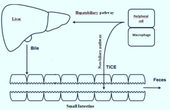
There are two main excretory pathways for cholesterol disposal from the body, which are named the hepatobiliary and non-biliary route (Temel and Brown 2012). The hepatobiliary route transfers cholesterol from peripheral cells and macrophage foam cells in the artery wall plaque to the liver for secretion (Glomset 1968; Rong et al. 2001), while in the non-biliary pathway the cholesterol is directly secreted into the intestine (Figure 1) (van der Velde et al. 2007). Therefore, both the liver and intestine are involved in cleansing the body from the excess cholesterol, and indeed, both the biliary and non-biliary pathways are part of the reverse cholesterol transport (RCT).
5
Figure 1. Schematic representation of the main pathways of cholesterol excretion. Adapted from (Brufau et al. 2011).
6
1.1.1 Hepatobiliary pathway
The classic model of cholesterol secretion, which is called the hepatobiliary pathway, was presented by John Glomset almost 40 years ago (Glomset 1968). In this process, cholesterol, from peripheral tissues and macrophage foam cells in the artery wall plaques, is returned to the liver via high-density lipoproteins (HDLs), for secretion into bile and excretion through the feces (Glomset 1968; Rong et al. 2001).
Different types of molecules such as transporters and/or receptors both in peripheral cells and hepatocytes (e.g., the HDL receptor), apolipoproteins (e.g., apoA1), and plasmatic enzymes (e.g., lecithin:cholesterol acyltransferase (LCAT)) are involved in this process. They are responsible for transporting the cholesterol from peripheral tissues, moving it through plasma, and finally delivering it to the liver for excretion (Jolley et al. 1998). In the following pages, this pathway will be explained in details.
1.1.1.1 Cholesterol influx into liver
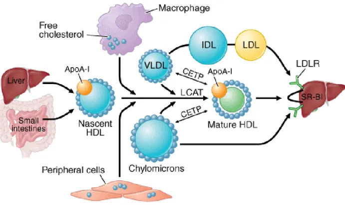
HDL is the main lipoprotein involved in removing excess cholesterol from cells and transporting it through the circulation into the liver. The apolipoprotein A1 (apoA1) is the main structural protein component of the HDL. HDL formation starts when apoA1 is synthesized and secreted as a lipid-poor protein mainly by the liver and also to some extent by the intestine (Figure 2) (Oram and Vaughan 2006). ApoA1 interacts with the membrane-embedded ATP binding cassette transporter A1 (ABCA1) and incorporates small amounts of unesterified cholesterol and phospholipids into the apoA1 molecule (Zannis et al. 2006). ABCA1 changes lipid-poor apoA1 to partially lipidated “nascent” lipoproteins. Then, these nascent HDLs become effective acceptors for cholesterol secreted by peripheral and foam cells (Oram and Vaughan 2006; Vaughan et al. 2006). ABCA1 mediates the rate-limiting step in HDL particle formation and maintains plasma HDL levels (Oram and Vaughan 2006). Maturation of these nascent HDLs happen in the circulation through the activity of the enzymes lecithin: cholesterol acyltransferase (LCAT) and phospholipid transfer protein (PLTP). LCAT converts free cholesterol of nascent HDL to cholesteryl ester and PLTP transfers phospholipids from remnant particles to HDL (Jolley et al. 1998). Then, ABC transporters (ABCA1 and ABCG1), in peripheral and foam cells, by interacting with HDL,
7
transfer the unesterified excess cholesterol (Wang et al. 2004; Tall et al. 2008). ABCG1 is highly expressed in macrophages whereas ABCA1 is more ubiquitous (Oram and Vaughan 2006). Targeted disruption of ABCA1 and ABCG1 resulted in a total ablation of cholesterol efflux in vitro, decreased reverse cholesterol transport into feces in vivo, and accelerated atherosclerosis (Out et al. 2006; Yvan-Charvet et al. 2007). Moreover, higher expression of ABCG1 was observed in cholesterol-loaded macrophages, providing an explanation for the reverse relationship between HDL levels and risk of atherosclerosis (Wang et al. 2004; Brufau et al. 2011). Mature HDL particles are remodeled by cholesteryl ester transfer protein (CEPT) in the circulation. CEPT facilitates the transport of cholesterol esters (CEs) and TG between the lipoproteins. It transfers CE molecules from HDL to very low density lipoprotein (VLDL) and chylomicron in exchange for TG (Curtiss et al. 2006). Ultimately, cholesteryl esters are delivered to the liver (Jolley et al. 1998).
8 1.1.1.2 Hepatic cholesterol uptake from circulation
The liver first clears the cholesterol from HDL at the basolateral side of the hepatocytes via scavenger receptor class B type 1 (SR-B1)-dependent selective uptake which is predominant pathway of cholesterol uptake in rodents (Jolley et al. 1998). It is important to note that in CETP containing species like human, monkey and rabbit a large portion of HDL’s CE cargo is transferred by CETP to apolipoprotein B (apoB)-containing lipoproteins, which are cleared by the liver through hepatic low-density lipoprotein (LDL) receptors (LDL-R) (Figure 3) (Morton et al. 2014; Temel et al. 2015). Approximately 70% of total LDL-R found in the body are present at the basolateral membrane of hepatocytes. The major function of LDL-R is mainly to bind ApoB and/or ApoE containing lipoproteins, such as LDL, VLDL, and chylomicron remnants to remove the highly atherogenic LDL particles from the circulation (Ikonen 2008). Therefore, hepatic LDLRs have a crucial role in the removal of LDL cholesterol particles from the circulation (Ouguerram et al. 2004). The LDL-R activity is downregulated post-transcriptionally by proprotein convertase subtilisin/kexin type 9 (PCSK9) (Abifadel et al. 2003).
9
Cholesterol may also be taken up from circulation through LDL receptor-related protein 1 (LRP1). LRP1, as a multifunctional receptor, is expressed in the liver and has close structural and biochemical similarities to LDL-R. LRP1 is responsible for the removal of VLDL remnants (IDL) and chylomicron remnants from the circulation. The lipoprotein remnants are enriched with cholesterol, therefore, their prolonged stay in the bloodstream could be atherogenic as well (Moon et al. 2012). LRP1 thus plays a key role in clearing these atherogenic particles along with LDLRs (Rohlmann et al. 1998). Hepatic LRP1 gene expression has been found to be negatively associated with intracellular cholesterol levels (Moon et al. 2011). Moreover, lower hepatic gene expression of LRP1 has been reported in HepG2 cells in a diabetic condition which was associated with the development of an atherogenic dyslipidemia (Moon et al. 2012). Sterol regulatory element-binding protein 2 (SREBP-2), as a nuclear receptor involved in cholesterol metabolism, regulates the gene expression of LDL-R, PCSK9 and LRP1 in the liver (Moon et al. 2011). When cellular cholesterol levels are low, SREBP-2 are transported to the Golgi, cleaved, and translocated to the nucleus in which gene targets are activated, while cellular cholesterol levels are high, the SREBP-2 remain uncleaved and attached to the endoplasmic reticulum and their gene expression is suppressed (Engelking et al. 2005).
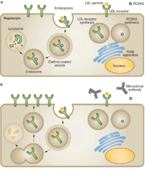
Interaction between PCSK9 and LDL-R
PCSK9, a member of the subtilisin serine protease family, is synthesized by the liver as a precursor in the endoplasmic reticulum (ER), and then transformed to an active protease in the Golgi apparatus and is subsequently secreted into circulation (Seidah et al. 2003). PCSK9 is coded as a natural inducer of LDL-R degradation (Maxwell et al. 2004). PCSK9 binds to the LDL-R, internalizes it, and the receptor along with the LDL particle are subsequently destroyed (Horton et al. 2009). Indeed, the tight binding of PCSK9 to LDL-R and its degradation in lysosome compartments prevents LDL-R recycling to the cell surface (Gent et al. 2004). Reduced LDL-R levels result in a reduction of LDL cholesterol uptake, which could lead to hypercholesterolemia. Therefore, PCSK9 has an important role in cholesterol metabolism (Maxwell et al. 2003). In the absence of PCSK9, the apoB–LDL receptor complex undergoes endocytosis then the LDL-R dissociates from the ligand. The
10
ligand is sent to the lysosome for degradation, and the LDL-R is recycled back to the cell surface to clear more LDL-C from the circulation (Figure 4) (Gent and Braakman 2004). It is important to note that there are different gene variants of PCSK9 which vary in their affinity for LDL-R and consequently leads to diverse changes in the circulating levels of LDL-C (Cunningham et al. 2007; Kwon et al. 2008). PCSK9 loss-of function (LOF) mutations are associated with lower plasma LDL-C levels and decreased risk of cardiovascular diseases. It means that PCSK9 does not bind LDLR to induce degradation. Therefore, LDLR can return to the cell surface and clear the cholesterol from circulation. Whereas PCSK9 gain-of function (GOF) mutations lead to higher LDL-C levels due to LDLR degradation and consequently, hypercholesterolemia and an increased risk of CVD (Abifadel et al. 2003; Cohen et al. 2006). PCSK9 shares a mutual regulatory pathway with LDL-R through the SREBP-2 (Maxwell et al. 2003). Indeed, both PCSK9 and LDL-R gene expression are up regulated transcriptionally by the transcription factor, SREBP-2 (Smith et al. 1990; Dubuc et al. 2004). Regulation of cholesterol metabolism is mainly modulated by SREBP-2. For instance, high dietary cholesterol prevents maturation of SREBPs and cuts off cholesterol and LDL receptor synthesis (Goldstein et al. 2006).
11
Figure 4. LDL-cholesterol metabolism in the presence (a) or absence of PCSK9 (b). Taken from (Dadu et al. 2014).
12
Transcriptional control of cholesterol levels in the liver
Cholesterol synthesis, uptake and clearance from the body are chiefly regulated via two nuclear receptors, sterol regulatory element binding proteins (SREBPs) and liver X receptors (LXRs) in the liver (Ikonen 2008).
The molecular mechanism of how hepatocytes maintain cholesterol homeostasis has become more precise with the discovery of the transcription factors sterol regulatory element binding proteins (SREBPs) (Weber et al. 2004). Sterol regulatory elements (SREs) are nucleotidic sequences in the gene promoters, encoding proteins involved in cholesterol homeostasis such as HMGCoA-r and LDL receptor (LDL-R). These sequences are recognized by a family of transcription factors called SREBP. The SREBP family members, SREBP-1 (a and c) and SREBP-2, are synthetized as membrane protein in the endoplasmic reticulum. SREBP-2 is considered to be largely involved in the regulation of cholesterol metabolism (Goldstein et al. 2006)
SREBP2 modulates cholesterol metabolism through the activation of transcription of genes involved in cholesterol synthesis and liver uptake from the circulation (Goldstein et al. 2006). 3-hydroxy 3-methylglutaryl coenzyme A reductase (HMGCoA-r) catalyzes the rate limiting step in cholesterol synthesis in the liver and its gene expression is regulated by SREBP2 (Horton et al. 1998). As mentioned above, LDLR, PCSK9, and LRP1 transcripts are also regulated by nuclear factor SREBP2.
LXR acts as whole body cholesterol sensors. Oxysterols as ligands activate LXR and its activation contributes in reverse cholesterol transport through stimulation of transcripts involved in cellular cholesterol efflux and hepatic cholesterol secretion (Tontonoz et al. 2003). ABC transporters have been identified as LXR target genes, including ABCA1, ABCG1, and also ABC transporters G5/G8 (ABCG5/G8). The ABCA1 and ABCG1 play a critical role in the efflux of excess cellular cholesterol to apoA1, the first step in reverse cholesterol transport. The ABCG5/G8 proteins form a dimer that resides in the apical membrane of the hepatocyte and functions to pump cholesterol into bile (Venkateswaran et al. 2000; Tontonoz and Mangelsdorf 2003). In addition to ABC transporters, cholesterol 7 alpha-hydroxylase
13
(CYP7A1) is also an LXR target gene. CYP7A1 catalyzes the rate-limiting step in bile acid synthesis in the liver (Peet et al. 1998).
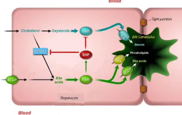
1.1.1.3 Cholesterol excretion from the liver
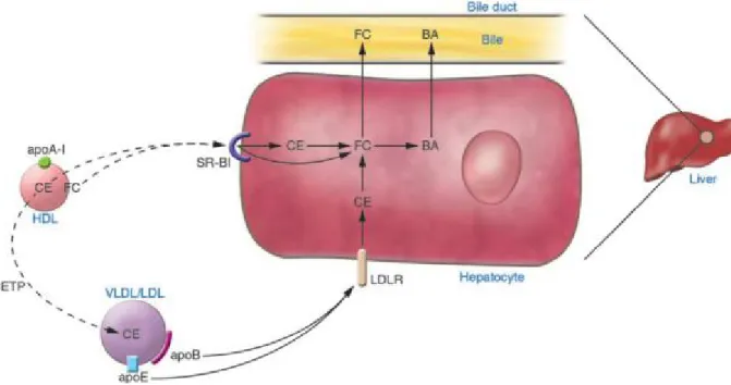
A large portion of cholesterol delivered to the liver can be directly discarded via ABCG5/G8 in the bile canaliculus (Berge et al. 2000). The presence of a biliary acceptor ‘micelle’ is necessary for cholesterol movement into bile. Micelles are assembled during the simultaneous transport of bile acids and phospholipids into bile by transporters such as the bile salt export pump (BSEP) and the multidrug resistance protein 2 (MDR2), respectively. MDR2 is also encoded by the ABCB4 gene (Voshol et al. 1998). Furthermore, cholesterol can be converted to bile acids by an array of enzymes including CYP7A1 and then secreted in the bile canaliculus (Figure 5) (Myant et al. 1977). The excess free cholesterol can also move to the endoplasmic reticulum (ER) in the liver where it is repackaged onto nascent apoB-containing lipoproteins that are ultimately secreted from the liver into the bloodstream (Pramfalk et al. 2005).
Figure 5. Cholesterol excretion from the liver. Adapted from (Jonker et al. 2009).
14
Once cholesterol is secreted into bile, a large portion of this pool is delivered to the lumen of the small intestine via the common bile ducts (Temel and Brown 2015). Cholesterol excretion in the form of neutral sterols or bile acids via the bile into the feces is the main mechanism of elimination of excess cholesterol from the body (Brufau et al. 2011). Therefore, the hepatobiliary pathway is an atheroprotective route that decreases the risk of atherosclerosis (Yu et al. 2002b; Meissner et al. 2011).
1.1.1.3.1 Bile acids formation and the enterohepatic circulation
Bile acids are amphipathic steroids that are formed from cholesterol in the liver. Conversion of cholesterol to bile acids is critical for maintaining cholesterol homeostasis and preventing cholesterol accumulation in liver. Primary bile acids are conjugated to either taurine or glycine to increase hydrophilicity. They are secreted into the bile and stored in the gallbladder. Upon ingestion of a meal, they are discharged into the small intestine to promote nutrient digestion and absorption in the proximal intestine. Bile acids are re-absorbed with an efficiency of 95% at the distal ileum and transported back to the liver through the portal vein and re-secreted into bile which results in the accumulation of a pool of bile acids (Russell 2003; Dawson et al. 2009). This bile acid pool cycles between the liver and the intestine and is called the enterohepatic circulation (Hofmann 2009) and the non-absorbed (5%) bile acids are eliminated from the body in the feces (Dawson et al. 2009)
Role of FXR in regulation of bile acid synthesis and transport
There is accumulated evidence indicating that farnesoid X receptor (FXR) exerts a key role in bile acid metabolism through the regulation of bile acid synthesis, bile acid secretion, intestinal bile acid absorption, and hepatic uptake of bile acids (Sinal et al. 2000; Eloranta et al. 2008; Lefebvre et al. 2009; Modica et al. 2010).
Bile acid absorption in the intestine is regulated by the nuclear factor FXR through the regulation of bile acid transporters from the intestine to the portal system (Wang et al. 1999; Matsubara et al. 2013). These acid transporters are involved in bile acid reabsorption at the apical and basolateral membranes of the ileum (Figure 6). The apical sodium-dependent bile
15
acid transporter (ASBT) is expressed at the apical membrane of enterocytes in the terminal ileum and mediates the reabsorption of bile acids from the ileum. The ileal bile acid binding protein (iBABP) shuttles the bile acids from the apical side to the basolateral membrane of ileum. Heteromeric organic solute transporter α and β (Ostα-Ostβ) are ileal basolateral bile acid transporters. They transport the bile acids from the basolateral side of the ileum toward the liver (Shneider 2001; Dawson et al. 2005). Bile acids are then re-circulated via the portal circulation to the hepatocytes, where a sinusoidal Na+- dependent taurocholate cotransport peptide (NTCP) takes them up into hepatocytes. NTCP transporters are involved in bile acid uptake at the basolateral membrane of the hepatocytes. It is the major uptake system to transport bile salts from the portal circulation into the liver cells (Stieger et al. 1994).
Figure 6. Transporters involved in bile acid reabsorption in the ileum. Adapted from (Schaap et al. 2014) E nte rohe pati c circ ula tio n of bi le a ci ds iBABP
16
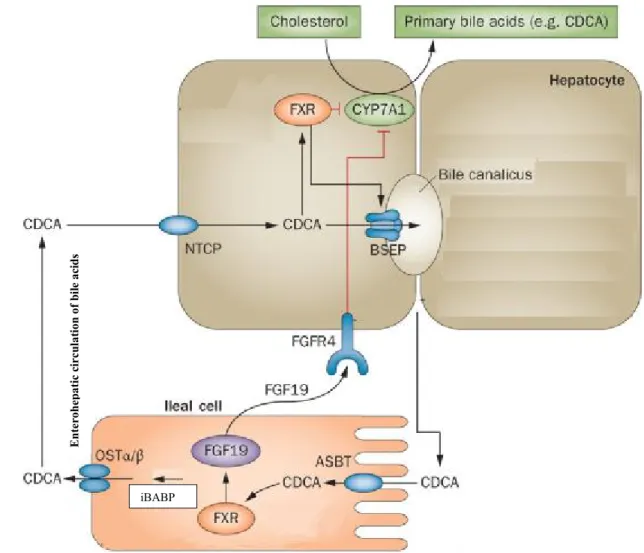
Bile acids function as natural ligands for the transcription factor FXR (also known as bile acid receptor or nuclear receptor subfamily 1 group H member 4) (Parks et al. 1999; Wang et al. 1999). Bile acids induce FXR activation both in the liver and the intestine which leads to a set of interactions resulting in suppression of bile acid biosynthesis (Wang et al. 1999). Activation of FXR is a major mechanism in suppressing bile acid biosynthesis by reducing the expression levels of CYP7A1. FXR-mediated induction of hepatic small heterodimer partner (SHP) and intestinal fibroblast growth factor 15/19 (FGF15/19) (FGF19 in humans) has been shown to be responsible for this suppression (Kerr et al. 2002; Holt et al. 2003).
a. Hepatic FXR/SHP/CYP7A1 pathway
FXR knockout mice showed an increase in bile acid synthesis and CYP7A1 gene expression suggesting FXR-mediated bile acid inhibition of CYP7A1 (Figure 7) (Sinal et al. 2000). CYP7A1 catalyzes the rate-limiting step of cholesterol conversion into bile acids in the liver (Jelinek et al. 1990). FXR inhibits CYP7A1 gene transcription by indirect mechanism. Bile acid-activated FXR induces SHP gene expression that inhibits the activity of liver related homologue-1 (LRH-1), and results in inhibiting CYP7A1 gene transcription (Goodwin et al. 2000). The FXR/SHP mechanism is supported by the finding that SHP and CYP7A1 mRNA expression levels have an inversed relationship, and CYP7A1 expression and bile acid synthesis are induced in SHP knockout mice. Paradoxically, bile acid feeding to SHP null mice inhibits CYP7A1 expression and bile acid synthesis suggesting that redundant pathways may exist for bile acid inhibition of CYP7A1 (Kerr et al. 2002; Wang et al. 2002).
SHP
The SHP gene is expressed in different tissues, including liver, heart, pancreas, kidney, adrenal gland, spleen, stomach, and small intestine (Lee et al. 1998). SHP is an atypical receptor without a DNA-binding domain, but has a putative ligand-binding domain, which makes SHP a member of the nuclear receptor family (Seol et al. 1996; Seol et al. 1997). Furthermore, SHP interacts with several nuclear receptor family members. Through these interactions, SHP is involved in diverse metabolic pathways, including cholesterol, bile acid,
17
triglyceride, and glucose homeostasis (Lee et al. 2007; Zhang et al. 2011). For instance, hepatic SHP overexpression in transgenic mice led to liver steatosis due to an indirect activation of SREBP-1c (Boulias et al. 2005). Besides, deletion of SHP decreased TG accumulation in ob/ob obese mice, which was associated with increased hepatic VLDL secretion and elevated expression of microsomal triglyceride transfer protein (MTP), the rate limiting enzyme in VLDL assembly and secretion (Huang et al. 2007).
Not only SHP can affect diverse biological responses, but also several genetic variations and mutations of SHP were identified in obese and diabetic subjects in population based studies showing that there might be a relationship between SHP genetic variations and increased risk of obesity and type 2 diabetes (Nishigori et al. 2001; Hung et al. 2003; Echwald et al. 2004; Enya et al. 2008).
b. Intestinal FXR/FGF15/19 /FGFR4 pathway
It has been suggested that intestine-derived FGF15/19 acts as an enterohepatic signal to activate hepatic fibroblast growth factor receptor 4 (FGFR4) signaling, which inhibits CYP7A1 expression in the hepatocytes (Figure 7) (Inagaki et al. 2005). Bile acids induce FXR activation in the intestine which results in stimulation of FGF15/19 and then, FGFR4 acts as a hepatic receptor for intestinal FGF15/19. FGFR4 mediates the effects of intestinal FGF15/19 on suppression of bile acid biosynthesis in liver (Holt et al. 2003). FGFR4 deficient mice showed higher mRNA levels of CYP7A1 suggesting that FGFR4 is also involved in suppression of bile acid biosynthesis (Kong et al. 2012).
Intestinal FXR-dependent mechanism is based on the observation that GW4064 (a synthetic FXR agonist) induces an intestinal hormone FGF15/19, which activates a hepatic FGFR4 signaling and through that inhibits bile acid biosynthesis in the liver (Holt et al. 2003). Intestinal FXR activation by bile acids leads to the release of FGF15/19 from the ileum and the expression of FGF15/19 mRNA is negatively correlated to the CYP7A1 mRNA expression levels in mouse liver (Inagaki et al. 2005).
18
Figure 7. The molecular mechanisms of FXR pathway in bile acid synthesis in liver and intestine. Adapted from (Ory 2004; Inagaki et al. 2005)
19
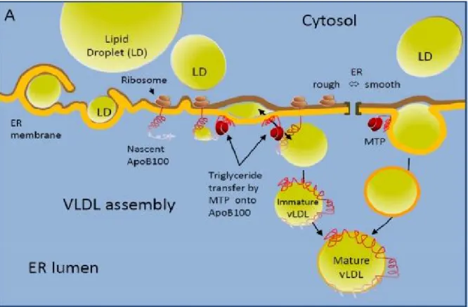
1.1.1.3.2 VLDL assembly
Since liver constantly takes up circulating triglycerides and cholesterol from both endogenous and exogenous sources, VLDL production and secretion by hepatocytes is a crucial step in preventing hepatic steatosis (Alger et al. 2010; Flamment et al. 2010).
When lipids are available in the ER, the newly synthesized apo-B polypeptide interacts co-translationally with MTP, which is a rate-limiting molecule in VLDL assembly and secretion, and transfers triglycerides (TG) into the apo-B (Figure 8) (Rava et al. 2006). ApoB-100 is an essential structural protein that translocates into the luminal side of the endoplasmic reticulum (Cianflone et al. 1990). Diacylglycerol acyltransferase 2 (DGAT2) plays a role in VLDL assembly by converting fatty acids into TG. DGAT2 catalyzes the last step of the synthesis of TGs that are going to be incorporated into VLDL. Acyl-CoA:cholesterol acyltransferase (ACAT-2) has also a key role in the hepatic storage and packaging of cholesteryl ester into apoB-containing lipoproteins (VLDL), by converting free cholesterol into cholesterol esters (Pramfalk et al. 2005; Chang et al. 2009). Cell death-inducing DNA fragmentation factor alpha (DFFA)-like effector B (Cideb) is a lipid droplet-associated protein contributing to further lipidation of lipoprotein particles after they exit the endoplasmic reticulum compartment (Ye et al. 2009). The last step of VLDL production is mediated by small GTPase a (Sar1a), which is required for Golgi trafficking events. Sar1a, an intracellular vesicular trafficking protein, facilitates the movements of VLDL particles toward the Golgi apparatus where they are secreted in the plasma (Asp et al. 2000). Since VLDL carries both TG and cholesterol into circulation, appropriate assembly and secretion of VLDL is important for both liver cholesterol contents and plasma cholesterol levels.
20
21
1.1.2 Non-biliary TICE pathway
Transintestinal cholesterol excretion (TICE) presents a non-hepatobiliary direct route of cholesterol secretion “from blood to gut” (van der Velde et al. 2007). The highest levels of TICE have been reported to take place in the proximal small intestine (Brown et al. 2008). Under normal physiological conditions the biliary route is a predominant pathway for cholesterol excretion while TICE accounts for nearly 20–30% of fecal neutral sterols in both human and animal (Temel and Brown 2012). However, there is evidence showing that the TICE pathway can be stimulated by both pathophysiologic and pharmacologic stimuli (Temel and Brown 2012). For example, pharmacological activation of liver X receptor (LXR) (van der Veen et al. 2009) or high fat diet (van der Velde et al. 2008) resulted in increased intestinal cholesterol disposal. There is evidence showing that the set point of cholesterol excretion is sustained by the crosstalk between biliary and non-biliary pathways (Kruit et al. 2005). In a recent study hepatic flavin monooxygenase 3 (FMO3) was identified as a key cholesterol regulator of both biliary and non-biliary RCT pathways (Warrier et al. 2015). Indeed, cholesterol disposal from the body requires contribution of both biliary and non-biliary TICE pathways (Temel and Brown 2015). Since TICE like the hepatobiliary route is involved in cholesterol removal from thebody, it can be considered an atheroprotective pathway.
However, to date our knowledge about the molecular mechanisms that define TICE is limited. The identification of non-biliary route largely stems from multiple observations indicating that biliary cholesterol excretion does not match with the amount of cholesterol in the feces. According to the classic biliary model, both biliary cholesterol secretion and fecal cholesterol loss should precisely be predictable by plasma HDL levels. In contrast, several studies showed that biliary and fecal sterol losses are quite normal in mice with extremely low HDL levels (Jolley et al. 1998; Groen et al. 2001; Xie et al. 2009). Similarly, biliary obstruction or diversion did not avoid the appearance of neutral sterols in the feces (Pertsemlidis et al. 1973; van der Velde et al. 2008). For instance, in hepatic ABCG5/G8 and MDR2 knockout mice, which lack the ability to normally secrete cholesterol into the bile, fecal cholesterol loss remains either unchanged or, in some cases, increased (Voshol et al. 1998; Yu et al. 2002a). These findings have led to the discovery of a non-hepatobiliary route of cholesterol excretion termed transintestinal cholesterol excretion (TICE) (van der Velde et
22
al. 2007). Earlier findings of the existence of an additional pathway of cholesterol excretion go back to the beginning of the last century. In 1927, Sperry reported that bile diversion in mice did not lessen fecal neutral excretion (Sperry 1927). However, this observation was only confirmed 50 years later by Pertsemlidis (Pertsemlidis et al. 1973).
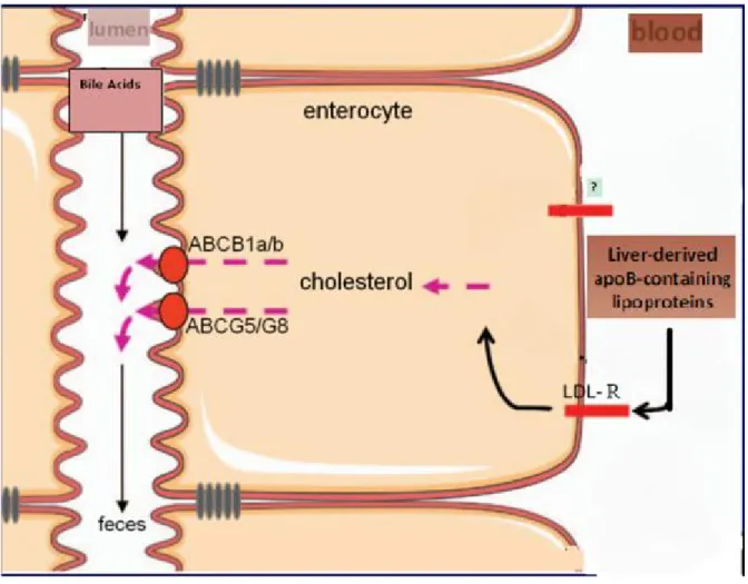
TICE as its name implies, relies on the intestine for cholesterol secretion. Through this pathway, cholesterol-derived from plasma lipoprotein is directly secreted via the small intestine into the lumen (van der Velde et al. 2008; Temel and Brown 2012). The intestine is responsible for receiving the cholesterol from the blood via its cholesterol receptors at the basolateral membrane. Possibly, there are molecules involved in transferring the cholesterol through the basolateral to the apical side of enterocytes, and finally there are cholesterol transporters at the apical membrane of enterocytes to discard cholesterol into the lumen. In order to address this pathway effectively, the molecules and receptors involved in the TICE pathway will be discussed in details in four steps.
1.1.2.1 Step1: The role of lipoproteins in the TICE pathway
TICE requires plasma lipoproteins to transport the cholesterol from either peripheral tissues and/or the liver to the small intestine for secretion (Temel and Brown 2012). However, it is still not clear which type of lipoproteins play the main role in this pathway.
As mentioned before, normal biliary and fecal cholesterol loss was observed in apoA-1 or ATP-binding cassette transporter A1 (ABCA1) null mice, although these mice present extremely low plasma HDL levels (Jolley et al. 1998; Xie et al. 2009). These findings imply two important points; firstly, the existence of non-biliary cholesterol excretion in addition to hepatobiliary route and secondly the non-biliary route does not depend on HDL to be proceeded. In line with these studies, recently Vrins et al showed that the rate of TICE did not change significantly in ABCA1 deficient mice with very low levels of HDL compared to the wild type (WT) littermates (Vrins et al. 2012). Moreover, intestinal perfusions of a modified Krebs solution supplemented with bile salts and phospholipids in B1-deficient mice, SR-B1 is a well-known HDL receptor, were significantly associated with a twofold increase in TICE suggesting that HDL might not have a main role in the TICE pathway (Acton et al.
23
1996; van der Velde et al. 2008). It thus seems that despite the predominant role of HDL in the hepatobiliary route (Tall et al. 2008), its role in non-hepatobiliary TICE pathway is unclear.
Based on these findings, it seems that liver-derived apoB-containing lipoproteins have a main role in delivering cholesterol to the proximal part of the intestine. The excess free cholesterol in the liver is shifted to the ER where it is packed onto nascent apoB-containing lipoproteins. The first apoB-containing lipoprotein secreted from the liver into the circulation is the VLDL. The VLDL then changes to other apoB-containing lipoproteins such as the IDL and/or the LDL. The liver-derived apoB-containing lipoproteins are then recognized by the proximal small intestine through lipoprotein receptors such as LDL-Rs and probably other receptors like LDL-Rs family (Temel and Brown 2015). For instance, Brown et al showed that liver-specific depletion of Acetyl-CoA acetyltransferase 2 (ACAT2) resulted in increased fecal sterol loss through non-biliary pathway without changes in HDL levels (Brown et al. 2008). ACAT2 converts free cholesterol into cholesterol esters to pack them in VLDL molecules. Liver-specific inhibition of ACAT2 via antisense oligonucleotide (ASO) prevents cholesterol esterification and secretion of CE in apoB-containing lipoproteins into the plasma. In this study, a reduction in ACAT2 protein and activity of 99.3% was observed and they found that even a small amount (less than 1% of normal) of ACAT2 may be sufficient to support packaging of hepatic CE into apoB-containing lipoproteins and these lipoproteins drives the cholesterol toward the proximal small intestine for secretion, suggesting that apoB-containing lipoproteins deliver cholesterol esters to the TICE pathway for excretion (Brown et al. 2008). In line with this study, Marshal et al recently showed that acutely reducing hepatic ACAT2 expression resulted in packaging the hepatic cholesterol onto nascent apoB-containing lipoproteins that feed cholesterol into the TICE pathway for fecal excretion (Marshall et al. 2014). All in all, these studies suggest that hepatic apoB-containing lipoproteins have a key role in delivering cholesterol into the TICE pathway for fecal excretion.
On the other hand, Le May et al. recently reported that both LDL and HDL can provide cholesterol for TICE in human and mice jejunal explants at the basolateral side of the enterocytes. They radiolabeled both LDL and HDL with 3H-free cholesterol (3H-LDL, 3 H-HDL) and observed that both these lipoproteins can deliver cholesterol to the proximal part of
24
the small intestine. According to Le May’s finding, both HDL and LDL can be involved in TICE (Le May et al. 2013).
Based on these findings, various lipoproteins might participate in TICE and provide cholesterol for secretion through this pathway. Regarding the role of lipoproteins as the cholesterol carriers in TICE, the liver function is undeniable in this pathway. Indeed, the liver plays a central role in TICE by providing requisite lipoproteins. Taking all together, these two organs, the intestine and the liver, collaborate for cholesterol secretion through the TICE pathway.
1.1.2.2 Step 2: Cholesterol receptors at intestinal basolateral membrane
Several studies have been conducted to investigate the role of potential intestinal basolateral receptors involved in cholesterol uptake from circulation in TICE route based on which lipoproteins are involved in delivering cholesterol to the basolateral side of the small intestine.
Recently, Le May et al reported that deletion of PCSK9 increases TICE. PCSK9 deletion means no degradation effect on LDL-R and that results in a higher number of intestinal LDL-R as a cholesterol acceptor in the TICE and consequently higher levels of TICE (Le May et al. 2013). This group had previously showed that PCSK9 deficient mice have higher amounts of LDL receptors in their intestine (Le May et al. 2009). Plasma PCSK9 induces LDL receptor degradation. Overall, this study showed that LDL receptors at the intestinal basolateral membrane are involved in cholesterol uptake from LDL (Le May et al. 2013). However, intestinal cholesterol uptake from apoB-containing lipoprotein does not merely depend on LDL receptors because there is evidence showing that LDL receptor deficient mice still have normal or increased levels of TICE (Brown et al. 2008; Le May et al. 2013).
Seemingly, other members of the LDL-R family can also play a role as cholesterol acceptors at the basolateral membrane of enterocytes. Other receptors including low-density lipoprotein receptor-related protein 1 (LRP1), very low-density lipoprotein receptor (VLDLR), and apolipoprotein E receptor 2 (apoER2), which are all members of the LDL receptor family, are also expressed in the gut (Herz et al. 1988; Garcia-Miranda et al. 2010). As a result, it
25
would be reasonable to assume that they can function as the main or secondary receptors for cholesterol delivering lipoproteins.
Based on these data, it can be concluded that LDL receptor and/or LDL receptor family, including LRP1, VLDLR, and apoER2 could participate in the intestinal cholesterol uptake from both apo E and/or apoB-containing lipoproteins.
1.1.2.3 Step 3: Cholesterol trafficking from the basolateral to the apical membrane of enterocytes
The trafficking itinerary of TICE-derived cholesterol within the enterocyte is not fully understood. It is probable that apoB-containing lipoproteins, which are the TICE cholesterol donor particles, are eventually degraded in lysosomes. It is tempting to assume that the trafficking itinerary would involve endosomal/lysosomal compartments (Temel and Brown 2012). Thus, TICE would need Niemann-Pick type C1 (NPC1) and NPC2 proteins to move the cholesterol out of the lysosomal compartment. These two cholesterol-binding proteins act in the removal of unesterified cholesterol from lysosomes (Vance et al. 2011; Temel and Brown 2012).
Vrins et al introduced other factors possibly involved in trafficking cholesterol. They showed that endosomal Rab protein 9 (Rab9) and lysosomal integral membrane protein-2 (LIMP2) could also play a role in intracellular trafficking of cholesterol derived from TICE (Vrins et al. 2009). Rab is a family of Ras-like small G proteins that control membrane trafficking (Ikonen 2008). The intestinal expression of these two genes and TICE were significantly increased in mice upon peroxisome proliferator-activated receptor delta (PPAR δ) activation (Vrins et al. 2009). Furthermore, earlier Van der Veen et al. showed that PPAR δ activation led to higher fecal neutral sterol excretion without affecting the hepatobiliary cholesterol secretion (van der Veen et al. 2005). As a consequence, increased expression of Rab9 and LIMP2 might be a sign of cholesterol trafficking. Nevertheless, the relationship between the proteins encoded by these genes and TICE is still not clear (Vrins et al. 2009).
26
1.1.2.4 Step 4: Cholesterol efflux via intestinal apical transporters into the lumen
After being moved to the intestinal apical membrane, cholesterol is excreted via apical transporters into the lumen. ABCG5/G8 transporters are involved in TICE, regarding to their indispensable role in cholesterol excretion. Van der Veen et al. showed that TICE is impaired in ABCG5 deficient mice, suggesting that ABCG5/G8 contributes to TICE. However, in this study ABCG5 deficient mice still had a substantial contribution of TICE, suggesting that other apical transporters might be involved (van der Veen et al. 2009). On the other hand, Le May et al observed that TICE was reduced by 26.5% in ABCB1a/b deficient mice (Le May et al. 2013). It seems, therefore, that both apical transporters, ABCG5/G8 and ABCB1a/b, contribute to the excretion of cholesterol into the gut lumen. It is reasonable to suppose that the secreted cholesterol into the gut requires luminal acceptors. A mixture of bile salts and phospholipids has been suggested as the luminal cholesterol acceptors in TICE (van der Velde et al. 2007). This finding is in the line with the presence of high concentration of bile-derived phospholipids in the proximal small intestine, a region of the gut that has been reported to have the highest levels of TICE (Temel and Brown 2012).
In summary, mainly plasma apoB-containing lipoproteins deliver cholesterol to the intestine for excretion through the TICE route. LDL receptor and/or LDL receptor family are responsible for cholesterol uptake from circulation at the intestinal basolateral membrane. The exact molecules involved in trafficking the cholesterol from basolateral to apical membrane, are unknown at the present time. ABCG5/G8 and ABCB1a/b are the main transporters at the intestinal apical membrane responsible for the secretion of cholesterol into the lumen in the TICE pathway (Figure 9).
Taken together, TICE is a non-biliary route of the cholesterol elimination. It directly discards the cholesterol into the lumen. Thus, TICE could be considered an anti-atherogenic pathway; however, a better understanding of this newly identified pathway will require further investigations.
27
Figure 9. A model of non-biliary transintestinal cholesterol excretion (TICE). Adapted from (Le May et al. 2013).