Modalities of exercise training on liver fat accretion and
inflammatory markers in ovariectomized rats
par
Abdolnaser Pighon
Département de kinésiologie
Thèse présentée à la Faculté des études supérieures en vue de l’obtention du grade de Philosophiae Doctor (Ph.D.)
en Sciences de l’activité physique option Physiologie de l’exercice
Mars, 2010
Faculté des études supérieures et postdoctorales
Cette thèse intitulée:
Modalities of exercise training on liver fat accretion and inflammatory markers in ovariectomized rats
Présenté par: Abdolnaser Pighon
a été évaluée par un jury composé des personnes suivantes:
Yan Burelle, président-rapporteur Jean-Marc Lavoie, directeur de recherche
Raynald Bergeron, membre du jury André Tchernof, examinateur externe Marielle Ledoux, représentant du doyen de la FES
Résumé
Les facteurs de risque des maladies cardiovasculaires, telle, que la détérioration du profil lipidique, deviennent plus prononcés après la ménopause, ce qui fait de la maladie coronarienne, l’une des principales causes de décès chez les femmes ménopausées. Une proportion importante de femmes prennent du poids après la ménopause en particulier dans la région abdominale entraînant par conséquent des perturbations métaboliques. Des données récentes suggèrent également que l’absence des œstrogènes observée à la ménopause favorise le développement de la stéatose hépatique. Cette dernière a été incriminée pour incriminée dans le développement de la résistance à l'insuline, et est de ce fait considérée comme une composante hépatique du syndrome métabolique. Il est impératif d'établir des stratégies visant à contrecarrer l'accumulation de graisse dans le foie et l’accroissement du tissu adipeux chez les femmes ménopausées, en tenant compte que l'utilisation de l'hormonothérapie substitutive est de nos jours moins soutenue. Les quatre études de la présente thèse ont été conduites pour tenter de fournir des informations sur le traitement et la prévention de l’augmentation de la masse graisseuse et de la stéatose hépatique qu’entraîne la suppression des œstrogènes, à travers les modifications du mode de vie (diète et exercice physique) chez la rate ovariectomizée (Ovx); un modèle animal de la ménopause.
Dans les deux premières études nous nous sommes concentrés sur l’augmentation de la masse graisseuse et sa reprise suite à une perte de poids. Dans la première étude, nous avons montré que les rates Ovx qui ont suivi un programme de restriction alimentaire (FR) ont diminué significativement (P < 0.01) leur poids corporel, leur contenu en graisses intra-abdominales ainsi que leurs triacylglycérols (TAG) hépatiques, comparativement aux rates Ovx nourries à la diète normale. De plus, l’entraînement en résistance (RT) a prévenu la reprise de poids corporel ainsi que l’accroissement du tissu adipeux et l’accumulation de lipides dans le foie des rates Ovx, après l’arrêt du régime amaigrissant. Les résultats de la deuxième étude ont confirmé l'efficacité de la restriction alimentaire associée à
l’entraînement en résistance (FR + RT) dans la réduction du poids corporel, des lipides dans le foie et le tissu adipeux chez les rates Ovx. Tenant compte des résultats de notre première étude, l’entraînement en résistance seulement a constitué un atout pour atténuer le poids corporel et la masse grasse reprise par les rates Ovx suite à un programme de perte de poids (FR + RT); bien que l'impact ait été moindre comparé au maintien seul de la restriction alimentaire. De la même manière que la supplémentation en œstrogènes, les résultats de la troisième étude indiquent que l'entraînement en endurance mené concurremment avec l’ovariectomie a significativement atténué l'accumulation de lipides dans le foie ainsi que dans le tissu adipeux. Toutefois, l’entraînement en endurance effectué avant l'ovariectomie n'a pas protégé contre l'accumulation des graisses qu’entraîne l'ovariectomie, si celui-ci est interrompu après l'ovariectomie. Enfin, pour compléter les résultats antérieurs, nous avons montré dans la quatrième étude que l’expression des gènes impliqués dans la synthèse de lipide; SREBP-1c, SCD-1, ChREBP, et ACC dans le foie a augmenté après le retrait des œstrogènes, tandis qu’une diminution (P < 0.01) des niveaux d'ARNm de PPAR-α a été observée. De plus, l'expression hépatique des gènes des cytokines pro-inflammatoires incluant IKKβ, IL-6 ainsi que le contenu protéinique de NF-кB étaient augmentés (P < 0.01) chez les rates Ovx par rapport aux rates ayant subi une Ovx simulée (Sham). Toutes ces perturbations ont été améliorées avec la supplémentation en œstrogènes seulement, ainsi qu'avec l'entraînement en endurance seulement.
Dans l'ensemble, nos résultats indiquent que l'exercice physique (en résistance ou en endurance) a un impact significatif sur la réduction de l'accumulation des lipides dans le foie et dans le tissu adipeux des rates Ovx. De plus, chez les rates Ovx, l’entraînement en endurance mimerait les effets des œstrogènes sur l'expression des gènes impliqués dans l'accumulation de lipides et l’inflammation préclinique dans le foie.
Mots-clés : Œstrogènes, Obésité ménopausique, Reprise de poids et de graisse, Foie gras, Restriction alimentaire, Entraînement en résistance, Entraînement en endurance, Bio-marqueurs de l'inflammation, Ovariectomie (Ovx), Rat
Abstract
Cardiovascular disease risk factors, such as lipid profile deterioration, become more pronounced after menopause making coronary heart disease a leading cause of death among postmenopausal women. A large proportion of women after menopause gain weight especially in the abdominal region resulting in several metabolic disturbances. Recent evidence also suggests that loss of estrogen function in menopause is associated with the development of a state of hepatic steatosis. Excessive fat accumulation in hepatocytes has been shown to play an important role in the development of insulin resistance and is even considered as a hepatic component of the metabolic syndrome. There is an important need to establish strategies to counteract fat accumulation in adipocyte and liver in postmenopausal women specifically considering the fact that utilization of hormone replacement therapy is now less supported. The four studies of the present thesis have been conducted in an attempt to provide information on the treatment and prevention of estrogen withdrawal-induced fat mass and hepatic steatosis via lifestyle modifications (diet and exercise training) in an ovariectomized (Ovx) rat model of menopause.
In the first two studies we focused on fat mass gain and regain following weight loss. In study 1, we showed that food restriction program (FR) decreased (P < 0.01) body mass, intra-abdominal fat pad weight, and liver triacylglycerol (TAG) levels as compared to normally fed Ovx rats. Moreover, resistance training program (RT) was useful in preventing body weight as well as adipose tissue and liver fat regain in Ovx rats, following diet-induced weight loss. Results of study 2 confirmed the efficiency of the FR + RT program in reducing body weight as well as liver and adipocytes fat accretion in Ovx rats. In line with the findings of our first study, continuation of only RT constituted an asset to attenuate body weight and fat mass regain in Ovx rats following a FR + RT weight loss program, although the impact was less than maintaining FR alone. Similar to estrogen supplementation, results of study 3 indicated that endurance exercise training conducted concurrently with the induction of ovariectomy significantly attenuated liver and adipocyte
fat accumulation. However, an endurance exercise training state acquired before ovariectomy did not provide any protective effects against ovariectomy-induced fat accumulation if exercise is discontinued after the ovariectomy. Finally, complementing previous findings we showed in study 4 that liver gene expressions of transcription factors SREBP-1c and ChREBP along with downstream lipogenic enzymes SCD-1 and ACC were increased with estrogens withdrawal conversely to reduced PPAR-α mRNA levels (P < 0.01). Furthermore, gene expressions of pro-inflammatory cytokines including IKKβ and IL-6 as well as protein content of NF-кB were higher (P < 0.01) in the liver of Ovx than in Sham animals. All of these responses were corrected with estrogen supplementation alone as well as with endurance exercise training alone in Ovx rats.
On the whole, our results indicate that exercise training (resistance or endurance) has a significant impact on reducing fat accumulation in liver and adipocytes in Ovx rats. In addition, it seems that endurance exercise training in Ovx rats stimulates estrogenic-like effects on the expression of genes involved in lipid accumulation and sub-clinical inflammation in the liver.
Keywords: Estrogen, Menopausal obesity, Weight and fat regain, Hepatic steatosis, Food restriction, Resistance training, Endurance exercise training, Inflammatory bio-markers, Ovariectomy (Ovx), Rat
Table of contents
Résumé ... i
Abstract ... iii
Table of contents ... v
List of tables ... viii
List of figures ... ix
Abbreviations ... x
Acknowledgements ... xiii
Introduction ... 1
Chapter 1: Review of literature ... 3
Emergence of metabolic syndrome and hepatic steatosis in menopausal hormonal state: treatment and prevention by exercise training ... 3
Cardiovascular disease in postmenopausal women ... 3
Menopausal obesity and the emergence of metabolic syndrome ... 5
Menopause, weight gain and fat redistribution ... 6
Menopause and hepatic steatosis ... 10
Prevention/treatment of adipose tissue and liver fat accumulation in menopausal hormonal state ... 30
The specific case of liver fat accumulation and exercise training ... 40
Pathogenic role of sub-acute systemic inflammation in obesity and insulin resistance .. 41
Inflammation ... 41
Circulating inflammatory markers in metabolic syndrome ... 43
Cellular inflammatory responses in obesity and insulin resistance ... 45
Physical activity and systemic low-level inflammation ... 50
The importance of inflammatory cytokines in fatty liver and insulin resistance ... 54
Inflammation and menopause ... 56
General objective of thesis and presentation of manuscripts ... 58
Abstract ... 63 Introduction ... 64 Methods ... 66 Results ... 69 Discussion ... 72 References ... 78 Legends ... 84
Chapter 3: Original article 2 ... 90
Abstract ... 92 1. Introduction ... 93 2. Methods ... 95 3. Results ... 98 4. Discussion ... 101 References ... 105 Legends ... 110
Chapter 4: Original article 3 ... 116
ABSTRACT ... 118 INTRODUCTION ... 119 METHODS ... 121 RESULTS ... 124 DISCUSSION ... 126 REFERENCES ... 131 LEGENDS ... 137
Chapter 5: Original article 4 ... 144
Abstract ... 146
Introduction ... 148
Methods ... 150
Results ... 155
References ... 163
Figure legends ... 171
Chapter 6: General discussion and conclusion ... 180
Conclusion ... 186
List of tables
Table 1. Taken from [Despres 1993] Features of the metabolic syndrome. ... 7
Table 2. Taken from [Loria, Lonardo et al. 2008] Possible pathophysiological bases for an association between NAFLD and accelerated atherosclerosis. ... 12
Table 3. Taken from [Wilfred de Alwis and Day 2007] Genes potentially involved in the pathogenesis of nonalcoholic fatty liver disease (NAFLD). ... 17
Table 4. Effects of estrogen withdrawal on liver fat accumulation. ... 29
Table 5. Taken from [Bruunsgaard 2005] Self-reported physical activity and physical performance in relation to low-level inflammation in epidemiological studies ... 52
Table 6. Estrogenic-like effects of exercise training on metabolic and inflammatory bio-markers in Ovx rats. ... 185
List of figures
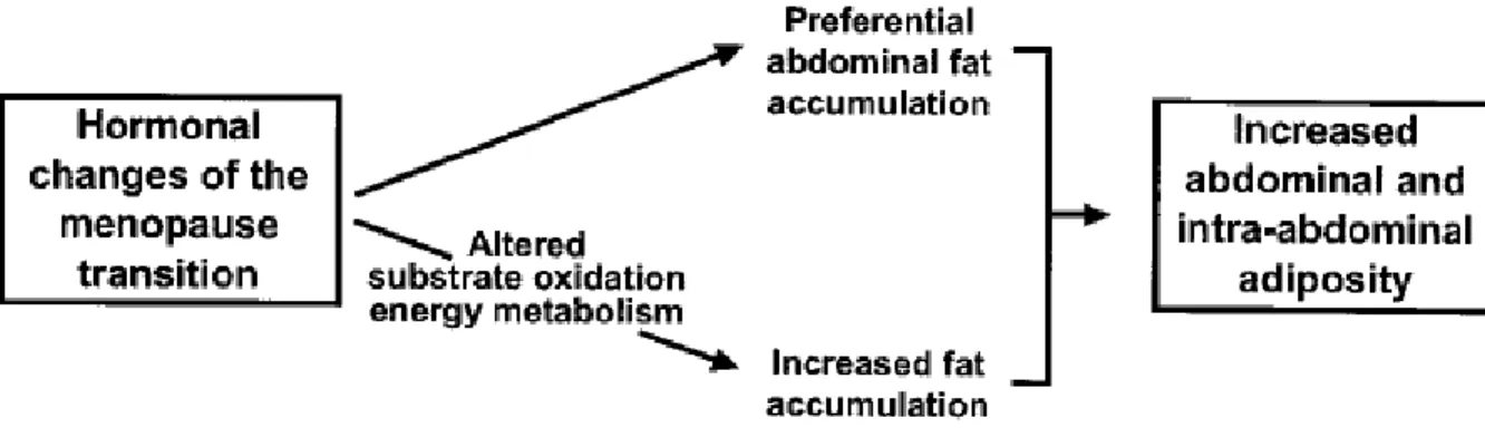
Figure 1. Potential mechanisms explaining menopause-related increases in abdominal and intra-abdominal adiposity... 9
Figure 2. Taken from [Lavoie and Gauthier 2006] Overview of the four main pathways involved in the development of nonalcoholic hepatic steatosis, and their regulatory factors. ... 15
Figure 3. Taken from [MacLean, Higgins et al. 2006] Metabolic state after weight-reduction (A), early in relapse (B), and after relapse (C). ... 39
Figure 4. Taken from [Shoelson, Herrero et al. 2007] Potential mechanisms for obesity-induced adipocyte inflammation. ... 47
Figure 5. Taken from [Chen 2006] Hypothetical model of metabolic stress, cellular inflammatory responses and effects on insulin signaling pathway. ... 48
Figure 6. Taken from [Shoelson, Lee et al. 2006] Local, portal, and systemic effects of inflammation in insulin resistance and atherogenesis. ... 51
Figure 7. Taken from [Rogers, Perfield et al. 2009] Ovx mice display early hepatic steatosis and inflammation. ... 59
Abbreviations
ACC : Acetyl-CoA carboxylase AMPK : AMP-activated protein kinase
CVD : Cardiovascular disease CHD : Coronary artery disease
CRP : C-reactive protein
ChREBP : Carbohydrate response element-binding protein E2 : 17β-estradiol (estrogen)
ER : Estrogen receptor FFA : Free fatty acid FAS : Fatty acid synthase
FR : Food restriction
HDL : High density lipoprotein HRT : Hormone replacement therapy
HSL : Hormone sensitive lipase
IKKβ : Inhibitor of kappa B kinase beta IL : Interleukin
IRS : Insulin receptor substrate LDL : Low density lipoprotein
LPL : Lipoprotein lipase
MCP-1 : Monocyte chemotactic protein 1 NAFLD : Nonalcoholic fatty liver disease
NF-κB : Nuclear factor-kappa B Ovx : Ovariectomy
PAI-1 : Plasminogen activator inhibitor 1
PGC : Peroxisome proliferator-activated receptor-γ coactivator PPAR : Peroxisome proliferator-activated receptor
RT : Resistance training
SCD-1 : Stearoyl coenzyme A desaturase 1
SREBP-1c : Sterol regulatory element-binding protein-1c T2DM : Type 2 diabetes mellitus
TAG/TG : Triacylglycerole TNF : Tumor necrosis factor VLDL : Very low density lipoprotein
To all my families (old, new, future) especially to my mother
Acknowledgements
I acknowledge MSRT (Ministry of Science, Research, and Technology) of Iranian government, CIHR (Canadian Institute of Health Research and the Natural Sciences) and NSERC (Engineering Research Council of Canada) for funding the studies presented in this thesis.
I truly thank the laboratory personnel, professorial staff, fellow graduate and undergraduate students, and administration staff of the Départment de Kinésiologie at the Université de Montréal for their contributions (intellectual, technical, administrative) to my formation and academic development, merci beaucoup!!!. I would also like to thank Dr. Jolanta Gutkowska and her laboratory team at CHUM Hôtel-Dieu. As well, I offer my regards and blessings to all of those who supported me in any respect during the last five years of my Ph.D. studies.
A special thanks to my thesis director Prof. Jean-Marc Lavoie. I am heartily thankful to him; whose encouragement, guidance and support from the initial to the final level enabled me to develop an understanding of the subject. One simply could not wish for a better or friendlier supervisor.
Finally and most importantly, I am very grateful to the wonderful support that I have received from my wife, Azin (Razieh Barsalani). She never stopped helping me at both home and university, although she was struggling with her own studies. Also, the support that I received from my family overseas funded me a great courage towards continuing my studies and I truthfully appreciate that.
Introduction
One of the subpopulations in which the prevalence of obesity and overweight is growing rapidly is postmenopausal women. Although it is not yet clear whether the menopausal transition itself leads to weight gain, it is known that the physiological withdrawal of estrogen brings about changes in fat distribution that increase the risk for the metabolic syndrome, diabetes, and cardiovascular disease [Dubnov-Raz, Pines et al. 2007]. In fact, with arrival of menopause, women experience an increase in body weight and alterations in body composition, with a tendency for intra-abdominal (visceral or central) fat accumulation [Brochu, Starling et al. 2000]. Increased intra-abdominal fat is strongly associated with insulin resistance and cardiovascular complications, while the same amount of lower body fat seems to have a protective effect [Kopelman 2000; Okura, Nakata et al. 2004]. Remarkably, postmenopausal women tend to accumulate fat outside the adipose tissue (mainly in the liver) referred to as ectopic lipid deposition, which may be the cause of deleterious metabolic complications [Volzke, Schwarz et al. 2007; Kotronen and Yki-Jarvinen 2008]. On the other hand, there is growing evidence of the metabolic and cardiovascular impact of liver lipid infiltration [Johnson, Sachinwalla et al. 2009] and interventions which reduce hepatic fat concentration are often accompanied with significant improvements in metabolic function such as insulin resistance and cardiovascular metabolic disturbances [Petersen, Dufour et al. 2005]. These observations highlight the importance of understanding the molecular and physiological mechanisms that underlie menopause-associated obesity and metabolic dysregulation. Therefore, it is relevant to investigate possible strategies and their underlying mechanisms for the prevention/treatment of adipocyte and liver fat accumulation in the estrogen-deficient state.
The four studies presented in this thesis have been conducted in an attempt to provide information on the treatment and prevention of estrogen withdrawal-induced fat mass increase and hepatic steatosis by lifestyle modifications (diet and exercise training) in ovariectomized (Ovx) rat model of menopause. Rodent ovariectomy is one approach to modeling human menopause and studying the metabolic consequences of loss of ovarian function. Studies in rodents consistently demonstrate that Ovx promotes obesity and its
metabolic complications. Using the Ovx model, we addressed several questions regarding regulation of adipocytes and liver fat accumulation.
In the first study, we tested the hypothesis that substituting food restriction (FR) by resistance training (RT) after a period of weight loss would maintain the decrease in fat accumulation in liver and adipose tissue that occurs with weight loss in Ovx rats. In line with this approach, the second study investigated the effect of maintaining RT or FR on body weight regain, fat mass, and liver lipid infiltration in estrogen deficient animals previously submitted to a FR + RT weight loss program. An interesting question related to exercise and estrogen withdrawal is whether women who exercise regularly during their reproductive period are protected against the deleterious metabolic effects of menopause. Therefore, in the third study, we addressed this question using an animal model that allowed us to test a complete design of trained and untrained animals before and after withdrawal of estrogens. In continuation with our third study, the aim of the fourth study was to test the hypothesis that exercise training reduces the expression of key molecules involved in lipid synthesis while favoring the expression of molecules involved in fat oxidation. The second objective of this last study was to investigate the effects of ovariectomy and exercise training on gene expression of inflammatory markers in the liver.
This thesis comprised seven chapters. The first section of chapter 1 presents a review of literature on the emergence of metabolic syndrome (intra-abdominal fat) and hepatic steatosis in the postmenopausal hormonal state, and their treatment and prevention by lifestyle modifications (exercise training). In the second part, a review on the pathogenic role of sub-acute inflammation in obesity and insulin resistance is presented. Chapters 2-5 introduce the experimental studies of this thesis that are presented according to the format required by the journals in which they are published or have been submitted to and have the references provided at the end of each study. Finally, chapter 6 presents a general discussion and conclusion on the studies presented in this dissertation. Chapter 7 presents thesis references.
Chapter 1: Review of literature
Emergence of metabolic syndrome and hepatic steatosis in
menopausal hormonal state: treatment and prevention by
exercise training
Cardiovascular disease in postmenopausal women
Gender differences in the development of cardiovascular disease (CVD) are well documented. Female gender is comparatively protected against CVD in the reproductive age range [Loria, Lonardo et al. 2008]. However, coronary heart disease (CHD) is a main and leading cause of death in women [Wingo, Calle et al. 2000]. Early epidemiological studies indicated higher incidence of the disease in postmenopausal women compared to women of reproductive age [Gordon, Kannel et al. 1978; Rosenberg, Hennekens et al. 1981; Colditz, Willett et al. 1987; Matthews, Meilahn et al. 1989]. Accordingly, cardiovascular diseases are more prevalent in men than in premenopausal women, but the incidence increases sharply in postmenopausal women [Wenger, Speroff et al. 1993]. Menopause is characterized by the progressive reduction of estrogens resulting to cessation of menses [Mastorakos, Valsamakis et al. 2010]. Strategies to prevent CVD in this population should therefore be a principal objective for healthcare providers.
Based on above evidence, the hypothesis that estrogens have protective effect against atherosclerosis has been put forward. Studies that have investigated the role of age at menarche and the calculated total lifetime exposure to endogenous estrogen, indicate that endogenous estrogens appear to play a protective role for the cardiovascular system [de Kleijn, van der Schouw et al. 2002; Jansen, Temme et al. 2002; Saltiki, Doukas et al. 2006]. These studies conclude that shorter lifetime exposure to endogenous estrogens is an
important risk factor for the presence and the severity of coronary heart disease. It is known that estrogens exert several protective effects on the cardiovascular system such as favorably modifying the lipid profile by increasing high density lipoprotein (HDL) and lowering low density lipoprotein (LDL) while also improving endothelial function [Lieberman, Gerhard et al. 1994]. The lack of these estrogenic effects at the time of menopause results in deleterious metabolic changes that not only negatively affect lipids but also fat redistribution and insulin resistance [Seed 2002]. Therefore, although we have to be careful on the effect of age, menopause can be considered a risk factor for CVD because estrogen withdrawal has a negative effect on cardiovascular functions and metabolism. Menopause negatively impacts upon many traditional risk factors for CVD, including changes in body fat distribution from a gynoid to an android pattern, reduced glucose tolerance, abnormal plasma lipids, increased blood pressure, endothelial dysfunction and vascular inflammation [Rosano, Vitale et al. 2007]. Based on these data, hormone replacement therapy in postmenopausal women was the first line prescription by physicians for many years and numerous observational studies suggested a cardiovascular benefit in women taking postmenopausal hormone replacement therapy [Psaty, Heckbert et al. 1994; Sidney, Petitti et al. 1997; Grodstein, Manson et al. 2000]. However, in recent years, large prospective and randomized trial studies such as the Women’s Health Initiative (WHI) reported no CHD benefit by hormone replacement therapy and even suggested a possible increased incidence of CVD [Rossouw, Anderson et al. 2002]. Although no specific mechanism has yet fully explained this paradox, several potential adverse consequences from estrogen therapy relative to CHD risk have been proposed, such as elevations in TAGs and C-reactive protein (CRP) along with increased likelihood of thrombus formation; all of which have been implicated in increasing CHD risk in women [Welty 2001; Alexander and Clearfield 2006]. Moreover, from a clinical standpoint, the protection conferred by initiating hormone replacement therapy soon after the menopause is small [2006]. Therefore, it seems that prevention of CVD or CHD in postmenopausal women have to mostly rely on well-established interventions such as diet and exercise
which should be vigorously emphasized by health care providers at the time of menopause [2006; Alexander and Clearfield 2006].
Menopausal obesity and the emergence of metabolic syndrome
The prevalence of obesity and type 2 diabetes mellitus (T2DM) is rapidly increasing worldwide. The public health consequences of this situation are devastating and recognized by practically every major international health organization. Obesity is first and foremost a problem of energy imbalance (energy intake exceeds energy expenditure); and unfortunately, women who can expect to live almost more than a third of their lives after menopause; are disproportionately affected by obesity and its co-morbidities such as aforementioned CVD. Review of the relevant literature and results from recent clinical trial studies indicate that 60% of postmenopausal women are considered overweight and obese and 43% present the metabolic syndrome [Ford, Giles et al. 2002]. In addition, postmenopausalstatus is associated with a 60% increased risk of the metabolicsyndrome, even after adjusting for confounding variables, such as age, body mass index, household income, and physicalinactivity [Park, Zhu et al. 2003]. Menopausal obesity-related CVD become a leading cause of morbidity and mortality in women after fifty years of age [Simoncig-Netjasov, Vujovic et al. 2008]. In parallel there is an increased prevalence of cardiometabolic abnormalities in the transition from pre- to postmenopause such as increased central (intra-abdominal/visceral/abdominal) body fat, a shift toward a more atherogenic lipid profile, increased blood pressure, and glucose intolerance along with reduced insulin sensitivity and high prevalence of nonalcoholic fatty liver disease (NAFLD); elucidating the noticeable increase of rate in CVD after menopause [Carr 2003; Clark 2006]. It has been suggested that there is a metabolic syndrome resulting from the menopause due to estrogen deficiency, as many of the risk factors are more prevalent in postmenopausal women [Kaaja 2008]. Eshtiaghi et al. recently reported that menopause can be a predictor of metabolic syndrome independent of age [Eshtiaghi, Esteghamati et al.
2010]. The features of the metabolic syndromeinclude the accretion of visceral adiposity, insulin resistance, hypertension, and dyslipidemia (hypertriglyceridemia,reduced HDL, and increased small dense LDL particles) (Table 1) [Despres 1993]. The emergence of these risk factorsmay be a direct consequence of ovarian failure or alternatively,an indirect result of the metabolic cost of central fat redistribution with estrogen deficiency [Carr 2003]. Nevertheless, the exact mechanism linking menopausal hormonal context and its resulting visceraladiposity to the downstream metabolic diseases remains unclear.
Menopause, weight gain and fat redistribution
The rate of weight gain during the menopausal period is not consistent between studies [Panotopoulos, Raison et al. 1997]. While it is still unclear whether the menopause transition itself brings about weight gain [Crawford, Casey et al. 2000; Dubnov-Raz, Pines et al. 2007], there is good evidence that menopause is associated with weight gain and changes in fat distribution that increase the risk of cardiovascular diseases [Astrup 1999; Milewicz, Demissie et al. 2003; Genazzani and Gambacciani 2006]. Rosano et al. reported that postmenopausal women tend to gain weight from the first year of menopause and experience a redistribution of body fat from a gynoid to an android pattern [Rosano, Vitale et al. 2007]. There are two patterns of fat distribution: accumulation of fat centrally as intra-abdominal fat (named android or apple shape); and accumulation of fat peripherally in the gluteo-femoral region (named gynoid or pear shape). Apple shape/android/intra-abdominal/central/visceral fat deposition is associated with a higher risk of hypertriglyceridemia, insulin resistance, diabetes, and CVD, independently of overall obesity [Kannel, Cupples et al. 1991; Despres 1993]. It seems that estrogen promotes the accumulation of gluteo-femoralfat [Krotkiewski, Bjorntorp et al. 1983]. This may, at least partially, explains that fluctuations in reproductive hormone concentrations throughout women’s lives uniquely predispose them to excess weight gain. For example, menopause is
Table 1. Taken from [Despres 1993] Features of the metabolic syndrome.
1. Central obesity
2. Insulin resistance
3. Dyslipidemia
a. Elevated TG
b. Small dense LDL particles
c. Reduced HDL
4. High blood pressure
5. Hypercoaguable state
one of the critical periods of a woman’s life during which weight gain and onset or worsening of obesity is favored [Pavon de Paz, Alameda Hernando et al. 2006]. Longitudinal and review of cross-sectional studies support the notion that the menopause transition, independently of aging process and total body fatness, is associated with an increase in abdominal and visceral adipose tissue accumulation [Tchernof, Calles-Escandon et al. 1998]. Alterations in regional adipose tissue metabolism along with positive energy imbalance resulting from hormonal changes of the menopause transition may be potential mechanisms for the menopause related acceleration in abdominal fat accumulation [Guthrie, Dennerstein et al. 2003] (Figure 1). Moreover, Lovejoy et al. in their recent observational-longitudinal study with annual measurements for 4 years reported that menopause onset is associated with reduced energy expenditure (both basal and physical activity) and fat oxidation that can predispose to obesity (total and visceral abdominal fat) if lifestyle changes are not made [Lovejoy, Champagne et al. 2008]. A menopause related decline in fat-free mass (muscle) is also reported which may be responsible for a decrease in energy expenditure [Colombel and Charbonnel 1997; Panotopoulos, Raison et al. 1997]. Intra-abdominal adipose tissue is thought to be the most important determinant for the constellation of metabolic disturbances, termed metabolic syndrome. Women with high amounts of visceral fat have an excess of cardiovascularmortality and associated metabolic abnormalities [Lapidus, Bengtsson et al. 1984]. Therefore, it is not surprising that review of the relevant literature and results from recent clinical trials indicate that metabolic syndrome may occur in at least 40% of postmenopausal women, which is largely determined by overweight status and obesity [Lobo 2008]. The prime emphasis in management of the metabolic syndrome and the prevention of CVD is to reduce underlying modifiable risk factors through lifestyle changes [Kaaja 2008]. Consequently, almost all concerned review studies support the importance of focusing on postmenopausal women, with the goal of weight reduction, increasing physical activity and encouraging healthy dietary choices to prevent weight and visceral fat gain in menopause transition [Sowers, Zheng et al. 2007; Kaaja 2008; Lobo 2008; Lovejoy, Champagne et al. 2008].
Figure 1. Potential mechanisms explaining menopause-related increases in abdominal and intra-abdominal adiposity.
Menopause and hepatic steatosis
Non-alcoholic fatty liver disease (NAFLD)
Liver lies below the diaphragm in the thoracic region of the abdomen. This organ plays a major role in metabolism and has a wide range of functions including production of biochemicals necessary for digestion, glycogen storage, decomposition of red blood cells, protein synthesis, hormone production, and detoxification [Maton, Hopkins et al. 1993]. Liver is particularly vulnerable to ectopic fat accumulation [Bruce and Byrne 2009] that could result in NAFLD characterized by hepatic lipid accumulation in the absence of significant alcohol consumption. NAFLD is the most frequent chronic liver disease in Western countries [Angulo 2002] and its incidence in both adults and children is rapidly rising in conjunction with the burgeoning epidemics of obesity and T2DM [Stein, Dong et al. 2009]. NAFLD is defined as fatty infiltration of the liver exceeding 5 to 10% by weight [Salt 2004]. It includes a wide spectrum of disorders ranging from simple steatosis described by hepatic lipid accumulation in the form of triglyceride (TG) to nonalcoholic steatohepatitis (NASH) described by the association of lipid accumulation with evidence of hepatocyte injury, inflammation and different degrees of fibrosis [Brunt and Tiniakos 2005]. NASH can also progress to cirrhosis and hepatocellular carcinoma. NAFLD is now considered the hepatic manifestation of metabolic syndrome and has insulin resistance as its feature [Musso, Gambino et al. 2003; Musso, Gambino et al. 2008]. Moreover, new evidence suggests that NAFLD is becoming a risk factor for diabetes and CVD; independently of insulin resistance, metabolic syndrome, plasma lipid levels, and other usual risk factors [Chitturi and Farrell 2007; Alkhouri, Tamimi et al. 2009]. It was reported that hepatic steatosis by itself is associated with a pro-atherogenic lipid profile [Cali, Zern et al. 2007] and increased production of pro-inflammatory markers [Wieckowska, Papouchado et al. 2008]. In support of this, recent epidemiological studies suggest that NAFLD may be dynamically involved in the pathogenesis of CVD, potentially through the increased release of pro-atherogenic markers from liver mainly inflammatory cytokines
[Targher, Marra et al. 2008]. Loria and et al. showed that many well-defined metabolic, haemodynamic, hormonal, pro-thrombotic and pro-inflammatory CVD risk factors play a major role in the complex pathophysiology of NAFLD (Table 2) [Loria, Lonardo et al. 2008]. Moreover, they conclude that lipotoxicity derived from NAFLD represents a potential biological mechanism accounting for increased CVD risk, and there appears to be a close link between deranged energy homeostasis, inflammatory changes in adipose and liver tissues and molecular mediators of atherogenesis.
On the other hand, NAFLD is involved in whole-body insulin resistance and dyslipidemia, although whether insulin resistance is a consequence of liver ectopic fat deposition or vice versa remains an unanswered question [Bruce and Byrne 2009]. Some researchers have proposed that with insulin resistance, the combination of increased plasma glucose and free fatty acids concentrations promote hepatic fatty acid synthesis and impair β-oxidation leading to hepatic steatosis [Marchesini, Brizi et al. 1999; Sanyal, Campbell-Sargent et al. 2001]. On the contrary, other investigators have projected that liver fat accumulation and hepatic insulin resistance can happen without the development of peripheral insulin resistance [Kraegen, Clark et al. 1991; Kim, Fillmore et al. 2001]. However, when considerable hepatic steatosis occurs, liver becomes insulin resistant and overproduces both glucose and very low density lipoprotein (VLDL) leading to hyperglycemia, hypertriglyceridaemia, and decreased HDL concentrations [Kotronen and Yki-Jarvinen 2008]. Moreover, the results of Samuel et al. support the hypothesis that hepatic steatosis leads to hepatic insulin resistance by stimulating gluconeogenesis and activating protein kinase C-ε (PKC-ε) and c-Jun N-terminal protein kinase 1 (JNK1), which may interfere with tyrosine phosphorylation of insulin receptor substrate 1 and 2 (IRS-1 and IRS-2) and impair the ability of insulin to activate glycogen synthase [Samuel, Liu et al. 2004].
The exact pathogenesis of hepatic lipid accumulation seems to be very complex and only partially understood. Nevertheless, it is a condition usually associated with obesity (particularly central abdominal obesity), diabetes, and insulin resistance. On the whole, the
Table 2. Taken from [Loria, Lonardo et al. 2008] Possible pathophysiological bases for an association between NAFLD and accelerated atherosclerosis.
The left-hand column lists the widely accepted genetic and environmental risk factors for atherosclerosis. Interestingly, evidence is mounting that the same factors also play a role in the development of NAFLD. LDL, low density lipoprotein; VLDL, very-LDL; HDL, high density lipoprotein; ALT, alanine aminotransferase; HCV, hepatitis C virus; T2DM, type 2 diabetes mellitus; PAI-1, plasminogen activator inhibitor-1; CRP, C-reactive protein.
general mechanism of liver fat accumulation involves an imbalance between lipid availability (from circulating lipid uptake or de novo lipogenesis) and lipid disposal (through fat oxidation or triglyceride-rich lipoprotein secretion) [Musso, Gambino et al. 2009]. Excessive fat accumulation in the liver can occur as a result of: (i) increased fat delivery into the liver (dietary fatty acids and plasma non-esterified fatty acids derived from adipose tissue), (ii) increased fat synthesis in liver, (iii) reduced fat oxidation, and (iv) reduced fat export in the form of VLDL (see Figure 2 for an overview). Considering the complexity and heterogeneity of the mechanisms involved, it is quite difficult to imagine that it would be possible to identify a single gene variation as the single cause of the disease [Petta, Muratore et al. 2009]. Therefore many genes, related not only to fat accumulation but also to different mechanisms implicated in the disease progression, have been evaluated, and some polymorphisms capable of increasing the severity of the disease have been identified (Table 3) [Wilfred de Alwis and Day 2007].
According to Petta et al. [Petta, Muratore et al. 2009] body fat, insulin resistance, oxidative stress and mitochondrial dysfunction, cytokine/adipokine interplay, and apoptosis are risk factors of NAFLD. Most interestingly, recent data indicates that intra-abdominal (visceral) fat likely plays a pivotal role in the pathogenesis of NAFLD [Thomas, Hamilton et al. 2005] affecting all above mentioned risk factors. In effect, visceral fat acts as an endocrine storage organ, secreting different molecular mediators such as FFA, adiponectin, leptin, TNF, IL-6, etc; and participating directly in NAFLD pathogenesis in different ways, dependently or independently of insulin resistance, therefore contributing to liver fat accumulation [Ronti, Lupattelli et al. 2006]. To date, the most effective treatments of NAFLD are lifestyle changes (diet, weight reduction, and exercise) [Williams, Sander et al. 2006]. However, the front-line therapy with lifestyle modifications resulting in weight loss through decreased caloric intake and exercise is often difficult to maintain on a long term basis [Stein, Dong et al. 2009]. Therefore, information regarding fat accumulation in liver and adipocytes is needed to establish the most effective strategies to prevent and treat NAFLD and to counteract the deleterious metabolic effects of NAFLD.
Figure 2. Taken from [Lavoie and Gauthier 2006] Overview of the four main pathways involved in the development of nonalcoholic hepatic steatosis, and their regulatory factors. Nonalcoholic hepatic steatosis is characterized by (1) an increase in the uptake of lipids by the liver, (2) an increase in hepatic de novo lipogenesis (DNL), and an insufficient elimination of excess liver triacylglycerols (TAGs) by means of (3) hepatic lipid oxidation and (4) very low density lipoprotein (VLDL) assembly and secretion. HSL, hormone-sensitive lipase; LPL, lipoprotein lipase; FAT/CD36, fatty acid translocase/cluster of differentiation 36; SREBP-1c, sterol-regulatory-element-binding protein 1c; ChREBP, carbohydrate-response-element-binding protein; LXR, liver X receptors; PPAR,
peroxisomal proliferator-activated receptors; SCD-1, stearoyl-CoA desaturase-1; AMPK, AMP-activated protein kinase; PGC-1α, peroxisome proliferator-activated receptor gamma coactivator-1 alpha; MTP, microsomal transfer protein; DGAT, diacyglycerol acyltransferase; ARF-1, ADP-ribosylation factor 1; ApoB, apolipoprotein B.
Table 3. Taken from [Wilfred de Alwis and Day 2007] Genes potentially involved in the pathogenesis of nonalcoholic fatty liver disease (NAFLD).
RXR, retinoid X receptor; LXR, liver X receptor; SREBP, sterol responsive element binding protein; ChREBP, carbohydrate responsive element binding protein; MTP, microsomal triglyceride transfer protein; PPARα, peroxisome proliferator-activated receptor α; PPARγ, peroxisome proliferator-activated receptor γ; RBP4, retinol binding protein 4; TNFα, tumor necrosis factor α; IL-6, interleukin-6; SOCS, suppressor of cytokine signaling; PPA2, protein phosphatase A2; SOD, superoxide dismutase; GST, glutathione transferase; GSH, glutathione peroxidase; TLR, Toll-like receptor; MnSOD, manganese superoxide dismutase; MAO, monoamine oxidase; AFABP, adipocyte fatty acid binding protein; IL-10, interleukin-10; MCP1, monocyte chemoattractant protein-1; TNFR, tumor necrosis factor receptor; KLF6, Kupper-like factor 6; CTGF, connective tissue growth factor; MMP, matrix metalloproteinase; TIMP, tissue inhibitor of matrix metalloproteinase.
NAFLD in postmenopausal women
Gender may influence the incidence and severity of NAFLD. Women are protected from the occurrence of CVD and NAFLD [Isidori, Giannetta et al. 2005; Lonardo, Carani et al. 2006]; however, similarly to CVD and atherosclerosis, the estrogen-related hepato-protective effect disappears after menopause [Carulli, Lonardo et al. 2006]. Population-based studies showed that nonalcoholic hepatic steatosis is more common in men than in women; however, following the menopause there is a reversal in gender distribution so that NAFLD is more common in females [Park, Jeon et al. 2006]. In fact, nonalcoholic hepatic steatosis is twice as common in postmenopausal compared to premenopausal women [Hagymasi, Reismann et al. 2009]. It seems that endogenous estrogens play a protective role against the hepatic steatosis. Basic and clinical studies support the hypothesis that estrogens might protect from the development of NAFLD [Carulli, Lonardo et al. 2006; Lonardo, Carani et al. 2006]. For instance, anti-estrogens increase the risk of nonalcoholic steatohepatisis [Bruno, Maisonneuve et al. 2005]. In addition, alterations in body composition, fat distribution and/or hormonal or metabolic changes that occur following menopause may influence the development and progression of NAFLD [Suzuki and Abdelmalek 2009]. Therefore, it is logical that several studies indicated that menopause as a natural state of estrogen deficiency is associated with hepatic steatosis [Clark, Brancati et al. 2002; Park, Jeon et al. 2006; Volzke, Schwarz et al. 2007]. The importance of this phenomenon is enlightened by the fact that excessive fat accumulation in liver plays an important role in the development of insulin resistance [Kadowaki, Hara et al. 2003]. Furtheremore, there is a widespread agreement that NAFLD predicts CVD [Bataller, Sancho-Bru et al. 2003]. Increased fat accumulation in the liver is accompanied by atherosclerosis and the metabolic syndrome [Brea, Mosquera et al. 2005; Villanova, Moscatiello et al. 2005; Targher, Bertolini et al. 2006; Targher, Bertolini et al. 2007; Tolman, Fonseca et al. 2007], even independently of intra-abdominal visceral adiposity [Nguyen-Duy, Nichaman et al. 2003; Thamer, Machann et al. 2007]. New findings even indicate that ectopic fat in liver may be more important than visceral fat in characterization
of metabolically benign obesity in humans with which and atherosclerosis has been proposed to exist [Stefan, Kantartzis et al. 2008; Messier, Karelis et al. 2010]. Moreover, Tarantino et al. claim that “hepatocytes are the last cells to be involved in the progressive chain of fat accumulation and probably the first cells to tell us that something is wrong” [Tarantino, Pizza et al. 2009]. While it is not completely clear yet, the general association between NAFLD and CVD has just been established by the fact that the liver is involved in regulating/secreting numerous CVD risk factors, notably a cytokine tumor necrosis factor-alpha (TNF-α), an acute-phase protein CRP, glucose, lipoproteins, coagulation factors (plasminogen activator inhibitor-1) and a substance which increases blood pressure (angiotensin II) [Tarantino, Pizza et al. 2009]. Therefore, due to the increasing prevalence and association with other metabolic disorders in postmenopausal women, it is important that clinicians gain a deep understanding of NAFLD and its clinical presentation as well as therapeutic options in the absence of ovarian secretions. Consequently, information relative to cellular and molecular mechanisms involved in the development of hepatic steatosis in menopausal status has clinical importance. It seems that hormone replacement therapy decreases the risk of steatosis [Hagymasi, Reismann et al. 2009] and the prevalence of NAFLD is lower in postmenopausal women taking hormone replacement therapy than in women not taking it [Clark, Brancati et al. 2002]. Nevertheless, although hormone replacement therapy appears safe in NAFLD, it is not recommended for liver protection because of the increased risk of cardiovascular events [Rossouw, Anderson et al. 2002; McKenzie, Fisher et al. 2006]. A recent review on NAFLD in older women reported that at present, there are no specific or effective pharmacological treatments available; and lifestyle modifications with weight loss and exercise are regarded as first line treatments [Frith and Newton 2010]; as this is the case for the management of metabolic syndrome.
General mechanisms of adipocyte fat accumulation and hepatic steatosis in rat model of menopause
Among the several endocrine factors that are accountable for the development of obesity, female ovarian hormones have been shown to play a major role [Picard, Deshaies et al. 2000]. Animal models and molecular markers are precious research tools to understand the process leading to adipocyte and liver fat accumulation in a postmenopausal hormonal context. The Ovariectomized (Ovx) rat model of menopause is a model resembling the decline in estrogen levels in postmenopausal women, which is at least partially responsible for the increase in osteoporotic fractures and cardiovascular diseases [Gallo, Zannoni et al. 2005]. In addition, the Ovx rat model may be considered as an experimental model of postmenopausal obesity that may also resemble the characteristic features of a metabolic syndrome occurring in menopause; therefore, Ovx rats can be used as models to reflect the lipid pathogenic changes in perimenopausal or postmenopausal women [Wang, Guo et al. 2004]. This will help to investigate possible lifestyle intervention or new pharmacological treatments.
It is now well established that in animals, Ovx leads to increased food intake and body weight [Latour, Shinoda et al. 2001] thus resulting in increased adipose tissue and liver fat accretion [Deshaies, Dagnault et al. 1997; Picard, Deshaies et al. 2000]. Data from observational and clinical trials evidently show that estrogens possess favorable metabolic effects and estrogen treatment has been shown to decrease body weight gain and fat accumulation in both animals and humans [Tchernof, Calles-Escandon et al. 1998; Seidlova-Wuttke, Hesse et al. 2003; Seidlova-Wuttke, Jarry et al. 2003]. Although hard to separate specifically, estrogens act through two general actions regarding the pathogenesis of Ovx induced fat gain: central effects of estrogens withdrawal (increased food intake and decreased energy expenditure resulting in adipocyte fat gain preferably in intra-abdominal region; since our focus is on hepatic fat accumulation, in our research work we call it ‘‘extra-hepatic effects of estrogens’’) and ectopic effects of estrogens withdrawal (or intra-hepatic: affecting ectopic tissue of liver at molecular level resulting in ectopic fat
accumulation). There might be interactions between two effects of estrogens in terms of adipocyte and ectopic fat accumulation. For example, central effects are indirectly involved in liver fat accumulation via increased fatty acid flow into the liver from circulation (arising from increased food intake and higher intra-abdominal fat depositions).
Mechanisms of estrogen action in brief
It is now well recognized that the effects of estrogens are not limited to the female reproductive system and almost all tissues are under estrogenic influence in both men and women [Ciocca and Roig 1995; Matthews and Gustafsson 2003]. Epidemiological and clinical evidence strongly suggest that estrogens, in particular 17β-estradiol (E2) the most potent and dominant estrogen in mammals, play an important regulatory role in the metabolism and regional distribution of adipose tissue [Wade and Gray 1978; Ohlsson, Hellberg et al. 2000; Mayes and Watson 2004]. Estrogen deficiency leads to increased fat, preferentially in visceral fat, which would link obesity to the susceptibility of related disorders [Pallottini, Bulzomi et al. 2008]. Estrogens promote subcutaneous fat depot after sexual maturation [Ohlsson, Hellberg et al. 2000]. Conversely, in postmenopausal women abdominal fat increases [Sjostrom, Smith et al. 1972]. It seems that E2 controls fat distribution by changing the lipolytic response into the two fat depots differentially, thus favoring fat accumulation in peripheral depots at the expense of the visceral depot [Pallottini, Bulzomi et al. 2008]. Estrogens also regulate activity of lipoprotein lipase (LPL), a major lipogenic enzyme in adipose tissue [Hamosh and Hamosh 1975]. It has been shown in several studies that ovariectomy in female rats results in increased LPL, while estrogen replacement decreased the LPL activity [Mayes and Watson 2004].
Moreover, in recent years it has become evident that estrogens’ role in adipose tissue biology and lipid metabolism may be broader and more complex than initially appreciated. It seems that active metabolic tissues, such as the liver, are particularly sensible to estrogen effects in terms of different functions including lipid metabolism. The
molecular and biological mechanisms underlying the metabolic actions of estrogen in liver are poorly understood. Estrogen is a steroid hormone mainly produced by ovaries. Its actions are predominantly mediated by genomic mechanisms through its nuclear receptors (ER) α or β [Bjornstrom and Sjoberg 2005]. ERs are ligand-activated transcription factors that mediate estrogens’ biological actions in liver and it was shown that ER diminishes sharply at postmenopause [Shimizu 2003; Meza-Munoz, Fajardo et al. 2006]. Outstanding advancements in recent years suggested that estrogens action in vivo is complex and often involves activation of cytoplasmic signaling cascades in addition to genomic actions mediated directly through estrogen receptors α and β; and might simultaneously activate distinct signaling cascades that function as networks to coordinate tissue responses to estrogen [Segars and Driggers 2002]. These orchestrating distinct signaling pathways which involves specific complexes of cytoplasmic proteins might supplement or augment genomic effects of estrogen that are attributable to transcriptional activation by bound receptors [Driggers and Segars 2002]. Therefore, it is not surprising that E2 has been shown to exert rapid non-genomic biological actions through membrane bound subpopulations of ER [Kelly and Levin 2001; Evinger and Levin 2005; Revankar, Cimino et al. 2005]. Interestingly, D’Eon et al. reported novel genomic and non-genomic actions of E2 that promote leanness in Ovx animals independently of reduced energy intake [D'Eon, Souza et al. 2005]. Moreover, it has been suggested that E2 reduces adiposity by promoting the use of lipid as fuel which is recognized by the stimulation of pathways that promote fat oxidation in muscle, by inhibition of lipogenesis in adipose tissue, liver, and muscle; and by improved rates of adipocyte lipolysis [Pallottini, Bulzomi et al. 2008].
Central (extra-hepatic) effects of estrogen withdrawal
Obesity results from an imbalance between energy intake and expenditure. Since hyperphagia is a well known response to Ovx and is prevented if estradiol is replaced, many of the effects attributed to estradiol may be explained primarily by changes in food
intake [Richard 1986]. In fact, one view of Ovx-induced obesity is that estrogen removal leads to a marked increase in body energy stores of the rat (via increased energy intake and food efficiency along with decreased energy expenditure), which leads to increased energetic efficiency [Picard, Deshaies et al. 2000; Lemieux, Picard et al. 2003]. This contributes to weight gain, especially as visceral or intra-abdominal fat, that has been reported in both Ovx animals [Paquette, Shinoda et al. 2007] and women during and after menopause [Simkin-Silverman and Wing 2000]. Consequently, determinants of lipid metabolism such as liver triacylglycerol (an index of long-term hepatic lipid accumulation) and adipose tissue lipoprotein lipase activity (the enzyme which hydrolyzes lipoprotein-bound triglycerides and favors tissue uptake of so released fatty acids) are altered in correspondence with increased energy flux [Lemieux, Picard et al. 2003]. In other words, Ovx-induced increased energy efficiency is accompanied by concomitant adaptations of peripheral lipid metabolism that include the induction of pathways implicated in fat accumulation [Deshaies, Dagnault et al. 1997]. Therefore, the central effects of estrogen withdrawal indirectly (i.e. via food intake and changes in insulin levels and its efficiency of action) affect liver fat accumulation in Ovx animals [Picard, Deshaies et al. 2000]. Briefly, central effects of estrogen supplementation in Ovx rats have been shown to lower food intake [Gray and Wade 1981; Pedersen, Bruun et al. 2001], decrease adipose tissue lipoprotein lipase (LPL) activity [Gray and Greenwood 1984], increase adipose tissue lipolysis [Darimont, Delansorne et al. 1997], increase spontaneous physical activity [Roy and Wade 1975], and increase energy expenditure [Heine, Taylor et al. 2000; Pedersen, Bruun et al. 2001]. In regard to central effects of estrogen, Picard et al. state that Ovx induces obesity by removing the catabolic actions of estrogens, which act upon as yet poorly defined central neuropeptidergic pathways that regulate energy balance [Picard, Deshaies et al. 2000]. For example, estrogen has been reported to have negative effects on feeding and energy expenditure through direct actions on the hypothalamus and/or through indirect actions by regulating adipose hormones such as leptin, adiponectin, and resistin [Cooke and Naaz 2004].
Intra-hepatic effects of estrogen withdrawal
Several conditions promote liver TAG accumulation among which an estrogen-deficient state is considerable [Paquette, Shinoda et al. 2007; Barsalani, Pighon et al. 2008; Corriveau, Paquette et al. 2008]. For instance, hepatic steatosis was reported to become evident in an aromatase-deficient mouse (which lacks the intrinsic ability to produce estrogen) and was diminished in animals after treatment with estradiol [Nemoto, Toda et al. 2000]. In addition, visceral obesity, metabolic syndrome with insulin resistance, and hepatic steatosis are the main features of the aromatase knockout (ArKO) mouse phenotype [Simpson, Jones et al. 2005]. Although many of the effects attributed to estrogens in the pathogenesis of Ovx-induced fat gain may be explained primarily by the central effects of estrogens mostly via changes in food intake, D’Eon et al. demonstrated that estrogen reduced adiposity in Ovx rodents which is not confounded by differences in food intake [D'Eon, Souza et al. 2005]. Their data are consistent with the phenotypes of both estrogen receptors-α (ERKO) knock-out and aromatase (and thus estrogen)-deficient mice, both of which exhibit increased adiposity with no reported differences in food intake [Heine, Taylor et al. 2000; Jones, Thorburn et al. 2000; Jones, Thorburn et al. 2001; Misso, Murata et al. 2003]. Moreover, the results of Beckett et al. suggest that estradiol regulates substrate metabolism in ectopic tissues such as skeletal muscles independent of changes in food intake [Beckett, Tchernof et al. 2002]. Taken together, these data show that the ovarian hormonal status has important ectopic effects at the molecular level in peripheral tissues such as the liver rather than only central effects of diet (amount or type) and energy expenditure [Barsalani, Pighon et al. 2008]. Fisher et al. reported that despite a similar food intake, Ovx-pair fed animals gained markedly more weight than did Sham animals and nearly as much as Ovx-ad libitum animals [Fisher, Kohrt et al. 2000]. Likewise, unpublished data from our lab indicate that pair-feeding in Ovx rats does not completely prevent liver fat accretion in rats. Therefore, there must be factors other than food intake in the pathogenesis of liver fat accumulation in estrogen-deficient states.
Some pathways leading to liver lipid infiltration in estrogen deprived states have been investigated. Increased lipid uptake by liver because of increased fatty acid flow from circulation coming from intra-abdominal fat deposition, attributed to the increased food intake after estrogen withdrawal, can primarily and partially explain hepatic fat accumulation. The portal/fatty acid flux theory suggests that visceral fat, via its unique location and enhanced lipolytic activity, releases toxic free fatty acids, which are delivered in high concentrations directly to the liver [Malavazos, Gobbo et al. 2009]. This leads to the accumulation and storage of hepatic fat and the development of hepatic insulin resistance [Xu, Barnes et al. 2003]. However, the portal/fatty acid flux theory has been questioned with the observation that the bulk of portal vein FFAs originate from subcutaneous adipose tissue in overnight-fasted obese individuals [Klein 2004]. Nevertheless, other mechanisms and pathways leading to hepatic steatosis in postmenopausal state need to be considered. Unfortunately, studies on the expression of lipid metabolism-related genes in the liver Ovx rats are limited.
Enhanced uptake mechanisms of lipids by the liver resulting from estrogen deficiency could also play a role yet to be explored. As estrogen levels decline, there may be increased lipogenesis and reduced fatty acid oxidation within the liver [Suzuki and Abdelmalek 2009]. Thus, another possible pathway leading to hepatic steatosis is de novo lipogenesis. Liver synthesizes fatty acids de novo through a complex cytosolic polymerization in which acetyl-coenzyme A is converted to malonyl-CoA by ACC and undergoes several cycles of metabolic reactions to form one palmitate molecule [Fabbrini, Sullivan et al. 2010]. Liver de novo fatty acid synthesis is mostly regulated by three known transcription factors: SREBP-1c, ChREBP, and PPAR-γ [Hashimoto, Cook et al. 2000; Bugianesi, Leone et al. 2002; Matsusue, Haluzik et al. 2003; Evans, Barish et al. 2004]. SREBP1-c activates FAS and SCD-1 genes that are responsible for lipogenesis in the liver [Reddy and Rao 2006]. D’Eon et al. investigated the expression of several genes involved in the regulation of lipogenesis in the liver of Ovx-control and Ovx-estrogen (E2) replacement mice [D'Eon, Souza et al. 2005]. Similar to their observations in adipose
tissue, estrogen supplementation in Ovx rats decreased hepatic expression of the lipogenic gene SREBP-1c, and its downstream targets ACC-1 and FAS compared to Ovx-control rats. Similarly, in another study from our lab, increased lipogenesis in liver of Ovx rats has been reported by changes in the expression and protein levels of lipogenic molecules such as SREBP-1c and SCD-1 [Paquette, Wang et al. 2008]. In addition, suppression of hepatic gene expressions involved in lipid oxidation such as PPAR-α was also reported in these Ovx rats. PPAR-α is a receptor for peroxisome proliferators that functions as a sensor for fatty acids (lipid sensor), and ineffective PPAR-α sensing can lead to reduced energy burning resulting in hepatic steatosis [Reddy and Rao 2006]. Moreover, very recently it has been reported that estrogen removal decreased the rate of fatty acid oxidation by 34% in liver tissue of Ovx rats [Paquette, Chapados et al. 2009]. Furthermore, Na et al. reported that estrogen deficiency in the liver of Ovx rats (high-fat fed) raises lipogenesis by increasing mRNA expression of FAS and PPAR-γ, while diminishing lipolysis by decreasing the expression of HSL and PPAR-α mRNAs [Na, Ezaki et al. 2008]. In line with this observation, in a very recent study of Rogers et al., liver from Ovx mice displayed visible steatosis even in a state of pair-feeding that was coincident with a remarkable elevation in hepatic PPAR-γ expression which is known to stimulate a program increasing lipogenic gene expression [Rogers, Perfield et al. 2009]. Accordingly, higher expression of two genes involved in lipogenesis; FAS and ACC was observed in this study. To confirm the role of estrogens in the regulation of hepatic lipid metabolism, it has been shown that 17β-estradiol (E2) replacement in an animal model completely prevented the accumulation of lipids in the liver of Ovx rats and normalized the disturbed lipogenesis and lipid oxidation in liver [Paquette, Wang et al. 2008; Paquette, Chapados et al. 2009].
Moreover, to our knowledge, the VLDL-TG production and secretion system under low estrogenic condition is not well established. However, very recent data (unpublished) from our laboratory revealed declined VLDL-TG production in Ovx rats [Barsalani, Chapados et al. 2010 submitted paper].
Despite growing number of evidences relating to menopause-associated metabolic disturbances; there are few studies with reference to the underlying pathways of estrogen-deficiency induced hepatic steatosis. Taken together, estrogen withdrawal can have direct effects on hepatocytes and cellular constituents of liver tissue (intra-hepatic effects), as well as central effects on food consumption, energy expenditure, and adipose deposition that contribute to the overall effects on liver fat accretion (Table 4).
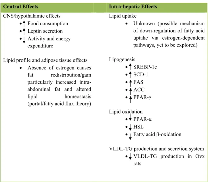
Table 4. Effects of estrogen withdrawal on liver fat accumulation.
Central Effects Intra-hepatic Effects
CNS/hypothalamic effects • Food consumption • Leptin secretion • Activity and energy
expenditure
Lipid profile and adipose tissue effects • Absence of estrogen causes
fat redistribution/gain particularly increased
intra-abdominal fat and altered
lipid homeostasis (portal/fatty acid flux theory)
Lipid uptake
• Unknown (possible mechanism of down-regulation of fatty acid uptake via estrogen-dependent pathways, yet to be explored) Lipogenesis • SREBP-1c • SCD-1 • FAS • ACC • PPAR-γ Lipid oxidation • PPAR-α • HSL
• Fatty acid β-oxidation
VLDL-TG production and secretion system • VLDL-TG production in Ovx
rats
Effects of estrogen withdrawal on liver lipid accumulation (hepatic steatosis) may be direct by affecting lipid uptake, de novo lipogeneis, lipid oxidation, and liver VLDL-TG production and secretion in liver; or secondary to its effects on the central nervous system (CNS) or fat gain, particularly intra-abdominal (visceral) fat accretion.
Prevention/treatment of adipose tissue and liver fat accumulation in
menopausal hormonal state
More than 60% of American postmenopausal women are overweight or obese [Mokdad, Serdula et al. 1999] and as mentioned earlier, it is well established that menopause is associated with weight gain, unfavorable alteration in body composition (elevated visceral fat deposition), and a state of hepatic steatosis [Astrup 1999; Faria, Ribeiro Filho et al. 2002; Volzke, Schwarz et al. 2007]. It seems that hormone replacement therapy (HRT) alleviates these symptoms of menopause [Hassager and Christiansen 1989; Arabi, Garnero et al. 2003; Green, Stanforth et al. 2004]. However, research on the safety of HRT is conflicting. The Women’s Health Initiative in the United States in 2002 and the Million Women Study in the UK in 2003 reported the evidence of increased risk of heart disease, stroke, venous thromboembolism, and breast cancer with HRT in postmenopausal women [Rossouw, Anderson et al. 2002; Beral 2003]. In general, although short-term use of HRT remains beneficial for severe menopausal symptoms, the uncertainty with the risks/benefits of HRT along with the well-publicized results of above two large-scale HRT trials, have led to the conclusion that HRT will not protect future health in postmenopausal women [McPherson 2004; Wegge, Roberts et al. 2004]. Therefore, it seems that the justification for HRT can no longer be applied for disease prevention or treatment, thus women continue to seek alternative options to improve their quality of life and reduce the risk of heart disease, osteoporosis, and breast cancer during postmenopause time [Cassidy 2005].
Interestingly, the most research recommended prevention/treatment for weight gain, elevated visceral fat deposition, and hepatic steatosis is identical which is weight loss through lifestyle interventions including exercise and/or diet. These lifestyle modifications can constitute an important alternative (or complementary) strategy to HRT in postmenopausal women to alleviate concerned disorders. Moreover, very recently Zanesco and Zaros in their review paper reported that in an attempt to reduce the incidence of CVD
in postmenopausal women, a variety of approaches have been used but the results are conflicting and changes in lifestyle have been proposed as a most effective preventive action [Zanesco and Zaros 2009]. It seems there is no substitute for an appropriate lifestyle [Dubnov-Raz, Pines et al. 2007]. Moreover, Hagey and Warren suggested that exercise and nutrition play important roles in the prevention and treatment of obesity, diabetes, and CVD in postmenopausal women [Hagey and Warren 2008]. Data from a 5-year randomized clinical trial known as the Women’s Healthy Lifestyle Project, demonstrated that weight gain and increased waist circumference during the peri- to postmenopause can be prevented by a long-term lifestyle dietary and physical activity intervention [Simkin-Silverman, Wing et al. 2003].
One of the most important components of lifestyle relates to physical activity which for a long time has been known to be a powerful low-risk means for the promotion of all aspects of human health including postmenopausal women [Pines and Berry 2007]. Postmenopausal women might demonstrate a greater response to exercise [Hagey and Warren 2008] since it was shown that even small increases in physical activity and exercise at the time of menopause can help prevent the atherogenic changes in lipid profiles and the weight gain experienced by menopausal women [Rainville and Vaccaro 1984]. Longitudinal and cross-sectional studies have shown that physical activity is associated with lower body fat and less central adiposity in postmenopausal women [Stevenson, Davy et al. 1995; Astrup 1999; Guo, Zeller et al. 1999; Irwin, Yasui et al. 2003; Sternfeld, Wang et al. 2004]. The results of a research by Hagberg et al. even indicated that numerous years of high-intensity endurance training had a greater effect on total and regional body fat values than HRT in postmenopausal women [Hagberg, Zmuda et al. 2000]. Furthermore, it has been shown that moderate-intensity exercise can result in improvements in coronary/metabolic risk factors such as insulin action in postmenopausal women [Ready, Drinkwater et al. 1995; Ready, Naimark et al. 1996; Asikainen, Miilunpalo et al. 2002; Asikainen, Miilunpalo et al. 2003; Frank, Sorensen et al. 2005]. Given that obesity is extremely prevalent and difficult to treat, prevention of weight gain during this time in a
women’s life is an important health goal. A successful model of weight gain prevention has yet to be established [Simkin-Silverman, Wing et al. 2003]. In a cross-sectional study by Hagmar et al., postmenopausal former elite endurance athlete women were investigated in terms of athlete’s heart being compared with age-matched sedentary controls. Authors suggested that intense training enhances cardiovascular performance in the aging female athlete [Hagmar, Hirschberg et al. 2005]. This may imply that previous exercise training (during the reproductive period) may be useful in the prevention of deleterious cardio-metabolic effects of menopause. Taken together, it seems that postmenopausal women with high levels of physical activity have lower body fat and abdominal fat and are less likely to gain fat (total and abdominal) during menopause than those with low levels of physical activity [Astrup 1999].
It has been shown that Ovx animal models can benefit from an exercise training program. In 1987, a reduction in fat gain with training has been reported [Richard, Rochon et al. 1987]. Then in 2002, Shinoda et al. showed that exercise training has a strong action upon reduction in body fat accumulation following a decrease in estrogen levels [Shinoda, Latour et al. 2002]. 8-wk endurance exercise training in the latter did not reduce overall weight gain and increased food intake brought about by an ovariectomy; suggesting a compensatory increase in muscle weight by training. On the other hand, although food restriction seems to prevent the Ovx-induced weight gain, this treatment suppresses muscle growth in Ovx rats [Fisher, Kohrt et al. 2000]. It appears that increase in body weight and organ weights including muscle mass subsequent to ovariectomy in rats [Booth and Tipton 1969; Santidrian and Thompson 1981] may be a compensatory mechanism to protect the bone loss when the estrogen levels are low. For instance, it has been reported that freely eating Ovx rats suffered less bone loss than did food restricted Ovx animals; suggesting that freely eating Ovx animals were partially protected from bone loss by their greater body weight [Wronski, Schenck et al. 1987]. In this regard, it was shown that muscle tissue hypertrophy induced by a progressive loading exercise program has a stimulatory effect on bone mass in Ovx rats as there are many studies showing beneficial effects of exercise on
![Table 1. Taken from [Despres 1993] Features of the metabolic syndrome.](https://thumb-eu.123doks.com/thumbv2/123doknet/2034945.4388/22.918.172.483.241.549/table-taken-despres-features-metabolic-syndrome.webp)
![Table 2. Taken from [Loria, Lonardo et al. 2008] Possible pathophysiological bases for an association between NAFLD and accelerated atherosclerosis](https://thumb-eu.123doks.com/thumbv2/123doknet/2034945.4388/27.918.184.830.223.1011/table-taken-lonardo-possible-pathophysiological-association-accelerated-atherosclerosis.webp)
![Figure 2. Taken from [Lavoie and Gauthier 2006] Overview of the four main pathways involved in the development of nonalcoholic hepatic steatosis, and their regulatory factors](https://thumb-eu.123doks.com/thumbv2/123doknet/2034945.4388/30.918.204.774.176.731/gauthier-overview-pathways-involved-development-nonalcoholic-steatosis-regulatory.webp)
![Table 3. Taken from [Wilfred de Alwis and Day 2007] Genes potentially involved in the pathogenesis of nonalcoholic fatty liver disease (NAFLD)](https://thumb-eu.123doks.com/thumbv2/123doknet/2034945.4388/32.918.177.830.219.1054/table-taken-wilfred-potentially-involved-pathogenesis-nonalcoholic-disease.webp)
![Figure 4. Taken from [Shoelson, Herrero et al. 2007] Potential mechanisms for obesity- obesity-induced adipocyte inflammation](https://thumb-eu.123doks.com/thumbv2/123doknet/2034945.4388/62.918.182.644.170.649/figure-shoelson-herrero-potential-mechanisms-obesity-adipocyte-inflammation.webp)
![Figure 5. Taken from [Chen 2006] Hypothetical model of metabolic stress, cellular inflammatory responses and effects on insulin signaling pathway](https://thumb-eu.123doks.com/thumbv2/123doknet/2034945.4388/63.918.206.807.167.587/figure-hypothetical-metabolic-cellular-inflammatory-responses-effects-signaling.webp)
![Figure 6. Taken from [Shoelson, Lee et al. 2006] Local, portal, and systemic effects of inflammation in insulin resistance and atherogenesis](https://thumb-eu.123doks.com/thumbv2/123doknet/2034945.4388/66.918.198.836.167.603/figure-shoelson-systemic-effects-inflammation-insulin-resistance-atherogenesis.webp)
![Table 5. Taken from [Bruunsgaard 2005] Self-reported physical activity and physical performance in relation to low-level inflammation in epidemiological studies](https://thumb-eu.123doks.com/thumbv2/123doknet/2034945.4388/67.918.180.848.242.917/bruunsgaard-reported-physical-activity-physical-performance-inflammation-epidemiological.webp)