VASCULAR CALCIFICATIONS IN
DIALYSIS PATIENTS
Pierre Delanaye, Service de Dialyse, CHU Sart Tilman, 4000 Liège Belgique Etienne Cavalier, Service de Chimie Médicale, CHU Sart Tilman, 4000 Liège, Belgique
A SERVICE FROM
In the two last decades, vascular calcifications have been identified as a major cardiovascular (CV) risk factor in chronic kidney disease (CKD) patients, especially in patients at the end-stage renal disease (ESRD) stage. In
the last century(1), calcifications were already known (and
feared) by nephrologists but they stressed on
“soft-tis-sue” calcifications(1 ;1-5)or on calciphylaxis(6)which can be
considered as an epiphenomenon of vascular calcifica-tions (very serious but relatively rare). Actually, there were relatively few articles on vascular or valvular calci-fications(7).
The interest on vascular calcifications has been highlighted since the following observations : firstly, dialysis patients have an exceptionally high CV mortality compared to the general population and this risk is not simply linked to
traditional CV risk factors(8). Secondly, epidemiological
studies have underlined the relationship observed between CV mortality and mineral metabolism markers (especially, phosphorus, calcium, calcium-phosphorus product and,
for some authors, PTH)(9-14). Lastly, it has been
demon-strated that a strong relationship does exist between sev-eral of these minsev-eral metabolism markers (the term “renal osteodystrophy” is replaced by “CKD-MDB” for Chronic
Kidney Disease-Mineral Bone Disorder(15)) and the
vas-cular calcifications(7 ;16-27). Studies establishing the link
between mortality and vascular calcifications are more difficult to do. However, even though data are rela-tively limited and methodologies sometimes question-able, several studies have suggested such a relationship between mortality (notably CV mortality) and
vascu-lar calcification(26 ;28-35). Nevertheless, we have to insist on
the fact that no direct proof of the “causal” link between vascular calcifications and CV mortality exists. More
pre-1. Introduction
2. Types and mechanisms
of vascular calcifications
3. Detection and
quantification of
calcifications
4. Impact of CKD-MBD
treatment options
5. Conclusions
AbbreviationsALP: alkaline phosphatase
BMP-2: bone morphogenic protein-2
CAC: coronary artery calcification
CaR: calcium sensing receptor
CaxP: calcium phosphate product
CKD: chronic kidney disease
CKD-MDB: chronic kidney disease – mineral bone disorder
CRP: C-reactive protein
CV: cardiovascular
Dp-uc-MGP: dephosphorylated uncarboxylated matrix Gla protein
EBCT: electron-beam-computed tomography
ESRD: end-stage renal disease Fet-A: fetuin-A
FGF-23: fibroblast growth factor 23 HD: hemodialysis
HDL: high-density lipoprotein
LDL: low-density lipoprotein
MGP: matrix Gla protein
MSCT: multi-slice computed tomography
OC: osteocalcin ON: osteonectin OPG: osteoprotegerin OPN: osteopontin PPi: pyrophosphate PTH: parathyroid hormone sHPT: secondary hyperparathyroidism
uc-MGP: uncarboxylated matrix Gla protein
VSMC: vascular smooth muscle cells
Introduction
in nephrology
cisely, we have no proof that decreasing vascular calcifications
is either possible(36)or even beneficial from a CV point of
view. Nevertheless, indirect proofs are advanced. Vascular calcifications even became important “endpoints” in clinical trials including patients receiving dialysis and are considered as a useful “surrogate marker” instead of CV mortality. Compared to the general population (where vascular calci-fications are, for example, present in older subjects), coro-nary calcifications in dialysis patients are more prevalent (up
to 90 % of dialysis patients(16)) and more severe (calcium
score 2.5 to 5-fold higher(20)) (16;20;23;25;37-40). They are also early
(often present in very young patients)(16;18-20;25;26;41;42)and more
rapidly progressive (16;20;41;43;44). The same conclusions could
be made for cardiac valve calcifications(20;23;24;45-47).
We still have to find out with more precision in what way vascular calcifications are harmful. Currently, there are some arguments to say that vascular calcifications in the intima, which are strongly linked to atherosclerosis (see below),
could actually stabilize this atherosclerosis plaque(5 ;48-51).
It seems that aorta calcifications (calcifications of the media, see below) could be more important, from a pathophysio-logical point of view, to explain the rather high CV mortal-ity in dialysis patients. Indeed, aorta calcifications induce vas-cular stiffness, increasing aortic pulse wave velocity (and
pulse pressure)(22;28;35;52;53)and inducing left ventricular
hyper-trophy and diastolic dysfunction that both could explain the
high prevalence of cardiac-related death(28;53-57). Of course,
cardiac valve calcification can induce per se functional
abnor-malities like rapid development of aortic stenosis(24;46). There
are also some data suggesting that the intensity of valve
cal-cification could predict CV mortality(58) although others do
not find such an association after adjusting for other risk fac-tors(31;59).
This “Up to Date in Nephrology” brochure reviews the recent literature on the types of vascular calcifications, the pathophysiological mechanisms responsible for vascular calcifications, the detection methods and the impact of CKD-MBD treatment options.
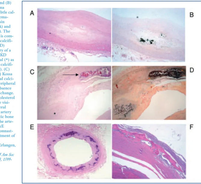
Figure 1. (A) and (B) Coronary intima plaque with subtle cal-cification in hema-toxylin and eosin (H&E) stain (A) and Kossa stain (B). The arterial media is com-pletely free of calcifi-cation.(C and D) Coronary artery of a patient with CKD showing intimal (*) as well as medial calcifi-cations (arrow). (C) H&E stain. (D) Kossa stain.(E) Medial calci-fication of a peripheral artery in the absence of any intimal change. No lipid or cholesterol depositions are visi-ble.(F) Peripheral muscular type artery with metaplastic bone formation in the arte-rial media (H&E stains). (Dr. Nonnast-Daniel, Department of Nephrology,
University of Erlangen, Germany)
(Amann. Clin J Am Soc Nephrol 2008, 3, 1599-1605).
2.1 Medial versus intimal calcifications
(Figure 1)(60)
Two types of vascular calcifications are classically
described(60). One type is linked to atherosclerosis lesions
(in the vicinity of lipid depositions) and is located in the intima of the vascular wall. The other type is located in
the media and is also known as Monckerberg sclerosis(60).
This type of calcification is highly prevalent in ageing, in
CKD(61)and in diabetic patients. The medial
calcifica-tions are thought to be important to explain that calcifications are early and severe in dialysis patients compared to general population. It is probable that molecular mechanisms involved in these two types of calcifications are different (notably the role of lipids), but some mechanisms could be com-mon (local inflammation, calcium and phosphate balance disturbances, cal-cification inhibitors and activators
bal-ance disturbbal-ances, see below)(60). In
the same view, pathological mecha-nisms explaining soft-tissue and car-diac valve calcifications are little
known(60 ;62). In the CKD context,
most of animal or in vitro studies have
been realized with media (or both media and intima) calcifications mod-els (60 ;63). The impact on CV mortality is also difficult to apprehend but one study suggests that the risk of CV mortality is higher with intima calci-fications. It should be noted that in this report, calcifications were only assessed by standard radiography, (see
below)(26) (Figure 2). However, as
intima calcification is strongly linked to atherosclerosis, this statistical asso-ciation with mortality does not imply
that calcification per se is the most
harmful in this context(48 ;60). This
topic is difficult and subject of debate. This is, at least in part, due to the lack of an easy method to make differen-tial diagnosis between intimal and
medial calcification (see below) (60).
This debate is also illustrated by the
poor correlation observed between coronary vascular cal-cifications by electron-beam computed tomography (EBCT), which cannot distinguish intimal and medial calcifications and lesions observed by coronary
angiog-raphy in one study(64). However, medial calcifications are
very frequent in dialysis patients and their pathogenic power is not linked to vascular obstruction. So, the results of this last study do not imply that calcium score
meas-urement is without interest from a CV point of view (65).
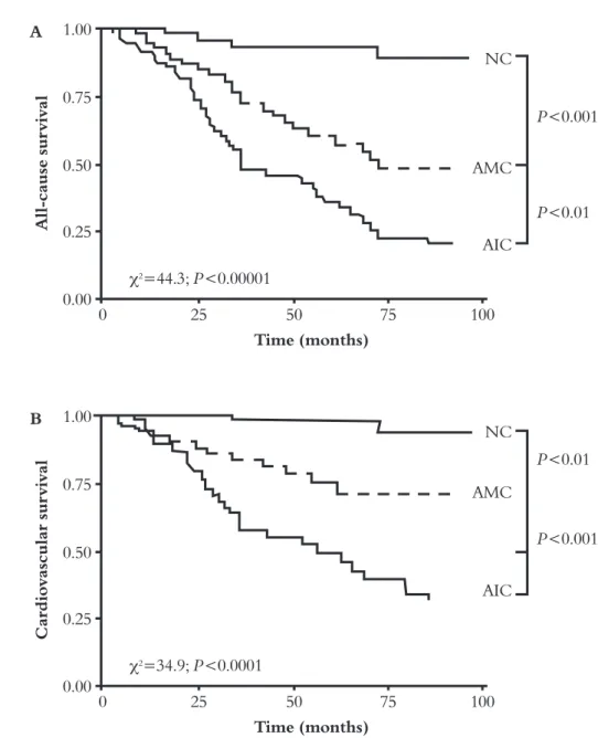
A All-cause su rvival 1.00 0.75 P<0.001 P<0.01 0.50 0.25 0.00 NC AMC AIC 0 25 50 75 100 Time (months) χ2=44.3; P<0.00001 B Ca rdiovascula r su rvival 1.00 0.75 P<0.01 P<0.001 0.50 0.25 0.00 NC AMC AIC 0 25 50 75 100 Time (months) χ2=34.9; P<0.0001
Figure 2. All-cause (A) and CV mortality (B) of ESRD patients as a function of their calcification status. NC: non-calcified, AMC: arterial medial calcification, AIC: arterial intimal calci-fication (London et al. Nephrol Dial Transplantation, 2003, 18,1731-1740).
TYPES AND MECHANISMS OF
in nephrology
2.2 Calciphylaxis
Calciphylaxis is a calcification syndrome associated with
ischemic cutaneous necrosis(66) (Figure 3)(67). Association
between vascular calcification and cutaneous gangrene has
already been described in 1899(68)and the term “calciphylaxis”
has been proposed by Hans Selye in 1962(69). Calciphylaxis or
“calcific uremic arteriolopathy” is histologically characterized by vascular calcification (especially calcification in the media), intimal proliferation, endovascular fibrosis and intravascular
thrombosis in small or medium arteries(66;70). The lesions are
usually located in two distinct patterns: distal with lesions of the lower extremities or proximal with the lesions on the
abdomen, inner thighs and buttocks(66;70;71). These calcifications
induce painful skin lesions progressing to ischemic necrosis. Calciphylaxis is a severe syndrome with a high mortality rate
(45 to 80%)(66;70-72;72). The details of the pathogenesis are still
unknown: is it simply a “sur-acute” form of vascular
calcifi-cation?(70;73). It seems that hypercalcemia/hyperphosphatemia,
low- or high bone turnover, steroids, warfarin and vitamin D
therapies could be implicated(17;66;70-73). Female gender, Caucasian
race, diabetes, and obesity are often proposed as risk fac-tors(70;71;73). Additional to wound care (notably with hyperbaric
Figure 3. Extensive cutaneous necrosis of the thighs with liveloid contour (Prey et al. Rev Med Interne, 2009, 30, 186-189).
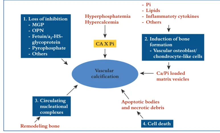
1. Loss of inhibition - MGP - OPN - Fetuin/a2 glycoprotein - Pyrophosphate - Others - Pi - Lipids - Inflammatoty cytokines - Others 3. Circulating nucleational complexes 4. Cell death 2. Induction of bone formation - Vascular osteoblast/ chondrocyte-like cells Ca/Pi loaded matrix vesicles Apoptotic bodies and necrotic debris
Remodeling bone Hyperphosphatemia Hypercalcemia CA X Pi Vascular calcification
Figure 4. Four non-mutually exclusive theories for vascular calcification. (1) Loss of inhibition as a result of deficiency of consti-tutively expressed tissue-derived and circulating mineralization inhibitors leads to default apatite deposition. (2) Induction of bone formation resulting from altered differentiation of vascular smooth muscle or stem cells. (3) Circulating nucleational com-plexes released from actively remodeling bone. (4) Cell death leading to release of apoptotic bodies and/or necrotic debris that may serve to nucleate apatite at sites of injury (Giachelli. J Am Soc Nephrol, 2004, 15, 259-2964).
oxygen(70)), the optimal therapy for calciphylaxis is not
stan-dardized. Calcium and phosphate control by non-calcium
phosphate binders, parathyroidectomy(66;74), sodium
thiosul-phate(75), bisphosphonates(76;77)and cinacalcet (see below) have
been proposed(70). Sodium thiosulphate (potent antioxidant
as well as chelator of calcium) and cinacalcet could be the most interesting therapies but we need more trials to proof this.
2.3 Vascular calcifications in CKD: an active
and complex process
Occurrence of vascular calcification is not new. Arterial cal-cification has been discovered in the “Iceman” who lived
5000 years ago(78)and scientists had already paid attention
to this phenomenon – and to its relation with renal disease
- in the 19thCentury(79). However, this pathology has only
been studied since the last two decades. Today, vascular cal-cification is considered as an actively regulated and complex process that remains not completely understood. We will describe hereafter some of the pathophysiological mecha-nisms. It is important to underline that these different
mech-anisms are not mutually exclusive (Figure 4)(80).
2.3.1 The role of calcium and phosphate and induction
of bone formation
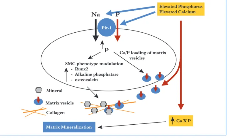
One major mechanism in the development of vascular cal-cifications is similar to that of bone formation. Indeed, vas-cular smooth muscle cells (VSMC) undergo osteogenic dif-ferentiation into phenotypically distinct osteoblast-like
cells(80-82). In this mechanism, phosphate has the most
important role(82 ;83). Actually, in vitro, high extracellular
phosphate concentrations induce a rise in intracellu-lar phosphate concentration which is actively medi-ated by Pit-1, a sodium dependent phosphate
co-trans-porter(83 ;84). This increasing phosphate concentration into
the VSMC will induce a phenotypic switch of VSMC
into osteoblast-like cells(80;83;85). The protein Cfba1/Runx2
is a specific and indispensable transcriptional regulator for this osteoblastic differentiation. Its expression is also enhanced in the presence of high extracellular phosphate (83;85;86). These “new” cells will express alkaline phosphatase (ALP), secrete bone-associated proteins (such as
osteo-pontin(87), collagen type 1, and bone morphogenic
protein-2 and osteocalcin(83 ;88)) under the control of Cfba-1, and
release mineralization-competent matrix vesicles in the
extracellular matrix(82 ;83 ;89). All these modifications will
favour for an optimal microenvironment for hydroxya-patite formation and calcification. Similar osteogenic
dif-ferentiation is also observed, in vivo, in animal and human
uremic models(81 ;85 ;90)(Figure 5)(80).
Na
Mineral Matrix Mineralization Elevated Phosphorus Elevated Calcium Matrix vesicle CollagenP
P
SMC phenotype modulation - Runx2 - Alkaline phosphatase - osteocalcinCa/P loading of matrix vesicles
Pit-1
Ca X P
Figure 5. Proposed model for the effects of elevated Ca and P on vascular smooth muscle cell (VSMC) matrix mineralization. Elevated Ca and P are proposed to stimulate vascular matrix mineralization in two ways. First, both Ca and P increase the activity of Pit-1: ele-vated P stimulates P uptake via Pit-1, and eleele-vated Ca induces expression of Pit-1 mRNA; Both mechanisms are proposed to enhance P uptake into VSMC as well as matrix vesicles. Elevated intracellular P then leads to VSMC phenotypic modulation, which includes upregulation of osteogenic genes (Runx2, osteocalcin, and alkaline phosphatase), and generation of a mineralization-competent extra-cellular matrix. In addition, increased Pit-1 in matrix vesicles promotes P loading of matrix vesicles, promoting nucleation of mineral within the extracellular matrix. Second, elevated Ca and/or P lead to increased Ca X P ion product, thereby promoting growth of apatite crystals in the matrix via thermodynamic mechanisms (Giachelli. J Am Soc Nephrol, 2004, 15, 259-2964).
in nephrology
In vitro exposure of VSMC to hypercalcemia also induced
overexpression of Pit-1 receptor and thus enhanced
intra-cellular phosphate concentration(89;91). Elevated
intracellu-lar calcium is also associated with an alteration of calcifi-cation inhibitors such as matrix Gla protein (MGP) and
fetuin A (see below)(89). If calcium may participate to the
pathogenesis of vascular calcification, phosphate has, once again, the pivotal role(80;82;83;89;92).
2.3.2 The role of bone turnover
As calcium and phosphorus have a pivotal role in the patho-genesis of vascular calcification, it is not surprising that bone-turnover disturbances can enhance vascular calcifi-cation because bone is the most important reserve of
cal-cium and phosphorus and could act as a buffer(93). Some
authors have written about the “bone-vascular” axis. From our point of view, the link between “low bone turnover” and risk of vascular calcifications is better illustrated by the current literature. However, we also think that the two “the-ories” of low and high turnover are not excluding each other
in a context of “bone as a calcium-phosphorus buffer”(5).
Low turnover : In the first article measuring vascular
cal-cifications, Braun et al. have already shown that coronary
calcifications are inversely correlated with bone mass(20).
Probably the most interesting study on this topic has been
published in 2004 by London et al. These authors have
actually compared vascular calcifications with bone his-tomorphometry by plain radiography, in 58 dialysis patients. They found that a high calcification score is inde-pendently associated with histomorphometry suggestive
of low bone turnover (or adynamic bone)(94). One
com-plementary study from the same group underlined that calcium load is particularly deleterious, in terms of vas-cular calcifications, for these patients with low bone
turnover(95). Studying both coronary calcifications and
bone histomorphometry in 101 dialysis patients, Barreto
et al. also found an inverse correlation between calcium
score and bone trabecular volume and trabecular
thick-ness in an univariate analysis(38). In 2009, Adragao et al.
studied the relationship between bone histomorphome-try and coronary calcifications in 38 dialysis patients. Contrary to precedent trials, low bone turnover was not associated with calcifications. However, lower bone
vol-ume was a risk factor for coronary calcification(96).
High turnover : We have already mentioned the
epi-demiological link between PTH and CV mortality in
dial-ysis patients, observed by some authors (10;11;13;14). Coen et al.
have demonstrated a relationship between high PTH levels (especially very high levels) and coronary
cal-cifications(12 ;97). Even though specific PTH fragments
(1-34 PTH) have been shown to inhibit vascular
cal-cifications in an animal model(98),Neves et al. have shown,
in a model of parathyroidectomized CKD rats, that
perfu-sion of PTH, per se, could induce vascular calcifications(99).
2.3.3 Balance between calcification inhibitors and
pro-moters
(Figure 6)In water, calcium and phosphate directly form insoluble pre-cipitates. This is not the case in serum, suggesting the
exis-tence of calcification inhibitors(100). In the last years, several
inhibitors have been actually described with a potential role in vascular calcifications. Chondrocytes, osteoblasts and osteoclasts have been identified in calcified atherosclerotic
plaques(101;102). These cell types can locally express
calcifica-tion activating proteins (osteonectin, osteo-calcin and bone morphogenic protein-2), inhibiting proteins (osteopontin, Matrix-Gla protein, pyrophosphate) and
regula-tory factors (osteoprotegerin system)(103).
Circulating proteins (Fetuin-A) could also participate in the vascular calcification process. We will summarize the most recent findings for some of the most important proteins.
Matrix Gla protein (MGP)
MGP is a 10-kDa protein expressed by chondrocytes and VSMC. Its role as cal-cification inhibitor has been illustrated by MGP knock-out mice who develop
extensive aortic calcifications(104). In 2002,
Moe et al. demonstrated a correlation
between vascular MGP expression and the calcifications of epigastric arteries in
dial-ysis patients(90;105). Of importance is the fact
Activators
Bone and mineral disorders FGF-23 Inhibitors Fetuin A Matrix-Gla protein Osteopontin Regulators OPG/RANKL Osteoblastic differentiation Phosphocalcic layer
Smooth vascular cells bALP
Osteoblasts
MGP OPN
BMP RANKL
Osteoid
Figure 6. Balance between calcification inhibitors and promotors (with the permission of Prof. Cristol, Department of Biochemistry, Montpellier).
that MGP requires vitamin K for its activation by
γ-car-boxylation(80 ;89 ;104 ;106). It has been shown that non fully
γ-carboxylated (but not γ-carboxylated MGP) is
associ-ated with vascular calcification(80 ;106 ;107). MGP would bind
and inactivate a pro-mineralization factor, BMP-2(108).
MGP also binds calcium crystals, inhibits crystal growth and plays a role in the normal phenotype of VSMC in
preventing the osteoblastic differentiation(86 ;109). MGP is
vitamin K dependent for its carboxylation and its activa-tion. This is the actual explanation for the observations that warfarin leads to extensive vascular calcifications in
animal and human studies(110-112). It is interesting to note
that vitamin K therapy partially reverses warfarin-induced
vascular calcifications in rats(112).
Osteoprotegerin (OPG)
OPG is a regulatory factor produced by bone marrow-derived stromal cells. OPG has a pivotal role in the regu-lation of the bone turnover inhibiting osteoclast differ-entiation and acting like a decoy receptor for the receptor
activator of NF-χB ligand (RANKL system)(113).
OPG-deficient mice will also develop both severe aortic
calcifi-cations and osteoporosis(114). However, the role of OPG in
vascular calcification remains unclear(115). On one hand, OPG
is considered to prevent vascular calcification as it blocks the bone remodeling process in the vascular tissue following the interaction between RANK (expressed by osteoclast-like cells) and RANKL (expressed by osteoblast-osteoclast-like cells). It is also a neutralizer of the pro-apoptotic actions of TRAIL (TNF-related apoptosis-inducing ligand), which
strongly activates vascular cell apoptosis(116). OPG is also
thought to inhibit ALP activity and to prevent vascular
calcifications(117). On the other hand, the inhibition of the
bone remodeling process by OPG could induce a calcium shift in vascular cells.
Osteopontin (OPN)
OPN is a phosphoprotein expressed in the mineral tissues which inhibits mineralization by blocking hydroxyapatite
formation and activating osteoclast function(118). OPN is
present in calcified vessels. OPN knock-out mice do not develop vascular calcification but, when these mice are bred with MGP knock-out mice, the vascular calcifications are
more important than in simple MGP knock-out mice(119).
OPN must be phosphorylated to act as a calcification
inhibitor(120). OPN inhibits mineralization of VSMC by
binding to the mineralized crystal surface(121). Contrary to
the fully phosphorylated OPN, cleaved OPN could act as a proinflammatory cytokine and a proangiogenic factor
facilitating vascular mineralization(118;122).
Pyrophosphate (PPi)
PPi is a small molecule made of two phosphate ions. It acts as a calcification inhibitor by inhibiting hydroxyapatite
crys-tal formation(123). Once again, knock-out mice (in fact,
knock-out mice for a precursor) develop vascular
calcifi-cations(124). Absence of PPi will promote VSMC
differen-tiation but the mechanism is not fully understood(125 ;126).
Absence of PPi combined with high phosphate levels and presence of type 1 collagen could facilitate the
develop-ment of calcification(92 ;127). It has been shown that dialysis
patients exhibit low serum PPi and that these are lowered
further during a hemodialysis session(128).
Fetuin-A (Fet-A)
Fet-A (60 kDa) is a potent calcification inhibitor produced by the liver. Contrary to other factors acting locally, Fet-A action is systemic. Its calcification inhibitory action is powerful and illustrated by knock-out mice developing
severe extraosseous calcifications(129). Even though the aorta
remains free from calcifications in this model, CKD and high phosphate diet will induce severe aorta calcifications in
these knock-out mice(130). Increasing Fet-A expression is
found in calcified arteries from dialysis patients(105). Fet-A is
thought to inhibit calcification by binding early calcium phos-phate crystals and by inhibiting crystal growth and mineral deposition. This could be facilitated by the formation of
large calciprotein particles(131 ;132). Fet-A could also
pro-hibit matrix vesicle calcification of the VSMC which take up circulating Fet-A in an extracellular
calcium-depend-ent way (133 ;134). Serum Fet-A concentration is decreased in
dialysis patients(135). The exact mechanism is still
hypo-thetical. It is probably related to chronic inflammation, as
Fet-A is a negative acute phase reactant(136).
Fibroblast Growth Factor 23 (FGF-23)
FGF-23 is a 30 kDa bone-derived protein that promotes renal phosphorus wasting and inhibits the conversion of 25-hydroxy-vitamin D to the active 1,25-di25-hydroxy-vitamin D form(137). In CKD patients, when the glomerular filtration rate decreases below 25-30 ml/min, the maintenance of normal phosphate levels is presumably accomplished by a
compen-satory rise in FGF-23(137;138). The action of FGF-23 on its
spe-cific receptor is mediated through the type-1 membrane-bound alpha-Klotho (Klotho). Indeed, Klotho is a FGF-23 receptor cofactor that directly interacts with the FGF-23 recep-tor. The importance of Klotho for FGF-23 activity is such that supraphysiological concentrations of FGF-23 have no impact
on mineral metabolism without the presence of Klotho(139).
In mice, high levels of FGF-23 have been shown to reflect a response to dietary phosphorus burden and thus can serve as a marker of arterial calcification(140). It is also evident that Klotho knock-out mice will develop vascular calcifications (this model
is considered as a model of ageing)(141). Interestingly, Klotho
has recently been described as a regulator of calcium home-ostasis, notably influencing the calcium transport across the
cell membrane(142;143). However, as Klotho is neither expressed
in the myocardium, nor in the blood vessels, its role as a pro-moter of vascular calcification has to be clarified in future studies. In the same way, knock-out mice for FGF-23 also
develop vascular and visceral calcifications(137;144;145). However,
this deletion is also associated with an increased serum phos-phate concentration, an increased expression of renal
1α-in nephrology
hydroxylase and an increased serum 1,25 vitamin D
concen-tration(137;144;145). Double knock-out mice (for FGF-23 and
1α-hydroxylase) develop neither hyperphosphatemia nor calci-fications, underlining the potential role of vitamin D in
vascular calcifications (see below)(146). So, a “direct”
(phos-phate and vitamin D independent) role of FGF-23 in the pathogenesis of vascular calcifications remains to be proven and a question still to be answered is: Is FGF-23 an inducer or a marker of vascular calcifications?(34;137;140;147).
Alkaline phosphatase (ALP)
ALP is a phenotypic marker of osteoblasts and is thought
to be essential for vascular calcifications(148). Its expression
in calcification seems to be under the control of Cfba-1(80).
ALP is expressed on the surface of differentiated cells and
could hydrolyze PPi, a calcification inhibitor(149).
Bone morphogenic protein-2 (BMP-2)
BMP-2 is an important molecule in the regulation of bone formation as well as vascular calcification. In bones, it
pro-motes osteoblast differentiation and mineralization(150).
Inhibition of BMP-2 inhibits osteoblast differentiation and
bone formation in vivo and in vitro(151)and protects against
atherosclerosis and vascular calcification(152).
Osteocalcin (OC)
OC, a vitamin-K dependent matrix protein that inhibits
cal-cium salt precipitation in vitro(153), shows a strong affinity for
hydroxyapatite and inhibits crystal growth(154). Even though
its role remains unclear, OC limits bone formation(155)and
it has been found in calcified atherosclerotic plaques and
cal-cified aortic valves(156). The role of OC in the pathogenesis
of vascular calcification clearly remains to be determined.
Osteonectin (ON)
ON, also called SPARC or BM40, is a calcium binding pro-tein involved in bone development that demonstrates
affin-ity for hydroxyapatite and collagen(157). ON has been found
in association with large calcifications in atherosclerotic plaques(158).
2.3.4 The role of inflammation, lipids and oxidative
stress
The uremic state is also characterized by increased oxida-tive stress. Oxidaoxida-tive stress has been shown to enhance ALP
in VSMC(159)and to promote differentiation of VSMC via
an activation of the Cfba-1/Runx2 protein(160;161). As
oxida-tive stress is also the result of inflammation, inflammatory cytokines have also been implicated in the pathogenesis of vascular calcification. Actually, tumour-necrosis factor-α
(TNFα) has been shown to induce differentiation of
VSMC and expression of ALP(162-165). Notably, oxidized LDL
and other lipid peroxidation products induce osteoblastic
differentiation in a dose-dependent manner(166)although
HDL inhibits it (but oxidized HDL promotes
differentia-tion, too)(167). Fet-A is both a negative acute-phase protein
and a calcification inhibitor and could thus be one link
between inflammation and calcification(163).
DETECTION AND
QUANTIFI-CATION OF CALCIFIQUANTIFI-CATIONS
3
3.1 Clinical Chemistry
According to their role in the pathogenesis of vascular cal-cifications, several biomarkers have been proposed to
pre-dict either vascular calcifications or CV mortality. In Table 1
we have compiled some of the most important trials on this topic in dialysis patients. However, interesting results have been recently published for CKD (non dialysis) patients(170;171).
2.3.5 Other factors
The concentration of leptin is increased in CKD patients(168).
Leptin has been shown to regulate osteoblastic differenti-ation and calcificdifferenti-ation of VSMC, which are known to
express leptin receptors(169).
In summary, there are two types of vascular calcifications; one type is located in the intima of the vascular wall, the other type is located in the media. Medial vascular calcifications are highly prevalent in patients with CKD. Calciphylaxis is a calcifica-tion syndrome associated with ischemic cutaneous necrosis and is histologically characterized by vascular calcification, intimal proliferation, endovascular fibrosis and intravascular thrombo-sis in small and medium arteries. Calcium and phosphate con-trol by non-calcium phosphate binders, parathyroidectomy, sodium thiosulphate, bisphosphonates and cinacalcet have been proposed as therapy for calciphylaxis but more trials are needed. Vascular calcification is associated with CV mortality in CKD patients. However, the pathogenesis of vascular calcifications is not fully understood. It is a rather complex process influenced by derangements of calcium and phosphate homeostasis, by a dysregulated balance between calcification promoters (ON, OC, and BMP-2) and calcification inhibitors (OPN, MGP, and PPi), by regulatory factors (OPG system) and by circulating proteins such as Fet-A, a calcification inhibitor with systemic action. A direct role of FGF-23 (phosphate and vitamin D independent) remains to be proven. ALP, a phenotypic marker of osteoblasts is thought to be essential for vascular calcification. Inflammatory cytokines have been involved in the pathogenesis of vascular calcification. Fetuin-A (Fet-A) is also a negative acute-phase protein and could be a link between inflammation and calcification. Oxidized LDL and other lipid peroxidation products induce osteoblastic differentiation and vascular calcifi-cation while HDL regulates it. Bone turnover disturbances can enhance vascular calcification. Leptin is increased in CKD patients and enhances calcification of VSMC.
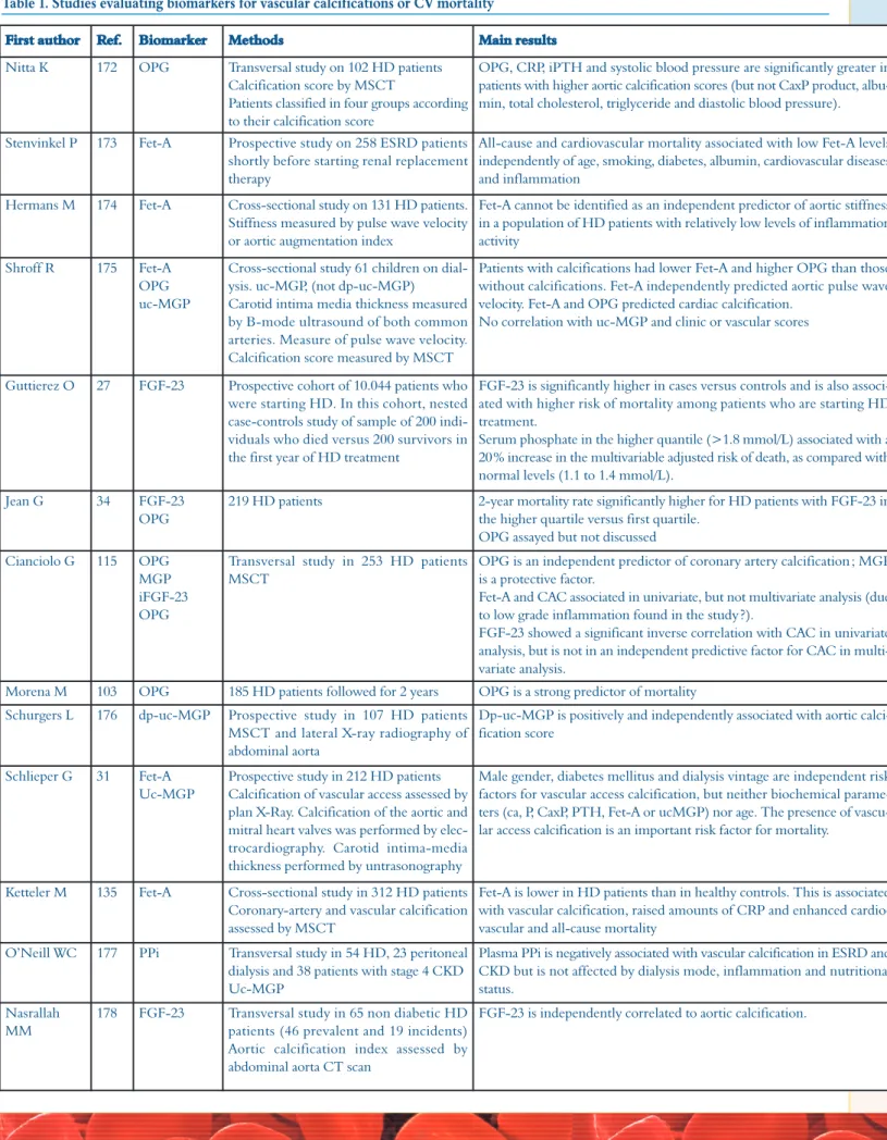
Table 1. Studies evaluating biomarkers for vascular calcifications or CV mortality
First author Ref. Biomarker Methods Main results
Nitta K 172 OPG Transversal study on 102 HD patients Calcification score by MSCT
Patients classified in four groups according to their calcification score
OPG, CRP, iPTH and systolic blood pressure are significantly greater in patients with higher aortic calcification scores (but not CaxP product, albu-min, total cholesterol, triglyceride and diastolic blood pressure).
Stenvinkel P 173 Fet-A Prospective study on 258 ESRD patients shortly before starting renal replacement therapy
All-cause and cardiovascular mortality associated with low Fet-A levels independently of age, smoking, diabetes, albumin, cardiovascular diseases and inflammation
Hermans M 174 Fet-A Cross-sectional study on 131 HD patients. Stiffness measured by pulse wave velocity or aortic augmentation index
Fet-A cannot be identified as an independent predictor of aortic stiffness in a population of HD patients with relatively low levels of inflammation activity
Shroff R 175 Fet-A OPG uc-MGP
Cross-sectional study 61 children on dial-ysis. uc-MGP, (not dp-uc-MGP) Carotid intima media thickness measured by B-mode ultrasound of both common arteries. Measure of pulse wave velocity. Calcification score measured by MSCT
Patients with calcifications had lower Fet-A and higher OPG than those without calcifications. Fet-A independently predicted aortic pulse wave velocity. Fet-A and OPG predicted cardiac calcification.
No correlation with uc-MGP and clinic or vascular scores
Guttierez O 27 FGF-23 Prospective cohort of 10.044 patients who were starting HD. In this cohort, nested case-controls study of sample of 200 indi-viduals who died versus 200 survivors in the first year of HD treatment
FGF-23 is significantly higher in cases versus controls and is also associ-ated with higher risk of mortality among patients who are starting HD treatment.
Serum phosphate in the higher quantile (>1.8 mmol/L) associated with a 20% increase in the multivariable adjusted risk of death, as compared with normal levels (1.1 to 1.4 mmol/L).
Jean G 34 FGF-23 OPG
219 HD patients 2-year mortality rate significantly higher for HD patients with FGF-23 in the higher quartile versus first quartile.
OPG assayed but not discussed Cianciolo G 115 OPG
MGP iFGF-23 OPG
Transversal study in 253 HD patients MSCT
OPG is an independent predictor of coronary artery calcification ; MGP is a protective factor.
Fet-A and CAC associated in univariate, but not multivariate analysis (due to low grade inflammation found in the study ?).
FGF-23 showed a significant inverse correlation with CAC in univariate analysis, but is not in an independent predictive factor for CAC in multi-variate analysis.
Morena M 103 OPG 185 HD patients followed for 2 years OPG is a strong predictor of mortality Schurgers L 176 dp-uc-MGP Prospective study in 107 HD patients
MSCT and lateral X-ray radiography of abdominal aorta
Dp-uc-MGP is positively and independently associated with aortic calci-fication score
Schlieper G 31 Fet-A Uc-MGP
Prospective study in 212 HD patients Calcification of vascular access assessed by plan X-Ray. Calcification of the aortic and mitral heart valves was performed by elec-trocardiography. Carotid intima-media thickness performed by untrasonography
Male gender, diabetes mellitus and dialysis vintage are independent risk factors for vascular access calcification, but neither biochemical parame-ters (ca, P, CaxP, PTH, Fet-A or ucMGP) nor age. The presence of vascu-lar access calcification is an important risk factor for mortality.
Ketteler M 135 Fet-A Cross-sectional study in 312 HD patients Coronary-artery and vascular calcification assessed by MSCT
Fet-A is lower in HD patients than in healthy controls. This is associated with vascular calcification, raised amounts of CRP and enhanced cardio-vascular and all-cause mortality
O’Neill WC 177 PPi Transversal study in 54 HD, 23 peritoneal dialysis and 38 patients with stage 4 CKD Uc-MGP
Plasma PPi is negatively associated with vascular calcification in ESRD and CKD but is not affected by dialysis mode, inflammation and nutritional status.
Nasrallah MM
178 FGF-23 Transversal study in 65 non diabetic HD patients (46 prevalent and 19 incidents) Aortic calcification index assessed by abdominal aorta CT scan
in nephrology
Even though the results observed in these studies are important, one should admit that they are sometimes con-tradictory. The discrepancies could be linked to several factors: the use of different assays and different techniques to evaluate the calcification scores and the presence of dif-ferent study designs
3.2 Imaging
3.2.1 Electron-beam computed tomography (EBCT)
and multislice CT-Scan (MSCT)
EBCT and MSCT are often considered as the reference methods for detecting and quantifying vascular (and
espe-cially coronary) calcifications(23;179-181)(Figure 7). EBCT uses
a gun of electrons to generate a beam focused on a tungsten ring target. The beam then sweeps from side to side along the tungsten ring generating a fan of X-rays. This allows for
excellent temporal resolution(182). EBCT was the first CT
scanner with sufficient time resolution to image the
mov-ing heart(181). Braun et al. were the first authors to study
coro-nary calcifications with EBCT in dialysis patients(20).
Quantification of calcifications is based on a score
pub-lished by Agatston et al.(183).
Electro-cardiographically triggered slices (ECG gating) with a distance of 3 mm are made usually starting approximately 2 cm below the carina and extending to the inferior
mar-gin of the heart(16 ;182). The “calcium
score” was determined by multiply-ing the area of calcification by a weighted density score : 1=130 to 199 Hounsfield units (HU), 2=200 to 299 HU, 3=300 to 399 HU and 4>400 HU. Individual scores are calculated for the left main coronary artery, the descending branch of the left coronary artery, the cir-cumflex branch of the left nary artery and the right coro-nary artery. The total corocoro-nary score is the sum of these indi-vidual scores(20 ;183). Sensu stricto, this score must be considered as semi-quantitative because the calcification areas are to be obtained, “by hand”, by radiologists. Nevertheless, com-pared to the other semi-quantitative scores with simple X-ray radiogra-phy (see below), the scores obtained with CT scans are obviously the most quantitative.
In the Agatston study, the inter-observers difference between scores
was 2.5±5.5 %(183). Still in the
nor-mal population, the good intra- and interobserver variability was
con-firmed thereafter (5-8 %)(184 ;185).
Maybe more intriguing is the repro-ducibility of coronary calcifications observed in some studies (two
dif-Figure 7. Electron-beam computed tomography (EBCT) of the coronary artery. A. Calcification in middle left coronary artery (68-year-old female volunteer). B. Extensive calcifica-tion in middle and distal left anterior coronary artery
(70-year-old women with ESRD) (Raggi et al. J Am Coll Cardiol
ferent scores at two different times in the same patient by
the same observer)(185): 7.2%(184), from 28% (for scores >
100) to 72 % (for scores < 10)(186)(the higher the
“cal-cium score”, the lower the variability, which is important in dialysis patients with high scores in the majority) and 35 %(187). So, interpreting EBCT results may not be easy in longitudinal studies. Comparing to EBCT, MSCT tech-nology is well known and more available. Moreover, its use is not limited to the quantification of vascular
calcifi-cations(182). Using both multi-detector row CT data and
volumetric score (relying on isotropic interpolation for better quantification) will improve precision and
repro-ducibility (182 ;185 ;187-189). The data regarding MSCT
repro-ducibility are relatively poor, especially in dialysis patients. One study with 50 non-dialysis subjects described a mean variability of 12 % with the Agatston score and of 7.5 %
with the volumetric score(189). One study has specifically
studied variability of calcium score measures by MSCT in 15 dialysis patients. The mean intra-observer variability was 0.9% after correction for the lowest (< 10) Agatston
scores (27% if the 3 scores below 10 are included)(182).
Besides coronary calcifications, valves and aortic calcifi-cation can also be detected and quantified by EBCT or
MSCT(20). However, the quantification methodology,
although based on the Agatston score, is less established and systematized than for the quantification of coronary artery calcifications(23;182).
If these methods seem the best, both to detect and quan-tify vascular calcifications, it does not allow making
dis-tinction between medial and intimal calcifications(2 ;21 ;181).
Moreover, these methods are relatively costly. EBCT is not
easy available, at least in Europe(21;180;181). We also have to keep
in mind that the irradiation dose is not negligible, especially with MSCT (total irradiation dose is 3- to 4-fold higher in
MSCT compared to EBCT(180))(181). These techniques are
thus still not recommended in clinical practice and are even-tually limited to clinical trials(190).
3.2.2 Ultrasonography
Echocardiography is a sensitive method for detecting car-diac valve calcification, although sensitivity seems less than
for EBCT or MSCT(59). This technology is widespread
over the world, relatively cheap and free of ionizing
radi-ation(21 ;45 ;180 ;181). The quantification of calcification
how-ever is more problematic. Highly echogenic plaques pro-ducing bright white echoes with shadowing were considered to be calcifications. Some authors only
differ-entiate patients with or without calcifications(58 ;59 ;191 ;192).
Interobserver variability is estimated at 4 %(31).
Vascular ultrasonography has also been proposed by the
London’s team to semi-quantify calcifications(22 ;28).
Calcifications are researched in four sites: common carotid artery, abdominal aorta, iliofemoral axis and in the legs. Once again, highly echogenic plaques producing bright
white echoes with shadowing were considered to be cal-cifications. The score for each site was 1 (calcification) or 0 (no calcifications) with a possible maximum score of 4. The score is very simple to obtain and among the 120 patients included in the study, only 5 have different scores when obtained by two different observers. With this methodology, the authors have well described the rela-tionship between CV mortality and vascular
calcifica-tions(22). Ultrasound technologies require a skilled and
consistent operator. Moreover, data derived from ultra-sound are qualitative. It is not known if this technique is
sufficiently sensitive to track changes over time(180). This
technology theoretically permits the distinction between
intima and media(2 ;5)but making distinction does not
appear to be so simple(180;181). One advantage for ultrasound
is the possibility to give some additional “functional” parameters (like elasticity) and to study the uncalcified plaque(180 ;181).
3.2.3 Standard radiography
Standard radiography is cheap, accessible and poorly irra-diating. However, the quantification remains, at best, semi-quantitative and observer-dependent. Comparing to EBCT and MSCT, the sensitivity is logically less per-forming. Different ways to apply standard radiography
have been proposed. London et al. proposed pelvic and
thigh radiography with patients in recumbent position(26).
Calcifications were then classified as discrete intimal-like plaques with irregular and patchy distribution or uniform linear railroad track-type in the media. The goal of these authors was to propose a simple method to distinguish intimal and medial calcifications. Two different observers analyzed the 202 patients included and the interobserver concordance was 92%. In this article, the authors showed
higher mortality linked to intimal calcification(26).
Although cheap, these calcification scores were not quan-titative, so limiting its interest, notably in trials studying
the impact of therapies on vascular calcifications(26 ;180).
Okuno et al. proposed a very simple score only based on
the presence (or not) of aorta calcifications observed on lateral abdomen radiography (at levels of the first four lumbar vertebrae). The presence of calcification was pre-dictive of CV mortality in this cohort of 515 dialysis patients(33).
In 2004, Adragao et al. proposed a simple calcification
score based on plain radiography of pelvis and hands. The pelvic radiography is divided into four sections by two imaginary lines (horizontal one over the upper limit of both femoral heads and a vertical one through the median of the vertebral column). Hand radiographies are divided by a horizontal line over the upper limit of the metacarpal bones. The presence of linear calcifica-tion (no distinccalcifica-tion done between medial and intimal calcifications) is counted as one in each section and the
in nephrology
maximum score is thus 8 (Figure 8)(32). With this
tech-nique, the authors found a statistically higher CV
mor-tality for patients with scores higher than 3(32). These
results will be confirmed in 2009(35).
In 2009, Jean et al. proposed another semi quantitative
score (1 to 3) based on 8 plain radiographies (front pelvis, profile lumbar and knee, right hand and arm, chest, skull
and orthopantogram). Score 1 is associated with light aortic or iliac calcification, score 2 with major aortic and iliac and femoral calcifications and score 3 with severe diffuse aortic, iliac, femoral, popliteal and arm calcifica-tions. In this study, the authors found significantly higher
1 year mortality for score 3 compared to score 1(34).
In 2008, Schlieper et al. proposed simple plain
radiogra-Figure 9. Abdominal aorta calcification (AAC) scoring. Grading of calcification was assessed at the anterior and the posterior
walls of the abdominal aorta adjacent to vertebrae L1-L4 and the composite score determined (Honkanen et al. Nephrol Dial
Transplantation, 2008, 23, 4009-4015).
Figure 8. Plain radiography of pelvis and hands. A. Calcification score is the sum of the presence (1) or absence (0) of parallel linear calcifications in each section. In this example, pelvis score = 1 + 1 + 1 + 1 = 4. B. Hands score in this example is 4; total score is the sum of pelvis and hands score (Adragao et al. Nephrol Dial Transplant, 2004, 19, 1480-1488).
IMPACT OF CKD-MBD
TREATMENT OPTIONS
4
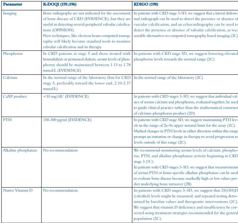
4.1 K-DOQI and KDIGO guidelines
In August 2009, the new Kidney Disease : Improving
Global Outcome (KDIGO) guidelines(190)replaced the
for-mer Kidney Disease Outcomes Quality Initiative
(K-DOQI) guidelines(195;196). In these new guidelines, a
com-plete chapter is dealing with vascular calcification. Other changes with a potential direct or indirect impact on
vas-cular calcifications are also summarized in Table 2.
4.2 Role of phosphate binders
In epidemiological studies, phosphorus is one of the most important mineral parameters associated with CV
mortality in dialysis patients(9-11;197). Moreover, in in vitro
or animal models, high phosphate and calcium concen-trations are often required to induce vascular
calcifica-tions(82;89;198). So, the impact of phosphate lowering
ther-apies on vascular calcifications is of interest. This is especially the case for phosphate lowering therapies that contain no calcium. Actually, calcium-based phosphate binders have been “accused” to participate to the occur-rence and progression of vascular calcifications in sev-eral (but not all) trials(16 ;18 ;22 ;26 ;44 ;94). Moreover, non cal-cium-based chelators (i.e. sevelamer) have been shown to prevent ectopic calcifications (and atherosclerosis) in
animal models(199 ;200). Four important randomised trials
have been published comparing the effect of calcium-based phosphate binders versus sevelamer on the
devel-opment of vascular calcifications : the Treat to Goal(41),
the RIND(30), the BRIC(201)and CARE-2 trials(202). The
conclusions of these studies are contradictory (two
favouring for sevelamer(30 ;41)and two showing no
dif-ference between sevelamer or calcium-based phosphate
binders(201;202)). Sensu stricto, due to differences in
method-phy of the vascular access to assess the presence of cal-cification (or not). This score interestingly and
inde-pendently predicts mortality(31). In all these studies, only
one observer calculates the scores and we have thus no
idea of intra- and interobservers variability(31-35).
A more “quantitative” and interesting method consists
of a lateral X-ray of the lumbar abdominal aorta(21 ;191 ;192).
This technique has been proposed for the general
popu-lation by Kaupilla et al. in 1997. This “calcium score”
has been shown to be predictive for CV morbidity and
mortality in the general population (193 ;194). A lateral X-ray
is obtained that includes the last two thoracic and the first two sacral vertebrae. The aorta is identified as the tubular structure coursing in front of the anterior sur-face of the spine. Only the segments of the aorta in front of the first to the fourth lumbar vertebra are considered. Points are assigned from 1 to 3 according to the length of each calcified plaque along the anterior and the pos-terior profile of the aorta in front of each vertebra. The
score could theoretically vary to a maximum of 24(193)
(Figure 9)(39). This technique has been used in a large epidemiological trial about vascular calcifications (the
CORD study) including 933 dialysis patients(39). The
calcium score has been calculated in 64 patients by two different observers with an excellent interobserver
agreement (χ=0.9). This technique was also used by
the London’s group in the trial studying correlation between vascular calcification, histomorphometry and
calcium load(95).
Both lateral X-ray of the lumbar aorta and echocardio-graphy have been shown to correlate well with EBCT
results(191 ;192). However, the ability of this combination
to predict EBCT coronary calcification is less impres-sive(191 ;192). Additional studies seem necessary before con-cluding about the interest of abdominal X-ray, notably in clinical practice.
In summary, several biomarkers (OPG, OPN, Fet-A, MGP, uc-MGP, dp-uc-MPG, FGF-23, PPi) have been proposed to predict either vascular calcifications or CV mor-tality. EBCT and MSCT are currently regarded as the most sensitive methods for detection and quantification of vascu-lar and especially coronary calcifications. However, they do not allow making distinction between medial and intimal calcifications. Moreover, they are costly and the irradiation dose, especially with MSCT is not negligible. As such, these methods are limited to clinical studies. The following meth-ods are cheaper but less sensitive and less quantitative. Echocardiography is used for the detection of cardiac valve calcification. Vascular ultrasonography has been proposed to semi-quantify calcifications. Standard radiography is a more simple method that allows distinguishing intimal and medial
calcifications. However, the calcification scores are generally not very quantitative. In 2004, a simple calcification score (1 to 8) based on plain radiography of pelvis and hands and in 2009, a semi quantitative score (1 to 3) based on 8 plain radiographies have been proposed. A more “quantitative” method is lateral X-ray of the lumbar abdominal aorta (score 1 to 24). Both, echocardiography and lateral X-ray of the lumbar aorta have been shown to correlate well with EBCT results. Additional studies are needed.
in nephrology
ologies and protocols, these four studies are difficult to compare. The fact that sevelamer also decreases LDL plasma levels could also explain the positive effect on
cardiac calcifications(185 ;203 ;204). So, polemic between
sup-porters of calcium-based versus non calcium-based
phos-phate binders is ferocious and not closed today(1 ;93 ;205-208).
However, one open label trial comparing sevelamer with calcium-based phosphate binders in 2013 dialysis patients found no difference in overall and CV mortality except in a subgroup analysis of overall mortality for patients
over 65 years(209).
However, it seems reasonable to avoid hypercalcemia
poten-tially induced by calcium-based phosphate binders(210).
4.3 Role of vitamin(s)
The role of vitamin D in the pathogenesis of vascular
cal-cifications is difficult to evaluate(54). Actually, both the
vita-min D receptor and the 1α-hydroxylase are expressed in
VSMC and cardiac myocytes(86). It also seems evident from
animal and in vitro studies that high doses of active vitamin
D will induce or favour vascular calcifications(54;63;211-216). We
have already talked about the role of vitamin D in
FGF-23 knock-out mice (see above)(137;144-146). However, it is not
clear if this vascular calcification development is depend-ent, or not, on vitamin D-induced hyperphosphatemia
and/or hypercalcemia(54;86;212-214;217). Moreover, low
“physio-Parameter K-DOQI (195 ;196) KDIGO (190)
Imaging Bone radiographs are not indicated for the assessment of bone disease of CKD (EVIDENCE), but they are useful in detecting several peripheral valvular calcifica-tions (OPINION).
New techniques, like electron beam computed tomog-raphy will likely become standard tools to monitor valvular calcification and its therapy.
In patients with CKD stage 3-5D, we suggest that a lateral abdom-inal radiograph can be used to detect the presence or absence of vascular calcification, and an echocardiography can be used to detect the presence or absence of valvular calcification, as rea-sonable alternatives to computed tomography based imaging (2C)
Phosphorus In CKD patients at stage 5 and those treated with hemodialysis or peritoneal dialysis, serum levels of phos-phorus should be maintained between 1.13 to 1.78 mmol/L (EVIDENCE)
In patients with CKD stage 5D, we suggest lowering elevated phosphorus levels towards the normal range (2C)
Calcium In the normal range of the laboratory (but for CKD stage 5, preferably toward the lower end, 2.10-2.37 mmol/L)
In the normal range of the laboratory (2C)
CaXP product <55 mg2/dL2(EVIDENCE) In patients with CKD stages 3–5D, we suggest that individual
val-ues of serum calcium and phosphorus, evaluated together, be used to guide clinical practice rather than the mathematical construct of calcium–phosphorus product (2D).
PTH 150-300 pg/ml (EVIDENCE) In patients with CKD stage 5D, we suggest maintaining PTH lev-els in the range of 2x-9x upper normal limit for the assay (2C). Marked changes in PTH levels in either direction within this range prompt an initiation or change in therapy to avoid progression to levels outside of this range (2C).
Alkaline phosphatase No recommendation We recommend monitoring serum levels of calcium, phospho-rus, PTH, and alkaline phosphatase activity beginning in CKD stage 3 (1C).
In patients with CKD stages 3–5D, we suggest that measurements of serum PTH or bone-specific alkaline phosphatase can be used to evaluate bone disease because markedly high or low values pre-dict underlying bone turnover (2B).
Native Vitamin D No recommendation In patients with CKD stages 3–5D, we suggest that 25(OH)D (calcidiol) levels might be measured, and repeated testing deter-mined by baseline values and therapeutic interventions (2C). We suggest that vitamin D deficiency and insufficiency be cor-rected using treatment strategies recommended for the general population (2C).
logical” doses of active vitamin D seem to inhibit vascular
calcification in some (but not all(216;218)) animal models(219).
The dose(216;218;219)and the type of active vitamin D (calcitriol
or analogs) could be of some importance(86;217). Large
stud-ies, although retrospective, have also shown that active vita-min D therapies could be associated with a reduction in mortality(220-222).
The role of native vitamin D could be different from active vitamin D in the vascular calcifications context. One recent retrospective study showed that patients (both CKD and non CKD were included) with low serum 25-OH-vitamin D were more likely to develop coronary
artery calcifications(223). Wolf et al. had already shown that
lower baseline 25-OH vitamin D levels were significantly associated with increased mortality in a cohort of 825 inci-dent dialysis patients but there was no data neither on CV
mortality nor on vascular calcifications(224). In a sample of
52 dialysis patients, London et al. did not find any
correla-tion between 25-OH vitamin D levels and calcificacorrela-tion
scores (ultrasonography and X-ray radiography(22)) but
25-OH vitamin D was negatively correlated with aortic pulse
wave velocity(225). In 233 dialysis patients (47 % treated
with low doses of active vitamin D), Matias et al. described
an association between 25-OH vitamin D levels and
cal-cification scores(32)in multivariate analysis(226).
Additional and interventional studies seem necessary before concluding about the role of active and native vitamin D on vascular calcifications.
In conclusion, the use of vitamin D must be reasonable. Yet, this therapy must induce neither hypercalcemia nor hyperphosphatemia. It could be also of interest not to induce low bone turnover with these therapies (see above)(67 ;94).
4.4 Role of calcimimetics
Calcimimetics (such as cinacalcet, Mimpara®) form a new
therapeutic class that modulates the calcium sensing recep-tor (CaR) in the parathyroid glands in order to diminish
PTH secretion(227;228). Contrary to other therapies for sHPT
(such as vitamin D), cinacalcet simultaneously reduces PTH, serum calcium and phosphorus. Moreover, very high PTH levels have been proposed as predictive of coronary
calcifications in dialysis patients(12). Lastly, it has been shown
that CaR is present in human VSMC and is probably down regulated in VSMC from dialysis patients which could be
related to the process of calcification (229-231). So, the
poten-tial interest of cinacalcet for the treatment of vascular
cal-cification does well exist. Several in vitro and animal
stud-ies have actually underlined the role of calcimimetics in
such a context(230). The in vitro study published by Alam et
al. suggested that calcimimetics may inhibit the
develop-ment of VSMC calcifications(229). One study in a CKD rat
model has shown that both calcimimetic and calcitriol
reduce PTH secretion. The effects on serum calcium were logically opposite. Rats treated with calcitriol developed aorta calcifications although rats treated with calcimimet-ics did not. Maybe the most interesting are the results on rats treated with combined therapy (calcimimetic and cal-citriol). Indeed, the hypercalcemia but not the
calcifica-tions are prevented by such a combination(212). The same
authors confirmed these results in another rat model with
lower vitamin D doses(218). These results will be confirmed
by other authors with the difference that combination ther-apy here leads to lower vascular calcifications than under calcitriol therapy alone (but higher than with a calcimimetic
alone)(232). Another author using a model of CKD rats with
high phosphate diet (thus without vitamin D) showed that vascular and cardiac calcifications could be prevented by
both calcimimetic and parathyroidectomy(233). In another
model of uremia-enhanced vascular calcifications (apoE
knock-out mice), Ivanovski et al. confirmed that
cal-cimimetics reduce the progression of vascular calcifications
both in the intima and media. They also showed in vitro the
direct inhibitory effect on the process of both vascular cal-cification and atherosclerosis. This last observation was
unexpected and mechanisms are unknown(234). Moe et al.
studied the effect of calcium (in the diet) and calcimimet-ics on vascular calcification occurrence. If both calcium and calcimimetics show efficacy in lowering PTH levels, both the combination (calcium+calcimimetics) and the calcium only group will favour for cardiac and vascular calcifica-tions whereas the group only treated with calcimimetic will
show benefit on extraosseous calcifications(235). Koleganova
et al. also showed that calcitriol (with non hypercalcemic
doses) increases medial calcification and proliferation of
VSMC compared to calcimimetics(216).
In humans, several case reports have been published suggesting the potential benefit of cinacalcet in
calci-phylaxis(67 ;236-238). A retrospective analysis of four randomised,
double blind and placebo controlled clinical trials in patients with secondary hyperparathyroidism suggested cardiovas-cular benefit with cinacalcet. Indeed, this treatment led to a 39 % risk reduction in CV events leading to hospitaliza-tion (infarctus, unstable angina or heart failure). There was
no effect on global mortality(239). Recently, a prospectively
designed observational study, including 19186 patients fol-lowed from November 2004 up to 26 months, found a sig-nificant all-cause CV survival benefit that was associated with cinacalcet prescription in hemodialysis patients
receiv-ing IV Vitamin D(240). One limited Japanese, not randomised,
not placebo controlled trial including 8 patients treated with cinacalcet and 60 controls suggested that cinacalcet is effec-tive in preventing the progression of coronary
calcifica-tions(241). Regarding this topic, the definitive results of the
ADVANCE trial are awaited with impatience. In this ran-domised study, 360 hemodialysis subjects were treated for 52 weeks with cinacalcet and low doses of active vitamin D
in nephrology
(for example, calcitriol at 0.125 µg/d) in one group and with variable doses of active vitamin D in the other group. Study
design and inclusion criteria are presented in Figure 10(40).
To avoid interferences in calcification progression, all patients were treated with calcium-based chelators and lipid lowering therapy should not be initiated. The pri-mary objective was the progression of coronary calcifica-tions measured by MSCT. Secondary objectives are
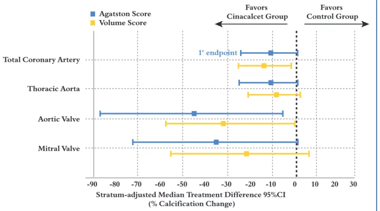
resumed in Figure 11. Preliminary results didn’t show any
statistical difference for the primary objective (24 % CAC progression in the cinacalcet group versus 31% in the flex-ible Vitamin D group; p = 0.073). However, after adjust-ment for the initial phosphorus concentrations (which were higher in the cinacalcet group), the difference in the primary objective was significant in favour of the cinacal-cet group (+26 % CAC progression versus +42 %,
p=0.031)(242). Furthermore, a consistent trend toward less
progression of CV calcification was observed at all sites evaluated in the cinacalcet group compared to the control
group (Figure 12). It has to be noted that the Vitamin D
sterol levels in the arm with cinacalcet plus low dose Vitamin D remained continuously above the initially pre-defined low doses of Vitamin D sterols (e.g. at least 0.2 µg/d calcitriol instead of 0.125 µg/d). Although the ADVANCE results suggest that treatment with cinacalcet may attenu-ate the progression of vascular calcifications, only the ongo-ing EVOLVE trial could provide a conclusive answer to the question whether cinacalcet treatment improves CV out-come and mortality in dialysis patients compared to
treat-ment without cinacalcet(243). Finally, we will only cite some
therapies recently proposed for vascular calcifications:
vita-min K(244), sodium thiosulfate(245)and bisphosphonates(246).
Although of some interest in some case reports or even in preliminary trials, more trials seem necessary before clin-ical use.
• On hemodialysis for
≥
3 months• iPTH > 300 pg/mL (31.8 pmol/L) OR
• iPTH
≥
150 pg/mL and≤
300 pg/mL (15.9 –31.8 pmol/L)
— and receiving treatment with vitamin
D analogs at time of PTH assessment — and corrected serum Ca x P >
50 mg2/dL2(3.9 mmol2/L2)
• Corrected serum Ca
≥
8.4 mg/dL (2.1 mmol/L)• Screening CAC score
≥
30— Subjects were subsequently
strati-fied:
≥
30–399,≥
400–999, and≥
1000 Day 1 W4 30 mg 60 mg 90 mg 120 mg 180 mg W8 W12 W16 W20 W28 MDCT Scan MDCT Scan W52 MDCT Scan S C R E E N I N G R A N D O M I Z A T I O N Cinacalcet group n=180 Control group N=180 Titration phase 20 wk Follow-up phase 32 wkFigure 10. ADVANCE study: study design, treatment schema and inclusion criteria. MDCT = multi-detector computed tomo-graphy (Floege et al. Nephrol Dial Transplantation, 2010, 25, 1916-1923).