HAL Id: tel-00911665
https://tel.archives-ouvertes.fr/tel-00911665
Submitted on 29 Nov 2013
HAL is a multi-disciplinary open access
archive for the deposit and dissemination of sci-entific research documents, whether they are pub-lished or not. The documents may come from teaching and research institutions in France or abroad, or from public or private research centers.
L’archive ouverte pluridisciplinaire HAL, est destinée au dépôt et à la diffusion de documents scientifiques de niveau recherche, publiés ou non, émanant des établissements d’enseignement et de recherche français ou étrangers, des laboratoires publics ou privés.
Role of the post-transcriptional regulators Pumilio1 and
Pumilio2 in murine hematopoietic stem cells
Fabio Michelet
To cite this version:
Fabio Michelet. Role of the post-transcriptional regulators Pumilio1 and Pumilio2 in murine hematopoietic stem cells. Human health and pathology. Université René Descartes - Paris V, 2013. English. �NNT : 2013PA05S013�. �tel-00911665�
Université Paris Descartes
Ecole Doctorale Biologie et Biotechnologie
INSERM U1016, Institut Cochin / Equipe Dusanter-CramerRole of the post-transcriptional regulators
Pumilio1 and Pumilio2 in murine
Hematopoietic Stem Cells
Par Fabio Michelet
Thèse de doctorat en Hématologie et Oncologie
Dirigée par Dr. Evelyne Lauret
Présentée et soutenue publiquement le 7 Novembre 2013
Devant un jury composé de :
Président: Pr. Lacombe Catherine (PU-PH)
Rapporteur: Dr. Simonelig Martine (DR2). Rapporteur: Dr. Mazurier Frédéric (DR2) Examinateur: Dr. Cohen-Tannoudji Michel (DR2) Directrice de thèse : Dr. Lauret Evelyne (CR1, HDR)
CONTENTS
ACKNOWLEDGEMENTS ... I
ABSTRACT ... V LIST OF FIGURES ... VII LIST OF ABBREVIATIONS ... IX
INTRODUCTION ... 1
1. STEM CELLS ... 3
1.1. DEFINITION OF STEM CELL POTENTIAL ... 4
1.1.1. Totipotent stem cells ... 4
1.1.2. Pluripotent stem cells ... 4
1.1.2.1. Embryonic stem cells ... 5
1.1.3 Multipotent stem cells and Adult stem cells ... 6
1.1.4. Progenitor cells ... 7
2. HEMATOPOIETIC STEM CELLS ... 8
2.1. MURINE HEMATOPOIETIC STEM CELL ASSAYS ... 11
2.1.1. In vitro assays ... 12
2.1.1.1. Colony-Forming Cell Assay ... 12
2.1.1.2. Long-term culture-initiating cell assay ... 13
2.1.2. In vivo Assays ... 13
2.1.2.1 Short Term in vivo assays ... 13
2.1.2.1.1. Colony-forming unit-spleen (CFU-S) cells assay ... 13
2.1.2.2 Long Term Repopulating Cells assay ... 14
2.1.2.2.1. Competitive repopulation assay ... 14
2.1.2.2.2. Limiting dilution assay ... 15
2.1.2.2.3. Serial transplantation assay ... 15
2.1.3. Immunophenotypical analysis of HSC/Progenitor cells ... 16
2.1.3.1. Thy1.1lo, Lin- Sca-1+ Cells ... 16
2.1.3.2. Lin c-Kit+ Sca-1+ Cells... 16
2.1.3.3. SLAM Family Members ... 16
2.1.3.4. Fluorescent dyes ... 17
2.1.4. Phenotype and isolation of HSC based on their cycling behaviour ... 17
2.2. HUMAN HEMATOPOIETIC STEM CELL ASSAY ... 19
2.2.1. In vitro assays ... 19
2.2.2. In vivo assays ... 19
2.2.3. Immunophenotypical analysis of human HSC/Progenitor cells ... 20
2.3. HEMATOPOIETIC STEM CELLS MAINTENANCE ... 21
2.3.1.1. Anti-apoptotic proteins ... 21
2.3.1.2. Transcription factors ... 22
2.3.1.3. Signal transducers ... 24
2.3.1.4. Polycomb group proteins ... 25
2.3.2. Hematopoietic stem cells niche and cell-extrinsic pathways ... 26
2.3.2.1. Cellular components of the HSC niche ... 27
2.3.2.2. Molecular regulators of HSC fate ... 29
2.3.2.2.1. Secreted ligands and their receptors ... 30
2.3.2.2.2. WNT signaling ... 32
2.3.2.2.3. Notch signaling ... 34
2.3.2.2.4. Hedgehog and TGFβ signaling ... 38
2.3.2.2.5. The extracellular matrix ... 39
2.3.2.2.6. Adhesion molecules ... 40
2.3.2.2.7. Chemical gradients ... 42
2.3.2.3. Induced HSC mobilization ... 44
2.3.2.4. The “progeny niche”: feedback regulation from HSC progeny ... 44
2.3.3. Post-transcriptional regulation in HSCs ... 45
2.3.3.1. microRNA ... 45
2.3.3.2. RNA binding proteins ... 46
3. PUMILIO ... 49
3.1. PUF FAMILY PROTEINS ... 49
3.1.1. Pumilio proteic domains ... 52
3.2. PUF PROTEINS: POST-TRANSCRIPTIONAL REPRESSORS ... 56
3.2.1. Mechanisms of action ... 56
3.2.1.1. Deadenylase recruitment ... 56
3.2.1.2. Inhibition of translation initiation ... 57
3.2.1.3. Inhibition of translation elongation ... 58
3.2.1.4. Cooperation with miRNAs ... 58
3.2.2. Non-canonical PUF mechanism of action ... 60
3.2.2.1. PUFs as activators of mRNA expression ... 60
3.2.2.2. mRNA localization by PUF proteins ... 61
3.3. PUF COMBINATORIAL REGULATION: TARGETS AND PARTNERS ... 63
3.3.1. Target mRNAs ... 63
3.3.2. Protein partners ... 64
3.4. PUF REGULATORS ... 71
3.5. PUF AND STEM CELLS MAINTENANCE IN INVERTEBRATES ... 72
3.6. MAMMALIAN PUF PROTEINS ... 73
3.6.1. Pumilio proteins and neuronal functions. ... 73
3.6.2. Pumilio proteins and spermatogenesis ... 74
3.6.3. Pumilio proteins and cancer ... 75
3.6.4. Murine Pum1 and Pum2 genes: structure and localization ... 75
3.6.5. Murine Pum1 and Pum2 isoforms ... 76
3.6.6. Expression of Pum1 and Pum2 in murine HSPCs ... 76
3.6.7. Pum proteins and stem cells maintenance in mammals ... 79
RESULTS... 83
5. RESULTS ... 85
5.1. PUM1 and PUM2 knockdown impair murine in vitro HSPC potential ... 85
5.2. PUM1 expression restores the function of shmP1-transduced LSK cells ... 89
5.3. Pum1 and Pum2 KD alter the cell cycle and cell survival of murine HSPCs. .. 90
5.4. Pum KD leads to loss of immature cells ... 92
5.5. Pum1 and Pum2 KD do not lead to increased senescence ... 93
5.6. Pum1 and Pum2 are not involved in genomic stress response ... 94
5.7. Pum1 and Pum2 knockdown impair the human HSPC potential... 96
5.8. PUM1 and PUM2 KD impair the in vivo reconstitution potential of murine and human HSPCs ... 98
5.9. Effects of Pum1 overexpression in murine HSCs ... 99
5.10. Implication of Pum1 and Pum2 in Delta4/Notch activity ... 101
5.11. Identification of genes implicated in Pumilio effects on HSCs. ... 103
5.12. Validation of the HP7 cell line as a model for murine HSPC studies. ... 104
6. MATHERIALS AND METHODS ... 108
6.1. Lentiviral constructs and transduction ... 108
6.2. Mice ... 108
6.3. Murine HSC purification ... 109
6.4. Isolation and immuno-labeling of human CD34+ cells ... 109
6.5. Culture experiments ... 110
6.6. Quantitative reverse transcriptase (RT)-PCR ... 111
6.7. Protein analysis. ... 111
6.8. CFC assays... 112
6.9. Detection of apoptotic cells ... 112
6.10. Immunophenotypical analysis of LSK cells ... 112
6.11. Senescence analysis ... 113
6.12. γH2AX foci analysis ... 113
6.13. Long-term competitive repopulation assays ... 113
6.14. Microarray ... 114
6.15. Intracellular localization of Pum1 and Pum2 in HP7 cell line ... 114
6.16. Statistical analysis ... 115
6.17. Acknowledgements ... 115
DISCUSSION ...117
7. DISCUSSION ... 119
7.1. Role of Pum1 and Pum2 in HSPC cell expansion ... 120
7.1.1. Impact of Pum1 and Pum2 KD on cell cycle ... 121
7.1.2. Impact of Pum1 and Pum2 KD on apoptosis ... 122
7.2. Role of Pum1 and Pum2 in HSPC hematopoietic potential. ... 122
7.2.1. In vitro hematopoeitic potential ... 122
7.2.2. In vivo reconstitution potential ... 123
7.3. Pum proteins and cellular stress ... 126
7.5. Effects of Pum1 overexpression in KLS cells ... 128
7.6. Identification of genes implicated in Pumilio effects on HSCs. ... 129
8. CONCLUSIONS AND PERSPECTIVES ... 131
ANNEX I ... 135
ANNEX II ... 177
i
ACKNOWLEDGEMENTS
Je tiens tout d’abord à remercier le Professeur Catherine Lacombe de me faire l’honneur de présider le jury de cette thèse.
J’aimerais remercier également le Docteur Martine Simonelig et le Docteur Fréderic Mazurier pour avoir accepté d’être rapporteur de ce travail de thèse et pour le temps consacré à la lecture de ce manuscrit.
Je remercie le Docteur Cohen-Tannoudji Michel pour avoir accepté d’être examinateur de ce travail de thèse.
Je remercie surtout et en particulier Evelyne Lauret, ma directrice de thèse, pour m’avoir suivi pendant ces quatre années. Elle m’a enseigné tout ce que je sais aujourd’hui sur l’hématopoïèse et sur les cellules souches. J’ai énormément apprécié sa manière de travailler, toujours motivée par la curiosité de trouver des réponses à chaque nouvelle hypothèse, et toujours prête à aller de l’avant et à dépasser chaque difficulté de manière simple et positive. Travailler avec elle était tout aussi enrichissant du point de vue personnel que scientifique, j’ai beaucoup aimé ses suggestions, toujours exposées clairement et directement, ainsi que son respect total de mon opinion et de mes idées. Merci Evelyne.
Je remercie énormément Isabelle Dusanter pour m’avoir accueilli dans son équipe, pour avoir toujours été agréable et prête à m’aider mais aussi pour tous ses conseils, grâce à ses amples connaissances scientifiques et techniques dans lesquelles j’ai puisées afin de concrétiser mon projet scientifique.
Je remercie Serge Fichelson, Isabelle Vigon et Ayda Miri-Nezhad pour leur travail de recherche sur le rôle de Pumilio dans le système humain.
Je remercie Aurore Hattabi pour son travail accompli jusqu’alors, pour son aide dans les expériences, pour sa gentillesse et pour toutes nos discussions tantôt très profondes tantôt très légères qui ont égayé nos soirées au laboratoire.
ii
Je remercie Patrycja Palikowska pour sa gentillesse, son aide et sa grande disponibilité.
Je remercie Van Thi Hong TO qui a collaboré à ce travail pendant son stage en m’offrant d’excellents résultats et en me laissant un très bon souvenir de cette période.
Je remercie donc tous les membres du laboratoire et de l’unité U1016 Inserm que j’ai connus pendant ces quatre années et qui ont rendu agréable le temps passé ici en leur compagnie.
Je remercie Azzedine, Jean-Marc et Alain pour leur sympathie sans faille, leur bonne humeur et pour toutes les histoires bizarres racontées pendant la pause déjeuner et pour toutes les discussions sur l’actualité qui m’ont aidé à mieux comprendre ce pays.
Je remercie Luana pour les beaux souvenirs des premières années.
Je remercie Katy, qui restera toujours la blonde la plus intelligente que j’ai jamais connue (après ma chef), Helen pour les moments divertissants passés ensemble, Fanny pour le « couscous » de minuit.
Je remercie Emilie pour sa sympathie, pour le « ménage » et les pauses goûter.
Je remercie les « New Entries » avec lesquels je me suis senti à l’aise dès le départ, Frédéric pour nos discussions scientifiques et non-scientifiques dont j’ai beaucoup appris, Alexandre, toujours optimiste bien qu’il parle trop tout seul, Audrey qui quant à elle parle trop aux cellules et Cécile pour sa contribution active au projet Pumilio et qui parle trop.
Un doctorat, ce n’est pas seulement une expérience de laboratoire mais aussi une expérience de vie, surtout s’il est réalisé dans une ville et un pays splendides comme Paris et la France, où j’espère revenir un jour.
iii
C’est pour cela que je tiens à remercier toutes les personnes qui ont fait de ces quatre années une expérience incroyablement positive qui m’a fait évoluer professionnellement mais surtout d’un point de vue personnel.
Je remercie avant tout ma famille et en particulier ma mère qui a fait tant de sacrifices pour me permettre d’arriver jusqu’ici, qui m’a toujours soutenu et à qui je dois vraiment tout. Grazie Mamma!
Je remercie Chiara à qui je dois mes premiers beaux souvenirs dans cette ville et sans laquelle je me serai probablement perdu dès le premier jour en sortant du train, et qui, armée de patience, est toujours restée près de moi.
Je remercie l’équipe du CFB avec laquelle j’ai passé sans aucun doute la plus belle année de ma vie et en particulier : Riccardo et les mille soirées passées ensemble, Roberto pour ses chansons a cappella, Chris pour le «Ring of fire », Harry pour la « pomme » magique, Alice pour avoir démontré qu’il est possible d’escalader la fontaine de Chatelêt, Catherine pour le tango et l’escrime en attendant le RER, Luane pour l’ «éponge » et Anna avec laquelle j’ai passé les soirées les plus folles et qui, sans le savoir, m’a énormément enseigné.
Je tiens aussi à remercier Sabrina avec laquelle j’ai passé des bons et amusants moments, dont j’ai un excellent souvenir et qui m’a fait me sentir un peu plus français en m’enseignant la vraie culture française… !
Merci Clara pour les gâteaux empoisonnés et pour les entraînements exténuants !
Un remerciement particulier à toutes les personnes du club de voile de Choisy pour les splendides week-ends passés tous ensemble, toujours gentils et disponibles pour me donner de nombreux conseils mais sans pitié sur l’eau ! Je remercie en particulier Ludivine, Clémentine et Chloé, vous me manquerez mais je reviendrai ! (Encore plus fort qu’avant)
iv
Derniers remerciements enfin, mais pas des moindres, à mes amis de toujours qui sont restés près de moi malgré la distance et avec lesquels je passe d’excellents moments à chaque fois que je rentre chez moi.
Je remercie Gary et Giulio pour tout le temps passé ensemble à la recherche de nouvelles expériences et grâce à qui c’est toujours comme si j’étais parti hier !
Je remercie Davide pour toutes les discussions, toutes les soirées et les mille pensées qui nous unissent depuis des années et qui sont toujours très importantes pour moi, mais surtout pour être toujours à mes côtés dans tout ce que j’entreprends.
Je remercie Matteo qui égaye mes journées avec ses histoires, et pour son dynamisme qui a fait qu’on a passé les soirées les plus incroyables ensemble.
Je remercie Alessandro pour sa sympathie contagieuse qui me rappelle toujours que le juste style de vie prévoit une grande équité de fêtes, d’entrainements et de régime sain !!
v
ABSTRACT
The central properties of stem cells are the pluripotency and the capacity of self-renewal. Hematopoietic stem cells (HSCs) posses such common features that allows them to generate all the cells of the hematopoietic compartments, maintaining in the same time the HSC pool. We develop approaches focused on ex vivo HSC expansion through activation by exogenous HOXB4 (human HSCs) or Notch/Dll-4 ligand (murine HSCs). Two independent transcriptomic analyses surprisingly converged toward an increased expression of two genes never identified sofar as crucial for HSC functions: Pumilio1 (Pum1) and Pumilio2 (Pum2). Pum1 and Pum2 are posttranscriptional regulators belonging to the Pumilio-FBF (PUF) family of RNA-binding proteins. Although it was established that the primordial role of PUF proteins is to sustain mitotic proliferation of stem cells in Invertebrates, so far nothing is known about the role of Pum1 and Pum2 in human and murine HSCs.
For these reasons, we have investigated the roles and mechanisms of action of Pum1 and Pum2 in murine and human HSCs through shRNA strategy. Pum1 and Pum2 knockdown (KD) in murine HSCs led to a decreased HSC expansion and clonogenic potential ex vivo, associated with an increased apoptosis and a cell cycle arrest in G0/G1 phase. KD of both Pum1 and Pum2 enhanced these effects, suggesting a cooperative effect. Expansion and clonogenic potential of KD Pum1 HSCs were rescued by enforced expression of Pum1 (insensitive to our shRNA), thus validating the specificity of our shRNA. Enforced expression of Pum1 could not rescue the functions of Pum2 KD HSCs, highlighting the non-redundant role of these proteins. Furthermore, when Pum1 or Pum2 KD HSCs were inoculated into lethally irradiated mice to follow the long-term hematopoietic potential, only rare bone marrow cells derived from Pum1 and Pum2 KD HSCs were evidenced after 4 months, contrary to control HSCs. Identical results were obtained with human Pum1 or Pum2 KD HSCs.
In conclusion, our results demonstrate the involvement of Pumilio factors in stemness maintenance, expansion and survival of murine and human HSCs. Identification of Pumilio factors and their targets as new regulators of HSCs expansion will allow consider them as new tools for therapeutic perspectives.
vii
LIST OF FIGURES
Figure 1. Differentiation of human tissues ... 5
Figure 2. Model of the hematopoietic hierarchy... 9
Figure 3. Myeloid bypass model and Conventional model ... 11
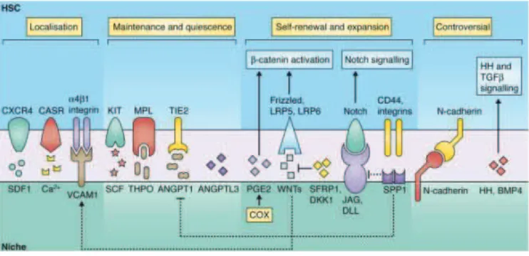
Figure 4. The HSC microenvironment ... 27
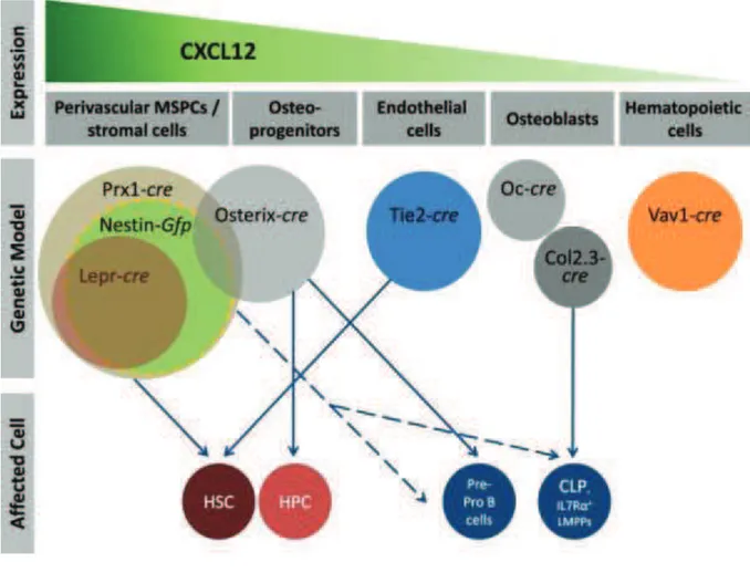
Figure 5. Distinct cellular sources and niches for CXCL12 in bone marrow ... 31
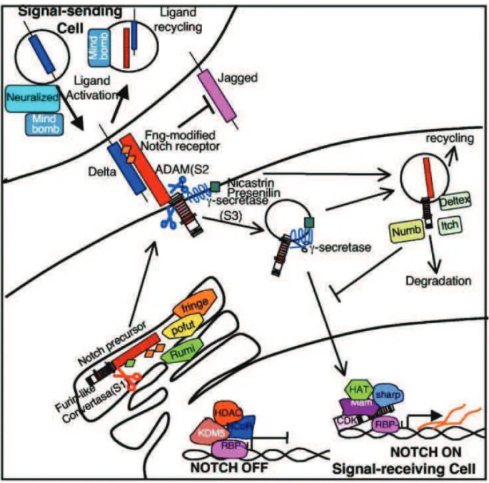
Figure 6. The Notch signaling pathway ... 35
Figure 7. Crosstalk between HSCs and their niche ... 43
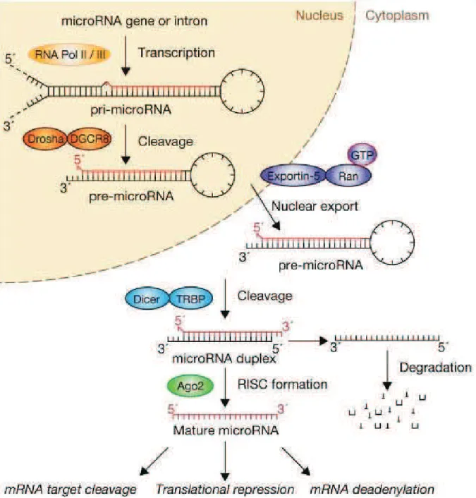
Figure 8. microRNA biogenesis and function ... 47
Figure 9. PUF proteins throughout eukaryotes ... 51
Figure 10. Diagram of the human Pum1 protein conserved domains. ... 52
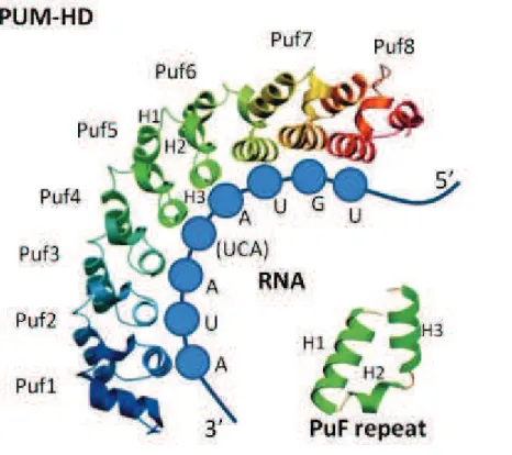
Figure 11. Pum Homology Domain structure and RNA binding ... 53
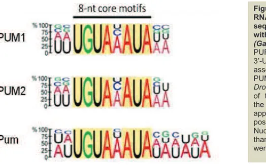
Figure 12. Analysis of RNA consensus sequence associated with PUM proteins ... 55
Figure 13. Deadenylase recruitment by yeast Puf5 ... 57
Figure 14. Pum2 mediated translational control in Xenopus ... 58
Figure 15. Pumilio binding alters local p27-3ʹ UTR structure and 221 and miR-222 accessibility ... 59
Figure 16. Possible mechanisms of mRNA activation by PUF proteins. ... 60
Figure 17. Working model of cooperation between FBF-2 and PGL-1 ... 62
Figure 18. mRNAs associated with human PUM proteins in HeLa S3 cells ... 63
Figure 19. Pum-NRE-Nos-Brat interaction surface in Drosophila ... 65
Table 1. mRNA targets and protein partners of model Puf proteins ... 68
Figure 20. Role of Pum, Nos and Brat in the Ovarian Stem Cell System ... 71
Figure 21. Pumilio1 and Pumilio2 isoforms ... 77
Figure 22. General experimental protocol for in vitro experiences ... 85
Figure 23. Validation of the shRNA efficiency in murine cells ... 86
Figure 24. mPum1 and mPum2 KD inhibit hematopoietic potential of murine HSPCs ... 88
Figure 25. mPum1 expression rescues the functions of shmP1-transduced LSK cells ... 89
Figure 26. PUM1 and PUM2 KD alter the cell cycle and induce cell apoptosis in murine HSPCs ... 91
Figure 27. Pum KD leads to loss of immature cells ... 92
Figure 28. Pum1 and Pum2 KD do not lead to increased senescence ... 94
Figure 29. Pum KD has no effect on protection from genomic stress or DNA repair . 95 Figure 30. PUM1 and PUM2 KD inhibit the in vitro hematopoietic potential of human HSPCs ... 97
Figure 31. PUM1 and PUM2 KD inhibit the in vivo reconstitutive potential of murine and human HSPCs ... 99
viii
Figure 33. Delta4/Notch pro-self-renewing action is partially maintained in Pum KD
cells... 102
Figure 34. Identification of genes implicated in Pumilio effects on HSCs ... 104
Figure 35. Validation of the HP7 cell line ... 105
Figure 36. Intracellular localization of Pum1 and Pum2 in HP7 cell line ... 106
Table 2. Sequences of RT-qPCR primers and list of shRNAs used to invalidate human and murine Pum1 and Pum2 ... 116
ix
LIST OF ABBREVIATIONS
Ab: antibody
AGM: aorta-gonad mesonephros Ago: Argonaute
Ang-1 : angiopoietin 1 ANGPT1: angiopoietin 1 ANGPTL3: angiopoietin like 3 APC: allophycocyanin
BCL2: B-cell lymphoma 2
BFU-E: burst-forming units-erythroid cell
BM: bone marrow
BMP : bone morphogenetic protein Brat: Brain Tumor
BrdU: bromodeoxyuridine CASR : Ca2+ -sensing receptor CD: cluster of differentiation cDNA: complementary DNA CDS: coding sequence CFC: colony-forming cell
CFU-E: colony-forming units erythroid cell
CFU-G: colony-forming units-granulocytes cell
CFU-GEMM: colony forming units-
granulocytes-erythrocytes-macrophages-megakaryocytes CFU-GM: colony forming units-granulocytes/macrophages cell CFU-M: colony-forming units-monocytes/macrophages cell
CFU-Mk: megakaryocyte-restricted cell CFU-S: colony-forming unit-spleen CLP: common lymphoid progenitor CMP: common myeloid progenitor CMRP: common myeloid restricted progenitor
CPE: cytoplasmic polyadenylation element
CPEB: cytoplasmic polyadenylation element binding protein
CRU: competitive repopulating unit CXCL12: chemokine (C-X-C motif) ligand 12
CXCR4: C-X-C chemokine receptor type 4
DAPI: 6-diamidino-2-phenylindole DAZ: deleted in azoospermia DC:dendtritic cell
DKK1 : dickkopf homolog 1 Dll: Delta-like
DNA: Deoxyribonucleic acid
dpc: days post coitum Dpp : Decapentaplegic EBF2 : early B-cell factor 2 EC: endothelial cells
ECM : extracellular matrix EGF: epidermal growth factor EP: erythrocyte progenitor EPO: Erythropoietin ES: embryonic stem cell EST: expressed sequence tag Etv6: Ets variant gene 6
FACS: fluorescence-activated cell sorting
FBF: fem-3 mRNA bindingfactor FITC: fluoresceine isotyocianate FL: fetal liver
Fmi: flamingo Fz: frizzled
G-CSF : granulocyte colony-stimulating factor
Gfi1: growth factor independence 1 GFP: green fluorescent protein GMP: granulocyte/macrophage progenitor
GO: gene ontology
GP: granulocyte progenitor GSC: germ stem cell hb: hunchback
Hes: hairy/enancher-of-split HH : hedgehog
HIF-1α : hypoxia-inducible transcription factor-1α
Ho 33342: Hoescht 33342 Hox: homeobox
HP: hematopoietic progenitor HP7: hematopoietic precursor 7 HSC: hematopoietic stem cells
HSPC : hematopoietic stem progenitor cell
ICM: inner cell mass IFNα : interferon α IL:interleukine
IPA: ingenuity pathway analysis iPS: induced pluripotent stem cell JAK-STAT: Janus family kinase-signal transducer and activator of
transcription KD: knockdown KSL: c-Kit+ Lin- Sca-1+ KO: knockout
x LAP : latency-associated protein
Lepr : leptine receptor
LIF: leukemia inhibitor factor Lin: lineage markers
LLC : large latent complex
LMPP: lymphoid multipotent progenitor LRP5/6: LDL receptor-related proteins 5 and 6
LTBP1 : latent TGF-β binding protein 1 LTC-IC: long-term culture-initiating cell LT-HSC: long-term repopulating
hematopoietic stem cells
LTRC: long-term reconstituting cell LT-RC: long-term reconstituting cell MacP: macrophage progenitor MAPK: mitogen-activated protein kinase
mbDll4: membrane bound Delta like 4 Mcl1: myeloid cell leukemia 1
MEP: megakaryocyte/erythrocyte progenitor
MERP: megakaryocyte erythroid restricted progenitor miRNA : microRNA MkP: megakaryocyte progenitor MkRP: megakaryocyte restricted progenitor MPP: multipotent progenitor mRNA: messenger RNA MSC: mesenchymal stem cell
MSPC : mesenchymal stem/progenitor cell
mTOR: mechanistis target of rapamycin
Mw: molecular weight
MyRP: myeloid restricted progenitor ncRNA : noncoding RNA
NK: natural killer
NOG: NOD/Shi-scid, IL-2Rγnull Nos: Nanos
NRE: nanos response element OB : osteoblast
Oc : osteocalcin OC : osteoclast OPN: osteopontin
ORF: open reading frame PBE: pumilio binding element PcG: Polycomb group
PCR: polymerase chain reaction PDGF: platelet derived growth factor
PDGFRα : platelet derived growth factor receptor α
PE: phycoerythrin
PGK: phosphorglicerate kinase
PRC1: Polycomb repression complex 1 Pten: phosphate and tensin homologue deleted on chromosome ten
PUF: Pum and FBF Pum: Pumilio
PUM-HD: PUM homology domain Raptor: regulatory-associated protein of mTOR
Ras: rat sarcoma protein RBC: red blood cell
RBP: RNA binding proteins Rh123: Rhodamine 123
RISC: RNA-induced silencing complex RNA: ribonucleic acid
ROS : reactive oxygen species RT-qPCR: Reverse transcription quantitative polymerase chain reaction RU: repopulating unit
S17: stromal 17 cell line Sca-1: stem cell antigen 1 SCF: stem cell factor SCID: severe combined immunodeficiency
SDF1: stromal cell-derived factor 1 shRNA: short RNA
SILAC: stable isotope labeling by amino acids in cell culture
siRNA : small interfering RNA
SLAM: signaling lymphocyte attractant molecule
SP: side population
SPP1: secreted phosphoprotein 1 ST-HSC: short-term repopulating hematopoietic stem cells
Tel: Translocation Ets leukemia TGF-β : transforming growth factor β TPO: thrombopoietin
UTR: untranslated region
VCAM1 : vascular cell adhesion molecule 1
VE: vascular endothelium
VEGF-A : vascular endothelial growth factor A
wt: wild type YS: yolk sac
1
3
1. STEM CELLS
During embryogenesis, a single fertilized oocyte gives rise to a multicellular organism whose cells and tissues have adopted differentiated characteristics or fates to perform the specified functions of each organ of the body. As embryos develop, cells that have acquired their particular fate proliferate, enabling tissues and organs to grow. Even after an animal is fully grown, however, many tissues and organs maintain a process known as homeostasis, where as cells die, either by natural death or by injury, they are replenished. This remarkable feature has ancient origins, dating back to the most primitive animals, such as sponges or hydrozoans. Throughout evolution, nature has exerted considerable fun and fancy in elaborating on this theme. Some amphibians, for instance, can regenerate a limb or tail when severed, and the neurons of bird brains can readily regenerate. While mammals seem to have lost at least some of this wonderful plasticity, their liver can partially regenerates providing that the injury is not too severe, and the epidermis and hair of their skin can readily repair when wounded or cut. Additionally, the epidermis, hair, small intestine, and hematopoietic system are all examples of adult tissues that are naturally in a state of dynamic flux: even in the absence of injury, these structures continually give rise to new cells, able to transiently divide, terminally differentiate and die.
The fabulous ability of an embryo to diversify and of certain adult tissues to regenerate throughout life is a direct result of stem cells properties. When a stem cell divides, each new cell has the potential either to remain a stem cell (through symmetric division) or become another type of cell (through asymmetric division) with a more specialized function. In other words stem cells are distinguished from other cell types by two important characteristics: self-renewal and potency.
4
Self-renewal consists in the ability to go through numerous cycle of mitotic cell division while maintaining the undifferentiated state. Potency is the capacity that allows unspecialized stem cells to differentiate into specialized cell types1.
1.1. DEFINITION OF STEM CELL POTENTIAL
Many of the terms used to define stem cells depend on the behavior of the cells in the intact organism or after transplantation in vivo or under specific laboratory conditions in vitro.
1.1.1. Totipotent stem cells
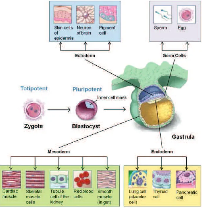
The fertilized egg is considered to be totipotent because it has the potential to generate all the cells and tissues that make up an embryo and that support development in utero. The fertilized egg divides and differentiates until it produces a mature organism. Adult mammals, including humans, consist of more than 200 kinds of cells. These include nerve cells (neurons), muscle cells (myocytes), skin (ephitelial) cells, blood cells (erythrocytes, monocytes, lymphocytes, etc.), bone cells (osteocytes), cartilage cells (chondrocytes) and other cells which are essential for embryonic development but are not incorporated into the body of the embryo as extraembryonic tissues, placenta, and umbilical cord. All of these cells are generated from a single, totipotent cell, the zygote, or fertilized egg (Figure 1).
1.1.2. Pluripotent stem cells
The term pluripotent, instead, is used to describe stem cells that can give rise to cells derived from all three embryonic germ layers: endoderm, mesoderm, and ectoderm. All of the many different kinds of specialized cells that make up the body are derived from one of these germ layers.
5 1.1.2.1. Embryonic stem cells
Emanating from the pioneering research of Martin Evans in the 1970s and cumulating with the successful parallels with human tissue, cells from the inner cell mass (ICM) of mammalian blastocysts can be maintained in tissue culture under conditions where they can be propagated indefinitely as pluripotent embryonic stem (ES) cells2. If injected back into a recipient blastocyst that is then carried to term in a female host, these cells can contribute to virtually all the tissues of the chimeric
6
offspring, including the germ cell compartment. To maintain cultured ES cells in their relatively undifferentiated, pluripotent state, they must both express the intrinsic transcription factor Oct4, and constitutively receive the extrinsic signal from the cytokine leukemia inhibitor factor (LIF)3,4. Upon LIF withdrawal, cultured ES cells spontaneously aggregate into embryo-like bodies, where they differentiate and spawn many cell lineages, including beating heart muscle cells, blood islands, neurons, pigmented cells, macrophages, epithelia, and fat-producing adipocytes5. Similarly, when ES cells are injected into nude mice, they differentiate into multicellular masses, called teratocarcinomas. Although the programs of gene expression in these structures often bear strong resemblance to the differentiation pathways typical of developing animals, the triggering of these programs is chaotic. These examples illustrate the importance of intercellular interactions and cellular organization in orchestrating development and embryo shape.
1.1.3 Multipotent stem cells and Adult stem cells
A multipotent stem cell is a stem cell that can give rise to several types of cells but it is limited in its ability to proliferate and differentiate.
Adult stem cells or somatic stem cells are considered as multipotent stem cells. They are found among differentiated cells in a tissue or organ. They can self-renew and differentiate to yield some or all of the major specialized cell types of the tissue or organ. The primary roles of adult stem cells in a living organism are to maintain and repair the tissue in which they are found. The origin of adult stem cells in some mature tissues is still under investigation. Adult stem cells have been identified in many organs and tissues, including brain, bone marrow, peripheral blood, blood vessels, skeletal muscle, skin, teeth, heart, gut, liver, ovarian epithelium, and testis. They are thought to reside in a specific area of each tissue (called the “stem cell
7
niche”). If the differentiation of adult stem cells can be controlled in the laboratory, these cells may become the basis of transplantation-based therapies.
1.1.4. Progenitor cells
A progenitor cell occurs in fetal or adult tissues and is partially specialized; it divides and gives rise to differentiated cells. Researchers often distinguish progenitor cells from adult stem cells in the following way: when a stem cell divides, one of the two new cells is often a stem cell capable of replicating itself again. In contrast, when a progenitor cell divides, it can form more progenitor cells or it can form two specialized cells, neither of which is capable of replicating itself. Progenitor cells can replace cells that are damaged or dead, thus maintaining the integrity and functions of the tissue. Progenitor cells are often classified depending on their potency. Multipotent progenitors can give rise to several different committed oligopotent progenitors that in turn can generate even more specified lineage restricted progenitors (often called precursors) able to differentiate only in mature effector cells. So, the definition of multipotent, oligopotent and lineage-restricted progenitors is strictly related to their degree of potency along the continuum of cells between the stem cell and the mature cells. Controversy about the exact definition remains and the concept is still evolving.
8
2. HEMATOPOIETIC STEM CELLS
The hematopoietic system represents a continuum of cells with changing phenotype and properties as they progress from stem to differentiated cells. While mature blood cells are produced at a rate of more than 1 million cells per second in the human adult 6, most of the hematopoietic stem cells (HSCs) from which they are derived cycle very infrequently and primarily reside in the G0 phase of the cell cycle under homeostatic conditions7. These facts present an interesting problem: how does the organism achieve a balance whereby an adequate pool of HSCs is maintained for the life of the organism, while at the same time HSCs consistently meet the organism’s enormous demand for continuous replenishment of mature blood cells, most of which are short lived? To answer this question we have to think at the HSC as a cell capable to continuously provide a series of intermediate progenitors whose potency is restricted to certain lineages, allowing for an enormous amplification in the numbers of terminally differentiated cells, while properly maintaining the HSC pool homeostasis throughout life by precisely balancing self-renewal and differentiation (Figure 2). The importance of this balance is underscored by the numerous examples where aberrant HSC development causes severe disease, when HSC differentiation into committed progenitors is not accompanied by the typical loss of self-renewal capacity, or HSC derived progenitors fail to fully differentiate into mature blood cells and may enter a preleukemic progression.
Anyway, even if consensus holds that HSCs give rise to multipotent progenitors (MPPs) of reduced self-renewal potential and that MPPs eventually produce lineage-committed progenitor cells in a stepwise manner, a recent elegant work by Yamamoto and colleagues demonstrated the existence of a differentiation pathway that suggest a revised model of hematopoietic differentiation8.
9
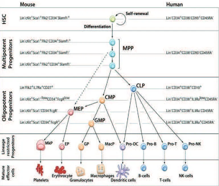
Figure 2. Model of the hematopoietic hierarchy (Seita and Weissman, 2010)
The HSC resides at the top of the hierarchy, and is defined as the cell that has both the self-renewal capacity and the potential to give rise to all hematopoietic cell types (multi-potency). Throughout differentiation, a HSC first loses self-renewal capacity, then loses lineage potential step-by-step as it commits to become a mature functional cell of a certain lineage. The cell surface phenotype of each population is shown for the mouse and human systems. Intermediate precursors between the first lineage committed progenitors and final mature cell, and different subsets of mature B- and T-cells are omitted. In the mouse system, heterogeneity of MPPs has been revealed by differences in cell surface marker phenotypes and functional differences of their subsets discussed. For example, evidence suggests that some of MPPs directly give rise to MEP without passing through CMP (dashed arrow). HSC: hematopoietic stem cell, CLP: common lymphoid progenitor, CMP: common myeloid progenitor, MEP: Megakaryocyte/erythrocyte progenitor, GMP: granulocyte/macrophage progenitor, MkP: Megakaryocyte progenitor, EP: erythrocyte progenitor, GP: granulocyte progenitor, MacP: macrophage progenitor, DC: dendritic cell, NK: natural killer, Lin: lineage markers
10
Using transgenic mice expressing Kusabira-Orange (KuO) fluorescent protein in all blood cell lineages, they unexpectedly found myeloid-restricted progenitors with long-term repopulating activity (MyRPs), which are lineage-committed to megakaryocytes, megakaryocyte-erythroid cells, or common myeloid cells (MkRPs, MERPs, or CMRPs, respectively) in the phenotypically defined HSC compartment (CD150high CD34- LSK) together with HSCs. To examine whether HSCs directly differentiate into these myeloid-restricted repopulating progenitors, they employed single cell sorting of HSCs, generating culture plates containing one HSC per well. After that, they let these cells accomplish one mitotic cycle in vitro to later transplant the two daughter cells in two different recipient mice. Strikingly, after single-cell transplantation, they were able to identify daughter cell pairs in which one was an HSC and the other was an MkRP as well as daughter cell pairs in which one was an HSC and the other was a CMRP, demonstrating that direct lineage commitment took place in an asymmetric manner at the HSC level and that HSCs can directly differentiate into lineage-restricted progenitors without passing through an MPP stage showing that loss of self-renewal and stepwise progression through specific differentiation stages are not essential for lineage commitment of HSCs (Figure 3). These myeloid bypass pathways could be essential for fast responses to ablation stress.
11
2.1. MURINE HEMATOPOIETIC STEM CELL ASSAYS
Hematopoiesis is now among the best-defined differentiation cascades in mammalian tissues due to the ease of access and morphologic distinctiveness of many of its members. The advent of antibody technology, culture capability, transplantation, and finally genetic engineering of mice has enabled a detailed understanding of processes involved in the commitment and differentiation of HSCs
Figure 3. Myeloid bypass model (left) and Conventional model (right) (Yamamoto et al. 2013)
HSCs self-renew and give rise to lineage-restricted progenitor cells. In the conventional hematopoietic differentiation model (right side), HSCs in the CD34- LSK population differentiate into multipotent progenitors (MPPs, Flt3- CD34+ LSK) of reduced self-renewal potential and MPPs eventually produce lymphoid-primed multipotent progenitors (LMPPs, Flt3+ CD34+ LSK) or lineage-committed progenitor cells in a stepwise manner. All mature blood lineages are considered to pass through the MPP and/or LMPP stage in the CD34+ LSK population. In contrast, in the myeloid bypass model (left side), the CD34- LSK cell population contains CMRPs, MERPs, and MkRPs in addition to HSCs. These MyRPs are produced by HSCs (LT-HSCs, IT-HSCs or ST-HSCs). MyRPs can clonally expand via self-renewal as in HSCs, B cells, and T cells. The CD34+ LSK population, which is downstream from the CD34- LSK population, also contains lineage-committed progenitors including myeloid-restricted progenitors, whereas ‘‘true’’ MPPs and lymphoid-primed multipotent progenitors (LMPPs) are minor populations in the CD34+ LSK population. Together, MyRPs, rather than ‘‘MPPs and LMPPs,’’ are considered to be the major suppliers of myeloid cells (platelets, erythrocytes, and neutrophils/monocytes) in the hematopoietic system at a single-cell level. LMPP fraction yields cells with various differentiation potentials and these progenitors might derive from ST-HSCs in the CD34- LSK population but not from MPPs in the CD34+ LSK population. Because cells in the CD34+ LSK fraction have oligopotent differentiation potentials rather than multipotent potentials, CD34+ LSK cells are considered to be OPPs.
12
into progenitor cells and ultimately mature blood cells. Lineage-committed progenitor cells differentiate in less than 3 weeks and five to 10 divisions in short-term assays; in contrast, stem cells and their immediate progeny have to accomplish a high number of divisions (>15) in “long-term assays” (>5 weeks) before they produce differentiated cells. Combining antibody-based subselection of cells with transplantation has made possible the identification of progenitors and HSCs. A range of different methods for characterizing stem and progenitor cell populations are now available for use in hematopoietic cell research. All assays measure two cardinal parameters of hematopoietic stem and progenitor cells: cell proliferation (measured by the number of cells produced) and differentiation potential (estimated by the number of different lineages represented). These two parameters can give us some “end points” that reflect the properties of these “unobservable” progenitors.
2.1.1. In vitro assays
2.1.1.1. Colony-Forming Cell Assay
The colony-forming cell (CFC) assays measure progenitor cells in a given population using semisolid agar- or, more commonly, well-defined methylcellulose-based culture media, which are commercially available. The majority of CFCs consist of lineage-restricted colonies: colony-forming units erythroid (CFU-E) which are more mature than the erythroid restricted burst-forming units-erythroid (BFU-E); megakaryocyte-restricted CFU-Mk; colony-forming granulocytes (CFU-G), colony-forming
units-monocytes/macrophages (CFU-M); and colony forming
units-granulocytes/macrophages (CFU-GM). The most immature (multipotent) CFC measurable contains granulocytes, erythrocytes, macrophages, and often megakaryocytes (CFU-GEMM). This CFC is also often called CFU-mixed, as it may
not always contain megakaryocytes but does contain erythroid and
13
difficult to assess, usually requiring specialized coculture systems9,10, and hence are not routinely used, although there are now commercially available methylcellulose-based colony assays to measure pre-B cells. While informative about the progenitor cell content of a population of interest, the CFCs do not measure HSCs.
2.1.1.2. Long-term culture-initiating cell assay
The long-term culture-initiating cell (LTC-IC) assay is a well-established in vitro assay used to enumerate primitive hematopoietic stem cells (HSCs) and relies on the two cardinal functions of HSCs: ability to self-renew and differentiation capacity. LTC-ICs present in minimally processed cell suspensions or in purified cell populations and cocultured on a supportive feeder layer are detected by their sustained ability to produce hematopoietic progenitors (CFC) after ≥ 4 weeks in culture. Refinements including the use of a defined stromal cell line, and extending the in vitro culture to 6 weeks allow detection of LTC-IC at similar frequencies to transplantable HSCs quantified using in vivo assays. An issue with these assays is the interlaboratory variability often observed due to varying feeder layers and specific culture conditions. They are useful, however, in limiting dilution format for quantitating primitive cells when other features such as homing capacity or other functions required for in vivo engraftment may compromise the reliability of transplant assays.
2.1.2. In vivo Assays
2.1.2.1 Short Term in vivo assays
2.1.2.1.1. Colony-forming unit-spleen (CFU-S) cells assay
Colony-forming unit-spleen (CFU-S) cells are cells that, once injected into an irradiated recipient, home to the spleen and form macroscopic colonies that provide very short-term (usually 1–3 weeks) in vivo repopulation of the mouse11. The CFU-S are therefore early engrafting cells, providing radioprotection to the mouse and
14
allowing it to survive more readily in the first 2–3 weeks posttransplantation when pancytopenia usually occurs. These progenitors are more immature than CFCs but are more mature than HSCs. The gold standard for measuring HSC is the long-term repopulating assay.
2.1.2.2 Long Term Repopulating Cells assay
Long-term reconstituting cells (LTRC) are a subpopulation of HSCs able to produce differentiated cells of multiple lymphoid and myeloid lineages for months in bone marrow and peripheral lymphoid organs. These long-lived clones are best identified by analyzing donor granulocytes, T- and B-lymphocytes in the peripheral blood of recipients 3 - 4 months after transplantation. Thus, in vivo, longevity, rather than multipotentiality, is the best criteria of stem cell ‘activity’.
2.1.2.2.1. Competitive repopulation assay
There are various types of long-term repopulating assays. The most common assay is the competitive repopulation assay12. This assay measures the functional potential of the unknown source of HSCs against a set known number of HSCs (usually whole bone marrow cells from congenic wild-type mice). The number of repopulating units (RU) in the donor cell population (source of unknown HSC content being measured) can be determined by the following formula: donor RU = % donor cells x C/(100 - % donor cells), where C = the number of competing RU and 1 x 10⁵ whole bone marrow cells = 1 competing RU13,14. While providing informations about the function of HSCs in their capacity to repopulate compared to the competing bone marrow, this study provides qualitative or at best semiquantitative information about the HSCs within a given population, but it cannot distinguish between the number of HSCs or their quality (progeny produced per HSC). Furthermore, caution should be used when designing competitive repopulation assays, as it has been shown that the reliability of
15
this assay is critically dependent on the numbers of HSCs present in the populations being assessed: when too few or too many HSCs (recipients of <1 x 10⁵ or >2 x 10⁷ bone marrow cells each from donor and competing sources) are present, the data may not be meaningful13.
2.1.2.2.2. Limiting dilution assay
The frequency of HSCs (from which the number of HSCs can be calculated) is commonly measured using the limiting dilution assay, which is a variation of the competitive repopulation assay. In this assay, a series of dilutions of the unknown source (donor ‘‘test’’ cells) are competed against a set number of competing bone marrow cells. The number of mice negative for reconstitution in each cell dose is then measured, and the frequency of HSCs (competitive repopulating units, CRU) is estimated using Poisson statistics15,16. Note that CRU, which measures the quantity of HSCs, is distinct from RU, which measures the functional quality of HSCs.
2.1.2.2.3. Serial transplantation assay
The most stringent test of HSC potential is the serial transplantation assay. The HSC compartment has been shown to be heterogeneous, comprising a hierarchy of HSCs that can be identified by their functional capacity. The most immature HSC in this hierarchy is capable of sustaining hematopoiesis throughout serial transplantation17–
19
. Hence, in this assay, the source of HSCs is transplanted into sequential serial transplant recipients, and the ability of this population to sustain hematopoiesis by presumptive self-renewing divisions is determined. Limitations to this assay are its dependence upon homing and engraftment processes that may be perturbed without altering stem cell function per se, in particular mutant mouse strains.
16
2.1.3. Immunophenotypical analysis of HSC/Progenitor cells
In the last 20 years, a number of different methods whereby HSCs and progenitor cells can be identified have emerged20–23. All rely on fluorescence-activated cell sorting (FACS)-based methods. The purity of the populations of HSCs achieved using these methods has increased within recent years, such that approximately 50%–96% of single cells in certain purified populations can give rise to long-term reconstitution after transplantation24,25. The most commonly used FACS-purified populations of HSC/progenitor cells include the following:
2.1.3.1. Thy1.1lo, Lin- Sca-1+ Cells
Short-term repopulating HSCs could be isolated based on their expression of stem cell antigen-1 (Sca-1), low expression of Thy1.1, and lack of expression of lineage markers21. The same population was shown to contain long-term repopulating HSCs in a subsequent study26.
2.1.3.2. Lin c-Kit+ Sca-1+ Cells
The latter population was further purified by the expression of the stem cell factor receptor, c-Kit, in 199227. This population is very heterogeneous and consists predominantly of progenitor cells with less than 10% of it representing HSCs. The lack of expression of CD3428 and Flt3 (CD135)29,30 has been used to further purify long-term repopulating HSCs (LSK+ CD34- Flt3-) from short-term repopulating HSCs (LSK+ CD34+ Flt3-) and multipotent progenitors (LSK+ CD34+ Flt3+)30.
2.1.3.3. SLAM Family Members
SLAM proteins are a family of cell surface glycoproteins in the immunoglobulin superfamily with specific SLAM antigens (CD150+ CD244- CD48- cells) identified as useful to purify a population of which approximately 50% of single cells reconstituted
17
lethally irradiated animals31. Unlike the limitations presented by those isolated using Thy1.1 or Sca-1 expression, the SLAM receptors appear to be expressed by many mouse strains31, and more faithfully detect HSCs in older, mobilized, or transplanted mice25.
2.1.3.4. Fluorescent dyes
Two different vital dyes, the mitochondrial-binding dye Rhodamine 123 (Rh123) and DNA-binding dye Hoescht 33342 (Ho 33342), have also been used either alone or in combination to isolate HSCs32–36. Both of these dyes are retained at very low intensity in HSCs due to high efflux of the dyes from HSCs, as shown by studies utilizing the drug verapamil. A more common method used in laboratories today is the use of the Hoescht 33342 (Ho 33342) to define the side population (SP)32. By analyzing Ho 33342 emission at two wavelengths simultaneously, the SP appear distinct from the main population in a distinct ‘‘tail’’ profile, which disappears with verapamil treatment32. The SP population is also not as pure as HSCs enriched by other methods such as LSK+ CD34- Flt3- cells, although it can be used in combination with other markers such as Sca-1, c-Kit, and CD34 to further purify HSCs with extreme efficiency24,37. Notably, cells with an SP profile have also been detected in many other organs but with inconsistent functional correlation with stem cell-like functions38. This method should not be assumed to yield stem cells in other tissue types or even in species other than the mouse.
2.1.4. Phenotype and isolation of HSC based on their cycling behaviour
Just before birth, cycling HSCs migrate from the liver to the developing bone marrow, where they engraft in small cavities of trabecular bone. By four weeks after birth, HSCs have fully matured and acquired a dormant status. A dormant status is necessary to preserve the self-renewal capacity of HSCs and to prevent stem cell
18
exhaustion39–41. In the healthy adult mouse, all long-term HSC activity is a feature of a small subset of LSK bone marrow cells. In comparison to progenitor populations, HSCs are recognized as ‘slow’ cycling42–45. Classical cell cycle analyses of highly purified HSCs that measure DNA content (Hoechst 33342 or 4’, 6-diamidino-2- phenylindole (DAPI)) alone, or combined with intracellular Ki67 expression (which is absent in cells at the G0 stage), have shown that more than 70% of CD34–CD48– CD150+ LSK HSCs are in the quiescent G0 stage of the cell cycle, whereas less than 10% of more differentiated multipotent progenitor cells (CD34+ LSK cells) are quiescent 46. Although such assays generate important informations regarding the cell cycle state of each cell following isolation, they do not reveal the cycling history of each cell over time or their precise physical location in the bone marrow. To address this, long-term label-retaining assays have been carried out, in which HSCs are labeled in vivo with the thymidine analogue bromodeoxyuridine (BrdU) or with a chromatinassociated green fluorescent protein (GFP) by incorporating a histone 2B– GFP fusion protein under the control of a doxycyclinregulated transgenic allele. After labeling, BrdU incorporation is stopped by removing the BrdU source, or GFP expression is repressed by the administration of doxycyclin. These conditions are continued in the subsequent chase period (up to one year), and during this time dividing cells gradually lose the accumulated label so that after more than four cell divisions the BrdU and GFP labels, respectively, are diluted out and are no longer detectable by flow cytometry. By contrast, cells that have undergone fewer than five cell divisions during the chase period remain labeled and are therefore called label-retaining cells (LRCs). Thus, the cell cycling history can be determined by measuring the loss of the label over a long time period. For example, in the CD34–CD48– CD150+ LSK HSC population, 22% still retained the label (that is, are LRCs) after 120 days of chase, 18% were LRCs after 213 days chase and some were LRCs after 380 days46. By contrast, in the hematopoietic progenitor cell populations (the CD34+ LSK
19
cells), few, if any, LRCs were found after only 100 days of chase. Different mathematical modeling strategies using the label decay kinetics data have revealed that the CD34–CD48–CD150+ LSK HSC population probably comprises two subsets: a dormant population (~30%) dividing only every 145-193 days and a ‘homeostatic’ quiescent population (~70%) dividing every 28–36 days. Although both populations could reconstitute the hematopoietic system in primary recipients, only dormant HSCs could be serially transplanted, suggesting that long-term self-renewal capacity is retained exclusively by dormant HSCs.
2.2. HUMAN HEMATOPOIETIC STEM CELL ASSAY 2.2.1. In vitro assays
To study the properties of human HSCs we can use exactly the same kind of assays used to characterize murine HSCs. The only different parameter is the time required to culture and to differentiate human HSCs both in CFC assay and in LTC-IC assay. Murine cells cultured on methylcellulose and supplied with cytokine are able to form colonies after 7-10 days of culture, instead human clonogenic progenitors need 2-3 weeks. LTC-IC assay for human cells can be extended from 5 to 12 weeks to individuate LTC-IC or even more primitive extended-LTC-IC.
2.2.2. In vivo assays
The study of human LT-RCs required the development of specific murine models with severe combined immunodeficiency (SCID). Immunodeficient mice are often used as recipients for human cells or tissues, because they can relatively easily accept heterologous cells due to lack of host immunity reducing rejection and allowing the engraftment of human cells, in our case HSCs. With the development of the NOD/scid mouse, an improved SCID mouse, it was possible to engraft more human cells and tissues than in the SCID mouse. However, many problems
20
remained before an in vivo humanized model could be achieved.
Mamoru Ito and his group at the Laboratory Animal Research Department in CIEA were successful in establishing an extremely severe combined immunodeficient mouse called the NOG (NOD/Shi-scid,IL-2Rγnull) mouse47 by combining the NOD/scid mouse and the IL-2 receptor-γchain (a common receptor for several cytokines) knockout (IL2rγKO) mouse. NOG mice lack T and B cells, Natural Killer cells (NK) and complement activity. Furthermore, they have reduced dendritic and macrophage functions.
The NOG mouse shows markedly better engraftment of human cells and human tissues than the NOD/scid mouse and also makes possible engraftment of human cancer cells, liver cells, etc. at high rates. In addition, after transplantation of human hematopoietic stem cells, human T cells can be developed in peripheral lymphoid tissues of the NOG mouse.
2.2.3. Immunophenotypical analysis of human HSC/Progenitor cells
The first CD34 surface glycophosphoprotein is commonly used for enrichment of human HSCs. CD34 is expressed on fetal liver hematopoietic cells, in cord blood cells and in the adult bone marrow cells. CD34+ bone marrow cells comprise only
1.5% of marrow mononuclear cells, but contain precursors for all
lymphohematopoietic lineages. CD34+ is a very heterogeneous compartment of cells constituted mainly by multipotent and oligopotent progenitors and only 1% of this population include CD34+ CD38- cells. CD34+ CD38- cells are highly enriched by LT-RCs. Recently, tracking the expression of several adhesion molecules in HSC-enriched subsets; CD49f was identified as a specific HSC marker. Single CD49f+ cells were highly efficient in generating long-term multilineage grafts, and the loss of CD49f expression identified transiently engrafting multipotent progenitors (MPPs).
21
2.3. HEMATOPOIETIC STEM CELLS MAINTENANCE
Hematopoiesis is a tightly regulated process in which a rare pool of hematopoietic stem cells (HSCs) gives rise to the lymphohematopoietic system. In order to maintain hematopoietic homeostasis throughout the lifetime of an animal, this pool of HSCs must be maintained. This is achieved by the processes of survival, quiescence and proliferation/self-renewal, a specialized cell division in which one or both of the daughter cells remain undifferentiated and retain essentially the same replication potential of the parent. One of the most important issues in stem cell biology and in regenerative medicine is to understand the mechanisms that regulate the properties of stem cells. Some examples of key regulators are listed below.
2.3.1. Cell-intrinsic pathways
2.3.1.1. Anti-apoptotic proteins
Stem cell maintenance requires that proliferation pathways remain functional while differentiation, senescence and cell death pathways are repressed. There is a large body of evidence suggesting that suppression of apoptosis is required for HSC survival. Studies using transgenic mice constitutively expressing BCL2 (B-cell
lymphoma 2) in all hematopoietic tissues provide evidence directly supporting this
theory. The forced expression of the oncogene Bcl2 resulted in increased numbers of transgenic HSCs in vivo and gave these cells a competitive edge over wild type HSCs in competitive reconstitution experiments48,49 suggesting that cell death plays a role in regulating the homeostasis of HSCs. Mcl1 (Myeloid cell leukemia 1), another anti-apoptotic Bcl2 family member, has been shown to be required for HSC survival50. In this study it was shown that inducible deletion of Mcl1 in mice resulted in a severe anemic phenotype due to a drastic loss of BM cells in a cell autonomous manner.
22 2.3.1.2. Transcription factors
The transcription factor Tel (Translocation Ets leukemia;also known as Etv6 [Ets variant gene 6]), an Ets (E-26 transforming-specific)-related transcriptional repressor, is also required for HSC maintenance. Conditional inactivation of Tel/Etv6 in HSCs rapidly leads to the depletion of Tel/Etv6-deficient HSCs. However, Tel/Etv6 is not required for the maintenance of committed precursors. When it is conditionally inactivated in most hematopoietic lineages, it does not affect their differentiation or survival51. The mechanism by which Tel/Etv6 modulates adult HSCs survival is not known.
The homeobox (Hox) genes encode transcription factors that regulate embryonic body patterning and organogenesis. They play a role in the regulation of hematopoiesis. The function of HOX genes in normal hematopoiesis has been widely studied using gene expression analysis and knockin or knockout studies in HSCs and early hematopoietic progenitors. Generally the overexpression of a HOX gene leads to an expansion of stem and progenitor cell populations together with a block on differentiation. Notable examples of this include the overexpression of murine Hoxb6, which resulted in the expansion of murine HSCs and myeloid precursors, together with the inhibition of erythropoiesis and lymphopoiesis52, and overexpression of murine Hoxb3 that resulted in several hematological abnormalities, such as a block of B- and T-cell differentiation as well as a delay in myeloid precursor proliferation53. Overexpression of human HOXC4 resulted in expansion of early and committed myeloid and erythroid progenitors54, and knockin of human HOXA5 caused an increase in the number of myeloid progenitors and blocked erythroid differentiation55,56. Other HOX genes are required for the maintenance of progenitor or stem cell status and promote their proliferation, especially HOXA9 and HOXB4. The former is the most preferentially expressed HOX gene in human CD34+ HSCs and early hematopoietic progenitors and is subsequently downregulated during
23
differentiation. Murine Hoxa9 and Hoxb4 overexpression enhances HSC expansion and myeloid progenitor proliferation57,58. Hoxb4 is also highly expressed in HSCs and downregulated during differentiation53,59. Our team contributed to demonstrate that its overexpression in murine and human cell lines results in a remarkable expansion of HSCs in vivo and in vitro without resulting in leukemia or lineage disturbances53,60. Indeed, the self-renewal ability of Hoxb4-transduced murine HSCs is 20–50-fold greater than untreated cells. In addition to the knockin and overexpression, knockdown and deletion studies in murine models and cell lines have also been used to evaluate the role of HOX genes in hematopoiesis. However, owing to the functional redundancy of HOX genes, the results of knockdown assays are sometimes difficult to interpret and do not always reflect the findings of studies where the gene has been overexpressed.
Gfi1 (Growth factor independence 1), a Zinc-finger repressor, has been implicated as a regulator of HSC self-renewal. Two groups working independently determined that Gfi1 controls self-renewal of HSCs by restraining their proliferative potential61,62. They showed that Gfi1-deficient HSCs display increased proliferation rates and are also functionally compromised in competitive repopulation and serial transplantation assays. Gfi1 might exert its effects on HSC proliferation by regulating the cell cycle inhibitor p21CIP1/WAF1. p21 mRNA expression levels are dramatically lower in the Gfi1-deficient HSCs62. p21CIP1/WAF1 itself has been implicated in the regulation of HSCs63. In its absence, HSCs have an impaired serial transplantation capacity.
The JAK–STAT (Janus family kinase–signal transducer and activator of transcription) pathway is a common downstream pathway of cytokine signaling that promotes hematopoiesis. Constitutive activation of the transcription factors of the Stat family, particularly Stat3 and Stat5, are frequently detected in leukemias, lymphomas and solid tumors64. Activation of Stat5 in HSCs led to the dramatic
24
expansion of multipotent progenitors and promoted HSC self-renewal ex vivo65. Deletion of Stat5 resulted in profound defects in hematopoiesis and markedly reduced ability of the mutant cells to maintain quiescence during steady-state hematopoiesis66 or repopulate the bone marrow of lethally irradiated mice. Another group show that transduction of adult mouse bone marrow cells with a constitutively activated form of Stat3 increased their regenerative activity in lethally irradiated recipients, whereas the transduction of these cells with a dominant negative form of Stat3 suppressed their regenerative activity67. These studies suggest that Stat proteins play a role in HSC self-renewal and potentially in other tissues, owing to the wide range of solid tissue and blood malignancies that harbor constitutively activated Stats.
2.3.1.3. Signal transducers
The Pten (phosphatase and tensin homologue deleted on chromosome ten) tumor suppressor, a modulator of several major signaling pathways, has very recently been implicated as a regulator of HSC self-renewal and an initiator of leukemogenesis68,69. It functions by inhibiting signaling through the AKT pathway. Although Pten deletion initially leads to a transient expansion of HSC numbers, the HSC pool then becomes depleted over time. Pten-deficient HSCs engraft normally in recipient mice, but are unable to sustain multilineage hematopoietic reconstitution. When Pten-deficient HSCs were transplanted into irradiated mice, they were only capable of short-term multilineage hematopoietic reconstitution and could not stably engraft irradiated recipients long term68,69.
Loss of Lkb1, another signal transducer involved in the Akt pathway, leads to impaired survival and escape from quiescence of HSCs, resulting in exhaustion of the HSC pool and in a marked reduction of HSC repopulating potential in vivo. Lkb1
25
deletion has an impact on cell proliferation in HSCs, but not on more committed compartments, pointing to context-specific functions for Lkb1 in hematopoiesis70–72. The mechanistic target of rapamycin (mTOR) is one of the proteins regulated by Pten and Lkb1 and it serves as a key sensor of cellular-energetic state and functions to maintain tissue homeostasis. Hyperactivation of the mTOR pathway impairs HSC function and is associated with leukemogenesis. The deletion of the mTORC1 component, regulatory-associated protein of mTOR (Raptor), in mouse HSCs leads to an impaired HSC regeneration in vivo73.
2.3.1.4. Polycomb group proteins
Polycomb-group proteins are a family of proteins that can remodel chromatin such that transcription factors cannot bind to promoter sequences in DNA. Recent studies have shown that Polycomb group (PcG) proteins and their interaction are important in the regulation of HSC self-renewal and lineage restriction. In particular, members of the PRC1 (Polycomb repression complex 1), such as Bmi1, Mel18 and Rae28, have been implicated. Bmi-1 plays an important role in regulating the proliferative activity of stem and progenitor cells. It is required for the self-renewal of both adult HSCs and neural stem cells74,75. Bmi1 enhances symmetrical expansion of the stem cell pool through self-renewal, induces a marked ex vivo expansion of multipotent progenitors, and increases the ability of HSCs to repopulate bone marrow in vivo76. Leukemic cells lacking Bmi1 undergo proliferation arrest, differentiation and apoptosis, leading to failure of leukemia in a mouse transplant mode77. In Bmi1-deficient bone marrow there is an upregulation of cell cycle inhibitors p16INK4A and p19ARF, and the p53-induced gene Wig1, and a downregulation of the apoptosis inhibitor AI-6. This suggests that a mechanism exists whereby Bmi1 functions by modulating proliferation and preventing apoptosis78. Bmi1 has also been shown to