Université de Montréal
Synthesis of Constrained Nucleosides
par
Juan Carlos Salinas Hernandez
Département de Chimie Faculté des Arts et des Sciences
Thèse présenté à la Faculté des Études Supérieures et postdoctorales en vue de l’obtention du grade de Philosophæ Doctor (Ph.D.) en chimie
Avril 2018
Résumé
La thérapie antisens, sous sa forme la plus basique, implique la liaison d'une séquence oligonucléotidique courte à un acide ribonucléique messager complémentaire (ARNm), ce qui peut finalement entraîner une prévention de la maladie génétique en stoppant la production de protéines pathogènes. Les oligonucléotides composés d'acides nucléiques naturels sont capables d'une reconnaissance de liaison à haute affinité à des séquences complémentaires d'ARN et d'ADN; cependant, ils subissent rapidement une digestion intracellulaire par l'action de nucléases et ne conviennent donc pas à la thérapie antisens. La présente thèse présente et détaille la synthèse de quatre nouveaux analogues d'acides nucléiques pouvant être utilisés comme agents antisens potentiels et dans des applications thérapeutiques associées.
Basé sur une stratégie à double contrainte, le chapitre trois explore la synthèse de TriNA 2, un analogue nucléosidique qui étend la structure bicyclique de LNA en un noyau tricyclique, limitant la rotation autour de l'angle de torsion γ.
Dans le quatrième chapitre, deux voies de synthèse pour la synthèse d'un analogue de nucléoside pipéridino bicyclique sont discutées; l'une basée sur un nucléoside comme point de départ et la seconde étant une synthèse qui utilise un sucre comme point de départ.
Au chapitre cinq, la conception et la synthèse d'un nouvel analogue de nucléoside phosphonate oxabicyclique sont discutées. La conception de ce nucléoside comporte un noyau perhydrofuropyranne qui limite la rotation autour des angles ɣ, δ et ε d'un nucléoside naturel. Le dernier chapitre décrit différentes approches de la synthèse de nucléotides à squelette macrocyclique basés sur des unités liées aux phosphonates. Notre première stratégie a utilisé
des H-phosphonates diastéréopure et leur alkylation stéréorétensive correspondante pour la construction du premier trinucléotide macrocyclique à cycle à 11 chaînons. Une approche de phosphoramidite complémentaire a fourni une voie complémentaire pour la synthèse du macrocycle à cycle à 11 chaînons et s’est avéré être un outil précieux pour la synthèse de macrocyles de différentes tailles.
Mots-clé : Thérapie antisens, acides nucléiques tricycliques, acides nucléiques bicycliques, acides nucléiques bloqués, restriction conformationelle, nucléosides, oligonucléotides, acides nucléiques, , la stabilité thermique des duplex
Abstract
In its basic form, antisense technology involves binding of a short oligonucleotide sequence to a complementary messenger ribonucleic acid (mRNA), which can ultimately result in prevention of genetic diseases prevention by stopping the production of pathogenic proteins. Oligonucleotides composed of natural nucleic acids are capable of high-affinity binding recognition to complementary RNA and DNA sequences; however, they rapidly undergo intracellular digestion through the action of nucleases and are thus unsuitable for antisense-based therapeutics. The present thesis reports and details the synthesis of four new nucleic acid analogues that can be used as potential antisense agents and in related therapeutic applications. Based on a double-constrain strategy, chapter three explores the synthesis of TriNA 2, a nucleoside analog that extends the bicyclic structure of LNA into a tricyclic core, restricting rotation around torsion angle γ.
In chapter four, two synthetic routes towards the synthesis of a bicyclic piperidino nucleoside analog are discussed; one based on a nucleoside as starting point and the second one being a carbohydrate-based synthesis.
In chapter five the conception and synthesis of a novel oxabicyclic nucleoside phosphonate analog is discussed. The design of this nucleoside features a perhydrofuropyran core which restricts rotation around angles ɣ, δ and ε of a natural nucleoside.
The last chapter describes different approaches toward the synthesis of macrocyclic backbone constrained nucleotides based on phosphonate linked units. Our first strategy used diastereopure H-phosphonates and their corresponding stereoretentive alkylation for the construction of the first 11-membered ring macrocyclic trinucleotide. A complementary phosphoramidite approach
provided a complementary route for the synthesis of the 11-membered ring macrocycle and showed a valuable tool for the synthesis of different size macrocyles.
Keywords : Antisense therapy, tricyclic nucleic acids, bicyclic nucleic acids, locked nucleic acids, conformational restriction, nucleosides, oligonucleotides, nucleic acids, duplex thermal stability.
Table of Contents
1 Chapter One – Introduction ... 1
1.1 DNA and RNA structure ... 1
1.2 DNA transcription ... 6
1.3 RNA processing ... 8
1.4 Protein synthesis – translation ... 9
2 Chapter Two – Antisense technology ... 11
2.1 History... 11
2.2 Molecular mechanism of action ... 11
2.2.1 RNA cleavage ... 12
2.2.2 Alternative mechanisms involving non-RNA cleavage ... 13
2.3 ASO modification ... 14
2.4 Constrained nucleosides... 18
3 Chapter Three... 22
Synthesis of TriNA 2 ... 22
3.1 Design of TriNA 2 ... 23
3.2 Retrosynthetic analysis of TriNA 2 ... 25
3.3 Synthesis of TriNA 2 ... 26
3.4 Duplex thermal stability measurements ... 31
3.5 Crystallographic and conformational analysis ... 33
3.6 Conclusions ... 34
4 Chapter Four ... 35
Synthesis of an azabicyclic nucleoside ... 35
4.1 Previous syntheses of azabicyclic nucleosides ... 36
4.2 Design of an azabicyclic nucleoside ... 38
4.4 Synthesis based on a nucleoside precursor ... 39
4.5 Retrosynthetic analysis based on a carbohydrate precursor ... 46
4.6 Synthesis based on a carbohydrate precursor ... 47
4.7 Conclusions ... 52
5 Chapter Five ... 53
Synthesis of a novel oxabicyclic nucleoside phosphonate ... 53
5.1 Previous syntheses of bicyclic perhydrofuropyran nucleosides ... 54
5.2 Design of a oxabicyclic nucleoside phosphonate surrogate of a phosphate ... 57
5.3 Retrosynthetic analysis ... 58
5.4 First synthetic route... 59
5.5 Second synthetic route ... 65
5.6 Duplex thermal stability measurements ... 74
5.7 Conclusions ... 75
6 Chapter six ... 76
Synthesis of backbone constrained nucleic acids ... 76
6.1 Macrocyclic nucleotides ... 77
6.2 General design of backbone constrained nucleic acids... 79
6.3 H-Phosphonate strategy ... 80
6.4 Copper-mediated coupling of H-phosphonates with alkynes ... 92
6.5 Chain extension by cross-metathesis ... 95
6.6 Phosphoramidite-Arbuzov method ... 98 6.7 Phosphoramidite method ... 101 6.8 Conclusions ... 105 7 Future Perspective ... 106 8 Experimental Procedures ... 108 8.1 General experimental ... 108 8.2 Experimental section ... 109 8.2.1 Synthesis of TriNA 2 ... 109
8.2.3 Synthesis of a oxabicyclic nucleoside phosphonate ... 144
8.2.4 Synthesis of backbone constrained macrocycles ... 172
9 X-Ray crystallographic data ... 201
9.1 Crystal and molecular structure of compound C19H24N2O8 (HAN489) ... 201
9.2 Crystal and molecular structure of compound C21H25N5O6 (ROBE39) ... 210
9.3 Crystal and molecular structure of compound C15H27O9P (ROBE52) ... 218
9.4 Crystal and molecular structure of compound C27H39N2O12P (HAN499) ... 225
Table of Figures
Figure 1. a) Generic nucleic acid double strand. b) Minor and major grooves of helix formed
by double strand nucleic acids1 ... 2
Figure 2. Torsion angles in nucleic acids4 ... 4
Figure 3. Sugar pucker of the furanose ring4 ... 5
Figure 4. Structures of A-type and B-type double stranded nucleic acids ... 6
Figure 5. General scheme for DNA transcription10 ... 7
Figure 6. RNA processing... 8
Figure 7. Translation process10 ... 10
Figure 8. RNase H active site19.The active site RNase in complex with the RNA/DNA hybrid. Active-site residues are shown in green; the RNA strand in pink, orange, and red; and the magnesium ions as yellow spheres. The water molecule (nucleophile) positioned to attack the scissile phosphate bond is indicated. Metal-ion coordination is shown as dashed yellow lines. ... 12
Figure 9. a) Generations of ASOs. b) Example of gap design oligonucleotide with MOE ... 15
Figure 10. Constraint of torsion angles ɣ and δ in selected nucleosides. ... 19
Figure 11. Restriction of torsion angles α and β in selected nucleosides. ... 21
Figure 12. Introduction of a methyl group at different positions of the LNA core. ... 23
Figure 13. Retrosynthetic analysis for the synthesis of TriNA 2. ... 25
Figure 14. Regioselective reductive cleavage of acetal 3.4. ... 28
Figure 15. Structural models of tricyclic nucleosides overlaid on oligonucleotide duplexes containing the corresponding (S)-cEt bicyclic modification. ... 33
Figure 16. Pseudorotation pathway of the furanose ring ... 34
Figure 17. Design rationale of the azabicyclic nucleoside ... 38
Figure 18. Retrosynthetic analysis of azabicyclic nucleoside I ... 39
Figure 19. a) Proposed transition state for the diastereoselective allylation of compound 4.8. b) X-Ray crystallographic analysis of compound 4.9. ... 44
Figure 21. Design rationale for the P-oxabicyclic nucleoside. ... 58
Figure 22. Retrosynthetic analysis of nucleoside I ... 59
Figure 23. Models for selective addition in the formation of phosphonates 5.4 and 5.26 ... 67
Figure 24. Conformational analysis of RNA versus nucleotide A ... 75
Figure 25. Macrocyclic nucleoside phosphates developed by Nielsen. The bold bond indicates the junction point between the terminal olefin precursors. *site of cleavage with ammonia. ... 77
Figure 26. General design of the backbone constrained nucleic acids ... 79
Figure 27. Comparison of macrocyclic phosphate synthesized by Nielsen and our proposed macrocyclic phosphonate ... 80
Figure 28. Mechanism proposed by Han for the copper-mediated formation of alkynylphosphonates ... 94
Table of Schemes
Scheme 1. Synthesis of homoallylic alcohol 3.2. ... 26
Scheme 2. Synthesis of bicyclic nucleoside 3.6. ... 27
Scheme 3. Synthesis of Weinreb amide 3.8 ... 29
Scheme 4. Synthesis of TriNA 2... 30
Scheme 5. The Wang synthesis of nucleoside D ... 36
Scheme 6. The Hanessian synthesis of a malayamycin analog K ... 37
Scheme 7. Synthesis of epoxide 4.3... 40
Scheme 8. Synthesis of nucleoside 4.5 ... 41
Scheme 9. Synthesis of aldehyde 4.8. ... 42
Scheme 10. Allylation of AZT ... 45
Scheme 11. Synthesis of epoxide 4.17... 48
Scheme 12. Synthesis of bicyclic sugar 4.22 ... 49
Scheme 13. Synthesis of nucleoside 4.27 ... 51
Scheme 14. The Hanessian synthesis of octosyl acid A ... 54
Scheme 15. Key steps in the synthesis of octosyl acid A by different groups ... 55
Scheme 16. Synthesis of bridged nucleic acid S ... 56
Scheme 17. Synthesis of α-hydroxyphosphonate 5.5. ... 61
Scheme 18. Attempts to form the tetrahydropyran core. ... 62
Scheme 19. Synthesis of oxabicyclic sugar 5.12 ... 63
Scheme 20. a) Synthesis of peracetylated sugar 5.14. b) Synthesis of naphthylidene acetal 5.15... 64
Scheme 21. Synthesis of nucleoside 5.20. ... 65
Scheme 22. Synthesis of diols 5.25 and 5.26. ... 66
Scheme 23. Synthesis of nucleoside 5.30. ... 68
Scheme 24. Synthesis of nucleoside 5.32 ... 70
Scheme 25. Attempts to form nucleotide dimer 5.35 ... 71
Scheme 27. Synthesis of nucleotide 5.48... 73
Scheme 28. Stereoretentive alkylation of H-phosphonates. ... 81
Scheme 29. Sulfurization of H-phosphonates ... 82
Scheme 30. Synthesis of precursors 6.10 and 6.13 ... 83
Scheme 31. Synthesis of RP and SP allyl phosphonates ... 84
Scheme 32. Stereospecific reactions of H-phosphonates ... 86
Scheme 33. Synthesis of trimeric nucleosides 6.19 and 6.20. ... 88
Scheme 34. Synthesis of 11-membered ring macrocycle 6.24. ... 89
Scheme 35. Synthesis of alkyl phosphonate 6.23. ... 91
Scheme 36. Copper-catalyzed coupling of a terminal alkyne with H-phosphonates. ... 93
Scheme 37. Synthesis of bromide 6.30 ... 96
Scheme 38. Attempts to functionalize compound 6.31 ... 97
Scheme 39. Attempted Grieco elimination of primary alcohols... 98
Scheme 40. Phosphoramidite method for the synthesis of oligonucleotides ... 99
Scheme 41. Attempted Arbuzov reaction of dimer 6.41. ... 100
Scheme 42. Synthesis of compounds 6.46 and 6.47 ... 102
Scheme 43. Synthesis of macrocycles 6.53 and 6.55 ... 103
Table of Tables
Table I. Duplex thermal stability of TriNA nucleotides. ... 32 Table II. Diastereoselective allylation of aldehyde 4.7. ... 43 Table III. Failed attempts of DMT group installation on alcohol 5.30 by Ionis Pharmaceuticals. ... 69 Table IV. Duplex stabilizing and mismatch discrimination properties of compound 5.48 ... 74
List of symbols and abbreviations
Å Ångström
Ac acetyl
Alloc allyl chloroformate
ASO Antisense Oligonucleosides
aq aqueous
B general placeholder for a nucleobase
Bn benzyl
BCNA backbone constrained nucleic acids
BOM benzyloxymethyl
BOP (benzotriazol-1-yloxy)tris(dimethylamino)phosphonium hexafluorophosphate
BRSM based on recovered starting material BSA bis(trimethylsilyl)acetamide
BusCl tert-butylsulfonyl chloride
CAN ceric ammonium nitrate CSA camphorsulfonic acid
d doublet
DBU 1,8-diazabicycloundec-7-ene DCC N, N'-dicyclohexylcarbodiimide
DCE dichloroethane (e.g., 1,2-DCE = 1,2-dichloroethane)
DCM dichloromethane
dd doublet of doublets
DDQ 2,3-dichloro-5,6-dicyanobenzoquinone
DEPBT 3-(diethoxyphosphoryloxy)-1,2,3- benzotriazin-4(3H)-one DIAD diisopropyl azodicarboxylate
DIBAL diisodbutylaluminium hydride DIPEA N,N-diisopropylethylamine DMAP 4-dimethylaminopyridine
DMI 1,3-dimethyl-2-imidazolidinone DMP Dess-Martin periodinane
DMPU 1,3-dimethyl-3,4,5,6-tetrahydro-2(1H)-pyrimidinone DMSO dimethyl sulfoxide
DMT 4,4’-dimethoxytrityl
EDC 1-ethyl-3-(3-dimethylaminopropyl)carbodiimide eq (equiv.) equivalent
ESI electrospray ionization
Et ethyl
g gram
h hour
HATU 1-[bis(dimethylamino)methylene]-1H-1,2,triazolo[4,5-b]pyridinium 3-oxide hexafluorophosphate
HIV human immunodeficiency virus HMPA hexamethylphosphoramide
HRMS high resolution mass spectrometry
Hz Hertz
i iso (as in i-Pr)
IBX 2-iodoxybenzoic acid
ImH imidazole
IR infrared spectroscopy
J coupling constant
Lev levulinyl
LICA ligand-conjugated antisense LNA locked nucleic acid
LRMS low resolution mass spectrometry
m multiplet
M molar (mol/L)
Me methyl
min minutes
mmol millimole
MOE 2’-O-methoxyethyl
MOP methoxypropyl
Ms methanesulfonyl
NaHMDS Sodium bis(trimethylsilyl)amide
Nap 2-naphthylmethyl
NIS N-iodosuccinimide
NMO N-methylmorpholine N-oxide NMR nuclear magnetic resonance
% percentage
PDC pyridinium dichromate
Piv pivaloyl
PMB p-methoxybenzyl
PNA peptide nucleic acid ppm parts per million
PPTS pyridinium p-toluenesulfonate
PS phosphorothioate
py pyridine
q quartet
RCM ring-closing metathesis
ROESY Rotating-frame Overhauser effect spectroscopy
r.t room temperature
s singlet
SDS solvent delivery system
t triplet
TBAF tetra-n-butylammonium fluoride TBAI tetra-n-butylammonium iodide TBDPS tert-butyldiphenylsilyl
TBS tert-butyldimethylsilyl
TES chlorotriethylsilane TFA trifluoroacetic acid
TFAA trifluoroacetic anhydride THF tetrahydrofuran
TLC thin layer chromatography TMS trimethylsilyl
Tr trityl
Ts p-toluenesulfonyl
UV ultraviolet
Acknowledgements
I would like to thank…
My supervisor Professor Stephen Hanessian for giving me the opportunity to pursue my graduate studies in his group and for his encouragement, guidance and support throughout all my studies. Thanks again Sir.
Michele Ammouche because you always gave me encouragement, plenty of advices and a tons of help when needed. Danke sehr!
Past and present members of the Hanessian group for their help in my chemistry and life. Special thanks to Oscar, Miguel, Vu, Leo, Eduardo, Rob, Jeremie, Shashi, Stephane, Etienne, JP, Gabrielle, Mike, Edouard, Sophiane, Lorenzo, JB, Gaetan, Julien, and Raj.
Drs. Punit Seth, Michael Migawa, Michael Østergaard and Eric E. Swayze at Ionis Pharmaceuticals for the interesting nucleosides projects, the valuable discussions, and all the help provided during my graduate studies.
My mom Alba, my dad Octavio, my sisters Lore and Haidy, the new member of the family Sophie and our cat Pacha. Gracias por todo el amor y el apoyo que me brindan dia a dia, los amo mucho.
1
1 Chapter One – Introduction
1.1 DNA and RNA structure
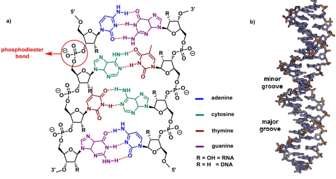
Nucleotides are the monomers of the polymeric macromolecules called nucleic acids. Each nucleotide is composed of three parts: the sugar backbone, the nitrogenous base and a phosphate residue. The sugar present in natural nucleic acids is a pentose, which depending on the substitution at the position 2', can be D-ribose (-CH-OH) or D-2-deoxyribose (-CH2-) (a in Figure 1). In DNA the deoxyribose is the constituent sugar of the nucleic acid. In RNA, ribose is the sugar present in the nucleic acid.
In both DNA and RNA, the pentose is attached to a phosphate group which acts as a linker to form a chain that constitutes the backbone of the nucleic acid. The phosphate bond is formed between the 3'-OH of one pentose to the 5'-OH of another pentose, thus forming a phosphodiester bond. In the anomeric position of each sugar (1'), there is a nitrogenated heterocycle called the “nucleobase”, which is cis (β face) to the hydroxymethyl group at the 5' position. The nucleobases are classified as purines and pyrimidines. Purines consist of a five-membered and a six-five-membered nitrogen-containing ring, fused together; Such as adenine and guanidine. On the other hand, pyrimidines such as cytosine, thymine and uracil, have a six-membered nitrogen containing ring. Adenine, guanine and cytosine are found in both DNA and RNA. Uracil is found exclusively in RNA. 5-Methyluracil (thymine) is present only in DNA.
2
Figure 1. a) Generic nucleic acid double strand. b) Minor and major grooves of helix formed by double strand nucleic acids1
Thanks to the X-ray diffraction pattern analysis data taken in the 50’s2,3 and to chemical analysis, it was demonstrated that the number of adenines (A) is always the same as the number of thymines (T). Similarly, the number of guanines (G) is always equal to the number of cytosines (C). It was concluded that DNA was a double strand, where the nucleobases of each chain form hydrogen bonds with its other chain counterparts. Further studies on the helical structure of DNA concluded that the pairs cytosine-guanine and adenine-thymine were exclusively always present along the double strand. This complementary behavior dictates that each strand of the DNA is exactly like the complementary strand but linked in an opposite direction.2
The interactions between the different nucleobases are unique. The pair guanine-adenine forms three hydrogen bonds, while the pair cytosine-thymine only forms two hydrogen bonds. As a result, DNA with a higher percentage of G-C becomes more stable. As the hydrogen bond is a
3
non-covalent bond, it is possible to separate the double strand. This process is reversible in solution; however, it is necessary to heat the DNA. The temperature at which fifty percent of the molecules of DNA are separated in 2 single strands, is called “the melting temperature (Tm)”. The Tm value is an indicator for measuring the stability of DNA and its derivatives. Several techniques have been developed for the measurement of the Tm. One of the most common is based on measuring the absorption of UV light by the nucleobases present in DNA, which exhibit a maximal absorption at 260 nm. Complementary pairs of nucleobases interact with each other pair of nucleobases by stacking of their pi-cloud electrons and by formation of hydrogen bonds. When the DNA is unwound to form two single strands, the stacking is weakened, increasing the absorption of light in a phenomenon called hyperchromicity.
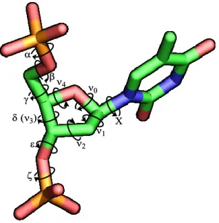
The duplex formed by two strands of DNA or two strands of RNA assumes a right-handed helix where the nucleobases are stacked in the inner part of the helix and the sugar-phosphate polymer is installed in the exterior of the helix. The RNA and DNA duplexes present a difference in the size and shape of the grooves formed by the helix (b in Figure 1). The DNA duplex forms a type B helix where the major groove is wider than the minor groove. The RNA duplex shows a more compact and stable helical structure, with a major groove smaller than in a DNA duplex. The conformation of a DNA strand is highly affected by the sugar-phosphate structural features. The most common way to describe the orientation of the sugar-phosphate backbone into the space is by the torsion angles which are defined by the rotation about each chemical bond (Figure 2). The standard nomenclature takes as a departing point the bond formed between the 5’-oxygen and the corresponding phosphorus, defined by the Greek letter α, followed by the bond O5’-C5’ which is defined as β, and continues through the nucleoside backbone until the bond formed between the oxygen at the 3’-oxygen and the phosphorus of the next nucleoside is
4
defined as ζ. Torsion angle Χ describes the rotation around the anomeric carbon-nitrogen bond which in turn describes the position of the nucleobase. The nucleobases can be positioned over the furanose core in a syn-conformation, or in an anti-conformation with the nucleobases pointing away from the sugar as in Figure 2.
Figure 2. Torsion angles in nucleic acids4
The angles defining the furanose ring are named from ν0 to ν4 and they help to describe the distribution of the atoms in the furanose core, being commonly known as the sugar puckering. As the furanose ring is a cyclic system, it is not possible to rotate freely, so a pseudorotation process takes place which helps to minimize the steric congestion of eclipsed groups in the sugar In nucleotides, the furanose ring can be classified into one of the possible conformations which are represented in the pseudorotation circle in Figure 3. Each conformation can be classified into the circle where E (envelop) and T (twist) nomenclatures are used to describe the furanose puckering. The superscript value describes which atom is in an endo conformation and the subscript value describes the atom located in an exo conformation.
5 Figure 3. Sugar pucker of the furanose ring4
When one of the atoms of the furanose ring is pointing above the reference plane in the same direction as the nucleobase, it is said that it is in an endo conformation as shown in Figure 3; if such atom is pointing in the opposite direction, it is in the exo conformation.
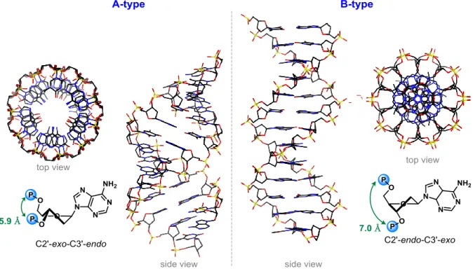
There are two main conformers with the lower energy profiles, C2’-endo-C3’-exo (also known as S-type) and C2’-exo-C3’-endo (N-type).5 The former one is encountered in DNA and the latter in RNA. The energy required to change between the different possible conformers is dependent on the substituents on the furanose sugar core but in most of the cases is ~2 KJ/mol. 6-9
The predominant sugar puckering in natural nucleic acids (N and S-type) affects the global helicity of the nucleic acid. For deoxyribose, as in DNA, the S-type conformation is lower in energy (≈ 0.6 Kcal/mol) over the N-type giving to DNA a so-called B-form, which is the most
6
common form in vivo. In the B-form, the predominant S-type conformation keeps adjacent phosphates at a distance of ~7 Å (Figure 4) and the base pairs are effectively perpendicular to and centered over the helical axis. In the ribose ring, the N-type conformation is predominant, giving DNA-RNA and RNA-RNA duplexes an A-type helix. The C2’-exo conformation forces adjacent phosphates to become close (~5.9 Å) forming a denser helix with a displacement of the bases away from the central helix.
Figure 4. Structures of A-type and B-type double stranded nucleic acids
1.2 DNA transcription
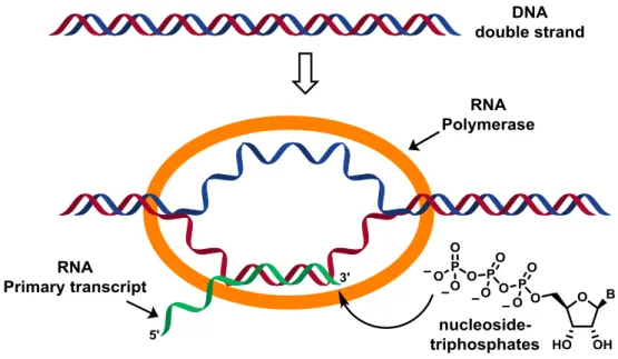
Transcription is the process of copying the genetic information found in the double-stranded DNA into a single-stranded RNA, which is complementary to the segment of DNA from which it was copied. In order to begin the transcription process, it is necessary to have the four
5'-7
triphosphate nucleosides (C, U, G, A), magnesium (II) and RNA polymerase (RNAp). RNAp is an enzymatic complex formed by several enzyme subunits which recognizes specific sequences in DNA (promoters) to start the transcription process. When the RNAp binds to the double-stranded DNA, it is necessary to unwrap a sequence consisting of about 14 base pairs. The gap formed will move through the DNA, as RNA opens and closes the double strand on its way through the DNA (Figure 5). RNA synthesis starts inside this transcription bubble in which each new nucleoside that joins the chain is chosen following the complementary base on DNA. The process of RNA synthesis continues until the DNA encodes a sequence (transcriptor terminator) and forms a loop in the RNA that helps to dissociate the RNAp transcription complex, and releases the new RNA strand (primary transcript).
8
1.3 RNA processing
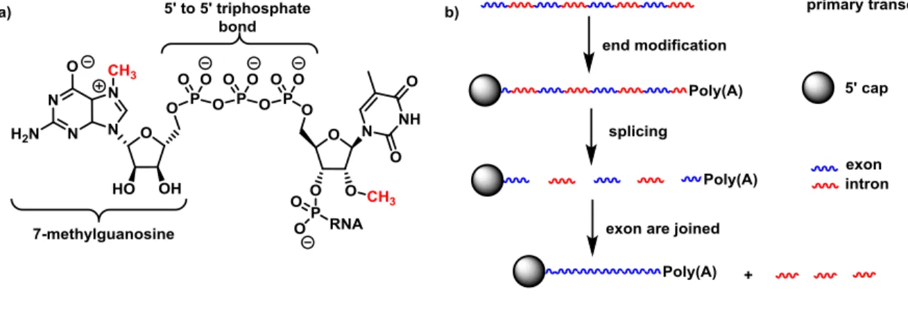
In eukaryotic cells, the RNA obtained from the transcription process (primary transcript) goes through some changes in its structure in order to be able to transmit the stored genetic information. The first transformation of the primary transcript is addressed by imparting stability of the newly synthesized RNA strand towards nucleases, which are enzymes that cleave the phosphodiester bond. Such transformation is achieved by capping at the 5' position with 7-methylguanosine linked through a 5'-5' triphosphate bond (a in Figure 6).11 Other modifications include the methylation of the 2'-OH of the first and second ribose units in the strand, and the addition of a long tail (between 100 and 200) of adenine ribonucleotides known as poly(A) at the 3' end of the RNA.12
The next step is the splicing of the stabilized RNA when some sequences, known as introns, are deleted (b in Figure 6). The number of sequences removed as introns depends on each species, but at the end all the remaining parts that codify for a gene (exons) are joined, forming the final mRNA responsible for translation of the genetic message.
9
1.4 Protein synthesis – translation
The synthesis of proteins (translation) is carried out in the rough endoplasmic reticulum (RER) and performed within the ribosome, which is a large macromolecular complex made of both RNA and protein.13 The amino acids that will form part of the newly synthesized protein are carried to the ribosome by transfer RNA (tRNA), a kind of RNA that is bonded to an amino acid and is able to recognize sequences of mRNA. Information is read from the sequences of nucleosides in groups of three (triads), which are called codons. Each codon is responsible for codifying a specific amino acid. One amino acid can be codified by different codons. Translation begins when the first molecule of tRNA interacts with the mRNA detecting the initiation codon and binding in the presence of the ribosome (Figure 7). With the ribosome reading the information and moving through the mRNA from the 5' to the 3' end, a second molecule of tRNA recognizes the next codon and brings the corresponding amino acid to the complex. The two amino acids react by forming a peptide bond, while the ribosome moves to the next codon, adding more amino acids and releasing the now empty tRNA. The rate of protein synthesis in eukaryotes can reach up to 14 to 18 amino acids/second per ribosome14. The process continues until a codon is reached which does not codify for an amino acid (stop codon). The last codon is recognized by a protein called “releasing factor”, which helps the synthetized protein to be expelled from the complex completing the translation process.15
10 Figure 7. Translation process10
11
2 Chapter Two – Antisense technology
2.1 History
In 1978, Stephenson and Zamecnik,16 working with chick embryo fibroblast cells infected with Rous sarcoma virus (RVS), achieved the inhibition of the production of the virus with a synthesized oligodeoxynucleotide. In the primary structure of the RVS, there were identical sequences of nucleotides next to the 5' cap and poly(A) 3' end. A 21-nucleoside sequence was the target of the study. A synthetic ribonucleoside was prepared complementary to a segment of the target sequence. The 13 nucleotide sequence d(AATGGTAAAATGG) bound to the target sequence, blocking the normal functioning of the virus by competitive hybridization.
Together with many subsequent studies, the idea of use RNA as a target was realized. The potential to treat different diseases was envisioned to be possible through malfunction of RNA, or blocking the translation process in the cell by introducing a complementary synthetic oligonucleotide that could hybridize with the mRNA. The complementary nucleotide chain is called the “antisense” chain. Binding to the target mRNA is a common mechanism of action for all antisense oligonucleotides (ASO).
2.2 Molecular mechanism of action
The multiple types of RNA and their multiple roles in the cell have become a growing area of interest with therapeutic potential in recent years. Historically important RNA include, mRNA, the small nuclear RNA (smRNA)17 that plays key roles in the splicing process, and the less
12
studied microRNA18 which seems to act as a natural gene regulator. Thus, the RNA molecule in general is a very attractive target for the development of new kinds of therapies for the treatment of diverse diseases, basically by preventing harmful proteins from being produced at the genetic level.
2.2.1 RNA cleavage
The destruction of the RNA chain by a family of enzymes called RNase H (ribonuclease H) is among direct approaches to inactivate its biological function. The RNase H enzymes found in mammals recognize the RNA-DNA duplex, hydrolyze the phosphodiester bond in the RNA and release the intact DNA strand.
Figure 8. RNase H active site19.The active site RNase in complex with the RNA/DNA hybrid. Active-site residues are shown in green; the RNA strand in pink, orange, and red; and the magnesium ions as yellow spheres. The water molecule (nucleophile) positioned to attack the scissile phosphate bond is indicated. Metal-ion coordination is shown as dashed yellow lines.
13
The cleavage necessitates enzyme recognition20 of both the RNA and DNA strands. The duplex fits in two grooves at the enzyme structure. The 2’-OH of five consecutive nucleotides of the RNA strand interacts with one groove in the active site of the enzyme.19 Different active–site residues of the enzyme, such as carboxylate and amino groups, form hydrogen bonds with the 3’ and 5’ sides of the phosphate bond to be cleaved (Figure 8). In the active site, two Mg2+ ions are chelated by four carboxylate groups and two molecules of water. One of the molecules of water attacks the phosphate linkage, assisted by one oxygen of the phosphate group which orients the attack and serves as a general base for deprotonation. After the attack, another metallic ion stabilizes the 3’-OH group from the cleaved phosphodiester bond.
The use of a designed ASO that binds a RNA target in order to destroy and therefore suppress the cellular function has been the subject of hundreds of studies.21,22 Depending on the structure of the nucleotides that form the ASO, the duplex ASO-RNA can facilitate RNase H activity resulting in the hydrolysis of the RNA target. Modifications focussing on increasing the metabolic stability and increasing the affinity towards the target RNA, have often resulted in a loss of RNase H activity.23 As an alternative, ASO hybrids have been developed composed of sections that support the RNase H activity and provide affinity to the complementary RNA (quod vide).
2.2.2 Alternative mechanisms involving non-RNA cleavage
Antisense oligonucleotides (ASOs) can be designed to work as competitive antagonists that bind to specific sequences in RNA and interrupt normal interactions with proteins, enzymes or other factors in the cell. One process that can be affected is maturation of the RNA primary transcript,
14
a fundamental step to obtain different functional RNAs such as mRNA and tRNA. Inhibition of 5’ capping on RNA may be performed by designing an ASO capable of binding to the 3’ end of the primary transcript. Another fundamental step in RNA processing is splicing. An ASO that binds to a sequence in RNA to be spliced, could create a barrier and supress splicing. Promising approaches of interrupting or modifying the RNA splicing process have been reported. For example, the use of ASOs by Kole24 corrected of an aberrant splicing in mutated RNA and restored the natural splicing site. ASOs designed to bind with high affinity to the 3’-splice sites in murine pre-mRNA have exhibited activity in deletion of specific exons, resulting in the inhibition of mRNA production and suppression of protein synthesis.25 In December 2016, the FDA approved Spinraza®, a medication used to treat spinal muscular atrophy (SMA). Spinal muscular atrophy is caused primarily by a genetic defect in the SMN1 gene which encodes a defective SMN protein, a protein required for the survival of motor neurons in the spinal cord. Spinraza® is an oligonucleotide that binds the pre-mRNA and promotes the inclusion of exon 7 in the final protein.26
2.3 ASO modification
Obtaining a higher resistance against the degradative action of nucleases was the first challenge in ASO design. Replacing a non-bridging oxygen atom in the phosphodiester linkage by a sulfur atom resulted in modified nucleotides27 which conferred resistance towards hydrolases. The new nucleotides received the name of phosphothioate (PS) nucleotides 2.1 (a in Figure 9). Upon RNA-PS nucleotide duplex formation, RNase H hydrolyzes the RNA strand and releases the PS nucleotide, blocking the production of the protein. Matsukura28 was the first to report that
15
oligonucleotides containing the phosphothioate modification could inhibit human immunodeficiency virus (HIV) in vitro.
Figure 9. a) Generations of ASOs. b) Example of gap design oligonucleotide with MOE
PS oligonucleotides present a similar affinity for RNA compared to natural nucleic acids (ΔTm ≈ -2°C per modification).29 They promote binding to plasma proteins, favoring the distribution
16
in the body. Overall, PS-ASOs are among the most common modifications in antisense technology, because they combine hydrolytic stability of the backbone with affinity for the complementary RNA strand.
The next generation of ASOs was designed to increase the affinity with the RNA and nuclease resistance. Methylation of the 2’-OH of the furanose gave rise to the 2’-O-methyl nucleoside (2.2 in Figure 9), which blocks the possibility of self-hydrolysis of the phosphodiester linkage by attack of the 2’-OH, and increase affinity vs. RNA (ΔTm ≈ 1°C per modification). The 2’-fluoro modification 2.3 (a in Figure 9), like the 2’-O-alkyl modifications, confers to the nucleoside a N- type conformation that shortens the distance between the 3’-5’ phosphate bonds, generating a more compact structure that assumes a type A helix conformation, which increases the affinity of the ASO to the RNA.30
A series of studies by Damha and coworkers have shown that introducing fluorine and methoxy groups at specific positions of the furanose ring, renders the puckering into an N-type conformation. Introduction of two fluorine atoms at the 2’ and 4’-position of furanose ring rendered nucleoside 2’,4’-diF-araU 2.431 into a N-type conformation, as a result of the anomeric effect between the antibonding orbital of C4’-F4’and the lone pair at oxygen of the furanose ring. The 4'-Cα-OMe-2'-F U nucleoside 2.5 also adopted an N-type conformation. When inserted into oligonucleotide sequences, 2.5 showed small changes in thermal affinity but gave high resistance towards the action of nucleases.32 Parenting compounds 2’-F,4’-OMe-araU 2.6 and 2’-OMe,4’-F-rU 2.7 presented high percentages in the N-type conformation as measured by 1H NMR spectrospcopy.33 It was suggested that the predominant effect responsible for N-type conformation in nucleoside 2.8 is the σC3’H3’ σ*C4’OMe hyperconjugation effect. Introduction
17
of 2.6 or 2.7 into DNA:DNA duplexes is destabilizing but it showed a slight improvement in thermal affinity when introduced into RNA:DNA duplexes.
The 2’-O-methoxyethyl (MOE) nucleosides 2.8 together with PS backbone modifications represent the most studied second generation of ASOs.34-37 The methoxyethyl group assumes a rigid conformation due to the gauche effect between the two oxygen atoms giving the furanose ring a N-type conformation. The MOE group is also suggested to form hydrogen bonds with water to protect the phosphodiester bond and increase resistance against nucleases38 by increasing hydration in the minor groove reducing interaction of the double strand with nucleases. In an attempt to enhance the RNA affinity and metabolic stability, scientists at Ionis Pharmaceuticals prepared oligonucleotides containing the 2’-O-(2S-methoxypropyl) modification (2S-MOP).39 The 2S-MOP modification (2.9 in Figure 9) was designed to further reinforce the gauche conformation and increase the hydrophobicity in the minor groove, however, the 2S-MOP modification did not improve the activity compared to the MOE ASOs. Such second generation of modifications resulted in a decrease in the toxicity associated with the first generation of ASOs,40 however, they presented some difficulties in the design of new oligonucleotides, due to incompatibility with the action of the RNase and the RNA cleavage mechanism. The former limitation was overcome by the use of a “gap” design23 (b in Figure 9), which combines 2’-O-alkyl nucleosides at the 3’ and 5’ end of the chain, with a central DNA region of PS nucleosides. In the gap design, the modified nucleosides located at the ends of the ASO increase the affinity for the RNA, and the PS internal chain supports the action of the RNase H for the cleavage of the target RNA. The success of the second generation of ASOs was shown by the approval in January 2013 of the gap designed oligonucleotide KYNAMRO™ by the FDA as a drug for the treatment of homozygous familial hypercholesterolemia.41
18
The third generation of ASOs contain different modifications on the carbon backbone. One of the more drastic changes was found in the peptide nucleic acid (PNA) 2.10 (Figure 9). Designed with a pseudopeptide backbone, consisting of N-(2-aminoethyl) glycine, PNA shows great affinity to RNA and DNA. Due to its pseudopeptidic structure, PNA is resistant to degradation by nucleases and proteases; however, it is not compatible with the RNase H mechanism of action. Such stability makes PNA a good candidate for use in the disruption of RNA processing by translation inhibition42 and splicing modulation mechanisms.43
2.4 Constrained nucleosides
Over the last 20 years, three distinct strategies have used covalent bonds to incorporate a conformational constraint into different nucleic acids. The first strategy is based on restriction of the furanose ring mobility by a bridge between C2’ and C4’, that locks the ring into an N-type conformation as exemplified in LNA and α-L-LNA. Locked nucleic acid (LNA)44 2.7 is a restricted analog of 2’-OMe nucleoside, in which the 2’ hydroxyl group methyl is tethered to the 4’-carbon (Figure 10). The resulting bicyclic structure confers a N-type conformation that presents an improved stacking between the nucleobases compared to that of the RNA duplex.45 ASO strands containing LNA modified nucleosides present a pronounced increase in duplex stability relative to the DNA-RNA duplex.46 The studies modifying the position of LNA in gap designed oligonucleotides concluded that a gap of at least 8 DNA nucleosides is necessary to induce efficient RNase H cleavage.47 Further studies have shown the successful use of a LNA-gap design.48
19
Figure 10. Constraint of torsion angles ɣ and δ in selected nucleosides.
Reported by Wengel, the hybridization properties of the eight isomers of LNA were investigated.49 It was found that α-L-LNA (2.8 in Figure 10) with the 2’,4’-bridge on the same side as the nucleobases and the 3’-hydroxy group inverted, presented comparable affinity for RNA as does LNA. Further studies showed that α-L-LNA assumed an S-type conformation when introduced into an ASO paired with RNA which produced a duplex with an intermediate character between A and B-type helix.50,51 As part of a continuing effort at Ionis Pharmaceuticals, a 6’-methylated analog of LNA was synthesised. The resulting constrained analog (S)-cEt-LNA (2.9 in Figure 10) increased the stability of duplex compared to those containing LNA.52 Exposure of a 10-mer poly T DNA oligomer with two (S)-cEt-LNA modifications at the 3’ end to snake venom phosphodiesterase, revealed that the LNA oligomer
20
was completely hydrolyzed in 1290 min while the (S)-cEt-LNA modified chains were >70% intact.53
The second strategy comprises the restriction of the torsion angles ɣ and δ and has been widely studied since reports by Leumann of the [3.3.0]bicyclo-DNA (bc-DNA) 2.10.54-56 In bc-DNA (Figure 10) the carbocyclic ring formed between the 3’and the 5’-positions of the furanose ring forced torsion angle ɣ into non-natural positions57 resulting in a poor affinity toward complementary DNA and RNA. A further constraint to bc-DNA was applied by introducing a cyclopropyl ring, forming tricyclo-DNA58 (tc-DNA) 2.11 which showed an in increased RNA affinity. Tc-DNA Duplex comprised exclusively of poly-A and poly-T strands, presented a highly stable self-pairing.59,60 X-ray crystallographic analysis of tc-DNA containing duplexes61 showed a change in torsion angle ɣ and β compared to bc-DNA forcing the furanose into a
C2’-exo conformation.
The six-membered ring constrained nucleoside [4.3.0]bicyclo-DNA 2.12 was synthesized and introduced into ASOs presenting similar thermal stability compared to natural DNA and RNA.62 With a similar strategy, scientists at Ionis Pharmaceuticals, in collaboration with the Hanessian group synthesized the bicyclic constrained nucleosides: cis-2.13 and trans-bicyclo-DNA 2.14, isomers of the previously synthesized bc4.3-DNA (Figure 10).63 Only incorporation of the monomer 2.13 into oligonucleotide sequences was possible, with the resulting ASOs presenting a slightly destabilizing effect upon duplex formation.
Combining the two approaches, the Hanessian group used a dual conformational restriction strategy featuring tricyclic nucleosides α-L-TriNA 164 2.15 and α-L-TriNA 265 2.16. In α-L-TriNA 1, a spiro-annulation around carbon C4’ restricts the torsion angle ɣ and locks the
21
furanose ring into a N-type conformation. In the α-L-TriNA 2, the N-type conformation is achieved as a result of a bridge between C2’ and C4’ and a fused 6-membered ring. Restriction of the torsion angles ɣ and δ is almost identical in both tricyclic nucleosides. α-L-TriNA 1 showed unprecedented duplex stabilizing properties versus DNA and RNA complements (8.3 °C/mod for RNA). In the case of oligonucleotide sequences containing α-L-TriNA 2, only a slight stabilizing effect was measured against RNA.
The third strategy of constraint was developed by Escudier, by restricting backbone torsion angles α and β (Figure 11). The dioxaphosphorinane-constrained nucleic acid66,67 (α,β-D-CNA in Figure 11) in which torsion angle α is found in a in a non-canonical value (+sc), promote the bend in a single-stranded DNA, preorganizing it into a looplike structure. Such a structure was used to act as a chain terminator for proof reading DNA polymerases.68 The synthesis of both P-isomers of phostone-constrained nucleic acids (P-CNA in Figure 11)69 features the formation of a six-membered ring cycle locked in a chair conformation which provides a restrain on the α and β torsion angles giving them atypical values. The dinucleotide containing LNA/ α,β-D-CAN (Figure 11) with the α,β-D-CAN in a (R)-C5’,RP configuration, forced the dimer into a A-type duplex as determined by NMR studies.70
22
3 Chapter Three
Synthesis of TriNA 2
23
3.1 Design of TriNA 2
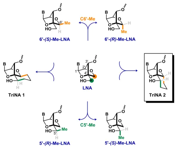
Starting from LNA as a template, a further restriction was applied by scientists at Ionis Pharmaceuticals, who introduced a methyl group into the C6’ position of LNA (Figure 12), to reduce rotation of the C4’-C5’ bond.52,53 Two isomers were obtained: (S)-Me-LNA and 6’-(R)-Me-LNA (Figure 12). After introduction into different sequences of oligonucleotides, the thermal stability values obtained for the ASOs were comparable to the values presented by LNA containing uracil as nucleobase (ΔTm = 4.5 °C/mod.). Replacement of uracil by thymine improved affinity of the (S)-isomer.
24
In a subsequent study, the effect of methyl group introduction at the C5’ position of LNA was studied.71,72 Oligonucleotides with a single modification containing 5’-(S)-Me-LNA (Figure 12) presented a similar thermal stability vs. RNA (ΔTm = 4.5 °C/mod). In contrast, 5’-(R)-Me-LNA completely reduced the stabilizing effect on duplex formation vs. both DNA and RNA.
These studies showed that introduction of methyl groups into the C5’ and C6’ positions were well tolerated, and a non-detrimental effects on thermal affinity was observed depending on the position and special orientation of the methyl group. To expand the constraining strategy, two dual-locked analogs were designed: TriNA 1 and TriNA 2 (Tricyclic Nucleic Acid) (Figure 12). Merging two of the methyl modifications into a single nucleoside with a new carbocyclic core, TriNA 1 was synthesized by Dr. Robert Giacometti in the Hanessian group and will not be discussed in detail in the present document.73
25
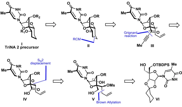
3.2 Retrosynthetic analysis of TriNA 2
Figure 13. Retrosynthetic analysis for the synthesis of TriNA 2.
The core of the TriNA 2 nucleoside I, is formed by locking the furanose ring of a LNA type bicyclic structure by a 6-membered ring. The synthesis of orthogonally protected nucleoside TriNA 2 precursor I was envisaged to originate from the selective reduction of enone II. Enone II would be synthesized by a ring-closing metathesis reaction between two appropriate partners at the C5’ and C6’ positions of bicycle III. Propargyl ketone III could be prepared by the addition of a Grignard reagent onto a Weinreb amide derived from acid IV. An intramolecular SN2 displacement involving the C2’-alcohol onto the 6’ carbon containing an appropriate leaving group would result in the formation of the constrained bicyclic nucleoside IV. At this point the configuration of the carbon bearing leaving group at 6’ would play an essential role in the synthetic strategy with a planned inversion. The formation of homoallylic mesylate V, could be achieved by the stereocontrolled addition of an allyl group to appropriate aldehyde at the C6’
26
position. Finally, we chose as a starting point an α-OTBDPS protected bis-hydroxymethyl thymidine VI which could be obtained in several steps from commercially available thymidine.
3.3 Synthesis of TriNA 2
The synthesis of TriNA 2 started from the previously prepared nucleoside 3.1, which had been used in the synthesis of (S)-cEt-BNA nucleoside.74 Accordingly, the required homoallylic alcohol (Scheme 1), was introduced through a two-step sequence comprising oxidation with Dess–Martin periodinane,75 followed by a Brown allylation reaction with (−)-Ipc
2B(allyl).76 The required (S)-alcohol 3.2 was obtained as a single diastereomer, with the selectivity arising from the attack of the allyl group in a chair-like transition stateǂ onto the aldehyde. In the six-membered transition stateǂ, the facial selectivity is determined by minimization of the steric interactions between the aldehyde and the methyl groups in the Ipc ligand (Scheme 1).
27
With alcohol 3.2 in hand, the corresponding methanesulfonate was synthesized and the cyclohexylidene acetal moiety was removed under acidic conditions to furnish diol 3.3 (Scheme 2).
Scheme 2. Synthesis of bicyclic nucleoside 3.6.
The formation of bicyclic core 3.6 from diol 3.3 required the selective attack of the 2’ instead of 3’ hydroxyl group. In principle, the formation of the tetrahydrofuran ring is favored over the oxetane ring; however, previous experience with the cyclization of similar substrates74 suggested protection of the C3'–OH of 3.3. The desired C3'-O-naphthylmethyl ether was successfully installed using a two-step sequence that involved protection of the diol moiety as a diastereoisomeric mixture of naphthylidene acetals 3.4, followed by selective deprotection of
28
the C2'-hydroxy group in the presence of TiCl4/NaBH3CN77 to furnish alcohol 3.5.* Base-promoted intramolecular displacement of the mesylate provided access to the bicyclic framework of 3.6 in 45% yield over three steps.
Figure 14. Regioselective reductive cleavage of acetal 3.4.
In the regioselective reductive cleavage of the naphthylidene acetal (Figure 14), titanium chloride (IV) acted as a Lewis acid that coordinates the less hindered oxygen of the acetal. Subsequent reduction of the generated oxocarbenium ion by sodium borohydride gave the 3’-Nap ether 3.5.
* Heating diol 3.3 with 2-naphthaldehyde in a Dean-Stark apparatus gave a low conversion to acetal 3.4. Instead,
29 Scheme 3. Synthesis of Weinreb amide 3.8
The N3-position of the nucleobase was protected as an N-benzyloxymethyl derivate to avoid potential side-reactions in the next steps (Scheme 3). Subsequent cleavage of the TBDPS group with TBAF provided alcohol 3.7 in 75% over two steps. As part of our strategy to extend the carbon chain at position C5’, we attempted to oxidize alcohol 3.7 into the corresponding aldehyde. To this end, several different side-products were obtained when the reaction was carried out in the presence of Dess–Martin periodinane, Swern conditions, or with a mixture of TEMPO and PIDA,78 a method developed for the synthesis of nucleoside-5’-carboxylic acids. To overcome the difficulties isolating the aldehyde, we opted to directly oxidize the primary alcohol to a carboxylic acid, before converting it to Weinreb amide 3.8. The oxidation was successfully accomplished under Corey-Schmidt conditions (PDC in DMF).79 HATU mediated coupling of the carboxylic acid with N,O-dimethylhydroxylamine hydrochloride afforded Weinreb amide 3.8 in good yield.
30 Scheme 4. Synthesis of TriNA 2.
Addition of 1-propynylmagnesium bromide to Weinreb amide 3.8 furnished propargyl ketone 3.9, which we hoped to selectively reduce to the enone. Since ring-closing metathesis reaction has several precedents in nucleoside synthesis,80-84 we decided to form the tricyclic core of TriNA 2 (Scheme 4) using such a strategy. Partial hydrogenation of 3.9 using Lindlar’s catalyst85,86 in the presence of 1,10-phenanthroline as a bidentate ligand minimized over-reduction.87 The α,ß-unsaturated ketone was converted to enone 3.10 using Grubbs’ II
31
generation catalyst, subsequent reduction of the enone under Luche conditions88 favoured formation of the desired (S)-alcohol 3.11 over the corresponding epimer, with a ratio of 2.1:1. Use of (R)-CBS gave the desired (S)-alcohol albeit in low yields (≈10%). Formation of the levulinate ester and subsequent olefin hydrogenation with concomitant Nap hydrogenolysis provided alcohol 3.12, which crystallized. Definitive evidence for the structure of TriNA 2 was obtained using X-ray crystallographic analysis. Transformation of 3.12 into the corresponding phosphoramidite and introduction into oligonucleotides sequences was performed at Ionis Pharmaceuticals.
3.4 Duplex thermal stability measurements
The duplex stabilizing properties of TriNA 1 and TriNA 2 versus complementary RNA were measured by scientists at Ionis Pharmaceuticals using a previously described oligonucleotide sequence.62 Modified nucleotides were incorporated into DNA oligonucleotides at four different locations to provide a position and sequence context for the Tm studies (Table 1). Moreover, the duplex–stabilizing properties of LNA, (S)-cEt, and (R)-cEt modified oligonucleotides were measured as additional controls. Relative to an unmodified DNA control, incorporation of TriNA 1 produced an average increase in duplex thermal stability of +4.4 °C/mod. TriNA 2 was found to be more stabilizing, producing an average increase in Tm of +6.2 °C/mod. By comparison, LNA, S-cEt, and R-cEt produced average Tm increases of +6 °C/mod. Consequently, TriNA 1, which has the 5′-(R),6′-(S) configuration stabilized less the duplex than TriNA 2 containing the 5′-(S),6′-(R) configuration.
32 Table I. Duplex thermal stability of TriNA nucleotides.
Sequence (5’ to 3’)
ΔT/mod (°C)
LNA S-cEt R-cEt TriNA 1 TriNA 2
GGATGTTCTCGA 6.3 6.3 6.6 5.3 6.8
GGATGTTCTCGA 5.6 5.1 5.6 4.0 6.1
GGATGTTCTCGA 7.0 6.9 7.0 4.5 6.4
GGATGTTCTCGA 5.0 4.7 4.6 3.6 5.5
Average ΔTm 6.0 5.8 6.0 4.4 6.2
To further understand the origins of the differential duplex stabilizing properties of TriNA 1 and TriNA 2, a structural model was developed based on an overlap with the crystal structure of a
S-cEt-modified A-form DNA duplex (Figure 15).89 Both TriNA analogues were well accommodated within the duplex, with the six-membered carbocyclic rings projecting into the minor groove for TriNA 1 and towards the edge of the major groove for TriNA 2. Visual analysis of the structures suggested that the (R)-5′-methylene group of the carbocyclic ring in TriNA 1 may experience a tight contact (2.7 Å) with one of the non-bridging oxygen atoms of the 5′-phosphodiester linkage. In contrast, the analogous distance for TriNA 2 is 3.2 Å, and the tightest contact (2.9 Å) is likely between the (S)-5′-methylene group and the oxygen atom of the 3′-adjacent nucleotide. This suggested that the incorporation of TriNA 1 into the duplex might produce subtle changes around torsion angles α and/or β, or enhance conformational mobility of the phosphodiester backbone, in order to relieve this tight spacing, and consequently lead to a smaller enhancement in duplex stability.
33
Figure 15. Structural models of tricyclic nucleosides overlaid on oligonucleotide duplexes containing the corresponding (S)-cEt bicyclic modification.
3.5 Crystallographic and conformational analysis
X-ray quality crystals analysis of ester 3.12 confirmed the absolute configuration of the molecule and provided parameters for the furanose sugar puckering and pseudorotation angle (P).5 In nucleotides, the pseudorotation angle value can be calculated from the values for the torsion angles of the furanose using the formula in Figure 16. Depending on the value obtained, the furanose can be classified into one of the possible conformations which are represented in the pseudorotation circle in Figure 16. For nucleoside 3.12, the value of P was 17°, which corresponded to a 2E C2-endo conformation. For comparison, LNA had a P value of 17°,90 confirming that TriNA 2 is constrained into a N-type conformation. The pucker amplitude (νmax) is defined as the amount by which the fifth atom is displaced from the plane defined by the remaining four atoms. In LNA the pucker amplitude is ≈60°91 and the measured value for nucleoside 3.12 using equation shown in Figure 16 was 59°.
34 Figure 16. Pseudorotation pathway of the furanose ring
The dual constraint affected the torsion angle γ which has an average value92 of 54° for double-stranded RNA-DNA duplexes. The value obtained from the X-ray crystallographic analysis for nucleoside 3.12 was 59.1°, and that obtained for LNA93 was ≈50°, similar to the canonical value.
3.6 Conclusions
When introduced into different positions of oligonucleotides constructs, TriNA 2 produced an average increase in the Tm value of +6.2 °C/mod, being more stabilizing than LNA and (S)-cEt-LNA.
The synthesis of compound 3.12 was successfully achieved in 17 synthetic steps and its absolute configuration was confirmed by X-ray analysis.
35
4 Chapter Four
Synthesis of an azabicyclic nucleoside
36
4.1 Previous syntheses of azabicyclic nucleosides
In 1999, Wang94 reported the synthesis of nucleoside D using azidothymidine (AZT) as starting point (Scheme 5). The synthesis took advantage the preinstalled azido functionality on AZT to obtain the protected 3’-amino thymidine A, which was oxidized under Moffatt conditions95 and reacted in a Reformatsky reaction.96 to afford a mixture of (R) and (S) isomers at 5’-OH. After O-tritylation and ester reduction, diols were separated and the (R)-alcohol C was tosylated. Intramolecular cyclization took place after removal of the benzyl carbamate group from the 3’nitrogen to give bicyclic nucleoside D.
Scheme 5. The Wang synthesis of nucleoside D
In a later publication,97 nucleoside D was introduced at two positions of a oligonucleotide, the duplex forming affinity of which was decreased against both DNA and RNA. No further exploration of the effect of the insertion of nucleoside D for other positions of oligonucleotide sequences was reported.
A related N-bicyclic nucleoside K was synthesized by Hanessian and coworkers as a ring-modified analog of malayamycin A (Scheme 6).98 The known olefin E99 was transformed into
37
alcohol F by oxidative cleavage and reduction of the aldehyde intermediate. The primary alcohol in F was mesylated and transformed to the corresponding azide. Subsequent hydrolysis of the 5,6-isopropylidene acetal gave diol G which was cleaved with sodium periodate. The resulting aldehyde was reduced to the alcohol which was mesylated to give H. Upon hydrogenation, cyclization took place to give piperidine I after protection of the nitrogen with a N-Bus100
(N-tert-butylsulfonyl) group. Acid mediated thioglycoside formation with thiophenol was followed
by protection as a pivaloyl ester. Treatment of J with a mixture of NIS/TfOH101 as source of iodonium ion promoted a stereoselective installation of the cytosine nucleobase. After a series of deprotections and introduction of the urea group, nucleoside K was obtained, but unlike malayamycin A did not exhibit any fungicidal activity.
38
4.2 Design of an azabicyclic nucleoside
The ability to restrict rotation around torsion angles ɣ, δ and ε, while maintaining canonical bond-geometries and sugar pucker, presents challenges in the design and synthesis of novel nucleoside analogs. Scientists at Ionis Pharmaceuticals proposed an azabicyclic nucleoside I in which the orientation of the 3’-phosphate simulated the A-form of RNA (Figure 17). Restriction of rotation around the torsion angle was achieved by forming a piperidine ring with an appended equatorial phosphate group. Previously used in a related compound (Scheme 5), the strategy was amended by incorporation of a 2’-OMe to provide an extra degree of constraint to assume the RNA-like C3’-endo sugar pucker.
Figure 17. Design rationale of the azabicyclic nucleoside
The introduction of the azabicyclic nucleoside into an ASO and measurement of the thermal affinity in complexation with RNA and DNA strands would provide information about the relationship between the structure of a modified nucleoside and its capacity to hybridize with complementary nucleic acids.
39
4.3 Retrosynthetic analysis based on a nucleoside
After a general analysis of the structure of the intended nucleoside I, the piperidine moiety was envisioned to arise from an intramolecular aza-Wittig reaction (Figure 18). The required partners for such a reaction could come from an iminophosphorane formed between the 3’-azido group in aldehyde II. Aldehyde II could arise from a diastereoselective allylation of aldehyde III to the (R)-homoallylic alcohol followed by oxidative cleavage. The secondary alcohol at the 5’-position would have the (R)-configuration. Core unit III would originate from uridine containing the 3’-azide with an α-orientation. Introduction of a nitrogen at the 3’-position of the furanose ring has usually required lengthy steps.102 A well-known strategy for the formation of azide III involves the regioselective opening of 2’,3’-epoxide IV by an inorganic azide. The required epoxide IV could be obtained by a multistep manipulation of a protected uridine.
Figure 18. Retrosynthetic analysis of azabicyclic nucleoside I
4.4 Synthesis based on a nucleoside precursor
Starting with uridine 4.1, the 2’,3’-epoxide was prepared by a reported sequence103-105 in which tritylation of the 5’-alcohol was followed by formation of 2’,3’-bis-mesylated 4.2 (Scheme 7). Treatment of the latter with aqueous NaOH triggered formation of the 2’,2-anhydro bridge as in
40
4.2a which underwent rapid hydrolysis to reveal alkoxide 4.2b followed by formation of epoxide 4.3.
Scheme 7. Synthesis of epoxide 4.3.
Treatment of 4.3 with NaN3 gave azide 4.4 as the major regioisomer (3:1 ratio) as a result of epoxide opening at the 3’-position (Scheme 8).106,107 The ring opening step is believed to proceed by activation of the epoxide in a SN2 like transition state where excess azide salt increases the ionic strength of the solution.102 Inversion of configuration at the 2’-OH was planned by displacement of a leaving group with an O-nucleophile, however, attempts using the corresponding triflate led to decomposition. Imidazylate (imidazole-1-sulfonate),108,109 a leaving group with similar nucleofugal properties as a triflate group developed in our group, was next investigated. Alcohol 4.4 reacted with sulfuryl chloride in the presence of imidazole, to provide chloride 4.4a in 86% yield. Treatment of alcohol 4.4 with N,N’-sulfuryldiimidazole resulted in elimination to form vinylazide 4.4b. After some experimentation, mesylation of the 2’-alcohol followed by treatment with KOBz was found to give ester 4.5 in good yield.105
41 Scheme 8. Synthesis of nucleoside 4.5
Installation of a methyl group at the 2’-OH position required the protection of the N3 position of the nucleobase by reaction with p-methoxybenzyl chloride (Scheme 9). After protection of nucleoside 4.5, the benzoate ester was cleaved under basic conditions to provide alcohol 4.6 in 92% yield over two steps. Treatment with dimethyl sulfate led to the 2’-O-methyl ether which was converted to primary alcohol 4.7 and oxidized under Swern conditions to give aldehyde 4.8 in good yield†.
42 Scheme 9. Synthesis of aldehyde 4.8.
With aldehyde 4.8 in hand, conditions were screened to favor formation of the (R)-homoallylic alcohol 4.9. Lewis acid mediated allylation with allyltrimethyltin gave different ratios of the desired isomer, with variable selectivities (Table II). Initial attempts favored the undesired (S)-isomer 4.9a, or gave a 1:1 mixture of alcohols. Employing MgBr2·Et2O led to a mixture of alcohols slightly enriched in the desired (R)-alcohol. Using the strained silacycle developed by Leighton,110,111 an almost equal mixture of alcohols was obtained. Gratifyingly, the use of Brown conditions,76 (+)-Ipc
2BAllyl, furnished the (R)-isomer as a single diastereomer in 90% yield after oxidative work-up. The absolute configuration of alcohol 4.9 was established by X-ray crystallographic analysis (Figure 19). The diastereoselectivity may be explained by a chair-like transition state (a in Figure 19) with the furanose ring positioned in an equatorial position to minimize a steric clash between the allyl chain and the methyl groups from the isopinocamphene auxiliary.