Cattle are a potential reservoir
of bubaline herpesvirus 1 (BuHV1)
S. S. Maidana,1,2,3F. Delgado,1L. Vagnoni,1A. Mauroy,4E. Thiry,4 S. Romera1,2,3,5
To cite: Maidana SS,et al. Cattle are a potential reservoir of bubaline herpesvirus 1 (BuHV1).Vet Rec Open 2016;3:e000162. doi:10.1136/vetreco-2015-000162
▸ Prepublication history for this paper is available online. To view these files please visit the journal online (http://dx.doi.org/10.1136/ vetreco-2015-000162). Received 21 April 2016 Revised 24 September 2016 Accepted 4 October 2016
For numbered affiliations see end of article.
Correspondence to Dr S. S. Maidana; [email protected]
ABSTRACT
In the present work, controlled experimental infection and transmission studies in domestic cattle (Bos taurus) and water buffaloes (Bubalus bubalis) were carried out to study the in vivo behaviour of bubaline herpesvirus 1 (BuHV1). Two bovine and two buffalo calves were infected with BuHV1 (20287N isolate) by intranasal aerosolisation. Two sentinel cattle did not receive the virus challenge, but were housed with infected buffaloes to evaluate horizontal transmission. All experimentally inoculated animals showed viral infection and respiratory clinical signs. BuHV1 experimentally infected calves showed intermittent viral excretion between 2 days and 18 days postinfection (dpi) with a maximum titre of excretion of 106TCID
50/
ml and moderate rhinitis between 2 dpi and 20 dpi. BuHV1 experimentally inoculated buffaloes showed mild respiratory signs, which consisted mainly of serous nasal secretions during the infection period. Sentinel calves showed mucosal specific IgG1
antibodies at seven days postcontact. Viral DNA was detected by PCR and sequencing in both buffaloes and sentinel calves, which could be associated with latency. In conclusion, this study showed the susceptibility of cattle to BuHV1 after both experimental infection and contact with infected buffaloes. These data increase the scarce knowledge on the pathogenesis in natural host and the susceptibility of cattle to BuHV1 experimental infection.
INTRODUCTION
The Herpesviridae family includes nearly 200 viruses isolated from various host species (E Thiry and others 2006). Host susceptibil-ity to herpesviruses indicates that the viruses have mainly co-evolved with their hosts, leading to a close adaptation (Davison 2002). However, ruminant α herpesviruses have been reported to cross the species barrier and adapt to other species (Julien Thiry and others 2006). Bubaline herpesvirus 1 (BuHV1) belongs to the cluster of ruminant α herpesviruses related to bovine herpesvirus 1 (BoHV1) (Julien Thiry and others 2006). The latter has only been associated with sub-clinical disease in water buffalo (Bubalus
bubalis) (St George and Philpott 1972,
Scicluna and others 2010). However,
Amoroso and others (2013) detected BuHV1 viral DNA on an aborted water buffalo fetus by means of PCR. In a previous study, the authors reported the molecular characterisa-tion of five BuHV1 field isolates obtained from asymptomatic water buffaloes of north-eastern Argentina for thefirst time (Maidana and others 2014). However, the susceptibility of cattle to BuHV1 infections as well as the ability of water buffaloes to transmit BuHV1 infections to cattle has not been studied yet.
In Argentina, water buffalo breeding repre-sents an important economic alternative to cattle breeding. This species, closely related to cattle, is mainly reared in the north-eastern part of the country, with a population of around 100,000 animals in mixed buffalo-cattle production systems (Maidana and others 2014). Considering these data, the aim of this study was to gain insights into BuHV1 experimental infections of buffaloes and cattle and into the epidemiological role of cattle in BuHV1 natural infections.
MATERIALS AND METHODS Viruses and cell culture
The BuHV1-20287N isolate used in this work was obtained from a nasal swab of buffalo (Maidana and others 2014). The virus was propagated in MDBK cells and viral stocks were produced after infection of MDBK cells at a low multiplicity of infection, as previously described (Romera and others 2014).
Experimental design of in vivo infections and sample collection
Four male calves and two female buffaloes, all aged six months, were randomly separated in three groups of two animals each: (a) buffalo test group (animals 174 and 992); (b) cattle test group (animals 216 and 224); and (c) sentinel cattle group (animals 223 and 229). Their naïve status for BuHV1, BoHV1 and BoHV5 exposure was verified
upon arrival and before experimental infection, by ELISA and seroneutralisation test (SNT), as described before (Romera and others 2014), as well as by virus iso-lation attempts from nasal samples. Before inocuiso-lation, all groups were strictly isolated from each other for two days. Animal care and experimental procedures were reviewed and approved by the Institutional Committee for Care and Use of Experimental Animals (CICUAE-CICVyA, National Institute of Agricultural Technology (INTA, Argentina), protocol No. 41/2012). Animals from both test groups were inoculated with 3 ml BuHV1 (20287N isolate, fourth passage in MDBK cells), containing a total dose of 107.5TCID50/ml by intranasal
aerosolisation (1.5 ml in each nostril). The sentinel cattle group received no virus challenge but was housed with infected buffaloes beginning at 24 hours postinocu-lation ( pi) to evaluate horizontal transmission. Animals were examined daily by a veterinarian who was not aware of the treatment received by each animal. During the next 21 days postinfection (dpi) or days postcontact (dpc), viral excretion, clinical signs such as loss of appe-tite, lesions of nasal, ocular and oral mucosa, and dis-charge from the nose or eyes, as well as rectal temperature and nervous signs, were checked. Rhinitis was scored as follows: 0=absent, 1=slightly serous, 2=severely serous, 3=seromucous, 4=mucopurulent. At 21 dpi and 20 dpc, animals were sedated with aceproma-zine (Acedan, Holliday Scott S.A., Argentina) by the intramuscular route and then euthanased by barbiturate overdose (Euthanyle, Brouwer, Argentina). Postmortem examination was performed immediately after euthan-asia and trigeminal ganglia and tonsil were collected. Pieces of approximately 100 mg of trigeminal ganglia and tonsil ganglia stored at −70°C were digested and DNA was extracted using the QIAamp DNA Mini kit (Qiagen, Tecnolab S.A., Argentina). DNA was subjected to B Glicoprotein (gB) PCR, as described before (De Carlo and others 2004), to examine the presence of viral DNA. Amplicons of PCR were purified and sequenced using BigDyeTerminator V.3.1 (Applied Biosystems) and an ABI3500xl sequencer (Applied Biosystems).
Blood samples were taken on the day of inoculation and weekly after that until 20 dpi. Serum antibodies against BuHV1 were measured by SNT as previously described, with some modifications (Romera and others 2014). Briefly, four replicates of six serial fourfold dilu-tions of each sample (1:4 to 1:1024) were mixed in 96-well plates with an equal volume of the BuHV1 refer-ence strain B6 containing 200 TCID50; this mixing led to
afinal neutralisation stage of 1:8 to 1:2048. Serum-virus mixtures were incubated for one hour at 37°C and then 100μl of the MDBK cell suspension at 200,000 cells/ml ±50,000 was added. After incubation for two to three days at 37°C, plates were read microscopically for cyto-pathogenic effects.
of the nasal cavity for 5 min and immediately dipped in 5 ml Eagle’s Minimal Essential Medium (E-MEM) con-taining 5000 IU penicillin/ml, 2500 µg streptomycin/ml and 10 µg amphotericin B/ml, as previously described (Romera and others 2014). Tampons were centrifuged and samples were stored at −80°C until used. Nasal samples were taken daily from 0 dpi to 21 dpi. Immediately after collection, nasal swabs were inoculated onto MDBK cell monolayers: 0.1 ml of nasal fluids was inoculated onto 96-well μ-titre plates and 10-fold serial dilutions were tested in 4 wells. Monolayers were inspected until cytopathic effects appeared and virus titres were calculated by the Reed and Muench method, as previously described (Romera and others 2014). IgG1
antibodies in nasal secretions from cattle were deter-mined by ELISA, as previously described (Romera and others 2014).
RESULTS
Clinical observations and BuHV1 in vivo characterisation
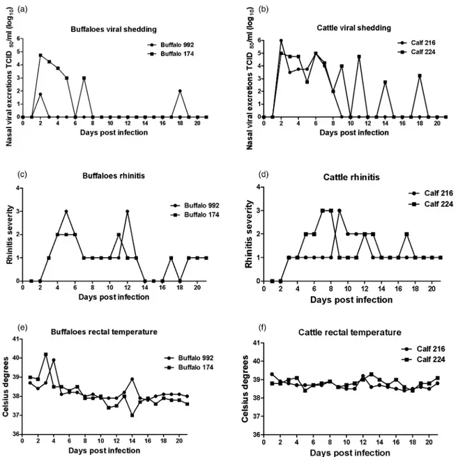
All experimentally inoculated animals showed signs of viral infection. Viral shedding, rectal temperature and rhinitis scores are shown in Fig 1. One of the buffaloes (174) shed virus continuously between days 2 and 5 pi and showed intermittent viral excretion at day 7 pi, with a maximum titre of excretion of 104.75TCID50/ml at day 2
pi. The other buffalo (992) showed viral excretion at days 2 and 18 pi, with maximum titres of 102.2 TCID50/ml at
day 18 pi. One of the clinical signs shown by infected buf-faloes was moderate rhinitis between days 2 and 20 pi. The peaks of rectal temperature were 40.2°C and 39.9°C for buffalo 174 at day 3 pi, and buffalo 992 at day 4 pi, respect-ively. In addition, infectious virus was isolated from only one calf of the sentinel group (cattle 223) at 18 dpc, with 101.75TCID50/ml viral titre. Sentinel cattle showed
moder-ate rhinitis and body temperature variations between 37.6°C and 39.5°C during the study period. Both cattle intranasally inoculated with BuHV1 (216 and 224) showed intermittent viral excretion for 16 dpi with peak titres of 106 TCID50/ml and 105 TCID50/ml at 2 dpi and 6 dpi,
respectively. These animals showed serous nasal discharge with varying degrees of severity, from mild to moderate rhinitis and body temperatures over 39°C at 12 dpi. Neither virus nor viral DNA was detected in vaginal secre-tions of buffaloes in the present work. To test the presence of latent BuHV1 DNA, PCR assays were performed using samples from trigeminal ganglia and tonsil dissected after euthanasia. Viral DNA was detected in the tonsil of buffalo 174 and in the trigeminal ganglia of sentinel calf 223 (Table 1). Sequence analysis of the amplification products obtained showed 100 per cent identity with the inoculated virus. In addition, trigeminal ganglia and tonsil were co-cultured with susceptible MDBK cells and monitored for BuHV1 replication. No cytopathic effects were observed in the cultures, indicating that the positive
Open Access
group.bmj.com
on February 1, 2018 - Published by
http://vetrecordopen.bmj.com/
FIG 1: Nasal virus shedding (a and b) rhinitis (c and d) and rectal temperature (e and f ) in two buffaloes (992 and 174) and two cattle (216 and 224) infected with bubaline herpesvirus 1 (BuHV1) (20287N strain). Virus shedding titres are expressed as log10 TCID50/ml nasal fluid. Rhinitis is scored as follows: 0, absence; 1, slightly serous; 2, severely serous; 3, seromucous; 4,
mucopurulent
TABLE 1: Humoral response after intranasal infection Animal
Humoral response Latent phase
Nasal IgG1titre (log) Seroconversion titre (log) Viral DNA in TG Viral DNA in Ton
Infected buffaloes 174 0 1.2 − + 992 0 0.9 − − Sentinel cattle 223 0.6 0 + − 229 0.6 0 − − Infected cattle 224 0.9 2.2 − − 216 0.9 2.2 − −
Detection of viral DNA in latent ganglia TG, trigeminal ganglia; Ton, tonsil ganglia
Humoral immune response
The humoral and mucosal responses specifically raised by animals after the experimental infection are shown in
Table 1. Both buffaloes seroconverted at 21 dpi, with titres of 1.2 (log10) and 0.9 (log10) for buffaloes 174 and 992, respectively.
Infected cattle seroconverted at 21 dpi, with maximum IgG titres of 2.2 (log10). In both experimentally infected cattle, IgG1antibodies were detected at 7 dpi, with titres
of 0.9 (log10), and samples taken at 14 dpi and 20 dpi were negative.
In sentinel cattle, humoral immune responses in serum at 7 dpc, 14 dpc and 20 dpc were negative. However, both animals showed mucosal IgG1 detected
only at 7 dpc, with titres of 0.6 (log10).
In summary, the data show that BuHV1 was able to replicate in experimentally infected buffaloes and calves and to be transmitted from infected buffalos to the naive calves of the sentinel group.
DISCUSSION
This study describes the in vivo behaviour of a previously identified BuHV1 isolate (20287N) from Argentina (Maidana and others 2014). The results obtained demon-strate the susceptibility of cattle to infection with BuHV1 by both experimental infection and transmission assays. Cattle experimentally infected with BuHV1 excreted virus from 2 dpi to 18 dpi. Moderate to severe rhinitis was observed at 18 dpi. Seroconversion (2.2 antibody titre) was detected at 21 dpi. The results of the present study are similar to those ofScicluna and others (2010), who, in a heterologous experimental infection assay, found that buffaloes were infected with BoHV1. Cattle suscepti-bility to BuHV1 was also shown by the transmission assay, in which viral excretion was detected in one of the two sentinel calves. Virus detection in only one animal and with low titre could be due to the gregarious behaviour towards individuals of the same species and/or to the low viral titre excreted by buffaloes. However, despite the low viral excretion of sentinel calves, viral transmission from buffaloes to cattle was corroborated by the detection of IgG1 mucosal antibodies in both animals. In addition,
viral BuHV1 DNA was detected in the trigeminal ganglia of one calf, demonstrating infection by the virus and onset of latency in the bovine species (Table 1). The pres-ence of mucosal antibodies in 223 cattle confirms contact infection. Its infection was able to generate an immune response, to make latency and reactivate at 18 dpc although the authors could not detect viral excretion during thefirst seven days after contact.
Although the viral load and behaviour that favour the efficient transmission of BuHV1 from buffaloes to cattle are still unknown, the results of the present study dem-onstrate the susceptibility of cattle to infection with BuHV1. This virus elicited mild to moderate disease
observed during the study period. Despite the previously reported high degree of genetic similarity between BuHV1 and BoHV5 (Maidana and others 2014), no nervous signs, which are characteristic of infections with the latter, were observed in cattle infected with BuHV1.
The authors reproduced BuHV1 experimental infec-tion of buffaloes by intranasal aerosolisainfec-tion. Similar to previous reports on BuHV1 pathogenesis (Montagnaro and others 2014), the respiratory signs after infection were mild, and consisted mainly of serous nasal secre-tions during the infection period. As for rectal tempera-ture, maximum values of 40.2°C were recorded at 3 dpi and 4 dpi, coinciding with the peak of viral excretion. These results are similar to those shown byMontagnaro and others (2014), although they observed a higher virus shedding period (10 days v 7 days). De Carlo and others (2004) detected viral DNA in nasal and vaginal secretions from day 5 to day 15 after immunosuppres-sion. However, in the present work, neither virus nor viral DNA was detected in vaginal secretions of buffaloes. Seroconversion was detected in buffaloes by SNT with 1 (log10) titre, coinciding with the results of Montagnaro and others (2014), who reported similar neutralising antibody titres at 15 days postchallenge.
In addition, in the present work, viral DNA of a BuHV1 was detected in infected buffalo tonsil, different from the results reported byScicluna and others (2010), who only detected the presence of viral DNA in the tri-geminal ganglion after BoHV1 infection of buffaloes. Although the authors have not detected viral DNA in latency organs (buffalo 992), it could be observed that the virus made latency before 20 dpi because the authors could detect viral DNA in the tonsil of buffalo 174. Hence the excretion in buffalo 992 at 18 dpi could also be more due to the reactivation of the virus rather than intermittency in viral excretion.
The present study and those of Montagnaro and others (2014)indicate that experimental infections with BuHV1 and BoHV1 in their natural hosts induce acute infections with pathogenicity features similar to those of other α herpesviruses. These data increase the knowl-edge on the pathogenesis of BuHV1, which has been scarcely studied, particularly regarding experimental infections in cattle. It would be interesting to know whether this event happens in mixed (cattle and buffa-loes) farms. This information will allow the rational design of sanitary measures that can decrease the risks of viral co-circulation and interspecies barrier crossings, with the consequent generation of genetic variants with unknown virulence in mixed herds.
Author affiliations
1Instituto de Virología, Centro de Investigaciones en Ciencias Veterinarias y
Agronómicas (CICVyA), Instituto de tecnología Agropecuaria (INTA), Castelar, Buenos Aires, Argentina
2Consejo Nacional de Investigaciones Científicas y Tecnológicas (CONICET),
Rivadavia 1917 (C1033AAJ), Ciudad Autónoma de Buenos Aires, Argentina
Open Access
group.bmj.com
on February 1, 2018 - Published by
http://vetrecordopen.bmj.com/
4Veterinary Virology and Animal Viral Diseases, Fundamental and Applied
Research on Animal Health center and Faculty of Veterinary Medicine, University of Liège, Liège, Belgium
5Cátedra de Inmunología, Universidad del Salvador, Pilar, Provincia de
Buenos Aires, Argentina
AcknowledgementsThe authors thank Mónica Florin-Christensen (INTA) for her help in drafting the English version of this manuscript, and Verónica Maldonado, Diego Soraire and Javier Leiva for their excellent technical assistance.
Contributors SSM and SR designed the experiments, analysed the data and drafted the manuscript together. SSM performed the experiments. FD and LV helped with postmortem examinations and histopathological analysis of animals. ET and AM participated in the interpretation of data and preparation of the manuscript draft. All authors read and approved the final manuscript. Competing interests None declared.
Provenance and peer review Not commissioned; externally peer reviewed. Data sharing statement The authors declare that the original research is available to the entire scientific community.
Open Access This is an Open Access article distributed in accordance with the Creative Commons Attribution Non Commercial (CC BY-NC 4.0) license, which permits others to distribute, remix, adapt, build upon this work non-commercially, and license their derivative works on different terms, provided the original work is properly cited and the use is non-commercial. See: http:// creativecommons.org/licenses/by-nc/4.0/
REFERENCES
Amoroso M. G., Corrado F., De Carlo E., Lucibelli M. G., Martucciello A., Guarino A., Galiero G. (2013) Bubaline herpesvirus 1 associated with
abortion in a Mediterranean water buffalo.Research in Veterinary Science94, 813–816
Davison A. J. (2002) Evolution of the herpesviruses.Veterinary Microbiology86, 69–88
De Carlo E., Re G. N., Letteriello R., Del Vecchio V., Giordanelli M. P., Magnino S., Fabbi M., Bazzocchi C., Bandi C., Galiero G. (2004) Molecular characterisation of a field strain of
bubaline herpesvirus isolated from buffaloes (Bubalus bubalis) after pharmacological reactivation.The Veterinary Record154, 171–174
Maidana S. S., Konrad J. L., Craig M. I., Zabal O., Mauroy A., Thiry E., Crudeli G., Romera S. A. (2014) First report of isolation and molecular characterization of bubaline herpesvirus 1 (BuHV1) from Argentinean water buffaloes.Archives of Virology159, 2917–2923
Montagnaro S., De Martinis C., Iovane V., Ciarcia R., Damiano S., Nizza S., De Martino L., Iovane G., Pagnini U. (2014) Bovine herpesvirus type 1 marker vaccine induces cross-protection against bubaline herpesvirus type 1 in water buffalo.Preventive Veterinary Medicine116, 56–62
Romera S. A., Puntel M., Quattrocchi V., Del Médico Zajac P., Zamorano P., Blanco Viera J., Carrillo C., Chowdhury S., Borca M. V., Sadir A. M. (2014) Protection induced by a glycoprotein E-deleted bovine herpesvirus type 1 marker strain used either as an inactivated or live attenuated vaccine in cattle.BMC Veterinary Research10, 8 Scicluna M. T., Caprioli A., Saralli G., Manna G., Barone A., Cersini A.,
Cardeti G., Condoleo R. U., Autorino G. L. (2010) Should the domestic buffalo (Bubalus bubalis) be considered in the epidemiology of Bovine Herpesvirus 1 infection?Veterinary Microbiology143, 81–88 St George T. D., Philpott M. (1972) Isolation of infectious bovine
rhinotracheitis virus from the prepuce of water buffalo bulls in Australia. Australian Veterinary Journal 48, 126
Thiry E., Muylkens B., Meurens F., Gogev S., Thiry J., Vanderplasschen A., Schynts F. (2006) Recombination in the alphaherpesvirus bovine herpesvirus 1.Veterinary Microbiology113, 171–177
Thiry J., Keuser V., Muylkens B., Meurens F., Gogev S., Vanderplasschen A., Thiry E. (2006) Ruminant alphaherpesviruses related to bovine herpesvirus 1.Veterinary Research37, 169–190
herpesvirus 1 (BuHV1)
Cattle are a potential reservoir of bubaline
Romera
S. S. Maidana, F. Delgado, L. Vagnoni, A. Mauroy, E. Thiry and S.
doi: 10.1136/vetreco-2015-000162
2016 3: Vet Rec Open
http://vetrecordopen.bmj.com/content/3/1/e000162 Updated information and services can be found at:
These include:
References
http://vetrecordopen.bmj.com/content/3/1/e000162#ref-list-1 This article cites 10 articles, 1 of which you can access for free at:
Open Access
http://creativecommons.org/licenses/by-nc/4.0/
non-commercial. See:
provided the original work is properly cited and the use is
non-commercially, and license their derivative works on different terms, permits others to distribute, remix, adapt, build upon this work
Commons Attribution Non Commercial (CC BY-NC 4.0) license, which This is an Open Access article distributed in accordance with the Creative
service
Email alerting
box at the top right corner of the online article.
Receive free email alerts when new articles cite this article. Sign up in the
Collections
Topic
Articles on similar topics can be found in the following collections(85) Open access (8) Infectious diseases (1) Herpesviruses (3) Epidemiology (4) Epidemiology (1) Bovine herpesvirus
Notes
http://group.bmj.com/group/rights-licensing/permissions To request permissions go to:http://journals.bmj.com/cgi/reprintform To order reprints go to:
http://group.bmj.com/subscribe/ To subscribe to BMJ go to: group.bmj.com on February 1, 2018 - Published by http://vetrecordopen.bmj.com/ Downloaded from