University of Montreal
Regulation of Lipocalin Prostaglandin-D
Synthase expression by interleukin-1β
in Human chondrocytes
By
Mostafa Esmael
Molecular Biology Department
Faculty of Medicine
Thesis submitted to Faculty of Graduate and Postdoctoral Studies in
partial fulfillment of the requirements of Master Degree in Molecular
Biology
August 2015
ii
Résumé
L'arthrose est une maladie multifactorielle complexe. Parmi les facteurs impliqués dans sa pathogénie, les certains prostaglandines exercent un rôle inflammatoire et d’autres un rôle protecteur. La prostaglandine D2 (PGD2) est bien connue comme une PG anti-inflammatoire, qui est régulée par l’enzyme «Lipocalin prostaglandine D-synthase». Avec l’inflammation de l'arthrose, les chondrocytes essaient de protéger le cartilage en activant certaines voies de récupération dont l'induction du gène L-PGDS. Dans cette étude, nous étudions la voie de signalisation impliquée dans la régulation de l'expression du (L-PGDS) sur les chondrocytes traités avec différents médiateurs inflammatoires.
Le but de projet: Nous souhaitons étudier la régulation de la L-PGDS dans le but de
concevoir des approches thérapeutiques qui peuvent activer la voie intrinsèque anti-inflammatoire.
Méthode et conclusions: In vivo, l'arthrose a été suivie en fonction de l’âge chez la souris
ou chirurgicalement suivant une intervention au niveau des genoux de souris. Nous avons confirmé les niveaux d’expression de L-PGDS histologiquement et par immunohistochimie. In vitro, dans les chondrocytes humains qui ont été traités avec différents médiateurs de l'inflammation, nous avons observé une augmentation de l’expression de la L-PGDS dose et temps dépendante. Nous avons montré, in vivo et in vitro que l’inflammation induit une sécrétion chondrocytaire de la L-PGDS dans le milieu extracellulaire. Enfin, nous avons observé la production de différentes isoformes de la L-PGDS en réponse à l'inflammation.
Mots clés: l'arthrose, Lipocalin prostaglandine synthase D, l'interleukine 1 bêta, la
iii
Abstract
Osteoarthritis is a complex multifactorial disease; many factors are involved in its pathogenesis, among those factors prostaglandins. Some prostaglandins have inflammatory role and some have anti-inflammatory role. Prostaglandin D2 (PGD2) is a well-established
anti-inflammatory PG which is synthesized by the Lipocalin prostaglandin-D synthase (L-PGDS) enzyme. Upon the initiation of the inflammatory process in osteoarthritis, chondrocytes try to save themselves by activating salvage pathways, among these pathways is the induction of L-PGDS gene. In this study we are addressing the signaling pathways involved in the regulation of (L-PGDS) gene expression, in chondrocytes treated with different inflammatory mediator.
Rationale: understanding the regulation of L-PGDS will allow us to design therapeutic targets
that can switch on the intrinsic anti-inflammatory pathway.
Method and findings: In vivo, osteoarthritis was induced in mice knees surgically or naturally
following aging, the development of osteoarthritis was then confirmed histologically. The expression levels of L-PGDS were detected by Immunohistochemistry. In vitro, human chondrocytes were treated with different inflammatory mediators (interleukin-1 beta, interleukin-17, hydrogen peroxide and tert-Butyl hydroperoxide). Interestingly, in most cases chondrocytes increased expression of L-PGDS in a dose and time dependent manner. We discovered that chondrocytes release L-PGDS into the extracellular space in response to inflammation, in vivo and in vitro. Lastly we observed different isoforms of L-PGDS generated in response to inflammation.
Key words: osteoarthritis, Lipocalin prostaglandin D synthase, interleukin 1 beta,
iv
Table of Contents:
Résumé ... ii Abstract ... iii Table of Contents: ... iv Table of figures: ... viList of abbreviations: ... vii
Acknowledgements: ... x
Chapter 1: Introduction ... 1
1.1 Osteoarthritis: ... 2
1.2 Epidemiology and risk factors: ... 4
1.3 Pathogenesis ... 7 1.4 Treatment: ... 11 1.5 Prostaglandins ... 21 1.6 PGD2 ... 24 1.7 L-PGDS ... 25 1.7.1 In the joint ... 29
1.7.2 In the central nervous system: ... 30
1.7.3 In the cardiovascular system: ... 31
1.7.4 Reactive oxygen species (ROS) ... 32
1.7.5 Nrf2 ... 33
1.7.6 Objectives and rationale: ... 36
v
2.1 Destabilization of medial meniscus surgery: ... 39
2.2 Aging associated OA: ... 39
2.3 Histology: ... 40
2.4 Immunohistochemistry: ... 41
2.5 Cartilage collection & Chondrocyte culture: ... 42
2.6 Western Blotting: ... 45
Chapter 3: Results ... 47
1. Histological changes in aging-associated and surgically-induced OA: ... 48
2. Expression of L-PGDS in aging-associated and surgically-induced OA: ... 50
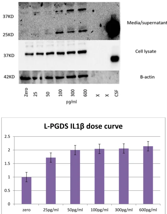
3. Expression of L-PGDS in chondrocytes treated with IL1β, dose-dependent and time course: ... 53
4. Expression of L-PGDS in chondrocytes treated with IL-17, dose-dependent and time course: .... 57
5. Expression of L-PGDS in chondrocytes treated with H2O2, dose-dependent and time course: .... 60
6. Expression of L-PGDS in chondrocytes treated with 4BHP, dose-dependent and time course: ... 64
7. Expression of L-PGDS in synovial fluid: ... 68
8. Expression of Nrf2: ... 71
Chapter 4: Discussion ... 75
Chapter 5: Conclusion ... 93
vi
Table of figures:
Figure 1: Normal anatomy and structure of knee joint. ... 3
Figure 2: Diagram of normal Knee versus osteoarthritic one showing pathological features of OA. ... 3
Figure 3: The synthesis of different PG’s from AA acid by COX and different PG synthases. ... 22
Figure 4: PGDS distribution in body tissues. ... 26
Figure 5: L-PGDS function. ... 27
Figure 6: L-PGDS 3D structure. ... 28
Figure 7: Nrf2 pathway and role of ROS in its activation. ... 35
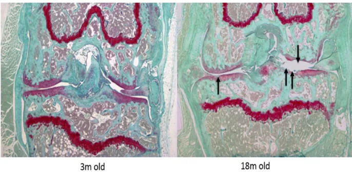
Figure 8: Histological changes in aging-associated osteoarthritis. ... 49
Figure 9 Histological changes in surgically induced osteoarthritis. ... 49
Figure 10 L-PGDS levels in spontaneous age associated osteoarthritis - immunohistochemistry. ... 51
Figure 11 L-PGDS levels in surgically induced osteoarthritis - immunohistochemistry. ... 52
Figure 12: L-PGDS expression in IL1B treated chondrocytes dose curve. ... 54
Figure 13: L-PGDS expression in IL-1B treated chondrocytes time course... 56
Figure 14: L-PGDS expression in IL-17 treated chondrocytes dose curve. ... 58
Figure 15: L-PGDS expression in IL-17 treated chondrocytes time course. ... 60
Figure 16: L-PGDS expression in H2O2 treated chondrocytes dose curve. ... 62
Figure 17: L-PGDS expression in H2O2 treated chondrocytes time course. ... 63
Figure 18: L-PGDS expression in 4BHP treated chondrocytes dose curve 24h. ... 66
Figure 19: L-PGDS expression in 4BHP treated chondrocytes dose curve 48h. ... 67
Figure 20: L-PGDS expression in synovial fluid samples ... 69
Figure 21: Nrf2 expression levels in chondrocytes treated with IL1β and 4BHP. ... 74
vii
List of abbreviations:
• 4BHP: tert-butyl hydroperoxide.
• AA: arachidonic acid.
• ARE: antioxidant response element.
• c-AMP: cyclic adenosine monophosphate.
• COX: cyclooxygenase.
• COX-1: cyclooxygenase-1.
• COX-2: cyclooxygenase-2.
• CSF: cerebrospinal fluid.
• C-DNA: complementary deoxynucleic acid.
• BMDM: bone-marrow-derived macrophages.
• DMM: destabilization of medial meniscus.
• dNTP: deoxynucleotide triphosphate.
• H2O2: hydrogen peroxide.
• Il-1β: interleukin 1 beta.
• Il-17: interleukin 17.
• IFNγ: interferon gamma.
• iNOS: inducible nitric oxide synthase.
viii • KD: kilodalton.
• L-PGDS: lipocalin prostaglandin D synthase.
• MAPK: mitogen-activated protein kinase.
• MMPs: matrix metalloproteinases.
• MMP-1 : matrix metalloproteinase-1.
• MMP-13: matrix metalloproteinase-13.
• MW : molecular weight.
• M-RNA: messenger ribonucleic acid.
• MgCl2: magnesium chloride 2.
• NO: nitric oxide.
• NSAIDs: non-steroidal anti-inflammatory drugs.
• Nrf2: nuclear factor erythroid 2-related factor 2.
• OA: osteoarthritis.
• O/N: overnight.
• PCR: polymerase chain reaction.
• Pg: picogramme.
• PKA: phosphokinase A.
• PKC: phosphokinase C.
ix • PG: prostaglandin.
• PGD2: prostaglandin D2.
• PGE2: prostaglandin E2.
• PGH2: prostaglandin H2.
• PGJ2: prostaglandin J2.
• RNA: ribonucleic acid.
• RPM: revolutions per minute.
• RT-enz: reverse transcriptase enzyme.
• RT-PCR: reverse transcriptase polymerase chain reaction.
• RT-qPCR: real-time quantitative polymerase chain reaction.
• ROS: reactive oxygen species.
• TNF-α: tumor necrosis factor alpha.
x
Acknowledgements:
At the start, I would like to thank Allah, the most beneficent, the most merciful, for the countless blessings he gave me.
I would like also to deliver a special thanks to Dr. Hassan Fahmi my research director and Dr. Daniel Lajeunesse my co-director, for their constant effort to guide me all through my master's journey.
Special thanks also go to Dr. Thierry Alquier and Dr. Guy Rousseau the jury members, for their time reviewing this work, and for their advice to improve it.
Every single member of Dr. Fahmi and Dr. Lajeunesse labs taught me something, either in science or in life, I owe them all many thanks.
Lastly, I would like to extend my thanks to the University of Montreal for all its support and for the scholarship. Also to the University of Montreal Hospital Research Centre (Cr-CHUM), osteoarthritis unit, Canadian Institutes of Health Research (CIHR) for all their help and directions and for funding this project.
1
2
A.
Introduction
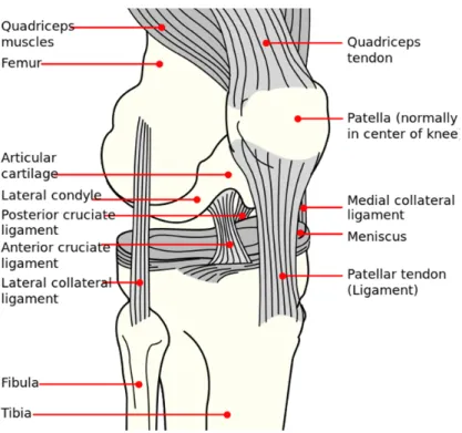
1.1 Osteoarthritis:Osteoarthritis (OA) is a chronic inflammatory degenerative disease that affects joints cartilage, ligaments and underlying bone. It was long thought to be an inevitable process of aging. Further studies on OA revealed the interplay between many pathophysiological factors namely: weight, age, sex, genes, biomechanical, biochemical, environmental and local factors in the joint itself. In normal joints, there is a balance between the anabolic and the catabolic processes, at the level of articular cartilage, bones and synovial membranes. In OA, this balance is disrupted leading to the classic features of OA like degradation of articular cartilage (leading to narrowing of joint space), subchondral bone remodeling (osteosclerosis) and cyst formation, osteophytes formation and synovial membrane inflammation and thickening. Normal anatomy of the knee joint is detailed in Figure 1. Figure 2 is a diagram showing the pathological changes in osteoarthritic knees versus normal knees.
3
Figure 1: Normal anatomy and structure of knee joint. Image source: http://commons.wikimedia.org/wiki/File:Knee_diagram.svg (open use).
Figure 2: Diagram of normal Knee versus osteoarthritic one showing pathological features of OA.
Image source: http://www.maurascollege.org/images/cervical-spondylosis-dizziness-treatment-information-disease-colorado-westminster-8933.jpg
4 1.2 Epidemiology and risk factors:
OA is the most common joint disease in North America, with an incidence of 10% in males and 15% in females above age 60. It’s expected that the total number of OA cases is going to grow with the aging of the population. There are many factors that affect the epidemiological distribution of OA cases, among these factors; the definition of what OA truly is, the method used to characterize it, the joints involved and the risk factors. For example, if we are going to check the prevalence among developed countries we may find obesity as a confounding factor raising the prevalence in this population. If we measure it among different age groups, the older the age group from which we take our samples the more prevalent the disease becomes. Most clinicians characterize OA radiologically according to the Kellgren and Lawrence scaling system where:
• Grade 0: no radiographic features of OA are present.
• Grade 1: doubtful joint space narrowing (JSN) and possible osteophytic lipping. • Grade 2: definite osteophytes and possible JSN on the anteroposterior
weight-bearing radiograph.
• Grade 3: multiple osteophytes, definite JSN, sclerosis, possible bone deformity.
• Grade 4: large osteophytes, marked JSN, severe sclerosis and definite bone deformity.
5
Regarding those patients radiologically characterized as OA, let us take the knee joint in people ≥ 40 years old as an example. In this population, the prevalence of knee OA was 19.2% according to the Framingham Study and 27.8% according to the Johnston County OA Project [1]. In the third National Health and Nutrition Examination Survey (NHANES III) researchers took a group of people >60 years old and characterized radiologically OA in their knees. They found that 37% of the study population had OA [1]. This suggests that different age groups have different prevalence. Conversely, if we take another factor such as which joint is involved, and we focus on hand OA for example, we will find the prevalence is 27.2% of the study subjects in the Framingham study. In addition, hip OA is less prevalent than knee OA or hand OA, with about 7% of women aged ≥65 years having a radiologically diagnosed OA. In Johnston County, the prevalence rate of hip OA reached 27% for individuals >45 years old [1]. Hence, in different age groups, the prevalence is totally different, as assessed by the Framingham study of hand OA (6.8%) and knee OA (4.9%) of the study subjects aged ≥26 year which is much different from the other study groups.
These differences in data reflect how different factors can affect the prevalence of OA, among those factors the definition of OA and the basis used to diagnose it i.e. clinically or radiologically, which joint is considered, and which age group is considered in the studies provided. OA is multifactorial in origin and factors such as age, gender, joint injury, body weight, bone density, collagen diseases (Ehlers-Danlos syndrome, Marfan syndrome) and muscle weakness around the joints are all important factors contributing to OA. Their modification determines the course of the disease. Genetic factors play a role also in the OA process [2, 3]. In a study to identify
6
the genetic loci that were associated with radiologically evident OA, researchers identified loci in 296 pedigrees on chromosomes 1, 7, 9, 13, and 19 particularly for OA of the hand [4]. OA of the hip also showed some genetic linkage to chromosome 11 [5]. Other chromosomes are also involved including 4 and 16 [6], 2 and 19 [7]. Chromosomes 2, 3 and 4 were shown to be involved in mutations in matrilin-3, a gene for non-collagenous matrix protein (MATN3). This mutation is found in 2% of hand OA patients in Iceland [8]. For patients with radiologically diagnosed OA of the knee single-nucleotide polymorphisms (SNPs) marker association within the gene for Leucine-Rich Repeats and Calponin Homology (LRCH1) on chromosome 13 was shown in studies from the UK and Canada [9]. In addition, the marker (rs91242a C/T) allele was detected more frequently in cases with significant knee OA versus control cases. Indeed, one out of 31 identified loci shows up more frequently in OA versus controls [9]. The function of the protein product of this gene is not fully known though it has a region similar to that of actin-binding protein that plays a role in cell shaping and intracellular signaling. Mutations which happen in structural components of articular cartilage and bone tissue potentiate the effect of other risk factors of OA and accelerate the pathology of the disease [3].
In conclusion the epidemiology and risk factors of OA are affected by the way you identify the cases, clinically versus radiologically, which joints are included, and which age group is targeted.
Age, race, acute and chronic repetitive trauma, are the main risk factors for OA, especially in the knee joint. To a lesser extent, they affect hip and hand OA. Age, obesity and gender, on the other hand, have a big role in the development of hip OA.
7
Bone mass in women, and whether they use hormonal replacement therapy or not, also play a role in the development of hip OA. Interestingly the higher the bone mass before and after menopause the higher the risk of developing OA of hip joints [10, 11].
Finally, obesity is the strongest modifiable risk factor for OA. Its strongest association is with the knee and hand OA while interestingly it’s less associated with hip OA [12, 13]. Given this association, a reduction in weight has a noticeable protective role against knee OA [14].
1.3 Pathogenesis:
OA is a complex multifactorial disease and the mechanisms involved in its pathogenesis are not fully uncovered yet. There are different mechanisms that could initiate OA [15]. The most common initiating factor for OA is the mechanical damage, either as a single event macro-trauma or repeated microtraumas [16]. These mechanical injuries initiate the inflammatory process in chondrocytes causing them to release degenerative enzymes that degrade articular cartilage [16]. Another less common mechanism is the defective collagen type 1 and 2 formations in certain diseases leading to articular cartilage failure under normal mechanical loads [17]. Once the initial factor (for example trauma) starts the OA process, many other factors get involved. These factors include (on the molecular level) proteases, proteases inhibitors and cytokines, affecting the cartilage degeneration and repair balance. On the non-molecular level, other factors also play a role like age, weight, joint alignment, physical activity, and systemic hormones and mineral deposition. The biochemical cell response to mechanical stressors is termed mechanotransduction [18]. This
8
process has been studied in different cell types including chondrocytes [19]. Studies showed an increase in aggrecan messenger ribonucleic acid (mRNA) and a decrease metalloproteinase 3 mRNA by quantitative polymerase chain reaction (qPCR) in healthy human chondrocytes when cultures were subjected to repetitive loading versus those grown on solid support [20]. Osteoarthritic cartilages respond differently from healthy ones to the same mechanical loads, as they show more deformability and more loss of fluids under the same load [21]. These different biomechanical responses make normal every day loads considered pathological for osteoarthritic cartilage [22]. Biomechanical signaling pathways become now therapeutic targets to ameliorate the response of osteoarthritic cartilage to mechanical load. The difference in response between normal chondrocytes and osteoarthritic chondrocytes to the same mechanical load is what lead to the use of autologous chondrocytes implantation in OA. This therapeutic technique is aiming to improve the inflammatory process by restoring the normal signaling pathway by using healthy chondrocytes [23].
Trauma, even blunt ones make the cartilage susceptible to OA and degeneration. Tumor necrosis factor alpha (TNF-α) levels were observed to be high in synovial fluid early after trauma. TNF-α enhances neutrophils mediated proteoglycans degradation and has pro-apoptotic effect under certain enabling conditions [24]. Interestingly exposing chondrocytes in vitro to TNF-α did not increase cell death rates or levels of prostaglandin E2 (PGE2) or prostaglandin D2 (PGD2). Trauma, on the other hand, increased these mediators, independently of TNF-α [24]. TNF-α was found to induce the expression of matrix metalloproteinase-1 (MMP-1) regardless of trauma,
9
contributing to cartilage degradation. These mediators clarify to some extent the biomechanical pathways involving trauma and cartilage damage.
The role played by mechanical load is not fully understood in the pathogenesis of OA. It was thought to cause wear and tear to the cartilage, accelerating OA. Further clinical and research data revealed that mechanical load in the form of exercise (in normal weight subjects) can cause, accelerate, ameliorate and even reduce the risk of OA [25, 26]. In 2013, a systematic review of clinical trials on the effect of exercise on osteoarthritic weight-bearing joints (knee mainly) was done [27]. This review included 60 clinical trials with 12 different exercise intervention programs [27]. The review concluded to a statistically significant effect for exercise in reducing pain and improving functionality in osteoarthritic patients, particularly aquatic exercise [27]. Fewer similar studies were done for hip joint OA, where it was shown that physiotherapy and home-based exercise programs don’t provide that much improvement in pain management and functionality [28]. Another study evaluated the effect of an intensive aerobic exercise program in patients with moderate to severe OA evidenced clinically and radiologically (grade III and IV). This study concluded that, there is no difference between exercise groups versus non-exercise groups regarding pain control and functionality [29]. In conclusion, exercise programs have to be tailored depending on many factors in each patient. We should always take into consideration the general medical condition of the patient particularly his cardiovascular capacity for exercise and whether his main complaint from OA is pain or limitation of movement. On the other hand, limitation of movement had put the osteoarthritic patients who are not following an exercise program at a higher risk for
10
cardiovascular diseases. This raises the question of screening sedentary osteoarthritic patient for cardiovascular diseases before assigning them to exercise programs. Excessive mechanical load induces biomechanical transformations in chondrocytes, for example cyclooxygenase 2 (COX-2) gene induction. These biomechanical transformations impair chondrocytes’ ability to handle oxidative stress, which leads to chondrocyte toxicity and eventually OA [30]. This mechanical load on chondrocytes was also found to induce the lipocalin prostaglandin D synthase (L-PGDS) gene. L-PGDS through its end products PGD2 and 15d-PGJ2 is hypothesized to contribute to the different processes initiated by mechanical load [30]. Figure 22 clarifies the biosynthesis of PGD2 and 15d-PGJ2.
11 1.4 Treatment:
Pharmacological and non-pharmacological management of OA mainly targets pain control, minimizing disability and improving the quality of life. General non-pharmacological management focuses on weight reduction and exercise programs.
Pharmacotherapy starts with acetaminophen for OA patients not showing inflammatory signs and symptoms [31]. Once there are symptoms of inflammation such as articular swelling, morning stiffness, night pain, joint effusion by examination, or synovitis on arthroscopic examination, non-steroidal anti-inflammatory drugs (NSAIDs) are indicated.
NSAIDs can be non-selective cyclooxygenase (COX) inhibitors including COX-1 and COX-2, or COX-2 selective inhibitors (coxib). NSAIDs should be started at the minimal dose that controls the inflammation to avoid the side effects of these drugs. NSAIDs and Capsaicin (topical analgesic) can be used topically in patients who can’t tolerate NSAIDs orally. Patients with gastritis, peptic ulcer or renal diseases are intolerant to NSAIDs.
Intra-articular glucocorticoids injection is the next step when there is an inadequate response to non-pharmacological management plus acetaminophen or NSAIDs. Failure of the initial therapeutic lines necessitates a re-evaluation of the joint to detect the probability of another underlying pathology such as crystal disease and other inflammatory arthropathies. This could be done by arthrocentesis and synovial fluid
12
examination. Current data suggests an added value for the use of glucosamine and chondroitin together versus the use of each of them alone in treating OA.
Resistant cases of OA are treated by another spectrum of medications designated for those cases particularly. Those medications include opioid analgesics, intra-articular hyaluronans, a trial of glucosamine or chondroitin, and finally colchicine. These management guidelines are in accordance with the guidelines from American College of Rheumatology [31].
American college of rheumatology (ACR) stated guidelines for managing OA according to joints involved and according to modality of treatment as follow [36]:
Non-pharmacologic management of hand OA:
1) Determine how OA affects the patient’s activities of daily living (ADLs).
2) Educate the patient about how to protect his joints.
3) Use supporting devices, like splints for patients with trapeziometacarpal OA.
4) Educate the patient about thermal techniques available for his condition.
Pharmacologic management of hand OA:
1) Topical capsaicin.
2) Topical NSAIDs.
3) Oral NSAIDs.
13
ACR recommends against the use of intra-articular therapies and opioid analgesics. Generally persons who are ≥75 years old should use topical anti-inflammatory rather than oral ones.
Non-pharmacologic management of knee OA:
1) Aerobic exercise.
2) Aquatic exercise.
3) Weight loss.
4) Self-management training.
5) Physiotherapy plus exercise.
6) Psychosocial guidance and support.
7) Knee protective gears according to the internal lesion, for example medially directed patellar taping, and medially wedged insoles for those who have OA in lateral knee compartment or laterally wedged subtalar strapped insoles for medial knee compartment OA patients.
8) Advised about thermal and transcutaneous electrical stimulation modalities available.
9) Use supporting devices like walking aids as needed.
10) Tai chi programs.
14
ACR didn’t provide specific recommendations regarding balance exercises, strengthening exercises, laterally wedged insoles, physiotherapy alone, knee braces or laterally directed patellar taping.
Pharmacologic management of knee OA:
1) Acetaminophen.
2) Oral NSAIDs.
3) Topical NSAIDs.
4) Tramadol.
5) Intra-articular corticosteroid injections.
6) Chondroitin sulfate.
7) Glucosamine.
8) Topical capsaicin.
Also, ACR didn’t have specific recommendations regarding the use of intra-articular hyaluronates, duloxetine, and opioid analgesics.
Non-pharmacologic management of hip OA:
1) Aerobic exercise.
2) Aquatic exercise.
15
4) Self-management training.
5) Physiotherapy plus exercise.
6) Psychosocial guidance and support.
7) Advised about thermal modalities and walking aids, as needed.
ACR didn’t have particular recommendations regarding balance exercises alone or with strengthening exercises, tai chi or physiotherapy alone.
Pharmacologic management of hip OA:
1) Acetaminophen.
2) Oral NSAIDs.
3) Tramadol.
4) Intra-articular corticosteroid injections.
ACR does not recommend the use of Chondroitin sulfate and Glucosamine and didn’t have specific recommendations for topical NSAIDs, Intra-articular hyaluronate injections, Duloxetine or opioid analgesics.
Many studies had evaluated the role of NSAIDs in OA and the mechanisms by which they work. The initial idea was that NSAIDs work only by inhibiting COX enzymes, but recently it was discovered that they downregulate the expression of the COX gene as well [32]. Figure 3 explains the synthesis of different PG’s from AA by COX enzyme. NSAIDs affect the synthesis of all prostaglandins (PG’s), the inflammatory as well as
16
the anti-inflammatory ones. This explains NSAIDs limited success in reversing or even stopping the progression of the disease. The moment we stop COX2 by NSAIDs we stop both the inflammatory and the salvation pathways. COX 1 and 2 activities are augmented by interleukin 1 beta (IL-1β) and arachidonic acid (AA) to produce different types of PG’s, as explained in Figure 3. The L-PGDS enzyme synthesizes PGD2 which contributes to the anti-inflammatory pathway through its end product 15-PGJ2. Via its inhibition of nitric oxide (NO) production PGD2 prevents cartilage degradation [33]. PGD2 even has a positive feedback effect on its own production through the stimulation of COX2; this is believed to be through its role in up-regulating other prostanoids such as PGF1α, PGF2α and thromboxane B2 (TXB2). PGE2 induces MMP1 and MMP13 which are inflammatory mediators in OA, and it inhibits proteoglycans promoting matrix degradation. PGD2, on the other hand, inhibits MMP1 & MMP13; this again suggests an anti-inflammatory role for PGD2. NSAIDs by blocking COX function and down-regulating it alter these entangled reactions, stopping the inflammatory and anti-inflammatory pathways. Figure 3 explains the synthesis of different PG’s from AA by COX enzyme.
Guidelines recommend the use of intra-articular glucocorticoids in cases of moderate to severe pain, especially after the failure of oral and topical management, or when they are contraindicated, like in cases of active peptic ulcer disease or renal failure [34-37]. This effect reaches its peak during the 1st two weeks after injection and then it wanes off. Even with the repeated injections, their efficacy is appreciated during the first year only [38]. No long-term benefits or harms had been proven for this modality of treatment [35]. The injected dose depends on the joint size, from 10mg for small,
17
20mg for medium sized and 40mg for large ones. All injection techniques should be done under complete aseptic conditions to minimize the risks of infection. Guidelines also recommend aspiration of any joint effusion before injection of steroids and sending the effusion fluid for analysis for better diagnosis and to exclude infection. Injection of steroids should be postponed until the culture results come back negative, especially if there is a high suspicion for infection like fever or rapidly accumulating joint effusion [39].
Studying the efficacy of this therapeutic modality showed a significant effect for intra-articular glucocorticoids injection versus placebo [34], even without a clinically evident joint inflammation [35]. Most studies were done on knee joint injections, then on the hip joint, and to a much lesser extent on carpometacarpal joints and other joints. Generally these injections cause pain relief rather than improvement of function or repair of joint damage.
Guidelines recommend the use of intra-articular hyaluronan injections in cases of OA (especially knee OA) which failed oral and topical therapeutic modalities, or in those patients who have a contraindication for the use of NSAIDs like active gastric ulcer or renal failure [31, 37]. Also, it’s the next step in cases of failure of intra-articular glucocorticoids injection [34-37]. This modality improves mainly pain symptoms and to some extent functionality but doesn’t prevent the progress of the disease or reverse it [40, 41]. Improvement of functionality in hyaluronan use is mainly through restoring the viscoelasticity of the synovial fluid that is usually lost in OA. These benefits were elaborated by many randomized trials and meta-analyses [42-46]. These injections
18
are usually given as one course per year (as one injection or five weekly injections according to the molecular weight (MW) of the drug). This is usually well tolerated at this rate with minimal to no side effects. Compared to glucocorticoids injections the hyaluronan compounds have similar efficacy with a slower onset of action, taking sometimes up to two months to reach its peak effect [47]. Studies compared also the effect of hyaluronan injections to that of NSAIDs and found almost similar effects [34, 42, 48, 49]. Hyaluronan showed some adverse reactions like flaring of the joint pain and swelling post injection in 1.5 to 5 percent of patients. These flares usually resemble septic arthritis with synovial fluid leukocytosis of 100,000 per mm3 [50].
These flares are related to injection techniques as well as to the MW of the injected drug, with higher rates in lateral approach versus medial one [51]. Also, higher rates of flares were found with the use of higher MW compounds compared to lower MW ones [52]. Generally no major side effects for these compounds were reported.
Surgical management is usually left as the last resort treatment of OA. There is a wide range of modalities available with different selection criteria for each modality. Arthroscopic intervention: it starts with joint irrigation [53] to improve visualization of the joint by removal of debris and blood. Following this, arthroscopy is used for debridement with or without arthroscopic synovectomy. Some hypotheses suggest that joint irrigation alone has a beneficial effect on the management of joint pain, by removal of tissue debris, crystalline materials and cartilage fragments, decreasing the burden on the joint and synovial membranes and slowing further destruction [54, 55]. Arthroscopic irrigation and lavage are an office procedure that can be done under local anesthesia and intravenous sedation.
19
Arthroscopic debridement is done mainly for meniscal tear usually with joint lavage. Some studies suggested that there is not much difference between arthroscopic debridement with lavage in addition to medical and physical therapy, compared to medical and physical therapy alone. This is regarding the pain level using WOMAC pain score [56]. Some randomized trials on patients with knee OA and meniscal tears showed no higher benefit with arthroscopic surgery, followed by physical therapy, compared to physical therapy alone [57-59].
Arthroscopic abrasion includes mainly drilling of sclerotic bone in affected joints. Randomized controlled trials did not show significant improvement with this technique compared to medical and physical therapy alone. Follow-up observations of patients who went through this procedure showed that 50 percent still needed a total knee replacement within 3 years [60].
No clinical trials were done to study the efficacy of arthroscopic synovectomy in patients with OA. This is usually reserved to those with severe inflammatory signs and symptoms unresponsive to initial NSAIDs, intra-articular glucocorticoid injections, arthroscopic irrigation, colchicine, and non-pharmacological measures.
Total joint replacement (arthroplasty) is another modality used as a final measure after the failure of other measures and in patients with significant limitation of activities [61]. Replacement surgery should be timed perfectly, ideally before significant joint deformity or instability, significant contractures or muscle atrophy. This aims to minimize the complications and to maximize outcomes [62, 63]. Preoperative medical management and postoperative rehabilitation affect greatly the surgical
20
outcomes [64, 65]. Total joint arthroplasty usually provides the best pain relief and functional improvement in patients with significant hip or knee OA [61, 66]. Improvement of functionality following surgery comes gradually, usually over a period of one year and lasts in average 5 years [67].
The joint resurfacing procedure is a treatment modality in which a metal cab or prosthesis replaces an arthritic femoral head; on the other side of the joint a metal acetabular component forms the articulation surface with the metal femoral head. This is known also as resurfacing arthroplasty, a common alternative for total hip replacement. This procedure shows some late complications, mainly femoral neck fracture and aseptic loosening. Other than these complications, this procedure is usually well tolerated and has excellent outcomes [68].
The chondrocyte grafting technique involves autologous chondrocytes implantation (ACI) into the defective areas of the cartilage aiming to fill these areas and restore the smooth surface as before. Chondrocytes are collected from the same patient from a non-weight bearing area. These are then allowed to grow in an in vitro culture media. Lastly, they are implanted into the cartilage defects under a cover of the periosteal membrane (1st generation, ACI-p); under a collagen type I/III membrane (2nd generation, ACI-c); or into a scaffold matrix (3rd generation, ACI-m). Usually, this technique is reserved to areas of confined and localized cartilage loss; this is usually seen in young patients who had joint trauma. Patients with advanced OA and wide areas of cartilage loss didn’t show promising results with this technique [69]. Magnetic resonance imaging confirmed a better filling of small chondral defects when done by
21
a graft technique than by any other technique. This technique is also less likely to produce osteophytes [70].
Carpometacarpal or trapeziometacarpal OA is a unique situation necessitating different surgical techniques, usually including trapeziectomy with or without ligament reconstruction and tendon interposition [71]. Interpositional arthroplasty and joint replacement are other modalities used for this type of OA. Comparative studies for the outcomes of those different procedures didn’t show any advantage for one technique over the others.
The demonstration of different treatment modalities, pharmacological and surgical ones, aims mainly to clarify the limitations that exist for current therapeutic modalities. Pharmacological modalities aren’t specific enough to spare the anti-inflammatory pathways, and surgical ones aren’t showing significant preference over the pharmacological ones. This justifies our search for more accurate understanding of this disease and for new therapeutic targets and accurate medications.
1.5 Prostaglandins:
PG’s are a family of physiologically active lipid compounds that are involved in many physiological and pathological processes all over the body. One of their major roles is in inflammation. PG’s can be both inflammatory and anti-inflammatory mediators. In this respect they are involved in the OA pathological process. The synthesis of PG’s starts with the release of AA from the cell membrane, a reaction catalyzed by phospholipases. The next reaction is catalyzed by a COX that leads to the
22
intermediate PGH2 from AA. Two isoforms of COX are known, COX-1 which is constantly expressed, and COX-2 which is induced by inflammatory mediators [72].
The unstable PGH2 is then metabolized into more bioactive forms of PG’s like PGE2, PGD2, PGF2α, PGI2, and thromboxane A2 by different synthases [73]. Figure 3
shows the pathways involved in the synthesis of different PG’s from AA.
Figure 3: The synthesis of different PG’s from AA acid by COX and different PG synthases.
Image source: http://vascular.free.fr/cox-pathway.jpg
PG’s act via specific G protein-coupled receptors. Each prostaglandin acts via multiple different receptors subtypes and these receptors can cross react together. Via these multiple receptors PG’s can multiply their coupling capacity to different signal transduction pathways leading to a more diverse range of downstream effects.
23
These effects can occur in functionally opposing directions within the same cell. PGE2 for example is widely expressed in the CNS. It has 4 receptors subtypes giving it a diverse functionality even inside the same cell. PGE2 can have neurodegenerative or neuroprotective effects depending on which receptor it binds to.
In articular cartilage, PGE2 plays a degenerative role. It inhibits proteoglycans synthesis, which are one of the main components of healthy cartilage. PGE2 also induces matrix metalloproteinase 13 (MMP13) and aggrecanase 5 (ADAMTS-5), which degrade cartilage proteins. The main effect of PGE2 on articular cartilage is mediated through a receptor known as EP4 [74].
Among different PG’s, PGD2 is an interesting one. It’s synthesized from PGH2 by PGD synthase and then gets enzymatically dehydrated into 15d-PGJ2. 15d-PGJ2 is the natural endogenous ligand of peroxisome proliferator-activated receptor gamma (PPAR-γ) nuclear receptor and transcription factor. PPAR-γ among other functions, is known to suppress inflammatory cytokines like COX-2, IL-1β, TNF-α, inducible nitric oxide synthase (iNOS) MMP1, MMP13 [75]. This function was tested with synthetic ligands of PPAR-γ before discovering 15d-PGJ2 [75]. This makes 15d-PGJ2 (and thus PGD2) one of the most important natural anti-inflammatory PG’s.
24 1.6 PGD2:
Many studies suggested a protective role for PGD2 in OA [73] [76]. Indeed, treating chondrocytes with PGD2increases the production of collagen type 2 and aggrecan, which are involved in cartilage repair [77]. In addition, chondrocytes treated with PGD2 showed lower rates of cell death [76]. PGD2has also a role in inhibiting the production of matrix metalloproteinase 1 and 13, which are involved in cartilage damage [78]. In light of these observations, it is noteworthy that studies noted increased levels of PGD2 during the resolution phase of the inflammation process. These high levels of PGD2 were lost by the use of COX-2 inhibitors suggesting a role for COX-2 in activating anti-inflammatory pathways [79]. Further exploring and confirming the role of PGD2 as anti-inflammatory, in vivo studies using PGD2 synthase knockout mice showed a failure of resolution of inflammation [80]. Another study confirmed the anti-inflammatory role of PGD2, using the delivery of PGD2 synthase into a murine air pouch model of monosodium urate monohydrate crystal-induced inflammation via a retroviral method [81]. PGD2has two main receptors, D Prostanoid receptor (DP1) and chemoattractant-receptor-like molecule (CRTH2), also known as (DP2) [82]. Activation of the DP1 pathway is the one responsible for the down-regulation of MMP 1 & 13 in chondrocytes [78]. PGD2 exhibits its anti-inflammatory effect through its potent anti-anti-inflammatory end product 15d-PGJ2 (15-deoxy-delta12, 14-PGJ2) [83]. 15d-PGJ2 has a significant anti-arthritic effect through downregulation of inflammatory mediators of OA [83]. This includes IL-1β, tumor necrosis factor-alpha (TNF-alpha), iNOS, and MMPs 1 and 13 [83]. 15d-PGJ2
25
induces its effect through nuclear PPAR-γ [83, 84] as well as other pathways independent ofPPAR-γ [84].
PPAR-γ is a nuclear receptor protein that works as a transcription factor for different genes including anti-inflammatory genes. This receptor is noticeably downregulated in osteoarthritic cartilage in vivo and was downregulated also in chondrocytes treated with IL-1β in vitro [85]. This down-regulation is interfering with the PGD2 protective function. PPAR-γ has a wide range of functions, besides being a potent anti-inflammatory effector through suppression of the previously mentioned mediators; it plays also a role in reversing insulin resistance. Studies found that PPAR-γ decreases insulin resistance and improves liver handling of glucose by upregulating insulin receptors [86], from which appeared the thiazolidinediones family of drugs as insulin sensitizers to treat type 2 diabetes. PPAR-γ also plays a key role in adipogenesis, therefore, PPAR-γ antagonists have been used to decrease steatosis and adipose tissue formation [87] [88].
1.7 L-PGDS:
PGD2 is formed from PGH2 by a PGD synthase. There are two isoforms of PGD synthase, lipocalin PGDS (L-PGDS) which is glutathione-independent and hematopoietic PGDS (H-PGDS) that is glutathione-dependent [89]. L-PGDS has a wide distribution all over the body especially within the central nervous system [90, 91], heart [92], retina [93], genital organs [94], and chondrocytes [95]. H-PGDS shows abundance mainly in hematopoietic tissues, mast cells [96], megakaryocytes [97],
26
and T-helper lymphocytes-2 [98]. Figure 4 explains the distribution of L-PGDS and H-PGDS in different tissues.
Figure 4: PGDS distribution in body tissues.
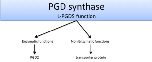
L-PGDS has a dual function; enzymatic as well as non-enzymatic. Figure 5 describes its known functions. Enzymatically, it synthesizes PGD2 from PGH2. Non-enzymatically, it acts as a transmembrane transporter protein for small hydrophobic molecules including PGD2, protecting it from metabolism and non-enzymatic dehydration into PGJ2. Also, PGDS has lipophilic ligand-binding properties by which it binds retinoid, thyroid hormones, and bile pigments including bilirubin and biliverdin. PGDS increases in cerebrospinal fluid (CSF) after subarachnoid hemorrhage to act as a scavenger for bilirubin. It plays a role also in cell migration and morphological
27
changes mainly through retinoid. Lipophilic ligand binding properties of this protein contribute to its anti-cancer function, as many lipocalin-ligands (like retinoid) are known to be involved in cell cycle regulation and cell proliferation. The main mechanism of this function is not fully deciphered yet.
Figure 5: L-PGDS function.
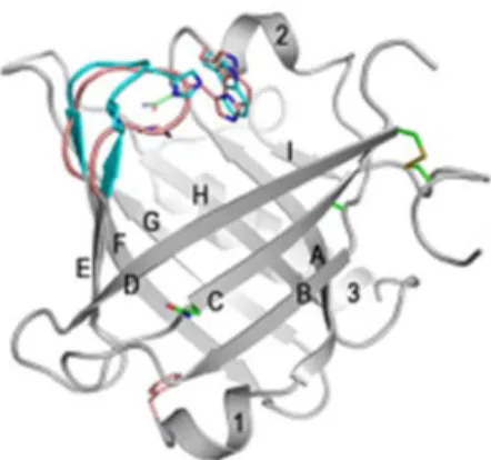
The three-dimensional structure of L-PGDS is formed of 9 β-Strands and 3 α-helices. They are labeled A-I and 1–3 respectively in Figure 6. L-PGDS is formed of 190 AA giving it a calculated MW of 21 kilodalton (KD). Though, it actually appears at a MW range of 25 to 37Kd. This is because of the post-translational modification of the protein, as this protein has multiple glycosylation and phosphorylation sites giving it a
28
wide range of MW and functionality according to which site is phosphorylated and which is glycosylated [99]. It has an enzymatic active site, at cysteine 65 (Cys65), an important glycosylation site at asparagine 51 (Asn51), and an important phosphorylation site at serine 106 (Ser106) [99].
Figure 6: L-PGDS 3D structure.
Image source: http://urade.wpi-iiis.tsukuba.ac.jp/research/analysis.php
Depending on L-PGDS location inside the cell or in body fluids, different modified isoforms of L-PGDS appear. This is very important because it indicates that L-PGDS is modified in different ways to adapt for its multiple functions and in different locations also. For the purpose of using L-PGDS as a therapeutic target, we need to identify exactly the enzymatically active form to use as a medication avoiding the side
29
effects that may arise from other forms. These post translational modifications suggest different functions for different L-PGDS isoforms.
L-PGDS is the key regulatory enzyme for PGD2 production from PGH2 therefore; it is the key regulatory enzyme for 15d-PGJ2 production also. This makes the regulation of L-PGDS production in chondrocytes the key for the regulation of natural anti-inflammatory pathways. The pathway we are working on in this study is described in
Figure 22 and explained in details in the discussion. 1.7.1 In the joint:
New studies started exploring the roles played by L-PGDS and H-PGDS as well as their regulation in cartilage. Higher abundance for L-PGDS mRNA and protein of about 20 fold was found in normal and osteoarthritic chondrocytes compared to H-PGDS [100]. L-H-PGDS is suggested to have a protective role in chondrocytes considering its end product, PGD2. Pursuing this protective role, L-PGDS is
expressed at higher levels in many pathological conditions. Among these conditions, multiple sclerosis [101], atherosclerosis and coronary artery diseases [92], diabetes mellitus [102], and hypertension [103]. An interesting study by Zayed et al. in 2008 showed a key role for IL-1β in the induction of L-PGDS, both at the mRNA and protein levels, in chondrocytes, in a dose and time-dependent fashion. This eventually led to higher levels of PGD2.
The increased levels of L-PGDS mRNA and protein were confirmed to be through gene induction. Different pathways were studied to identify those involved in L-PGDS gene induction. The successful blocking of IL-1β induction of L-PGDS using
30
translation inhibitors like cycloheximide, C-jun N-terminal kinase (JNK), NF-κB, p38 MAP kinase and Notch, confirmed the involvement of these pathways in L-PGDS gene induction by IL-1β [100]. Interestingly, inhibition of the extracellular signal-regulated kinase/mitogen-activated protein kinase (ERK/MAPK) pathway didn’t show any significant effect on blocking the induction of L-PGDS by IL-1β [100]. The end product PGD2 was found to have a negative feedback inhibitory effect on L-PGDS
induction byIL-1β in chondrocytes [100].
1.7.2 In the central nervous system:
L-PGDS is a major protein and it has many roles. It is used as a biomarker in patients with idiopathic normal pressure hydrocephalus (iNPH), a degenerative disease characterized by progressive dementia, gait problems and urinary incontinence. L-PGDS level is decreased in patient with iNPH, indicating the death of arachnoid cells responsible for its production. In the same disease, it plays a role as a scavenger of amyloid beta (Aβ) which has a neurotoxic effect leading to Alzheimer's disease, a major comorbid condition of iNPH [104]. Multiple sclerosis (MS) is an autoimmune disease leading to multifocal, recurrent, demyelination of the white matter of the central nervous system. L-PGDS generally plays a protective role in the CNS. In multiple sclerosis where there are plaques of degenerated myelin sheaths from the autoimmune process responsible for the pathology of the disease; L-PGDS is present at high levels in oligodendrocytes and astrocytes in the shadow plaques of MS. This is in contrast with other places within the white matter where there are no MS plaques, and at the same time of white matter from normal individuals. The observed
31
increased production of L-PGDS is a stress response in those cells and was not found to happen in parallel in the CSF itself [101]. L-PGDS also acts as a scavenger of Biliverdin in the CSF in cases of subarachnoid hemorrhage (SAH) [105]. Biliverdin is a metabolite of hemoglobin which is released from red cell corpuscles after their hemolysis in cases of subarachnoid hemorrhage. Hemolysis is due to low CSF osmolarity.
1.7.3 In the cardiovascular system:
In the cardiovascular system and in atherosclerosis particularly, L-PGDS plays a role as a marker for coronary artery diseases. This was based on particular findings linking degree of atherosclerosis with the levels of L-PGDS. In atherosclerosis, higher levels of PGE1 and its key enzyme “microsomal prostaglandin E synthase-1” (mPGES-1) and lower levels of PGD2 and its key enzyme (L-PGDS) were found in symptomatic plaques [77]. The opposite was found in asymptomatic plaques [77]. This suggests that a balance exists between those two types of PG’s upon which the pathology of the disease predominates [77]. This also suggests that the shift towards PGD2 may have a protective function in different tissues. Most recent studies used blood tests to detect L-PGDS levels in patients with different degrees of atherosclerosis and coronary artery disease. These studies were aiming to use L-PGDS serum levels as a diagnostic marker for the severity of atherosclerosis or a predictive tool of the probability and severity of coronary artery disease. Authors concluded that there were no significant differences in serum levels of L-PGDS
32
between different patients, suggesting a poor diagnostic value for L-PGDS level in atherosclerosis generally and coronary artery diseases specifically [106].
1.7.4 Reactive oxygen species (ROS):
Considering the fact that ROS activate Nuclear factor erythroid 2-related factor 2 (Nrf2) [107] and that Nrf2 induces L-PGDS by a recognized binding site for Nrf2 on the L-PGDS promoter region [108], it is reasonable to assume that ROS can induce L-PGDS in chondrocytes. This is part of a salvation pathway to protect chondrocytes against oxidative stress. Based on this hypothesis, ROS generators hydrogen peroxide (H2O2) and tert-butyl hydroperoxide (4BHP) were included in this study to
determine their effect on L-PGDS expression levels. Fukuhara, et al. in 2012 [109] had shown an anti-apoptotic effect for L-PGDS in neuronal cells where L-PGDS was induced in neuroblastoma cells by oxidative stress using H2O2 (0, 50, 100 μM) for
24H. L-PGDS levels were increased in a dose-dependent manner and cell viability assays showed decreased cell death with a direct correlation to the level of L-PGDS. They noticed that the Thiol group at Cys65 of L-PGDS got oxidized into sulfonic acid after treating neuroblastoma cells with H2O2. This suggested a ROS scavenger function for L-PGDS. These results were obtained by measuring the MW of L-PGDS before and after H2O2 treatment using MALDI-TOF (matrix-assisted laser desorption ionization - time of flight) mass spectrum. H2O2-treated L-PGDS proteins showed a 32Da higher MS difference.
The structure of L-PGDS is composed of 9 stranded antiparallel β-sheet folded on itself to form a hydrogen bonded β-barrel and α-helix [110]. Human L-PGDS have two
33
thiol groups Cys65 and Cys167. Cys65 is the active catalytic domain of the enzyme [111]. Thiol groups had been found to react with different reactive O2 species and
thus scavengers those harmful radicals, making those thiol groups key players in cell defense against free radicals [112, 113]. This suggests that the L-PGDS scavenger function also protects the cells against inflammation along with its role in the production of PGD2. L-PGDS also functions as lipid transporter [114] which is not affected by H2O2 treatment, as its binding capacity to lipophilic molecules was the same before and after treatment [109].
1.7.5 Nrf2:
Kim et al. in their study in 2013 [108] showed a role for Nrf2 in L-PGDS induction by PGD2. In bone marrow-derived macrophages, PGD2 showed a positive feedback effect on L-PGDS gene induction, increasing its expression through activation of Nrf2, a well-known antioxidant transcription factor [108]. A specific binding site for Nrf2 was identified on the promoter region of L-PGDS gene. This paper concluded a positive feedback effect for PGD2on L-PGDS gene induction in macrophages.
Kim et al. didn’t use a blocker of Nrf2 to confirm its involvement in PGD2mediated induction of L-PGDS gene but instead they used an Nrf2 Knockout (KO) mice approach to address this issue. This approach failed to induce L-PGDS gene in macrophages of KO mice treated with PGD2. Further confirming this role, macrophages from KO mice were transfected with Nrf2 which restored the lost response [108]. Nrf2 is normally latent within the cells until it gets activated by one of its activators, usually oxidative stressors. At that point, Nrf2 migrates to the nucleus
34
and binds to promoter regions of specific genes known as the antioxidant response element (ARE). These genes are the master regulators of the whole antioxidant system in cells. Nrf2 is mainly activated through free radicals, other activators were found to be involved also, PGD2 for example [108]. Studies have shown a higher antioxidant protective effect by induction of intrinsic antioxidant pathway through Nrf2 compared to the use of exogenous antioxidants like vitamin C or E [115]. This makes Nrf2 and its agonist’s potential therapeutic targets in OA, both for the Nrf2 role in L-PGDS induction and in activating intrinsic antioxidant pathways (ARE).
Infection and inflammation induce oxidative stress in cells and increase the production of ROS. Cells then activate their natural antioxidant system to maintain cell homeostasis. Nrf2 is a key transcription factor for the genes of ARE.
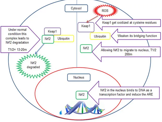
In normal conditions, Nrf2 is found bound to Kelch-like ECH-associated protein 1 (Keap1) which in turn is bound to actin in the cytosol. Keap1 promotes binding of Cul3-dependent E3 (Cul3) ubiquitin ligase complex to Nrf2 as a part of a post-translational modification of the protein. This leads to Nrf2 degradation under normal conditions making its half-life around 13-21 minutes [116, 117]. This keeps cellular Nrf2 levels relatively low. Keap1 is rich in the amino acid cysteine which makes it works as a sensor for the redox status of the cell.
Under oxidative stress, intracellular ROS and electrophiles increase leading to increasing oxidation of Keap1 cysteine residues (C151, C273, C288, C613) that limits its activity as a bridge binding ubiquitin ligase complex to Nrf2 and thus limits its role in Nrf2 degradation. Here Nrf2 half-life extends to 100-200 minutes [117]. Once Nrf2 is
35
released from the Keap1 complex, and survives degradation, it migrates to the nucleus where it plays its role as a transcription factor for ARE genes [118]. Nrf2 binds to promoter regions of many cytoprotective genes including L-PGDS, as recently a Nrf2 binding site was identified on the promoter region of L-PGDS gene as explained before [108]. Figure 7 explains the Nrf2 pathway and the role of ROS in its function.
Figure 7: Nrf2 pathway and role of ROS in its activation.
ROS leads to oxidation of cysteine residues on Keap1 leading to loss of its function as a bridge for ubiquitin. This leads to longer t1/2 for Nrf2 which then migrates to the nucleus and binds to promoter regions of the ARE genes.
36
A recent study in 2013 used coffee alkaloid trigonelline (trig) as an inhibitor of Nrf2 pathway in pancreatic cancer cells to block its protective effect and its anti-apoptotic effect. They had promising results controlling cancer growth [119]. Estrogen-related receptor beta (ERRβ) is a family of nuclear receptors that were found to block the Nrf2 pathway, particularly the (SFhERRβ) member of this family [120]. Nrf2 siRNA can also be used to silence this transcription factor to test its involvement in IL1-β induced L-PGDS [121].
To summarize, PGD2 had a positive feedback on L-PGDS induction through Nrf2, so
it may activate the intrinsic ARE as well as Nrf2. This suggests that PGD2 can
contribute to anti-inflammatory actions in two different ways, through 15d-PGJ2 or Nrf2 induced ARE. Nrf2 has a specific binding site on L-PGDS gene promoter region and induces it in macrophages. This opens the door for the hypothesis that the Nrf2 pathway may be involved in L-PGDS induction in chondrocytes also. IL-1β has a known role as an inflammatory mediator in many tissues including chondrocytes. It induces many inflammatory pathways including those producing free radicals, the main activator of Nrf2. This may explain the mechanism by which I1β induces L-PGDS, through Nrf2.
1.7.6 Objectives and rationale:
15d-PGJ2 is a major anti-inflammatory PG. It works through PPAR-γ to downregulate major inflammatory genes like MMP1, MMP13, iNOS, TNF-α, IL1β and COX-2. 15d-PGJ2 is formed by a dehydration reaction from PGD2. PGD2 is formed by PGDS from PGH2, the main precursor for all PG’s. PGH2 is in turn formed from AA by the
37
COX enzyme. This makes PGDS the rate-limiting step for 15d-PGJ2 synthesis and so for a major intrinsic anti-inflammatory pathway. Figure 22 explains this pathway in details. Considering the abundance of L-PGDS versus H-PGDS in chondrocytes, both normal and OA, we are focusing in this study on L-PGDS. Therefore, the main objective of this study is to understand the regulation of L-PGDS expression in chondrocytes. We are aiming in this study to identify chondrocytes’ response to different inflammatory conditions in vivo and in vitro, regarding L-PGDS. To do so, we are going to analyze the effect of IL-1β and interleukin 17 (IL-17) (as inflammatory cytokines), as well as H2O2 and 4BHP (as ROS generators) on L-PGDS expression levels in human chondrocytes in vitro. ROS generators are used in this study for two main reasons, first, ROS induce inflammation and second, ROS increase the half-life of Nrf2 and so enable its translocation into the nucleus to express ARE genes (see
Figure 7). Considering that Nrf2 has a binding site on L-PGDS gene promoter region
and plays a role in its regulation as explained before, among our objectives is to study the effect of ROS generators on L-PGDS expression levels. In vivo also, we are going to analyze L-PGDS expression levels by chondrocytes in mice OA models, both surgically induced and naturally induced (by aging), using immunohistochemistry techniques.
38
Chapter 2: Materials and
Methods
39
2.1 Destabilization of medial meniscus surgery:
The destabilization of medial meniscus (DMM) surgeries were done in 10-12 weeks old male mice, strain C57BL/6, to induce an OA model. These mice were sacrificed at 8 weeks of ages post-operatively; this time selection was based on observations from previous work done in our lab for the time needed to develop OA post DMM. Their knees were examined for OA changes histologically and for L-PGDS expression by immunohistochemistry.
The medial meniscus is fixed to the tibial plateau by the medial meniscotibial ligament (MMTL). A 3 mm longitudinal incision was made on the distal side of the patella with a # 15 blade exposing the joint capsule. The joint capsule was then opened by micro-iris scissors and a dissection was performed on the meniscotibial ligament. MMTL was dissected in a proximo-lateral direction with a micro-surgical knife. This led to the destabilization of the medial meniscus which eventually led to a medial displacement of the meniscus. The joint capsule was then closed by continuous sutures using 8-0 Vicryl®, and for the subcutaneous tissues and skin 7-0 Vicryl® was used. Sham surgeries were performed in animals following the same steps without dissecting the meniscotibial ligament.
2.2 Aging associated OA:
We followed the normal aging process in a large number of mice to determine the spontaneous appearance of OA compared to the DMM model. We sacrificed the mice and collected their knees at 3 or 18 months of age; this was also based on
40
was to obtain knee joint samples at different age points to study the changes in the articular cartilages, bones, synovial membranes, genes and proteins at different time points.
2.3 Histology:
Histology blocks were made in collaboration with other labs at Notre Dame Hospital. Blocks were cut using Leica® rotary microtome into 5 micrometers slices. They were then put on histology glass slides, and kept in the incubator for 48h at 37°C.
For histological staining we used a protocol of different washing and staining steps, in which the slides were processed as follow:
First we washed the slides in Xylene for 5 minutes twice. The slides were then washed again in ethanol 100% for 3 minutes twice. Then, one more wash with double distilled water (ddH2O) for 2 more minutes.
Slides were then stained with hematoxylin weigert 50% (Sigma-Aldrich®) for 4 minutes, then washed in ddH2O for 2 minutes, incubated in ferrous chloride for 1 minute, and finally washed in ddH2O for 2 more minutes.
Fast green staining (Sigma-Aldrich®) was done by incubating the slides in the dye for 30 seconds, then in acetic acid 1% for 5 seconds, followed by safranin-O (Sigma-Aldrich®) for 4 more minutes.
The final step was washing the slides with ethanol 100% for 1 minute, 3 times and then xylene for 2 minutes, two times. Slides were ready at that point and were covered with glass cover slips and glued.
41 2.4 Immunohistochemistry:
For immunohistochemistry, we washed the slides in Xylene for 5 minutes, 3 times and then in ethanol for 5 minutes, 3 more times (at concentrations of 100%, 95% and 75%). A final wash was performed in ddH2O for 1 minute twice. The sections on the slides were then marked with histology PAP pen (Abcam®), incubated with one drop on each section of peroxidase (Dako®) for 10 minutes at room temperature on a humid rack. This last step was the 1st blocking step. Slides were then washed with ddH2O for 1 minute, twice. They were then blocked again with 50µl of 1% BSA for 1h at room temperature. The 1st antibody was then incubated with the slides overnight (O/N) at 4°C in BSA 1% at different concentrations. For L-PGDS, the concentration was 1:100. Slides were then prepared for day 2 where they were washed in ddH2O for 1 minute twice, and then 1 drop of biotinylated link antibody (Dako®) was added to each slide for 30 minutes at room temperature. Slides were then washed again in ddH2O for 1 minute twice. Streptavidin HRP was added to each slide for 1 hour at room temperature. Another wash cycle was done. For the next step we prepared a diaminobenzidine (DAB) solution (Dako®), as 1 drop of DAB in 1ml of its buffer in a dark Eppendorf. The DAB solution forms a brown stain in peroxidase-based immunohistochemical reactions. We added 1 drop of the DAB preparation to each section and followed the change of the color under the microscopy. Once the tissues get the DAB brown stain we stopped the reaction by washing the slides in ddH2O to avoid oversaturation of the signal. Lastly, the slides were counterstained with Eosin. Slides were washed in ddH2O for 1 minute 4 times, then in 80% ethanol for 30 seconds, and then stained with Eosin for 2 seconds. Slides were then washed in
42
100% ethanol 3 times (5 minutes, 3 minutes and another 3 minutes). Lastly, they were washed in xylene for 2 minutes, after which we glued the slides with a glass cover. Each group of slides was treated under the same conditions and for the same periods so we can compare the results from different slides in the same group and within different groups. Expression levels of L-PGDS were quantified by counting cells stained with the DAB brown stain versus the total number of cells per high power microscopic field. This counting was done with at least 3 slides for each condition tested and the median of each condition was taken. The medians were compared in aged versus young mice, and in DMM versus the sham surgery group to quantify the increase in L-PGDS expression.
2.5 Cartilage collection & Chondrocyte culture:
Knee specimens were collected from patients undergoing joint replacement on the same day of operation or 1 day after. Normal knee specimens were collected from cadavers shortly after death. Synovial fluid samples were collected from patients with OA and normal subjects. All specimens were kept at 4°C until digestion to ensure the viability of chondrocytes.
Cartilage pieces were cut from the knees under aseptic conditions and kept them in cold sterile Phosphate buffered saline (PBS). The cartilage pieces were then washed twice with sterile PBS then kept in (penicillin/streptomycin) 5X in PBS for 1 hour at room temp. Cartilage pieces were then washed again twice with regular sterile PBS (without antibiotics), to remove any traces of the antibiotics. We started then digesting the cartilage pieces by incubating them in Pronase 1mg/ml in DMEM+ 10% FBS+ 1%
43
P/S Sigma-Aldrich® in a water bath at 37° for 1 hour with gentle agitation. Cartilage pieces were then washed with warm (37°C) sterile PBS twice. A collagenase digestion was then performed using 600units/ml or 2mg/ml of collagenase type I (Sigma-Aldrich) following its addition to the cartilage pieces for 5 hours incubated at 37°C in a water bath with gentle agitation. The fluid (containing chondrocytes) was then collected into 50ml falcon tubes and centrifuged at 1500 revolutions per minute (rpm) for 10 minutes. The supernatant was discarded, cells were re-suspended in DMEM 10% FBS 1% P/S, then seeded into a 175ml flask. The flasks were then incubated at 37°C in humidified 5% CO2 incubator, for 2 weeks until they reach full
confluence. The remaining cartilage pieces were then digested one more time by applying 50% diluted collagenase from the previous preparation, incubated at 37°C in a water bath with agitation O/N. The digestion fluid then went through the same previous steps to collect 2nd day chondrocytes and seed them into another 175ml flask. Both flasks were incubated for 2 weeks until they reached full confluence. Chondrocytes were then collected from the 175ml flask by trypsinization using 5ml trypsin 0.05% Multicell® per flask and kept in the incubator for 5 min. After the 5 minutes, the trypsinization reaction was stopped by adding 10ml DMEM media containing 10% FBS and 1% P/S. The fluid was then centrifuged at 1500rpm for 10 min, cells were re-suspended in different volumes of DMEM 10% FBS 1% P/S according to the final cell count needed per ml. After counting cells, they were seeded into 6 well plates at a density of 750,000 cells per well in 2ml media or into 12 well plates at a density of 250,000-300,000 cells per well in 1ml media. Cells were left 24h
44
to attach to the wells and then serum starved in a media containing 1% FBS for another 24h. At that point, chondrocytes were ready to be treated.
Chondrocytes were treated with:
• IL-1β (an inflammatory cytokine) at different concentrations from 0-600pg/ml for 48h, or at 100pg/ml for time course assays.
• IL-17 (also an inflammatory cytokine) at different concentrations from 0-100ng/ml for 48h, or at 50ng/ml for time course assays.
• H2O2 (as a ROS generator) at different concentrations from 0-50µM/ml for 48h
(re-added every 24h due to its instability), or at 20µM/ml for time course assays, (re-added also every 24h).
• 4BHP (also a ROS generator) at different concentrations from 0-50µM/ml for 24h and 48h, or 20µM/ml for time course assays.
For cell lysis, we removed the growth media from the plates from the previous step and washed the cells with cold sterile PBS 1ml/well twice. We then lysed the cells with a lysis buffer composed of Tris 20mM PH7.4, NaCl 150mM, EDTA 1mM PH 7.4, PMSF 1mM, protease inhibitors at concentration 1µg/ml and SDS 0.5% in double distilled water using 200µl/well for the 12 wells plates, or 400µl/well for the 6 well plates. The cells were incubated with the lysis buffer for 10min at room temperature with gentle shaking. Samples were then collected in Eppendorf tubes and sonicated for 10sec on ice. The lysate was then centrifuged at 13,000 rpm for 5min. Proteins