R E S E A R C H
Open Access
Duplex quantitative real-time PCR assay for the
detection and discrimination of the eggs of
Toxocara canis and Toxocara cati (Nematoda,
Ascaridoidea) in soil and fecal samples
Jean-Francois Durant
1, Leonid M Irenge
1,2, Renata Fogt-Wyrwas
3, Catherine Dumont
4, Jean-Pierre Doucet
5,
Bernard Mignon
6, Bertrand Losson
6and Jean-Luc Gala
1,2*Abstract
Background: Toxocarosis is a zoonotic disease caused by Toxocara canis (T. canis) and/or Toxocara cati (T. cati), two worldwide distributed roundworms which are parasites of canids and felids, respectively. Infections of humans occur through ingestion of embryonated eggs of T. canis or T. cati, when playing with soils contaminated with dogs or cats feces. Accordingly, the assessment of potential contamination of these areas with these roundworms eggs is paramount.
Methods: A duplex quantitative real-time PCR (2qPCR) targeting the ribosomal RNA gene internal transcribed spacer (ITS2) has been developed and used for rapid and specific identification of T. canis and T. cati eggs in fecal and soil samples. The assay was set up on DNA samples extracted from 53 adult worms including T. canis, T. cati, T. leonina, Ascaris suum (A. suum) and Parascaris equorum (P. equorum). The assay was used to assess the presence of T. cati eggs in several samples, including 12 clean soil samples spiked with eggs of either T. cati or A. suum, 10 actual soil samples randomly collected from playgrounds in Brussels, and fecal samples from cats, dogs, and other animals. 2qPCR results on dogs and cats fecal samples were compared with results from microscopic examination. Results: 2qPCR assay allowed specific detection of T. canis and T. cati, whether adult worms, eggs spiked in soil or fecal samples. The 2qPCR limit of detection (LOD) in spiked soil samples was 2 eggs per g of soil for a turnaround time of 3 hours. A perfect concordance was observed between 2qPCR assay and microscopic examination on dogs and cats feces.
Conclusion: The newly developed 2qPCR assay can be useful for high throughput prospective or retrospective detection of T.canis and/or T. cati eggs in fecal samples as well as in soil samples from playgrounds, parks and sandpits.
Keywords: Duplex real-time PCR, ITS2, Toxocara, Eggs, Fecal, Soil, Samples
* Correspondence:jean-luc.gala@uclouvain.be
1Centre de Technologies Moléculaires Appliquées, Institut de Recherche Expérimentale et Clinique, Université catholique de Louvain, Clos chapelle-aux-champs, 30 B1.30.24, 1200 Brussels, Belgium 2
Defense Laboratories Department, ACOS Ops&Trg, Belgian Armed Forces, Martelarenstraat, 181, 1800 Peutie, Belgium
Full list of author information is available at the end of the article
© 2012 Durant et al.; licensee BioMed Central Ltd. This is an Open Access article distributed under the terms of the Creative Commons Attribution License (http://creativecommons.org/licenses/by/2.0), which permits unrestricted use, distribution, and reproduction in any medium, provided the original work is properly cited.
Background
Toxocarosis is a zoonotic disease caused by the larvae of Toxocara, a worldwide distributed roundworm genus of the ascaroid group. Toxocara species of human and ani-mal health significance are essentially represented by T. canis and T. cati, parasites of canids and felids, respect-ively [1]. According to recent data, the prevalence of T. canis or T. cati is variable but remains high [2]. This widespread prevalence of Toxocara spp. in dogs and cats is associated with the contamination of playgrounds, municipal parks and households with eggs [3-5]. Red foxes (Vulpes vulpes) are also frequently infected by T. canis, an observation to consider in the light of recent epidemiological studies, which point out the progressive increase in the number of foxes in European urban environments over the last few years [6,7]. Children are most likely infected through ingestion of embryonated eggs of T. canis or T. cati when playing on soils con-taminated with dogs or cats feces containing Toxocara eggs. After ingestion, Toxocara eggs hatch in intestine and release larvae (juvenile worms) that penetrate the small intestine wall to enter the bloodstream. They sub-sequently travel through the bloodstream to all the major organs. Although most infections are asymptom-atic, two well-defined syndromes are classically recog-nized in humans: visceral larva migrans (VLM), a systemic disease caused by larval migration through major organs, and ocular larva migrans (OLM), a disease limited to the eye and optic nerve. Less severe syn-dromes have been described, in children (covert toxocar-iasis) and in adults (common toxocartoxocar-iasis) [8-10].
Accordingly, monitoring the presence of Toxocara eggs in dogs and cats feces, as well as in playgrounds and municipal parks likely to be contaminated by animal stools is critical in the control of toxocarosis.
Microscopic examination of dog and cat stools or soil samples is commonly used for identification of Toxocara eggs. The method includes an enrichment pre-analytical step through the use of centrifuge-flotation techniques [11]. However, the method displays poor sensitivity, due to the low recovery of Toxocara eggs especially from soil samples. Furthermore, microscopic examination can fail to unambiguously discriminate eggs of Toxocara species because they are fairly similar [12,13]. Other important roundworms include Baylisascaris procyonis, the com-mon intestinal roundworm of raccoons responsible for a severe human neurologic disease [14] and possibly Toxo-cara vitulorum (T. vitulorum), a cattle roundworm, which has been linked to a low level zoonosis alleged to affect children. There is, however, much uncertainty about the zoonotic potential of this species, as infections attributed to T. vitulorum could be due to T. canis or T. cati [2]. Owing to their high sensitivity and high specifi-city, PCR methods have been highlighted to improve the
detection and identification of Toxocara species of human significance. Numerous PCR methods for the de-tection and identification of T. canis and T. cati were reported to identify T. canis and T. cati in dog, fox and cat stools [15], as well as in soil samples [16,17]. The DNA-based methods take advantage of the high genetic variability within molecular markers such as ITS2 for the discrimination of T. canis and T. cati from their closely-related neighbors, namely T. leonina, T. vitu-lorum and T. malaysiensis [2,17]. However, despite these achievements, many drawbacks actually preclude their implementation in routine screening of Toxocara spp of clinical significance, either in dog, cat and fox stools or in soil samples from playgrounds and parks. These pitfalls include the risk of carry over contamin-ation, the low throughput of samples analysis, the dif-ficulty of automation and the lack of standardization. Accordingly, real-time PCR has the potential to cir-cumvent the drawbacks of endpoint PCR. Moreover, real-time PCR is rapid and can allow analysis of many samples in a short time without any need of add-itional post-PCR manipulations, often responsible for carry-over contamination. Consequently, the develop-ment of a real-time PCR assay for detection of Toxo-cara eggs could improve diagnosis of toxocariosis and thus improve health status of children in contaminated areas.
In the current study, we have developed a 2qPCR assay for rapid and specific identification of T. canis and T. cati eggs in fecal samples as well as in soil samples from sandpits and playgrounds. Results suggest that the 2qPCR assay is sensitive and specific for detecting T. canis and/or T. cati both in fecal and soil samples.
Methods
Biological and environmental samples
Three adult worms (one representative each from T. canis, A. suum and P. equorum), were provided by the Parasitology unit of the Faculty of Veterinary Medicine (FMV) of the University of Liège and subsequently used throughout the study as positive and negative controls. Fifty worms previously identified as T. canis (n = 30), T. cati (n = 14) or T. leonina (n = 6) by macroscopic and microscopic examination were kindly provided by Dr. R. Fogt (Department of Biology and Environmental Protection, University School of Physical Education, Poznań, Poland). Egg suspensions of T. cati and A. suum recovered from two adult worms were pro-vided by FMV.
Microscopic observations
Suspensions of T. cati and A. suum eggs were kindly provided by B. Mignon. For egg quantification, smears of 20 μL of egg suspension were observed under light
microscopy and counted. This process was performed in triplicate and the number of eggs in suspension was cal-culated as the mean from the three counts.
Microscopic examination for Toxocara egg identifica-tion was performed on enriched fecal samples through the flotation technique, as described previously [11].
Spiking of soil samples withT. cati or A. suum eggs
Twelve control soil samples, each made of 5 g of clean sand were spiked with known amounts of T. cati eggs (from 100 ± 30 to 5 ± 2) or A. suum eggs (from 1020 ± 280 to 5 ± 2).
Molecular analysis DNA extraction
DNA extraction was carried out on a suspension containing known amounts of T. cati and A. suum eggs. Briefly, the eggs in suspension were incubated with 100 μL of buffer G2 (Qiagen, Hilden, Germany) and with 20μL of protein-ase K (Qiagen, Hilden, Germany) at 56°C during 2 hours. DNA was purified with BioRobot EZ1 (Qiagen, Hilden, Germany), using a DNA tissue kit, according to the manu-facturer's instructions. The DNA was finally eluted in 100 μL of buffer and stored at −20°C until use.
DNA was extracted from 53 adult worms. Briefly, a piece of ~0.2 cm long was cut from each worm, minced with a scalpel blade on a sterile glass slide, and re-suspended in 500 μL NucliSENSW lysis buffer (Nucli-SENSW lysis magnetic extraction reagents, NucliSENSW miniMAG System, Biomérieux bv, Boxtel NL). The DNA was extracted from the suspension using the NucliSENSW miniMAG system and reagents according to the manufac-turer’s instructions. DNA was eluted in 100 μL and stored at−20°C until use.
The QIAampW DNA Stool (QiagenW, Leusden, The Netherlands) commercial kit was used to extract DNA from 45 animal fecal samples. In total ~250 mg of each fecal sample was processed according to the recommenda-tions of the manufacturer. DNA solurecommenda-tions were eluted in 200μL of buffer and stored at −20°C.
Total DNA was extracted from 5 g of soil samples (both spiked and actual soils) using the PowerMaxW soil DNA isolation kit and re-suspended in a final volume of 5mL of elution buffer as recommended by the manufacturer. The 5 mL of DNA extract solution was then reduced to 50μL fol-lowing an ethanol precipitation protocol recommended by the PowerMaxW soil DNA isolation kit manufacturer and stored at−20°C until use.
The 2qPCR real-time assay
Internal transcribed spacer 2 (ITS2) was selected as a tar-get for the amplification the T. canis and T. cati. Briefly,
the 2qPCR amplification was performed to specifically identify T. canis or T. cati. Primers and probes were designed manually in the T. canis and T. cati- specific part of ITS2 after multiple-alignment of the following ITS2 sequences: T. canis [Genbank: AB110034], T. cati [Genbank: AB110033], T. leonina [Genbank:Y09490], T. vitulorum [Genbank:EU189085] and T. malaysiensis [Genbank:AM231609]. ITS2 duplex- amplification was based on the use of two forward primers specific for T. canis (5’-GCGCCAATTTATGGAATGTGAT-3’) and T. cati (5’-ACGCGTACGTATGGAATGTGCT-3’) respect-ively, and a consensus reverse primer common to both species (5’-GAGCAAACGACAGCSATTTCTT-3’). More-over, the 2qPCR use two specific probes targeting T. canis (5’-FAM-CCATTAC CACACCAGCATAGCTCACCGA -3’-BHQ1) and T. cati (5’-Cy5-TCTTTCGCAACGTG CATTCGGTGA-3’-BHQ3). The selected primer candi-dates and the probes were tested in silico against all the publicly available nucleotide sequence databases by using BLASTN [18]. The expected amplicon sizes for T. canis and T. cati were 141-bp and 155-bp, respectively. Primers and probes were purchased from Eurogentec (Ougrée, Belgium).
Each 2qPCR was carried out in 25 μL of a reaction mixture containing 2.5 μL of extracted DNA as tem-plate, 300 nM of each primer, 100 nM of each probe and 12.5 μL of LightCyclerW 480 Probes Master 2x (Roche Diagnostics GmbH, Mannheim, Germany). Amplifica-tion was performed on a Roche LightCyclerW 480 Sys-tem Real-Time PCR sysSys-tem (Roche Diagnostics GmbH, Mannheim, Germany). The reaction was initiated at 95°C for 5 min followed by 45 cycles of denaturation at 95°C for 10 s, annealing at 60°C for 15 s and extension at 72°C for 5 s. Each sample was tested in triplicate and data were recorded as crossing points (Cq) on a Roche LightCyclerW 480 System, using the analytical software LCS480 1.2.9.11 from the same manufacturer.
Standard curves were constructed from serial dilutions of T. canis and T. cati DNA. Cq values obtained by 2qPCR assay were plotted against the logarithm of DNA amount to assess the dynamic range.
A sample was considered as positive when all wells within the triplicate were associated with an exponential fluorescence with a Cq value <40.00. The specificity of the test was investigated by performing the 2qPCR ana-lysis on DNA samples from worms (n =53) and negative controls from fungal and bacterial DNA (n = 33) and human DNA (n = 16). In order to define the LOD, a standard curve was constructed of 10:10 serially diluted DNA of these 2 DNA solutions which were used as a template for 2qPCR assays. Cq values obtained were plotted against the logarithm of copies to assess the dynamic range. The efficiency of 2qPCR assays was calculated as described by Wong and Medrano [19].
In-house qPCR assay
An in-house qPCR previously designed at CTMA and targeting the conserved region of the 18S rRNA in a wide range of Ascaridoidea was carried out on DNA batches extracted from all samples used in this study. This PCR was used as an internal quality control for DNA extraction while being also used to detect the pres-ence of Ascaridoidea in T. canis and T. cati-negative soil or fecal samples.
Primers and the probe were designed manually after a multiple alignment of the 18S sequences including T. canis [Genbank:U94382)], T. cati [Genbank:AF480059], T. leonina [Genbank:U94383], T. vitulorum [Genbank: EF180078], A. suum [Genbank:AF036587] and P. equorum [Genbank:U94378]. The 18S rRNA amplification was based on the use of a pair of primers (primer forward: 5’-CTACCACATCCAAGGAAGGCA-3’; primer reverse: 5’-TTATTTTTCGTCACTACCTCCTCATG-3’) and a probe (5’-CAGGCGCGCAAATTACCCACTCTC-3’) la-beled by the tandem Reporter-Quencher Red610-BHQ2.
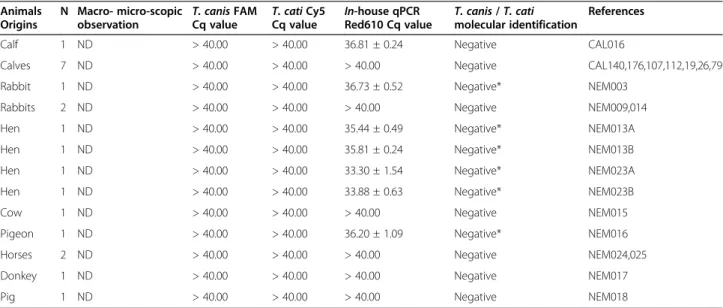
The specificity of the assay was further tested on several fecal samples from animals other than cats and dogs. These included randomly collected fecal samples from calves and/or cows (n = 9), horses (n = 2), rabbits (n = 3), hens (n = 4) as well as a single fecal sample from a donkey, a pig and a pigeon (Table 1). Noteworthy, these animals are not hosts of T. canis and T. cati, though they are com-mon hosts of other roundworms species. For each nega-tive sample, PCR inhibition was assessed as previously described [20]. This process also included the testing of a panel of DNA from bacteria (n = 25) and fungi (n = 8) (Table 2) and human DNA (n = 16).
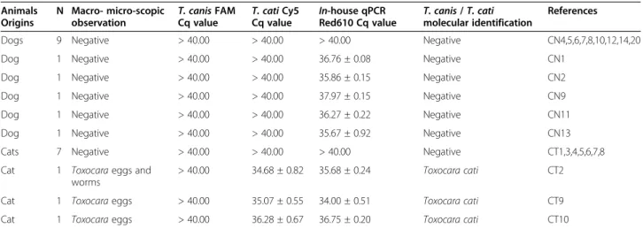
The test phase for the presence of T. canis and T. cati in fecal samples was carried out on a panel of 24 feces from dogs (n = 14) and from cats (n = 10) collected by the Clinivet veterinary centre (Table 3). Molecular results were compared with results of the light micro-scopic examination when these were available.
The 2qPCR assay was also used to assess the presence of T. canis and/or T. cati in 10 soil samples collected from sandpits and playgrounds across various areas of Brussels city (Belgium).
Statistical analysis
Statistical analyses were performed using the SPSS stat-istical package release 12.0 for Windows (SPSS, Inc., Chicago, IL). Concordance between 2qPCR and micro-scopic observation was calculated using the Kappa sta-tistics of Cohen, to assess the degree of agreement between these different methods.
Results
Limit of detection ofT. canis and T. cati DNA by 2qPCR
The PCR efficiencies were 100% and 95.8% for T. canis and T. cati, respectively (Figure 1). Significant and repro-ducible fluorescence signals generated by T. cati or by A. suum were consistently obtained with a DNA solution equivalent to 2 eggs per g of soil (Table 4). Regarding T. canis, the LOD estimation was based on DNA extracted from adult worms and calculated at 10 fg per assay. Note-worthy, prior to these experiments, the potential contam-ination of the soil sample to be spiked with Ascaridoidea eggs was ruled out by performing the 2qPCR as well as the in-house qPCR on these 12 soil samples.
Table 1 2qPCR results from fecal samples animals other than dogs and cats
Animals Origins
N Macro- micro-scopic
observation T. canis FAMCq value T. cati Cy5Cq value In-house qPCRRed610 Cq value T. canis / T. catimolecular identification
References
Calf 1 ND > 40.00 > 40.00 36.81 ± 0.24 Negative CAL016
Calves 7 ND > 40.00 > 40.00 > 40.00 Negative CAL140,176,107,112,19,26,79
Rabbit 1 ND > 40.00 > 40.00 36.73 ± 0.52 Negative* NEM003
Rabbits 2 ND > 40.00 > 40.00 > 40.00 Negative NEM009,014
Hen 1 ND > 40.00 > 40.00 35.44 ± 0.49 Negative* NEM013A
Hen 1 ND > 40.00 > 40.00 35.81 ± 0.24 Negative* NEM013B
Hen 1 ND > 40.00 > 40.00 33.30 ± 1.54 Negative* NEM023A
Hen 1 ND > 40.00 > 40.00 33.88 ± 0.63 Negative* NEM023B
Cow 1 ND > 40.00 > 40.00 > 40.00 Negative NEM015
Pigeon 1 ND > 40.00 > 40.00 36.20 ± 1.09 Negative* NEM016
Horses 2 ND > 40.00 > 40.00 > 40.00 Negative NEM024,025
Donkey 1 ND > 40.00 > 40.00 > 40.00 Negative NEM017
Pig 1 ND > 40.00 > 40.00 > 40.00 Negative NEM018
Assessment of the specificity of the 2qPCR
The 2qPCR assay generated specific 6-FAM fluorescence signals with all DNA samples extracted from T. canis adult worms (n = 31), whereas DNA samples extracted from T. cati adult worms (n = 14) or eggs (n = 2) gener-ated specific Cy-5 signals. No single T. cati DNA sample generated 6-FAM fluorescence, nor did any T. canis sample generate Cy-5 fluorescence. Sequence analysis of
each amplified target confirmed a 100% identity with the corresponding worm-ITS2 molecular targets. The 2qPCR also remained negative with DNA from A. suum (one adult worm and eggs solutions), from P. equorum (1 adult worm) and from T. leonina (6 adult worms). The assay also remained negative with human DNA as well as all bacterial DNA tested. No PCR inhibition was observed when assaying both fecal and soil samples.
Test phase on fecal samples
From the 24 cat and dog fecal samples examined (Table 3), three cat samples (CT2, CT9 and CT10) dis-played real-time PCR signals consistent with the pres-ence of T. cati. In each of these samples, T. cati eggs were visualized by microscopic examination. In addition, adult worms could be seen in feces CT2. The 14 feces samples from dogs (Table 3) remained negative as also were the 21 feces samples from other animals (Table 1). Nonetheless, while these samples were negative for T. canis and T. cati, several of them, (CAL016, NEM003, NEM013A, NEM013B, NEM023A, NEM023B, and NEM016) displayed a positive signal with the in-house qPCR (suggesting the presence of non-T. cati/canis Ascaridoidea eggs in these samples). Results of the 2qPCR assay and microscopic examination on the 24 cat and dog fecal samples were identical. Sensitivity and spe-cificity of the assay were calculated in comparison with microscopic examination (considered as gold standard) on the 24 cat and dog fecal samples and both displayed a 100% value. The kappa score of Cohen, a measure of agreement between microscopic observation and 2qPCR, was 100%.
Applicability of the assay assessed in actual soil samples
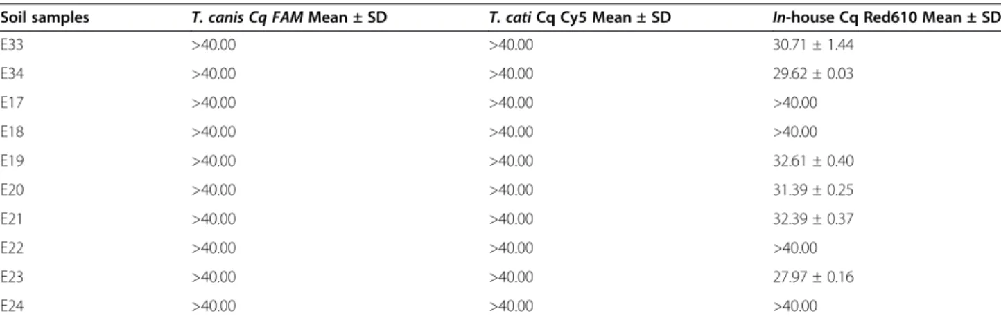
The assay has been used to assess 10 soil samples from sandpits and playgrounds collected in different areas of Brussels city (Belgium). While all the 10 samples remained negative for T. cati and T. canis, 6 out of 10 samples displayed positive signals with the in-house qPCR (Table 5), thus suggesting the presence of non-Toxocara Ascaridoidea eggs in these samples. The latter results as well as the previous ones from spiking Toxo-cara eggs in soil show that the assay is applicable for monitoring of the presence of Toxocara eggs in soil samples.
Discussion
We report here the development of a sensitive and spe-cific 2qPCR assay allowing rapid and reliable identifica-tion of eggs of T. canis and T. cati in clinical and environmental samples. Increasing populations of dogs, cats and foxes in the urban areas prompt indeed the need for standardized and high throughput analytical methods for studying the prevalence of Toxocara spp
Table 2 Bacterial and fungal DNA used for the setting up of the 2qPCR assay
Species Strain and isolate references
Bacillus anthracis CEB 9434
Bacillus cereus DSM 345
Enterococcus casseliflavus DSM 10255
Staphylococcus aureus ATCC 35884
Streptococcus oralis DSM 20627
Streptococcus pneumoniae ATCC 6314-D
Streptococcus pyogenes ATCC 12344-D
Acinetobacter calcoaceticus DSM 30006 Citrobacter freundii DSM 30039 Escherichia coli R453 Escherichia coli R456 Escherichia coli R457 Haemophilus influenzae DSM 4690
Klebsiella oxytoca ATCC 700324-D
Klebsiella pneumoniae ATCC 700721-D
Legionella pneumophila DSM 7513
Moraxella catarrhalis DSM 9143
Neisseria gonorrhoeae ATCC 53420-D
Neisseria meningitidis DSM 10036 Providencia stuartii DSM 4539 Pseudomonas aeruginosa DSM 50063 Pseudomonas fluorescens DSM 50090 Pseudomonas syringae DSM 1241 Serratia marcescens DSM 30121 Stenotrophomonas maltophilia DSM 8573
Alternaria alternata CTMA 07-022
Aspergillus fumigatus CTMA 07-035
Botrytis cinerea CTMA BC/07/31
Cladosporium cladosporoides CTMA 07-019
Cladosporium herbarum CTMA CH/07/41
Epicoccum nigrum CTMA 07-086
Pleospora herbarum CTMA 07-077
Trichophyton rubrum CTMA MYC001
ATCC: American Type Culture Collection.
CEB: Centre d’Etude du Bouchet, Vert le Petit, France.
DSM: Deutsche Sammlung von Mikroorganismen und Zellkulturen GmbH. CTMA: Center for Applied Molecular Technologies, private DNA sample collection.
eggs, mainly T. canis and T. cati, in fecal and environ-mental samples [9,17,21,22]. Over the last decade, sev-eral methods have been reported for the identification of Toxocara spp in environmental samples. These include light and scanning electron microscopy [13] and mo-lecular identification approaches for genotyping and identifying Toxocara spp. [15,16].
The aim of this study was to exploit the existing know-ledge and expertise in the field of real-time PCR and semi DNA extraction methods for the development of a high throughput method for the identification of Toxo-cara spp in fecal and soil samples. The analysis of stool
samples both by 2qPCR and microscopic observation showed a perfect correlation. As illustrated by the current molecular results, combining DNA extraction and 2qPCR contributes to a better standardization regarding the pre-analytical and analytical steps while allowing swift identification of T. canis or T. cati eggs. Compared with other conventional assays, the real-time PCR technology appears as a high throughput method for detecting Toxocara eggs in fecal and soil samples. Accordingly, this molecular assay can be used to assess the contamination of parks, playgrounds and sandpits with eggs of T. canis or T. cati. In the current study 5 g
Table 3 2qPCR results from dog and cat fecal samples
Animals Origins
N Macro- micro-scopic
observation T. canis FAMCq value T. cati Cy5Cq value In-house qPCRRed610 Cq value T. canis / T. catimolecular identification
References
Dogs 9 Negative > 40.00 > 40.00 > 40.00 Negative CN4,5,6,7,8,10,12,14,20
Dog 1 Negative > 40.00 > 40.00 36.76 ± 0.08 Negative CN1
Dog 1 Negative > 40.00 > 40.00 35.86 ± 0.15 Negative CN2
Dog 1 Negative > 40.00 > 40.00 37.97 ± 0.15 Negative CN9
Dog 1 Negative > 40.00 > 40.00 36.27 ± 0.22 Negative CN11
Dog 1 Negative > 40.00 > 40.00 35.67 ± 0.92 Negative CN13
Cats 7 Negative > 40.00 > 40.00 > 40.00 Negative CT1,3,4,5,6,7,8
Cat 1 Toxocara eggs and
worms
> 40.00 34.68 ± 0.82 35.68 ± 0.24 Toxocara cati CT2
Cat 1 Toxocara eggs > 40.00 35.07 ± 0.55 34.00 ± 0.51 Toxocara cati CT9
Cat 1 Toxocara eggs > 40.00 36.28 ± 0.67 36.75 ± 0.20 Toxocara cati CT10
Figure 1 2qPCR standard dilution curves forToxocara canis and Toxocara cati. Mean Cq values (Y axis) plotted against logarithm of DNA amount used for amplification (X axis). Continuous line represents the dilution curve for T. canis 2qPCR amplification whereas the dashed line corresponds to the dilution curve of T. cati 2qPCR amplification. E = PCR efficiency; R² = Square of linear correlation coefficient.
of soil was directly processed in the assay, yielding a LOD of 2 Ascaridoidea eggs per assay. As highlighted in this work, direct processing of 5 to 10 g of soil followed by a DNA concentration process significantly improves the detection threshold in soil samples while avoiding the use of cumbersome techniques for soil enrichment. Nevertheless, since only a limited number of samples were processed and considering that these samples were predominantly negative, a validation on a large panel of potentially contaminated soil samples could help to con-firm the usefulness of the 2qPCR. Our results also showed that animal feces (hens, pigeons, rabbits and calves) and soil samples, presumably harbored worms other than T. canis and T. cati [23]. This is in line with our positive in-house qPCR but negative 2qPCR results, hence confirming the specificity of our real-time PCR
assay. Noteworthy, pigeon and rabbit feces are com-mon in municipal parks and other play grounds, along with cat and dog feces. It should also be stressed that while the in-house qPCR was used as a simplex real-time PCR, it can be easily scaled up to the 2qPCR and thus constitute a triplex real-time PCR with-out any impact on the sensitivity of the assay, provided that the real-time PCR platform used is adapted to multiplexing.
The 2qPCR assay appears, therefore, to be a specific, sensitive and reliable tool for identifying T. canis and T. cati and discriminating them, a result that may be easily overlooked when using microscopic light exam-ination [13]. Additionally, the 2qPCR assay may help discriminating clinically relevant and non-relevant Toxocara spp eggs.
Table 4 2qPCR andin-house qPCR assays on soil samples spiked with T. cati and/or A. suum eggs
Samples Eggs origins Amount of eggs Mean ±
SD T. canis Cq FAM Mean ±SD T. cati Cq Cy5 Mean ±SD In-house Cq Red610 Mean ±SD
E25 T. cati 100 ± 30 > 40.00 25.18 ± 0.06 25.83 ± 0.05 E26 T. cati 100 ± 30 > 40.00 25.29 ± 0.02 25.97 ± 0.03 E27 T. cati 10 ± 3 > 40.00 31.12 ± 1.03 32.00 ± 0.76 E28 T. cati 10 ± 3 > 40.00 31.49 ± 0.53 29.53 ± 0.77 E29 T. cati 5 ± 2 > 40.00 30.62 ± 0.84 31.49 ± 0.53 E30 T. cati 5 ± 2 > 40.00 > 40.00 > 40.00 E31 T. cati 5 ± 2 > 40.00 30.13 ± 1.01 31.10 ± 0.75 E9 A. suum 1020 ± 280 > 40.00 > 40.00 25.93 ± 0.06 E10 A. suum 102 ± 28 > 40.00 > 40.00 30.08 ± 0.22 E32 A. suum 102 ± 28 > 40.00 > 40.00 29.66 ± 0.15 E11 A. suum 10 ± 3 > 40.00 > 40.00 33.70 ± 1.47 E12 A. suum 5 ± 2 > 40.00 > 40.00 > 40.00 E13 No egg spiked 0 > 40.00 > 40.00 > 40.00
Cq values higher than 40.00 are considered as negative results.
Table 5 Molecular assays on actual soil samples collected from playgrounds and sandpits
Soil samples T. canis Cq FAM Mean ± SD T. cati Cq Cy5 Mean ± SD In-house Cq Red610 Mean ± SD
E33 >40.00 >40.00 30.71 ± 1.44 E34 >40.00 >40.00 29.62 ± 0.03 E17 >40.00 >40.00 >40.00 E18 >40.00 >40.00 >40.00 E19 >40.00 >40.00 32.61 ± 0.40 E20 >40.00 >40.00 31.39 ± 0.25 E21 >40.00 >40.00 32.39 ± 0.37 E22 >40.00 >40.00 >40.00 E23 >40.00 >40.00 27.97 ± 0.16 E24 >40.00 >40.00 >40.00
The samples E33, E34, E19, E20, E21 and E23 are negative forT. canis and T. cati eggs and positive for non-Toxocara Ascaridoidea, whereas samples E17, E18, E22 and E24 are negative for any Ascaridoidea.
Although microscopic examination gave identical results to our 2qPCR assay, the identification process was all but a straightforward process. Eggs from frozen feces were particularly difficult to identify, owing to morphological modifications, and prompting reliance on a highly trained operator. It is of note that none of our soil samples were examined by light microscopy, but one can predict that observation of Toxocara spp eggs in this type of sample would have been even more challenging.
Lastly, though not assessed during this work, the assay is also expected to achieve accurate identification of Toxocara spp in tissue larva migrans. It has been reported indeed that during larva migrans, Toxocara spp larvae undergo morphological modifications which make species morphological-based identification nearly impos-sible [24].
Conclusion
In the present study, a molecular method was developed for allowing a reliable surveillance of fecal and soil sam-ple contamination with eggs of T. canis and T. cati. Compared to the conventional microscopic examination, used as gold standard, the real-time PCR approach appears to be rapid, displays a high throughput proces-sing rate, while achieving a sensitivity equivalent to the gold standard. Therefore, the current 2qPCR assay appears to be a very promising tool for assessment of contaminated sandpits and playgrounds by Toxocara spp eggs.
Competing interests
The authors report no conflicts of interest. The authors alone are responsible for the content and writing of the paper.
Authors’ contributions
DJF, ILM and GJL conceived the study; FWR, DJP, MB and LB provided worms, eggs suspensions and fecal samples, DJF, DC and MB carried out microscopic examination. DJF, DC and ILM carried out molecular analyses. ILM and GJL wrote the first draft of the paper, and all authors contributed to the final manuscript which they approve.
Acknowledgments
This work was funded by the Ministère de la Région Wallonne (DGTRE Division de la Recherche et de la Coopération Scientifique - project RESPIBAC no. 616313) supporting research and development and supported by the Department Management of Scientific and Technological Research of Defence (IRSD-RSTD, Royal High Institute for Defence). We thank Elodie Carlier (IRSDRSTD), PierreAlain Fonteyne (Université catholique de Louvain -UCL) and Françoise Maréchal (Université de Liège - Ulg) for their outstanding contribution to this work.
Author details
1Centre de Technologies Moléculaires Appliquées, Institut de Recherche Expérimentale et Clinique, Université catholique de Louvain, Clos chapelle-aux-champs, 30 B1.30.24, 1200 Brussels, Belgium.2Defense Laboratories Department, ACOS Ops&Trg, Belgian Armed Forces, Martelarenstraat, 181, 1800 Peutie, Belgium.3Department of Biology and Environmental Protection, University School of Physical Education, Królowej Jadwigi 27/39, 61-871 Poznań, Poland.4Royal Military Academy, Avenue de la Renaissance 30, 1000 Bruxelles, Belgium.5Clinivet, clinique vétérinaire de Gosselies, rue pont à Migneloux 39, 6041 Gosselies, Belgium.6Département
des Maladies Infectieuses et Parasitaires, Faculté de Médecine Vétérinaire, Université de Liège (Ulg), boulevard de Colonster, 20, B43, 4000 Liège, Belgium.
Received: 24 August 2012 Accepted: 23 November 2012 Published: 7 December 2012
References
1. Smith H, Holland C, Taylor M, Magnaval JF, Schantz P, Maizels R: How common is human toxocariasis? Towards standardizing our knowledge. Trends Parasitol 2009, 25(4):182–188.
2. Chen J, Zhou DH, Nisbet AJ, Xu MJ, Huang SY, Li MW, Wang CR, Zhu XQ: Advances in molecular identification, taxonomy, genetic variation and diagnosis ofToxocara spp. Infect Genet Evol 2012, 12:1344–1348. 3. Deplazes P, Van Knapen F, Schweiger A, Overgaauw PA: Role of pet dogs
and cats in the transmission of helminthic zoonoses in Europe, with a focus on echinococcosis and toxocarosis. Vet Parasitol 2011, 182(1):41–53. 4. Dado D, Izquierdo F, Vera O, Montoya A, Mateo M, Fenoy S, Galván AL,
García S, García A, Aránguez E, López L, Del Águila C, Miró G: Detection of zoonotic intestinal parasites in public parks of Spain, Potential epidemiological role of microsporidia. Zoonoses Public Health 2012, 59 (1):23–28.
5. Mattia S, Colli CM, Adami CM, Guilherme GF, Nishi L, Rubinsky-Elefant G, Marchioro AA, Gomes ML, Falavigna-Guilherme AL: Seroprevalence of Toxocara infection in children and environmental contamination of urban areas in Paraná State, Brazil. J Helminthol 2011, 25:1–6. 6. Brochier B, De Blander H, Hanosset R, Berkvens D, Losson B, Saegerman C:
Echinococcus multilocularis and Toxocara canis in urban red foxes (Vulpes vulpes) in Brussels, Belgium. Prev Vet Med 2007, 80(1):65–73.
7. Robardet E, Giraudoux P, Caillot C, Augot D, Boue F, Barrat J: Fox defecation behaviour in relation to spatial distribution of voles in an urbanised area: An increasing risk of transmission ofEchinococcus multilocularis? Int J Parasitol 2011, 41(2):145–154.
8. Despommier D: Toxocariasis: clinical aspects, epidemiology, medical ecology, and molecular aspects. Clin Microbiol Rev 2003, 16(2):265–272.
9. Otranto D, Eberhard M: Zoonotic helminths affecting the human eye. Parasit Vectors 2011, 4:41.
10. Chen J, Xu M-J, Zhou D-H, Song H-Q, Wang C-H, Zhu X-Q: Canine and feline parasitic zoonoses in China. Parasit Vectors 2012, 5:152. 11. Reinhard KJ, Confalonieri UE, Herrmann B, Ferreira LF, De Araujo AJG:
Recovery of Parasite Remains From Coprolites and Latrines: Aspects of Paleoparasitological Technique. Homo 1986, 37(4):217–239.
12. Borecka A, Gawor J: Modification of gDNA extraction from soil for PCR designed for the routine examination of soil samples contaminated with Toxocara spp. eggs. J Helminthol 2008, 82:119–122.
13. Uga S, Matsuo J, Kimura D, Rai SK, Koshino Y, Igarashi K: Differentiation of Toxocara canis and T. cati eggs by light and scanning electron microscopy. Vet Parasitol 2000, 92:287–294.
14. Wise ME, Sorvillo FJ, Shafir SC, Ash LR, Berlin OG: Severe and fatal central nervous system disease in humans caused by Baylisascaris procyonis, the common roundworm of raccoons: a review of current literature. Microbes Infect 2005, 7(2):317–323.
15. Jacobs DE, Zhu X, Gasser RB, Chilton NB: PCR-based methods for identification of potentially zoonotic ascaridoid parasites of the dog, fox and cat. Acta Trop 1997, 68:191–200.
16. Fogt-Wyrwas R, Jarosz W, Mizgajska-Wiktor H: Utilizing a polymerase chain reaction method for the detection ofToxocara canis and T. cati eggs in soil. J Helminthol 2007, 81:75–78.
17. Li MW, Lin RQ, Chen HH, Sani RA, Song HQ, Zhu XQ: PCR tools for the verification of the specific identity of ascaridoid nematodes from dogs and cats. Mol Cell Probes 2007, 21:349–354.
18. Altschul SF, Madden TL, Schäffer AA, Zhang J, Zhang Z, Miller W, Lipman DJ: Gapped BLAST and PSI-BLAST: a new generation of protein database search programs. Nucleic Acids Res 1997, 25(17):3389–3402.
19. Wong ML, Medrano JF: Real-time PCR for mRNA quantification. Biotechniques 2005, 39:1–11.
20. Lecouvet F, Irenge L, Vandercam B, Nzeusseu A, Hamels S, Gala JL: The etiologic diagnosis of infectious discitis is improved by amplification-based DNA analysis. Arthritis Rheum 2004, 50:2985–2994.
21. Epe C, Meuwissen M, Stoye M, Schnieder T: Transmission trials, ITS2-PCR and RAPD-PCR show identity ofToxocara canis isolates from red fox and dog. Vet Parasitol 1999, 84:101–112.
22. Saegerman C, De Blander H, Hanosset R, Berkvens D, Losson B, Brochier B: Evaluation des risques liés à la présence d’Echinococcus multilocularis et deToxocara canis dans la population vulpine en région bruxelloise. Epidémiol. et santé anim 2006, 50:97–104.
23. Ruff MD: Important parasites in poultry production systems. Vet Parasitol 1999, 84:337–347.
24. Bouchaud O, Houze S, Schiemann R: Cutaneous larva migrans in travelers: a prospective study, with assessment of therapy with Ivermectin. Clin Infect Dis 2001, 31:493–498.
doi:10.1186/1756-3305-5-288
Cite this article as: Durant et al.: Duplex quantitative real-time PCR assay for the detection and discrimination of the eggs ofToxocara canis and Toxocara cati (Nematoda, Ascaridoidea) in soil and fecal samples. Parasites & Vectors 2012 5:288.
Submit your next manuscript to BioMed Central and take full advantage of:
• Convenient online submission
• Thorough peer review
• No space constraints or color figure charges
• Immediate publication on acceptance
• Inclusion in PubMed, CAS, Scopus and Google Scholar
• Research which is freely available for redistribution
Submit your manuscript at www.biomedcentral.com/submit