Plant lipidomics: Discerning biological function by profiling plant complex lipids using mass spectrometry
Ruth Welti1, Jyoti Shah1, Weiqi Li2, Maoyin Li3,4,, Junping Chen5, John J. Burke5, Marie-Laure Fauconnier6, Kent Chapman7, Mee-Len Chye8, Xuemin Wang3
1Kansas Lipidomics Research Center, Division of Biology, Kansas State University, Manhattan, KS 66506, 2Kunming Institute of
Botany, Chinese Academy of Sciences, Kunming 661100, China, Department of Biology, Honghe University, Mengzi, Yunnan 661100, China, 3Department of Biology, University of Missouri, St. Louis, MO 63121, Donald Danforth Plant Science Center, St.
Louis, MO 63132, 4Department of Biochemistry, Kansas State University, Manhattan, KS 66506, 5Plant Stress and Germplasm
Development Unit, USDA-ARS, 3810 4th St., Lubbock, TX 79415, 6 Plant Biology Unit, Gembloux Agricultural University,
Passage des Déportés 2, B-5030 Gembloux, Belgium, 7Center for Plant Lipid Research, Department of Biological Sciences,
University of North Texas, Denton, TX 76203, and 8Department of Botany, The University of Hong Kong, Pokfulam, Hong Kong
TABLE OF CONTENTS 1. Abstract
2. Introduction
3. Stress-induced changes in plant lipid composition 3.1. Cold-induced lipid alterations
3.2. Lipid changes during adaptation to heat
3.2.1. Lipid profiles reveal that the dgd1-2 and dgd1-3 are leaky mutations of digalactosyldiacylglycerol synthase 1.
3.2.2. Acquired thermotolerance induced by heat acclimation in wild-type Arabidopsis is associated with increases in GalGalDG, GalGalDG to GalDG ratio, and saturation of GalGalDG.
3.2.3. The dgd1-2 and dgd1-3 mutants failed to increase GalGalDG level and GalGalDG to GalDG ratio in response to 38°C heat acclimation.
3.3. Wounding-induced alterations in plastidic lipids
3.4. Changes in phospholipids and galactolipids under phosphorus deficiency 4. Developmental changes in plant lipid composition
4.1. Lipid changes in cotton fibers during development 4.2. Alterations in potato lipids during tuber aging 5. Elucidation of gene function by analysis of plant lipid composition
5.1. Role of fatty acid desaturases and DHAP reductase in systemic acquired resistance 5.1.1. Lipid profiling as an aid in identification of mutated genes
5.2. Roles of phospholipases in response to freezing
5.2.1. Different roles of PLDs in freezing-induced lipid hydrolysis 5.3. Role of PLDzeta in lipid changes during phosphorus deficiency 6. Improvement of food quality by manipulation of lipid metabolizing genes 7. Perspective on the future
8. Acknowledgements 9. References
1. ABSTRACT
Since 2002, plant biologists have begun to apply mass spectrometry to the comprehensive analysis of complex lipids. Such lipidomic analyses have been used to uncover roles for lipids in plant response to stresses and to identify in vivo functions of genes involved in lipid metabolism.
2. INTRODUCTION
Innovative application of mass spectrometry technology in the 1990s demonstrated the potential for rapid, comprehensive, and quantitative profiling of complex lipid molecular species from biological samples without chromatographic separation (e.g., 1, 2). Electrospay ionization (ESI) mass spectrometry (MS) has proved to be particularly well-suited to complex lipid
analysis, and direct infusion of a biological extract combined with internal standards allows for straightforward quantification of polar lipid species from the mass spectra. The technical aspects of routine complex lipid analysis by ESI-MS have been reviewed recently (3, 4).
The methodology that has been most used in plant lipidomics utilizes an ESI triple quadrupole mass spectrometer (MS/MS) to collect a series of mass spectra. Each spectrum is specific for lipid molecular species with a common polar lipid head group, because lipids in a class produce a common head group-derived fragment in the mass spectrometer. Scanning in the precursor or neutral loss mode allows the production of the head group-derived fragment to be used as the criterion for detection (2, 5, 6). A series of head group-specific scans constitutes a lipid
profile, with lipid molecular species identified as to head group and mass, which is correlated with the number of acyl carbons in one or multiple acyl chains and the total number of acyl double bonds. Many of the mass spectral scans for plant lipids are the same as those used for analysis of animal lipids, (2), but several additional scans have been devised for plant-specific lipid classes (5, 7).
In plant biology, high-throughput lipid profiling is being used to follow metabolic changes in response to developmental, environmental, and stress-induced physiological changes. Rapid analysis of individual lipid molecular species from normal and genetically altered cells or organisms also can provide the detailed information needed to elucidate the functions of genes affecting lipid metabolism and lipid signaling. In addition, lipidomic analyses can be used to examine the effect of genetic manipulations performed for the purpose of improving the quality of plant-based foods. In this review, through a series of vignettes describing recent work, we summarize findings derived from mass-spectrometry based analyses of complex plant lipids and highlight some new approaches to plant lipid analysis.
3. STRESS-INDUCED CHANGES IN PLANT LIPID COMPOSITION
Membrane lipids undergo many changes in plants exposed to various stress conditions. Detailed, quantitative lipid profiling reveals lipid alterations, and examination of these changes often suggests potential mechanisms for the stress-induced changes. Here, we will examine alterations revealed by profiling of lipids from plants undergoing exposure to cold, heat, mechanical wounding, and phosphorus deficiency.
3.1. Cold-induced lipid alterations
When plants are exposed to low, non-freezing temperatures, the degree of fatty acid unsaturation and the content of phospholipids typically increase. Such lipid changes enhance membrane fluidity and reduce the propensity of cellular membranes to undergo freezing-induced non-bilayer phase formation, thus enhancing membrane integrity (8, 9). To analyze changes in specific lipid molecular species of Arabidopsis as a result of low temperature stress, lipid profiling was used (5). Phosphatidylcholine (GPCho), phosphatidylethanolamine (GPEtn), phosphatidylglycerol (GPGro), monogalactosyldiacylglycerol (GalDG), and digalactosyldiacylglycerol (GalGalDG) species that contain two polyunsaturated acyl species, such as 36:5- linoleoyl/linolenoyl (18:2/18:3-) and 36:6- (di18:3-) GPCho, 36:5- (18:2/18:3-) and 36:6- (di18:3-) GPEtn, 34:6- (18:3/hexadecatrienoyl (16:3)-) GalDG, and 36:6- (di18:3-) GalGalDG, were shown to increase in the plants during cold acclimation. At the same time, the level of more saturated species in these lipid classes, such as 36:2- (stearoyl (18:0)/18:2 and di-oleoyl (18:1)-) and 36:3- (18:1/18:2 and 18:0/18:3-) GPCho decreased. The data indicate an increase in desaturation activity and concomitant remodeling of both extraplastidic (GPCho and GPEtn) and plastidic (GalDG, GalGalDG, and GPGro)
lipid species. On the other hand, the content of phosphatidylinositol (GPIns) molecular species was unchanged in response to cold exposure. Cold exposure did induce significant increases in specific molecular species of the lipid metabolites lysoGPCho, lysoGPEtn, and phosphatidic acid (GPA). These data suggest that phospholipases, such as phospholipase A (PLA) and phospholipase D (PLD), which produce lysophospholipids and GPA, respectively, are activated during cold acclimation. GPA and lysophospholipids are minor phospholipids with potential regulatory functions, such as activation of target signaling proteins, regulation of cytoskeletal organization, and regulation of ion channel function (10). The observed changes in profiles imply that lipids play both structural and regulatory roles in plant adaptation and survival during low temperature exposure.
3.2. Lipid changes during adaptation to heat
Plant cellular membranes have long been proposed to be one of the prime targets of temperature stress (11-13). The chloroplast membranes that are host to the photosynthetic apparatus are thought to be highly vulnerable to damage caused by heat stress, as well as cold stress (11, 14, 15). As mentioned above, plants often adjust their cellular membrane properties through regulation of membrane lipid composition and fatty acid saturation levels, in response to temperature stress, to maintain membrane stability and optimize photosynthesis and other cellular processes (16-20).
To identify the various mechanisms of thermotolerance in plants, a forward genetic approach was employed to isolate and characterize a series of Arabidopsis
thaliana thermo-sensitive mutants (atts) that fail to acquire
thermotolerance after a brief exposure to 38ºC, a condition that induced acquired thermotolerance in wild-type seedlings and enabled the wild-type plants to survive the otherwise lethal high temperature treatment (21). One of the identified loci, defined by two allelic mutants, atts02 and atts104 (renamed as dgd1-2 and dgd1-3, respectively), was shown by map-based cloning to encode digalactosyldiacylglycerol synthase 1 (DGD1) (22). Mutations in DGD1 resulted in plants both defective in acquired thermotolerance and susceptible to moderately elevated temperatures (reduced level of basal thermotolerance). However, reverse transcriptase-polymerase chain reaction (RT-PCR) results indicated that the transcript level of DGD1 gene in these dgd1 mutants was not affected. Furthermore, the susceptibility of these mutants was not due to the induction of heat shock genes, a well known mechanism involved in acquired thermotolerance since no difference in the induction levels of heat shock protein genes was detected between mutants and wild-type plants. Bioinformatic analysis indicated that mutations of DGD1 caused by a single amino acid substitution in dgd1-2 and dgd1-3 caused localized conformational changes in DGD1, suggesting that thermosensitive phenotypes of these mutants could be the result of changes in DGD1 function in these mutants. This information suggested that there would be a close association of changes in thermotolerance and galactolipid profiles in plants.
3.2.1. Lipid profiles reveal that the dgd1-2 and dgd1-3 are leaky mutations of digalactosyldiacylglycerol synthase 1.
DGD1 is the major enzyme that catalyzes the conversion of GalDG to GalGalDG in chloroplast. A null mutant of DGD1 (dgd1-1) has less than 10% of the GalGalDG content of the wild-type plants and shows stunted growth, pale green leaf color and reduced photosynthetic capability under normal conditions (23). DGD1 knock-out mutants also show defective growth phenotypes. In contrast to dgd1-1 and the knockout mutants, the growth and development of 2 and
dgd1-3 mutant plants are similar to wild-type plants under
normal conditions (22). To determine if DGD1 function is indeed affected by the mutations in dgd1-2/dgd1-3, their lipid and fatty acid profile changes were examined in comparison to those of wild-type plants under normal growth conditions, using ESI-MS/MS (5, 6). The results are summarized in Table 1. The dgd1-2/dgd1-3 mutant plants grown at normal temperature retained 61% and 66%, respectively, of the GalGalDG levels of the wild-type. There was no significant change in overall GalDG level detected; however, the relative contribution of plastid-derived GalDGs was decreased while the endoplasmic reticulum (ER)-derived GalDG was increased at normal temperature. The observed reduction in GalGalDG lipid in these mis-sense dgd1 mutants was contributed by both plastid- and ER-derived GalGalDGs. As a result, the ratio of GalGalDG to GalDG was lowered in these mutants, 0.173 in dgd1-2, and 0.197 in dgd1-3, compared with 0.273 in wild type. The substantially higher amount of the GalGalDG in these mutants compared to the null mutant of
1 indicates that the mis-sense mutations in dgd1-2/dgd1-3 mutants altered rather than eliminated DGD1
function.
Concurrent with the reduction in GalGalDG, increases in mol% of GPCho and GPEtn were observed in leaf tissues of dgd1-2 and dgd1-3 mutants (Table 1). At 21°C, mutant plants showed a 24% to 36% enhancement in GPEtn and GPCho (dgd1-2 and dgd1-3, respectively) compared to wild-type plants, while no changes were detected in GPGro and GPIns between wild-type and
dgd1-2 or dgd1-3 mutants.
3.2.2. Acquired thermotolerance induced by heat acclimation in wild-type Arabidopsis is associated with increases in GalGalDG, GalGalDG to GalDG ratio, and saturation of GalGalDG.
To examine possible roles of GalGalDG and GalGalDG to GalDG ratio in acquired thermotolerance, first, lipid compositional changes in leaf tissue of wild-type
plants were analyzed before and after a 24 h 38°C heat
acclimation. The galactolipid profile changes were investigated in relation to the ability of the plants to acquire thermotolerance. Consistent with previous findings (24, 25), a significant increase in the relative amount of GalGalDG following heat acclimation treatment was found (Table 1). The mol% of GalGalDG in wild-type plants increased by 23% (13 mol% to 16 mol%) following the heat treatment, whereas, GalDG decreased from 47 mol%
to 38 mol%, resulting in a significant increase in the ratio
of GalGalDG to GalDG, from 0.27 at 21°C to 0.42 upon
acclimation at 38°C. The observed 55% increase in
GalGalDG to GalDG ratio owing to the temperature up-shift was contributed mainly by changes in galactolipids derived from the plastidic pathway. In addition, small increases in other major phospholipid species (GPGro, GPCho, GPEtn, and GPIns) were also observed.
A previous study showed that bean plants (Phaseolus vulgaris) increased in GalGalDG to GalDG ratio in response to elevated temperature treatments, and the ratio adjustment was postulated to play an important role in acquisition of thermotolerance (24). The observed sharp increase in GalGalDG to GalDG ratio in wild-type plants suggests that changes in galactolipid profile play a crucial role in a plant's ability to tolerate and adapt to high temperatures. GalGalDG molecule has a large polar head group and forms bilayers in aqueous environments, in contrast to GalDG, which forms HexII structure due to its smaller head group (13, 25). Thus, the increase in GalGalDG to GalDG ratio may help to maintain chloroplast membrane integrity and normal membrane protein function at high temperatures.
Consistent with the established role of fatty acid saturation in heat tolerance (17, 19, 26), an increased saturation level was observed in galactolipids from wild-type plants that were responding to heat acclimation as indicated by the double bond index (DBI) in Table 1. The DBI of GalGalDG decreased from 2.58 to 2.34 while the DBI of GalDG decreased from 2.95 to 2.84 after a 24 h heat acclimation at 38°C. Increases in the saturation level of other major polar lipids in response to heat treatment were also observed in this study (data not shown). Taken together, the results (22) suggest that acquired thermotolerance induced by heat acclimation in wild-type Arabidopsis is associated with an increase in relative amount of GalGalDG, a dramatic increase in GalGalDG to GalDG ratio, and a moderate increase in saturation of fatty acids.
3.2.3. The dgd1-2 and dgd1-3 mutants failed to increase GalGalDG level and GalGalDG to GalDG ratio in response to 38°C heat acclimation.
Although dgd1-2 and dgd1-3 mutant plants grow relatively normally under optimal conditions, they failed to develop green cotyledons and are unable to continue growth at moderately high temperatures of 30°C or above (22). They also failed to acquire thermotolerance after a 90 min exposure at 38°C, a condition that induced acquired thermotolerance in type seedlings and enabled wild-type plants to survive the otherwise lethal 45°C treatment. To determine if the thermosensitive phenotype of dgd1-2 and dgd1-3 is associated with the galactolipid changes, their lipid profile changes in response to heat acclimation were compared with those of wild-type plant (Table 1). In contrast to the increase in wild-type Arabidopsis, the mol% of GalGalDG in these mutants did not increase after the treatment. Instead, a slight decrease in GalGalDG was observed in both mutants. Leaves of dgd1-2 and dgd1-3 mutants contained only 41% the level of GalGalDG
Table 1. Lipid composition in leaves of the wild-type and the dgd1 mutants before and after high temperature treatment at 38°C for 24 h RLD Wild-Type dgd1-2 dgd1-3 Lipid Class 21°C 38°C 21°C 38°C 21°C 38°C GalDG 47.40 +/- 2.46 38.22 +/- 2.21 45.77 +/- 3.23 33.42 +/- 2.21 43.64 +/- 2.90 31.92 +/- 1.63 (34:x1) (38.67) (29.50) (31.15) (16.88) (26.93) (16.53) (36:x2) (8.65) (8.70) (14.23) (16.31) (16.65) (15.33) DBI 2.94 2.84 2.93 2.71 2.95 2.67 GalGalDG 12.96 +/- 0.34 15.99 +/- 0.56 7.92 +/- 1.05 6.54 +/- 0.17 8.59 +/- 1.11 6.50 +/- 0.50 (34:x) (3.60) (5.67) (1.65) (1.61) (1.84) (1.42) (36:x) (9.28) (10.23) (6.22) (4.87) (6.66) (5.04) DBI 2.58 2.34 2.68 2.57 2.69 2.65 Ratio of GalGalDG to GalDG 0.273 0.418 0.173 0.196 0.197 0.204 GPGro 14.46 +/- 0.62 16.79 +/- 0.51 14.01 +/- 1.14 16.82 +/- 1.48 16.59 +/- 1.34 17.47 +/- 1.08 GPCho 13.29 +/- 1.74 14.16 +/- 1.22 17.28 +/- 2.78 23.86 +/- 3.30 16.95 +/- 0.87 25.37 +/- 0.77 GPEtn 7.93 +/- 0.52 9.48 +/- 1.62 10.78 +/- 0.44 12.77 +/- 1.18 9.81 +/- 0.86 12.29 +/- 0.87 GPIns 3.09 +/- 0.29 4.31 +/- 0.33 3.11 +/- 0.15 4.82 +/- 0.15 3.28 +/- 0.17 4.80 +/- 0.35 GPSer 0.60 +/- 0.04 0.77 +/- 0.09 0.73 +/- 0.08 0.87 +/- 0.07 0.81 +/- 0.09 0.98 +/- 0.09 GPA 0.16 +/- 0.05 0.17 +/- 0.05 0.26 +/- 0.05 0.66 +/- 0.34 0.20 +/- 0.05 0.43 +/- 0.09
lyso (GPGro + GPCho + GPEtn) 0.11 0.10 0.16 0.24 0.14 0.23
Values are given as mole percentage and are the mean +/- SE of four independent plant samples. 134:x GalGalDG contains an 18 carbon and a 16 carbon fatty acid; x is the total number of double bonds. These species are largely derived from the plastidic pathway in wild-type plants. 236:x GalGalDG contains two 18 carbon fatty acids; x is the total number of double bonds. These species are derived from the ER pathway. DBI = Double bond index, the number of double bonds per fatty acid in a galactolipid molecule
observed in wild-type plants. Following heat acclimation, GalDG levels decreased in both wild-type and dgd1-2 and
dgd1-3 mutant plants, with no significant difference in the
extent of the GalDG decrease between wild-type and mutant plants. Thus, these dgd1 mutant plants failed to increase the ratio of GalGalDG to GalDG during heat acclimation, while wild-type plants increased it from 0.27 to 0.42 (Table 1). After one day of heat acclimation at 38°C, the ratio of GalGalDG to GalDG in the dgd1 mutants was less than half of that observed in wild-type plants. In addition, dgd1 mutants did not increase the saturation levels of their galactolipids.
Results from genetic, physiological, and lipidomics studies combined to reveal that GalGalDG and/or GalGalDG/GalDG ratio play an essential role in both basal and acquired thermotolerance in Arabidopsis. As observed in other plants, GalGalDG content and GalGalDG to GalDG ratio increase in wild-type Arabidopsis during heat acclimation, and this alteration is proposed to stabilize chloroplast membranes under heat stress. The dgd1-2/dgd1-3 mutants contain about 60% of the GalGalDG level observed in wild-type plants and no increase in GalGalDG was observed during heat acclimation. The GalGalDG level in the mutants appears to be sufficient for chloroplast function under normal temperatures since the growth and development of the mutants are similar to wild-type plants. However, because the mutant plants do not develop green cotyledons at 30°C and failed to acquire thermotolerance during heat acclimation, these results imply that a higher GalGalDG level and/or GalGalDG to GalDG ratio are required to maintain the thermostability of chloroplast membranes and their associated activities at high temperatures.
3.3. Wounding-induced alterations in plastidic lipids
Although considerable recent evidence suggests that oxylipins are components of plastid-localized polar complex lipids in Arabidopsis thaliana (27-29), the biosynthetic pathway by which these lipid species are made has not been defined. Recently, several mass spectrometry techniques have been combined to analyze these lipids during the plant response to wounding (30). ESI collision-induced dissociation-time of flight mass spectrometry was introduced to identify acyl chains. This technique involves using the collision cell of a quadrupole time-of-flight MS to fragment unselected ions produced by an ESI source, producing a profile of the fatty acyl species present in a mixture. The advantages of this technique over traditional methods of fatty acyl analysis are its simplicity and the fact that oxygenated fatty acyl species can be analyzed simultaneously with normal chain species.
Once the acyl species present were identified, ESI-MS/MS in the precursor mode was utilized to identify the nominal masses of complex polar lipids containing each acyl chain, and ESI quadrupole-time of flight mass spectrometry to confirm the identifications. Seventeen species of oxylipin-containing GPGros, GalDGs, and GalGalDGs were identified, including polar lipid species containing the oxylipins, (9S,13S)-12-oxo-phytodienoic acid (OPDA), dinor-oxophytodienoic acid ((7S,11S)-10-oxo-phytodienoic acid, dnOPDA), 18-carbon ketol acids, and 16-carbon ketol acids (30).
The accumulation of five OPDA- and/or dnOPDA-containing GalDG and two OPDA-containing GalGalDG species was monitored as a function of time in
Figure 1. Levels of the major oxylipin-containing GalDG and GalGalDG species during response to wounding in Arabidopsis
leaves. Precursor scanning for fatty acyl species was used to quantify OPDA- and dnOPDA-containing galactolipids. Wounded leaves were collected at the time points indicated, lipids were extracted, and galactolipids containing esterified oxylipins were analyzed using multiple acyl precursor scanning. A. GalDG species. B. GalGalDG species. The zero time point represents unwounded plants. n = 5. Error bars are standard deviation. Reprinted from with permission from American Society of Plant Biologists (30).
mechanically wounded leaves by measuring the precursors of the fatty acyl species (Figure 1). To perform this analysis, precursor ions of dnOPDA, OPDA, and normal chain fatty acids were measured by ESI-MS/MS, and data for each oxylipin-containing galactolipid species were compiled from the spectra. In unwounded leaves, the levels of all oxylipin-containing complex lipid species were low, but some of the oxylipin-containing species increased several hundred-fold within 15 minutes after mechanical wounding of the Arabidopsis leaves. In particular, polar lipid species that contained two oxylipins in the same molecule, such as diOPDA and OPDA-dnOPDA species increased very quickly and to a greater extent than lipid species that contained one oxylipin and one normal fatty acid. While the meaning these data have not been clarified, the authors suggest that the observed pattern of formation of oxylipin-containing lipid species is consistent with conversion of normal-chain galactolipids to di-oxylipin-containing galactolipids without hydrolysis of the fatty acids from the complex lipids (30).
3.4. Changes in phospholipids and galactolipids under phosphorus deficiency
Phosphorus is an essential macronutrient that often limits plant growth and development. When plants are subjected to a growth condition with limited
phosphorus, they show lower levels of phospholipids with increased levels of galactolipids, mostly in the form of GalGalDG, than under normal phosphate conditions (31). Quantitative lipid profiling has been used to analyze detailed changes in major and minor lipid molecular species affected by phosphorus deficiency in Arabidopsis
(32). Under phosphorus starvation conditions, the decrease
in phospholipids and increase in galactolipids are more drastic in roots than in rosettes. GalGalDG increased 10-fold in phosphorus-starved roots, but only 72% in rosettes. GPCho decreased 51% in phosphorus-starved roots, but 17% in rosettes. Interestingly, the gain in galactolipids quantitatively replaces the loss of phospholipids in phosphorus-starved rosettes. In normally grown Arabidopsis rosettes, the total concentration of phospholipids, including GPCho, GPEtn, GPIns, GPGro, GPSer, and GPA, was 86.7 nmol/mg DW; the total concentration of galactolipids, including GalDG and GalGalDG, was 78.5 nmol/mg DW. Thus, the total concentration of lipids, including phospholipids and galactolipids, was 165 nmol/mg DW in normally grown rosettes. In phosphorus-starved rosettes, however, the total phospholipids and galactolipids were 67.0 and 97.7 nmol/mg DW, respectively, but the total concentration of lipids was still 165 nmol/mg DW. The homeostatic response, in which membrane lipid concentrations are maintained, highlights the important role of membranes in
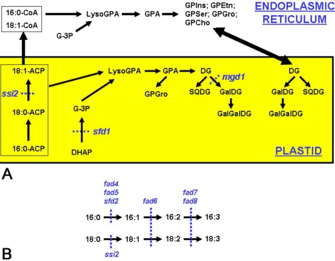
Figure 2. Schematic of lipid biosynthesis in Arabidopsis thaliana. A. Lipid biosynthesis in Arabidopsis leaves occurs in two
compartments – in the plastids and the endoplasmic reticulum. Fatty acid synthesis and desaturation of 18:0-ACP to 18:1-ACP occurs exclusively in the plastid. Desaturation of 16:0 and further desaturation of 18:1 occurs on acyl chains conjugated to glycerol backbone. B. Mutations in genes that affect specific steps in lipid biosynthesis are indicated in blue. fad6, fad7 and fad8 affect desaturation of 16C and 18C acyl chains. In contrast, the fad4, fad5 and sfd2 mutations primarily affect desaturation of 16C acyl chains. Abbreviations not used in the text are: lysoGPA: lysophosphatidic acid, SQDG: sulfoquinovosyldiacylglycerol. cellular function. However, in roots, the amount of
phospholipids lost is more than four times that the amount of galactolipids gained. Thus, galactolipids do not quantitatively replace phospholipids in roots as they do in rosettes during phosphorus starvation.
The molecular species of eight lipid classes, including GPIns, GPGro, phosphatidylserine (GPSer), GPEtn, GPCho, GPA, GalGalDG and GalDG, were profiled in rosettes and roots under normal and phosphorus starvation conditions (33). Lipids in Arabidopsis are synthesized through distinguishable routes, the prokaryotic pathway and the eukaryotic pathway (Figure 2). The prokaryotic pathway is localized on the plastid inner envelope, and the eukaryotic one is localized on the ER. GalGalDG can be considered as being in three pools: the “plastidic pool” is derived from the prokaryotic pathway and is located in the plastid, the “ER-extraplastidic pool” is derived from the eukaryotic pathway and is located outside the plastid, while the “ER-plastidic pool” is derived from the eukaryotic pathway but
is located inside the plastid. In rosettes and roots, the major molecular species, such as 34:2, 34:3, 36:4, 36:5, and 36:6, were generally lower in phospholipids during phosphorus starvation. The increase in these species, but not the plastid-derived 34:6 species, in GalGalDG suggests that hydrolysis of phospholipids supplies DG moieties for GalGalDG synthesis during phosphorus starvation. The data indicate that the eukaryotic pathway contributes to GalGalDG accumulation during phosphorus starvation, whereas neither 34:6-GalGalDG nor 34:5-GalGalDG increases during phosphorus starvation in rosettes and roots, suggesting that newly formed GalGalDG might not originate from the prokaryotic, plastidic pool.
4. DEVELOPMENTAL CHANGES IN PLANT LIPID COMPOSITION
4.1. Lipid changes in cotton fibers during development
Cotton fibers are single cells that initiate on the epidermal surface of ovules at anthesis (34). Fiber cell
elongation occurs primarily during the first 15 days after anthesis and cells achieve a length of more than two cm by this time. As fiber cells achieve their full length, there is a transition from rapid cell expansion to intense secondary cell wall synthesis and cells increase in thickness over the period of about 15 to 40 days after anthesis, a process defined as fiber maturation (35, 36). Ultimately the cell cytoplasm collapses and mature fibers desiccate when the fruit opens (about 45 days after anthesis). Fiber cell elongation is associated with rapid plasma membrane and vacuolar expansion and so these cells synthesize a considerable amount of membrane lipid to accommodate this intense period of cellular growth. The maturation phase is accompanied by a shift toward cellulose deposition adjacent to the plasma membrane surface and this requires the synthesis and trafficking of carbohydrate through the plasma membrane (37). Hence, this overall cellular system allows for a relative synchronous population of cells to be collected and evaluated for principal components that may impact key stages in cell enlargement and differentiation (36). The importance of membrane lipid metabolism to the development of cotton fibers, while theoretically clear, has been relatively unstudied. As a first step toward gaining a better understanding of lipid function in fiber cell formation, the glycerolipid content and composition of elongating and maturing fibers were determined recently by ESI-MS/MS (38).
High throughput quantification of the major polar lipid classes demonstrated that elongating fibers (on a dry weight basis) contained more lipid than maturing fibers
(38), as might be expected due to the considerable
accumulation of cellulose mass during fiber maturation. The polar lipid class composition was generally similar between elongating and maturing fibers, with phospholipids, not galactolipids, making up the majority of the polar lipid fraction. GPCho, GPEtn and GPIns were the principal phospholipid classes in all stages of fiber cell development, together accounting for more than 70% of the total polar lipid. GPGro, GalDG, and GalGalDG were relatively minor polar lipid constituents. More than 70 glycerolipid species were quantified in both elongating and maturing cotton fiber cells. Subtle, but significant, differences were quantified in the molecular composition of the major phospholipids. Generally, phospholipids of maturing fibers were more saturated than those in elongating fibers. For example, there was a greater proportion of 34:2 GPCho and 34:2 GPEtn in maturing fibers (compared to elongating fibers), whereas there was relatively more 36:6 GPCho and 36:4 GPCho and 34:3 GPEtn and 36:6 GPEtn in elongating fibers (compared to maturing fibers), perhaps reflecting a change in FAD3 desaturase activity with development. These types of metabolite analyses, while initially descriptive, provide a basis for associating other types of large-scale database information such as that from genomics and proteomics efforts.
Part of the value of profiling the glycerolipid molecular species in elongating cotton fibers was the availability of large DNA databases of expressed genes (available as ESTs: expressed sequence tags) at these same
stages of development (http://www.tigr.org/). By reconciling metabolite data with ESTs for lipid-metabolism enzymes, it was possible to delineate at the molecular level, in most cases, the pathways likely to be important for membrane lipid synthesis in cotton fiber cells (38). For example, the predicted pathway and DNA sequences corresponding to each enzymatic step (Figure 3) could be identified for the most abundant GPCho molecular species in both elongating and maturing fibers, namely 34:3 GPCho (16:0/18:3 GPCho). Moreover, new questions about metabolic relationships were suggested by comparison of metabolite and sequence database information. For example, is the phosphatidic acid precursor, 16:0/18:3 GPA (the major GPA species identified in extracts), utilized for the synthesis of the major GPIns molecular species (34:3 GPIns)? Surprisingly, no EST candidates were identified for a cytosine diphosphate (CDP):diacylglycerol (DG) synthase or a GPIns synthase, two enzymes that are clearly important in developing a comprehensive lipidomics map of cotton fiber metabolism. No doubt, high-throughput lipid profiling capabilities coupled with molecular genetic and biochemical information will be important in resolving and defining the importance of lipid metabolism in cotton fiber formation.
4.2. Alterations in potato lipids during tuber aging
Potato tubers are model organs for aging studies because they can survive up to three years after harvest, if they are stored at low temperature. Various studies have been undertaken to understand the role of lipid peroxidation in membrane damage and, in general, in the aging process
(39-42). Focusing particularly on the sprouting phase (42),
it has been demonstrated that the period during which apical dominance is lost, resulting in the development of multiple sprouts, is correlated with a clear increase in particular oxygenated fatty acids, or “oxylipins”, specifically the 9-hydroperoxide of linoleic acid and colneleic acid (42). Analysis of phospholipids and galactolipids by electrospray ionization tandem mass spectrometry showed a decrease in the level of GPCho, GPEtn, GPIns, GalGalDG, and GalDG during this particular period. The decrease in the amount of linoleic acid in the complex lipids correlated well with the amount of its metabolites 9-hydroperoxy linoleic acid and colneleic acid. During sprouting in potato tubers, the lipoxygenase pathway is activated: galactolipases and phospholipases liberate free fatty acids, among which linoleic acid which is transformed to its hydroperoxide by lipoxygenase. This last product is converted by the divinyl ether synthase to colneleic acid which is degraded to 9-oxo-nonanoic acid. Additionally, it has been demonstrated that a part of colneleic acid is esterified in phospholipids (43), although the physiological significance of this remains unknown.
5. ELUCIDATION OF GENE FUNCTION BY ANALYSIS OF PLANT LIPID COMPOSITION
5.1. Role of fatty acid desaturases and DHAP reductase in systemic acquired resistance
Systemic acquired resistance (SAR) is an inducible defense mechanism in plants that confers resistance to a broad-spectrum of pathogens (44-46). Prior
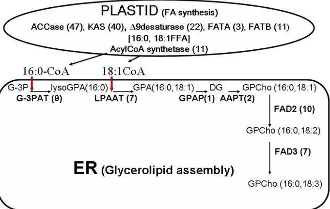
Figure 3. Compartmentation of metabolites and deduction of pathways for lipid metabolism in cotton fiber cells by combining
lipid profiling and DNA database information. Summary scheme for the pathway leading to the synthesis of GPCho 34:3 (16:0/18:3), a major glycerolipid molecular species quantified in both elongating and maturing cotton fiber cells. The diagram is based on both metabolite profiles and EST occurrences (numbers in parentheses) for enzymes in these pathways (collected from DNA database information posted at http://www.tigr.org/) in the same cell type at the same developmental stage. This scheme for elongating cotton fibers is consistent with the synthetic pathways described for extraplastidial GPCho in other types of plant cells. The fatty acids are assembled de novo in the plastids of fiber cells and exported to the ER for glycerolipid assembly and subsequent modification (e.g. introduction of additional double bonds). The diacylglycerol base for other phospholipids, such as GPEtn and GPIns, can be synthesized using the same machinery. Abbreviations not used in the text are: ACCase: acetylCoA carboxylase, KAS: keto-acyl-ACP synthase, FATA: Fatty acid thioeserase A, FATB: fatty acid thioeaterase B, FFA: free fatty acid, G-3PAT: G-3P acyltransferase, lysoGPA: lyso-phosphatidic acid, LPAAT: lysoGPA acyltransferase, GPAP: GPA phosphatase, AAPT: aminoalcohol phosphotransferase, FAD: fatty acid desaturase. Reprinted from with permission from AOCS press (38).
exposure to a necrotizing pathogen is required for the activation of SAR. SAR is constitutively expressed in the
Arabidopsis thaliana ssi2 mutant, which exhibits
heightened resistance to bacterial, viral and oomycete pathogens (47, 48). In addition, the ssi2 mutant plant is a dwarf and spontaneously develops lesions containing dead cells. The SSI2 gene encodes a stearoyl-acyl carrier protein (ACP) desaturase that catalyzes the conversion of ACP conjugated stearic acid (18:0) to oleic acid (18:1) in the plastids (49). The oleic acid moiety of 18:1-ACP is subsequently channeled into the synthesis of glycerolipids in the plastids. Oleic acid is also shunted to the cytosol as 18:1-CoA, which is then incorporated into glycerolipids in the ER. Lipid biosynthesis in Arabidopsis thaliana is summarized in Figure 2. ESI-MS/MS analysis showed that the ssi2 mutation resulted in an overall reduction in the
content of glycerolipids that are synthesized and localized in the plastids (50). In addition, some changes were also observed in the composition of extraplastidic lipids. Although the primary block in the ssi2 mutant is in the conversion of 18:0-ACP to 18:1-ACP, several compensatory changes were also observed. For example, the polar lipids, GPCho, GPEtn, and GPIns containing a 34:2 acyl combination were present at lower levels in the leaves of the ssi2 mutant than the wild type. In contrast, the level of 36:2 and 36:3-GPCho, -GPEtn, and -GPIns species were higher in the ssi2 mutant plant. Several studies have indicated that the effect of the ssi2 mutation on plant defense is not due to the accumulation of elevated 18:0 levels (49-54). Rather, it may be associated with the impact of the ssi2 mutation on the generation of a signal that modulates plant defense.
In contrast to the constitutive expression of SAR in the ssi2 mutant, SAR is compromised in the sfd1 mutant
(55). The SAR defect in sfd1 is due to its inability to
generate and/or translocate the phloem mobile signal in SAR. ESI/MS-MS analysis indicates that the sfd1 mutant has altered composition of plastidic glycerolipids, especially that of galactolipids, suggesting that SFD1 is required for lipid biosynthesis in the plastids. For example, level of the plastidic lipid species, 34:6-monogalactosyldiacylglycerol (GalDG), which is the most abundant complex lipid in Arabidopsis leaves, was lower in the sfd1 mutant than the wild type. In contrast, the level of 36:6-GalDG, another plastidic lipid that is believed to be derived from DG shunted into the plastids from the ER, was higher in the sfd1 mutant than the wild type, suggesting that an increase flux of ER-synthesized, acylated glycerol species into the plastid may compensate for the lipid composition deficiency in the sfd1 mutant. The sfd1 mutant was identified in a screen for suppressors of ssi2-conferred phenotypes (50).
ssi2-conferred dwarfing, cell death and SAR-like phenotypes
were suppressed in the ssi2 sfd1 double mutant plant (50). Presence of the sfd1 allele restored the lipid content and the 18:1 mol% in ssi2 containing plants. In addition, presence of the sfd1 mutant allele also resulted in lowered levels of hexadecatrienoic acid (16:3)-containing lipids. The increases in 18:1 mol% in these plants, or alternatively, the lowered content of 16:3-containing lipids could account for the suppression of
ssi2-conferred phenotypes. Mutations in the SFD2 and FAD7 genes, which are also associated with lipid
metabolism, also compromised SAR (K. Krothapalli, R. Chaturvedi, A. Nandi, R. Welti and J. Shah, unpublished). In addition, the sfd2 and fad7 mutations suppress the ssi2-conferred enhanced resistance against the bacterial pathogen,
Pseudomonas syringae pv maculicola, in the ssi2 sfd2 and ssi2 fad7 double mutant plants, providing further support for the
notion that a lipid-derived molecule is involved in activation of SAR. Like the sfd1 mutant, the sfd2 and fad7 mutants are also compromised in the synthesis and/or translocation of a phloem-mobile factor in SAR (R. Chaturvedi, K. Krothapalli and J. Shah, unpublished). A lipid composition defect that is common to the sfd1, sfd2 and fad7 mutants is the change in composition of plastid synthesized galactolipids, suggesting the involvement of a galactolipid in SAR. Indeed, SAR is also compromised in the mgd1 mutant (R. Chaturvedi, K. Krothapalli and J. Shah, unpublished). MGD1 catalyzes the synthesis of GalDG from DG in the plastids. These results suggest the involvement of a galactolipid or a product thereof in SAR.
5.1.1. Lipid profiling as an aid in identification of mutated genes
Metabolite profiling can expedite the cloning of genes affecting biochemical processes. The cloning of
SFD1 is an excellent example of this. ESI-MS/MS
profiling of polar lipid composition in the ssi2 sfd1 mutant plant had suggested that the SFD1 gene was involved in plastid lipid biosynthesis (50). This was further confirmed by lipid profiles of the sfd1 single mutant plant (55). Amongst the approximately 300 annotated genes predicted to be in the location on chromosome 2 to which SFD1 mapped, At2g40690 was predicted to encode a protein that could be involved in lipid metabolism. At2g40690 was
annotated as a putative dihydroxyacetone phosphate (DHAP) reductase. DHAP reductases catalyze the interconversion of DHAP and glycerol-3-phosphate (G-3P). G-3P provides the glycerol backbone for glycerolipid synthesis in plants. Sequencing of this locus in the sfd1-1 and sfd1-2 mutant alleles confirmed the presence of single site mutations within this gene in both mutants (55). Complementation experiments in Arabidopsis and Escherichia coli, confirmed the identity of SFD1, its biochemical function as a DHAP reductase, and its involvement in plant defense. In a similar manner, lipid profiling aided in the identification of the gene that contained the sfd4 mutation. The sfd4 mutant, which was also identified as a suppressor of ssi2 (50), has high levels of plastidic galactolipids that contain the monunsaturated fatty acids palmitoleic acid (16:1) and 18:1, but lower levels of galactolipids containing polyunsaturated 16C and 18C acyl chains, suggesting the SFD4 may encode a ω6-desaturase that is involved in the desaturation of 16:1 and 18:1 to hexadecadienoic acid (16:2) and 18:2, respectively, in plastid synthesized galactolipids. The lipid profile of sfd4 was very similar to that of the fad6 mutant, which lacks a functional ω6-desaturase. Sequencing of the FAD6 locus from the sfd4 mutant confirmed the presence of a mutation in the FAD6 gene in the sfd4 mutant.
5.2. Roles of phospholipases in response to freezing
Lipid changes are mediated by enzymes catalyzing lipid hydrolysis, biosynthesis, and/or modification. Phospholipases are major groups of enzymes that mediate phospholipid catabolism. The enzymes are classified into four major classes, PLD, phospholipase C (PLC), PLA1 and PLA2, based on the site of cleavage. Of these, PLD is the most prevalent family of phospholipase in plants (10). PLD generates GPA, and increases in GPA have been a common change observed under many stress conditions. In Arabidopsis, there are 12 PLD genes that are divided into six types, PLDalpha (3),
beta (2), gamma (3), delta, epsilon, and zeta (2). Each of
the PLDs characterized exhibits distinguishable biochemical properties, such as different requirements for Ca2+, phosphoinositides, and oleic acid for activity and/or different phospholipids preferences as substrates. These findings raise questions about the roles of different PLDs in stress responses.
Comparative profiling of wild-type and PLD mutants have provided valuable insight into the cellular functions of different PLDs. Combination of lipid profiling with genetic manipulation of PLDs has proven to be a powerful platform to gain information on lipid species changes and the role of different PLDs in producing lipid alterations and in plant responses to specific stress conditions. While most studies performed thus far on
Arabidopsis have focused on leaves or rosettes, a couple of
recent studies (section 5.3; 6, 32) have involved profiling of polar lipids of other organs, including roots (6, 32), stems, siliques, flowers, and seeds (6).
5.2.1. Different roles of PLDs in freezing-induced lipid hydrolysis
Compared to cold acclimation (section 3.1), a sublethal-freezing temperature induces more drastic
changes in membrane lipids. The change comes mostly from hydrolysis as the content of total membrane lipid decreases. At −8°C, a sublethal-freezing temperature for cold acclimated Arabidopsis, the loss of lipids resulted primarily from the loss of GPCho, GPEtn, and GPGro, with the 36:4 and 36:5 species contributing the most to the decline in GPEtn and GPCho levels (5). In contrast, the change in GPIns molecular species was minimal, and some GPIns species actually increased. GalDG levels tended to decline, but no loss of GalGalDG occurred in Arabidopsis exposed to −8°C. The levels of lipid metabolites GPA, lysoGPCho, and lysoGPEtn, increased dramatically. The large decline in phospholipids, but not galactolipids suggests that phospholipases are activated to a greater extent than galactolipases. The low level of the PLA products, lysophospholipids, in comparison with the PLD product, GPA, indicates that PLD is more active than PLA in frozen plant tissue. In addition, comparison of the molecular species profile of GPA with other lipid classes helps identify the substrates that give rise GPA. Upon freezing, most of the GPA species that increased match with those of the GPCho species that decreased.
One major lipid change in plant response to low temperatures is the increase in GPA molecular species. Despite the presence of 12 PLDs in Arabidopsis, suppression of PLDalpha1 resulted in a higher level of GPCho and a lower level of GPA, indicating that GPCho is the major in vivo substrate for PLDalpha1 under freezing conditions. PLDalpha1 is responsible for about 50% of GPCho hydrolyzed under the freezing conditions tested (5). The lack of difference between wild-type and PLDalpha1-deficient plants in the level of GPEtn and GPGro suggest that enzymes other than PLDalpha1 are responsible for the freezing-induced hydrolysis of GPEtn and most of the GPGro species. By comparison, genetic manipulation of PLDdelta has no major effect on membrane lipid hydrolysis
(56), suggesting that, unlike PLDalpha1, PLDdelta activity
does not contribute to substantial lipid hydrolysis. These metabolic differences in lipid changes indicate different functions in freezing for PLDalpha1 and PLDdelta. Indeed, phenotypic analysis shows that Arabidopsis plants deficient in PLDalpha1 and PLDdelta display opposite phenotypes in freezing tolerance (5, 56). Whereas suppression of PLDalpha1 rendered Arabidopsis plants more tolerant to freezing, knockout of PLD-delta made plants more sensitive to freezing (56).
5.3. Role of PLDzeta in lipid changes during phosphorus deficiency
As described in Section 3.4., the level of phospholipids in plants decreases under phosphorus-limited growth conditions. The decrease in phospholipids presumably allows phosphate to be used for other cell functions and also makes the lipid moiety DG available for galactolipid biosynthesis. Then, what causes the loss of phospholipids under phosphorus-limited growth? Expression profiling reveals substantial increases in the transcript levels of PLDzeta2 (56, 57). The loss of
PLDzeta2, but not PLDzeta1, led to a decreased
accumulation of GPA in root under phosphorus-limited conditions and compromised the plant’s ability to
hydrolyze phospholipids and to increase galactolipids. Plants depleted of PLDzeta1 and zeta2 displayed shorter primary roots than wild-type plants under the same phosphorus-limited conditions. These results indicate that
PLDzeta1 and zeta2 play a role in regulating root
development in response to nutrient limitation.
Three phosphorus conditions have been examined so far (32, 33). Under a standard growth condition (500 µM Pi), disruption of PLDzeta1, PLDzeta2, or both did not result in the differences in the concentrations of GPCho, GPA, GalGalDG and root growth in contrast to wild type. Under a phosphorus-limited condition (25 µM Pi), disruption of both PLDzeta1 and PLDzeta2 results in a lower concentration of GPA in roots, a retarded primary root growth, and unchanged concentrations of GPCho and GalGalDG in roots (33). Under the phosphorus-free condition (0 µM Pi), disruption of both PLDzeta1 and PLDzeta2 results in a lower concentration of GPCho with a correspondingly increased concentration of GalGalDG in roots. However, PLDzeta1 and PLDzeta2 mutants exhibit no alteration in the concentration of GPA or primary root elongation. These results indicate that, under moderate phosphorus
deficiency conditions (25 µM Pi), PLDzeta1 and
PLDzeta2 might function to modulate root growth for
better nutritional absorption by increasing GPA to stimulate root growth. During severe phosphorus
starvation (0 µM Pi), however, PLDzeta1 and PLDzeta2
might function to regulate lipid turnover between phospholipids and galactolipids for efficient using of internal phosphorus stores. Thus, PLDzetas play both signaling and metabolic roles in plant response to different severities of phosphorus deficiency (32).
6. IMPROVEMENT OF FOOD QUALITY BY MANIPULATION OF LIPID METABOLIZING GENES
Lipidomic analysis of complex lipids has also been used to understand function of lipid metabolizing genes in transgenic plants, with the goal of manipulating complex lipid metabolism in order to produce long-chain fatty acids, particularly omega-3 species, in plants (58). The goal is to produce in seeds fatty acids that are usually found primarily in fish oils and are beneficial in the diet. This requires production of a number of heterologous enzymes that must work together and with the plant metabolic enzymes to produce the desired fatty acids. Thus, after expression of the transgenes, the substrates and products of the new enzymes in the host plants must be evaluated, and it is obviously helpful to have a detailed analysis at the level of lipid molecular species. ESI-MS/MS analysis of the GPCho from linseed expressing heterologous desaturases and an elongase under the control of seed-specific promoters was carried out. In this example, it was demonstrated that GPCho acted as a substrate for the desaturases, and analysis of the lipid profiles in combination with traditional lipid analyses indicated that the transgenic enzymes acted on all GPCho molecular species, rather than on a subset of GPCho species.
7. PERSPECTIVE ON THE FUTURE
Analysis of the lipids from of T-DNA-tagged knock-out mutants or plants with alterations in genes involved in lipid metabolism should improve our understanding of the in vivo function of many of these genes. An example of a project currently underway involves a family of acyl-CoA binding proteins (ACBPs), of which only small (10-kDa) cytosolic acyl-CoA binding proteins that function in the storage and intracellular transport of acyl-CoA esters for β–oxidation and glycerolipid biosynthesis (59) have been well-characterized in eukaryotes other than plants. In Arabidopsis thaliana, a gene family encodes six ACBPs (60). While in vitro binding assays using Escherichia coli-expressed proteins have shown that they have varying affinities to different fatty acyl-CoAs (60), the physiological function of the six gene products remains unknown, and lipidomic analysis of knockout mutants are underway. In this study and others, the lipidomics approach has great potential to increase our understanding the gene function. Similarly, it is undoubtedly true that lipidomics will continue to enhance our understanding the role of membrane lipids in plant adaptation and tolerance to stresses. Hopefully, in the near future, application of comprehensive lipidomics technologies developed for triacylglycerols (61) and being developed for plant sphingolipids (62) will expand the sphere of plant lipidomics.
8. ACKNOWLEDGEMENTS
Work from authors’ laboratories was supported by grants from the National Science Foundation (MCB 0110979, MCB 0455318, DBI 0520140, DBI 0521587, and IOB-0454866) and the U.S. Department of Agriculture (2005-35818-15253). Support of the Kansas Lipidomics Research Center Analytical Laboratory was from National Science Foundation's EPSCoR program, under grant EPS 0236913 with matching support from the State of Kansas through Kansas Technology Enterprise Corporation and Kansas State University, as well as from NIH grant P20 RR016475 through the INBRE program of the National Center for Research Resources. This is contribution 07-25-J from the Kansas Agricultural Experiment Station.
9. REFERENCES
1. Han, X, and R. W. Gross: Electrospray ionization mass spectroscopic analysis of human erythrocyte plasma membrane phospholipids. Proc. Natl. Acad. Sci. USA. 91, 10635-10639 (1994)
2. Brügger, B, G. Erben, R. Sandhoff, F. T. Wieland, and W. D. Lehmann: Quantitative analysis of biological membrane lipids at the low picomole level by nano-electrospray ionization tandem mass spectrometry. Proc.
Natl. Acad. Sci. USA 94, 2339-2344 (1997)
3. Welti, R, and X. Wang: Lipid species profiling: a high-throughput approach to identify lipid compositional changes and determine the function of genes involved in lipid metabolism and signaling. Curr. Opin. Plant Biol. 7, 337-344 (2004)
4. Han, X, and R. W. Gross: Shotgun lipidomics: electrospray ionization mass spectrometric analysis and quantitation of cellular lipidomes directly from crude extracts of biological samples. Mass Spectrom. Rev. 24, 367-412 (2005)
5. Welti, R, W. Li, M. Li, Y. Sang, H. Biesiada, H. E. Zhou, C. B. Rajashekar, T. D. Williams, and X. Wang: Profiling membrane lipids in plant stress responses. Role of phospholipase D-alpha in freezing-induced lipid changes in Arabidopsis. J. Biol. Chem. 277, 31994-32002 (2002)
6. Devaiah,S. P, M. R. Roth, E. Baughman, M. Li, P.
Tamura, R. Jeannotte, R. Welti, and X. Wang: Quantitative profiling of polar glycerolipid species from organs of wild-type Arabidopsis and a PHOSPHOLIPASE
Dalpha1 knockout mutant. Phytochem. In press. (2006)
7. Welti, R, X. Wang, and T. D. Williams: Electrospray ionization tandem mass spectrometry scan modes for plant chloroplast lipids. Anal. Biochem. 314, 149-152 (2003) 8. Uemura, M, R. A. Joseph, and P. L. Steponkus: Effect of cold acclimation on the lipid composition of the inner and outer membrane of the chloroplast envelope isolated from rye leaves. Plant Physiol. 109, 15-30 (1995)
9. Thomashow, M.F: Plant cold acclimation: Freezing tolerance genes and regulatory mechanisms. Annu. Rev.
Plant Physiol. Plant Mol. Biol. 50, 571–599 (1999)
10. Wang, X, S. P. Devaiah, W. Zhang, and R. Welti: Signaling functions of phosphatidic acid. Prog. Lipid
Research 45, 250-278 (2006)
11. Armond, P. A, O. Bjorkman, and L. A. Staehelin: Dissociation of supramolecular complexes in chloroplast membranes. A manifestation of heat damage to the photosynthetic apparatus. Biochim. Biophys. Acta 601, 433-443 (1980)
12. Berry, J. A, and O. Bjorkman: Photosynthetic response and adaptation to temperature in higher plants. Annu. Rev.
Plant Biol. 31, 491-543 (1980)
13. Quinn P. J: Effects of temperature on cell membranes.
Symp. Soc. Exp. Biol. 42, 237-258 (1988)
14. Mohanty, P, B. Vani, and J. S. Prakash: Elevated temperature treatment induced alteration in thylakoid membrane organization and energy distribution between the two photosystems in Pisum sativum. Z. Naturforsch (C) 57, 836-242 (2002)
15. Weis, E, and J. A. Berry: Plants and high temperature stress. Symp. Soc. Exp. Biol. 42, 329-346 (1988)
16. Barkan, L, P. Vijayan, A. S. Carlsson, S. Mekhedov, and J. Browse: A suppressor of fab1 challenges hypotheses on the role of thylakoid unsaturation in photosynthetic function. Plant Physiol. 141, 1012-1020 (2006)
17. Falcone, D. L, J. P. Ogas, and C. R. Somerville: Regulation of membrane fatty acid composition by temperature in mutants of Arabidopsis with alterations in membrane lipid composition. BMC Plant Biol. 4, 17 (2004) 18. Gorver, A, M. Agarwal, S. Katiyar-Argarwal, C. Sahi, and S. Argarwal: Production of high temperature tolerance transgenic plants through manipulation of membrane lipids.
Curr. Sci. 79, 557-559 (2000)
19. Larkindale, J, and B. Huang: Changes of lipid composition and saturation level in leaves and roots for heat-stressed and heat acclimated creeping bentgrass (Agrostis stolonifera). Environ. and Exp. Bot. 51, 57-67 (2004)
20. Marcum, K. B: Cell membrane thermostability and whole plant heat tolerance of Kentucky bluegrass. Crop
Sci. 38, 1214-1218 (1998)
21. Burke, J. J, P. J. O'Mahony, and M. J. Oliver: Isolation of Arabidopsis mutants lacking components of acquired thermotolerance. Plant Physiol. 123, 575-588 (2000) 22. Chen, J, J. J. Burke, Z. Xin, C. Xu, and J. Velten: Characterization of the Arabidopsis thermosensitive mutant
atts02 reveals an important role for galactolipids in
thermotolerance. Plant, Cell and Environment 29, 1437-1448 (2006)
23. Dormann, P, S. Hoffmann-Benning, I. Balbo, and C. Benning: Isolation and characterization of an Arabidopsis mutant deficient in the thylakoid lipid digalactosyl diacylglycerol. Plant Cell 7, 1801-1810. (1995)
24. Suss, K.-H, and I. Yordanov: Biosynthetic cause of in
vivo acquired thermotolerance of photosynthetic light
reactions and metabolic responses of chloroplasts to heat stress. Plant Physiol. 81, 192-199 (1986)
25. Webb, M. S, and B. R. Green: Biochemical and biophysical properties of thylakoid acyl lipids. Biochim.
Biophys. Acta 1060, 133-158 (1991)
26. Alfonso, M, I. Yruela, S. Almarcegui, E. Torrado, M. A. Perez, and R. Picorel: Unusual tolerance to high temperatures in a new herbicide-resistant D1 mutant from
Glycine max (L.) Merr. cell cultures deficient in fatty acid
desaturation. Planta 212, 573-582 (2001)
27. Stelmach, B.A, A. Müller, P. Hennig, S. Gebhardt, M. Schubert-Zsilavecz, and E. W. Weiler: A novel class of
oxylipins,
sn1-O-(12-oxophytodienoyl)-sn2-O-(hexadecatrienoyl)-monogalactosyl diglyceride, from
Arabidopsis thaliana. J. Biol. Chem. 276: 12832-12838
(2001)
28. Hisamatsu, Y, N. Goto, K. Hasegawa, and H. Shigemori: Arabidopsides A and B, two new oxylipins from Arabidopsis thaliana. Tetrahedron Lett. 44, 5553-5556 (2003)
29. Hisamatsu, Y, N. Goto, M. Sekiguchi, K. Hasegawa, and H. Shigemori: Oxylipins arabidopsides C and D from
Arabidopsis thaliana. J. Nat. Prod. 68, 600-603 (2005)
30. Buseman, C. M, P. Tamura, A. A. Sparks, E. J. Baughman, S. Maatta, J. Zhao, M. R. Roth, S. W. Esch, J. Shah, T. D. Williams, and R. Welti: Wounding stimulates the accumulation of glycerolipids containing oxophytodienoic acid and dinor-oxophytodienoic acid in Arabidopsis leaves. Plant Physiol. 142, 28-39 (2006) 31. Härtel, H, P. Dörmann, and C. Benning: DGD1-independent biosynthesis of extraplastidic galactolipids after phosphate deprivation in Arabidopsis. Proc. Natl.
Acad. Sci. USA 97: 10649-10654 (2000)
32. Li, M, R. Welti, and X. Wang: Quantitative profiling of Arabidopsis polar glycerolipids in response to phosphorus starvation: Roles of PLDzeta1 and PLDzeta2 in
phosphatidylcholine hydrolysis and digalactosyldiacylglycerol accumulation in
phosphorus-starved plants. Plant Physiol. In press (2006)
33. Li, M, C. Qin, R. Welti, and X. Wang: Double knockout of phospholipases D-zeta1 and -zeta2 in Arabidopsis affect root elongation during phosphate-limited growth but do not affect root hair patterning. Plant
Physiol. 140, 761-770 (2006).
34. Stewart, J.D: Fiber initiation on the cotton ovule (Gossypium hirsutum). Am. J. Bot. 62, 723-730 (1975) 35. Schubert, A.M, J. D. Benedict, J. D. Berlin, and R. J. Kohel: Kinetics of cell elongation and secondary cell wall thickening. Crop. Sci. 13, 704-709 (1973)
36. Kim, H.J., and Triplett, B.A. (2001) Cotton fiber growth in planta and in vitro. Models for plant cell elongation and cell wall biogenesis, Plant Physiol. 127, 1361-1366.
37. Paredez, A.R, C. R. Somerville, and D. W. Ehrhardt: Visualization of cellulose synthase demonstrates functional association with microtubules. Science 312, 1491-1495 (2006)
38. Wanjie, S. W, R. Welti, R. A. Moreau, and K. D. Chapman: Identification and quantification of glycerolipids in cotton fibers: reconciliation with metabolic pathway predictions from DNA databases. Lipids. 40, 773-785 (2005)
39. Zabrouskov, V, and N. R. Knowles: Lipid metabolism during aging of high alpha-linolenate phenotype potato tubers. Arch. Biochem. Biophys. 402, 136-148 (2002) 40. Zabrouskov, V, and N. R. Knowles: Changes in lipid molecular species and sterols of microsomal membranes during aging of potato (Solanum tubersosum L.) seed-tubers. Lipids 37, 309-315 (2002)
41. Fauconnier, M.-L, J. Rojas-Beltran, J. Delcarte, F. Dejaeghere, M. Marlier, and P. du Jardin: Lipoxygenase pathway and membrane permeability and composition during storage of potato tubers (Solanum tuberosum L. cv Bintje and Désirée) in different conditions. Plant Biol. 4, 77-85 (2002)
42. Fauconnier, M.-L, R. Welti, E. Blée, and M. Marlier: Lipid and oxylipin profiles during aging and sprout development in potato tubers (Solanum tunberosum L.).
Biochim. Biophys. Acta 1633, 118-126 (2003)
43. Fauconnier, M.-L, T. D. Williams, M. Marlier, and R. Welti: Potato tuber phospholipids contain colneleic acid in the 2-position. FEBS Letters 538, 155-158 (2003)
44. Durrant, W. E, and X. Dong: Systemic acquired resistance. Annu. Rev. Phytopathol. 42, 185–209 (2004) 45. Shah, J: Lipids, lipases and lipid modifying enzymes in plant disease resistance. Annu. Rev. Phytopathol. 43, 229-260 (2005)
46. Chaturvedi, R, and J. Shah: Salicylic acid in plant disease resistance. In "Salicylic acid-A plant hormone" ed. S. Hayat and A. Ahmad, Springer, Dordrecht, The Netherlands. In press. (2006)
47. Shah, J, P. K. Kachroo, A. Nandi, and D. F. Klessig: A recessive mutation in the Arabidopsis SSI2 gene confers SA- and NPR1-independent expression of PR genes and resistance against bacterial and oomycete pathogens. Plant
J. 25, 563-574 (2001)
48. Sekine, K.T, A. Nandi, T. Ishihara, S. Hase, M. Ikegami, J. Shah, and H. Takahashi: Enhanced resistance to Cucumber mosaic virus in the Arabidopsis thaliana ssi2 mutant is mediated via an SA-independent mechanism.
Mol. Plant-Microbe Interaction 17, 623-632 (2004)
49. Kachroo, P, J. Shanklin, J. Shah, E. J. Whittle, and D. F. Klessig: A fatty acid desaturase modulates the activation of defense signaling pathways in plants. Proc. Natl. Acad.
50. Nandi, A, K. Krothapalli, C. Buseman, M. Li, R. Welti, A. Enyedi, and J. Shah: The Arabidopsis thaliana sfd mutants affect plastidic lipid composition and suppress dwarfing, cell death and the enhanced disease resistance phenotypes resulting from the deficiency of a fatty acid desaturase. Plant Cell 15, 2383-2398 (2003)
51. Kachroo, A, L. Lapchyk, H. Fukushige, D. Hildebrand, D. F. Klessig, and P. Kachroo: Plastidyl fatty acid signaling modulates salicylic acid- and jasmonic acid-mediated defense pathways in the Arabidopsis ssi2 mutant. Plant
Cell 15, 2952-2965 (2003)
52. Kachroo, A, S. C. Venugopal, L. Lapchyk, D. Falcone, D. Hildebrand, and P. Kachroo: Oleic acid levels regulated by glycerolipid metabolism modulate defense gene expression in Arabidopsis. Proc. Natl. Acad. Sci. USA 101, 5153–5157 (2004)
53. Kachroo, P, S. C. Venugopal, D. A. Navarre, L. Lapchyk, and A. Kachroo: Role of salicylic acid and fatty acid desaturation pathways in ssi2-mediated signaling.
Plant Physiol. 139, 1717-1735 (2005)
54. Nandi, A, W. Moeder, P. Kachroo, D. F. Klessig, and J. Shah: The Arabidopsis ssi2-conferred susceptibility to
Botrytis cinerea is dependent on EDS5 and PAD4. Mol. Plant-Microbe Interact. 18, 363-370 (2005)
55. Nandi, A, R. Welti, and J. Shah: The Arabidopsis
thaliana dihydroxyacetone phosphate reductases gene SUPPRESSOR OF FATTY ACID DESATURASE DEFICIENCY1 is required for glycerolipid metabolism and
for the activation of systemic acquired resistance. Plant
Cell 16, 465-477 (2004)
56. Li, W, M. Li, W. Zhang, R. Welti, and X. Wang: The plasma membrane-bound phospholipase Ddelta enhances freezing tolerance in Arabidopsis thaliana. Nature
Biotechnol. 22, 427-433 (2004)
57. Misson, J, K. Raghothama, A. Jain, J. Jouhet, M. Block, R. Bligny, P. Ortet, A. Creff, S. Somerville, N. Rolland, P. Doumas, P. Nacry, L. Herrerra-Estrella, L. Nussaume, and M. C. Thibaud: A genome-wide transcriptional analysis using Arabidopsis thaliana Affymetrix gene chips determined plant responses to phosphate deprivation. Proc.
Natl. Acad. Sci. USA 102, 11934-11939 (2005)
58. Abbadi, A, F. Domergue, J. Bauer, J. A. Napier, R. Welti, U. Zahringer, P. Cirpus, and E. Heinz: Biosynthesis of very long-chain polyunsaturated fatty acids in transgenic oilseeds: Constraints on their accumulation. Plant Cell 16, 2734-2748 (2004)
59. Faergeman, N. J, and J. Knudsen: Role of long-chain fatty acyl-CoA esters in the regulation of metabolism and cell signaling. Biochem. J. 323, 1-12 (1997)
60. Leung, K.-C, H.-Y. Li, G. Mishra, and M.-L. Chye: ACBP4 and ACBP5, novel Arabidopsis acyl-CoA-binding proteins with kelch motifs that bind oleoyl-CoA. Plant Mol.
Biol. 55, 297-309 (2004)
61. Han, X, and R. W. Gross: Quantitative analysis and molecular species fingerprinting of triacylglyceride molecular species directly from lipid extracts of biological samples by electrospray ionization tandem mass spectrometry. Anal. Biochem. 295, 88-100 (2001)
62. Markham, J. E, J. Li, E. B. Cahoon, and J. G. Jaworski: Plant sphingolipids: separation and identification of major sphingolipid classes from leaves. J. Biol. Chem. 281, 22684 - 22694 (2006)
Abbreviations: ACP: (stearoyl-) acyl carrier protein, CDP:
cytosine diphosphate, CoA: coenzyme A, DG: diacylglycerol, DHAP: dihydroxyacetone phosphate, dnOPDA: (7S,11S)-10-oxo-phytodienoic acid,, ESI: electrospray ionization, ESTs: expressed sequence tags, ER: endoplasmic reticulum, GalDG: monogalactosyldiacylglycerol, GalGalDG: digalactosyldiacylglcerol, G-3P: glycerol-3-phosphate, MS:
mass spectrometer or mass spectrometry, MS/MS: triple quadrupole mass spectrometer, OPDA: (9S,13S)-12-oxo-phytodienoic acid, GPA: phosphatidic acid, GPCho: phosphatidylcholine, GPEtn: phosphatidylethanolamine, GPGro: phosphatidylglycerol, GPIns: phosphatidylinositol, GPSer: phosphatidylserine, PLA: phospholipase A, PLC: phospholipase C, PLD: phospholipase D, RT-PCR: reverse transcriptase-polymerase chain reaction, SAR: systemic acquired resistance, 16:0, palmitoyl or palmitic acid, 16:1, palmitoleic acid, 16:2, hexadecadienoic acid, 16:3, hexaceatrienoyl or hexadecatrienoic acid, 18:0, stearoyl or stearic acid, 18:1: oleoyl or oleic acid, 18:2: linoleoyl or linoleic acid, 18:3, linolenoyl or linolenic acid
Key Words: Arabidopsis thaliana, cotton, Solanum tuberosum L., potato tuber, phospholipid, galactolipid,
glycerolipid, phosphatidylcholine, phosphatidylethanolamine, phosphatidic acid, freezing tolerance, phosphate deprivation, stress response, abiotic and biotic stress, mass spectrometry, oxylipin, colneleic acid, fatty acid hydroperoxide, lipoxygenase, phospholipase, Review
Send correspondence to: Dr. Ruth Welti, Kansas
Lipidomics Research Center, Division of Biology, Kansas State University, Manhattan, KS 66506, 785-532-6241; Fax: 785-532-6653, E-mail: [email protected]