Arch Virol (1996) 141= 1067-1075
_Archives
V ology
© Springer-Verlag 1996 Printed in Austria
Determination of the taxonomic position and characterization
of yam mosaic virus isolates based on sequence data of the
5'-terminal part of the coat protein cistron*
O. Duterme, D. Colinet, J. Kummert, and P. LepoivreFacult~ Universitaire des Sciences Agronomiques, Laboratoire de Pathologie V6g6tale, Gembloux, Belgium
Accepted January 22, 1996
Summary. The sequences of the N-terminal part of the coat protein cistron from
six isolates of yam mosaic virus (YMV-TOG, YMV-COT, YMV-12, YMV- CAR, YMV-BU1 and YMV-BU2) were determined. The analysis of the deduced amino acid sequences revealed the presence of consensus motifs characteristic of the potyvirus genus supporting the classification of YMV as a potyvirus member. The alignment of the N-terminal part of the coat protein of YMV-TOG, YMV-COT, YMV-12 and YMV-CAR showed that they were identical in size (152 aa) while YMV-BU1 and YMV-BU2 were shorter (140 aa) due to a deletion of 12 aa. These amino acid sequences exhibit an overall sequence identity ranging from 70.4% to 97.4% while the identity level with the other potyviruses sequenced in the considered region is below 50%, confirming that YMV is a distinct member of the potyvirus genus. The detailed analysis of the amino acid sequence alignment and of the identity levels observed between the N-terminal part of the coat protein of the six YMV isolates lead us to suggest that they have to be considered as distantly related strains of YMV rather than closely related but distinct viruses.
Introduction
Yam mosaic virus (YMV) was first described by Thouvenel and Fauquet [22-24]. This virus causes economically important losses in yams
(Dioscorea
spp.)
and the main symptoms consists of interveinal mosaic or vein banding. YMV has been reported in several African countries: Ivory Coast [22, 23],* The sequence data reported in this paper have been submitted to the EMBL database under the following accession numbers: YMV-TOG: Z48172, YMV-COT: Z48155, YMV- 12: Z48173, YMV-CAR: Z48174, YMV-BUI: Z48156, YMV-BU2: Z48157.
Nigeria [21] a n d T o g o [12]. A m o r p h o l o g i c a l l y similar virus, serologically related to YMV, has been r e p o r t e d in the C a r i b b e a n [8].
Y M V particles are flexuous a n d r o d shaped, a b o u t 785 n m long a n d com- posed of a single-stranded R N A molecule associated with one species of a 34 K D a coat protein (CP). This agent is t r a n s m i t t e d by aphids in the non- persistent m a n n e r [23]. D u e to these biological, microscopical a n d serological properties, Y M V was thus classified in the potyvirus g r o u p [24]. S h u k l a et al. [20] classified Y M V as a possible m e m b e r of the Potyvirus genus.
T h e use of sequence i n f o r m a t i o n for the classification of potyviruses is generally considered as m o r e reliable t h a n criteria based o n biological a n d serological properties. Moreover, C P a m i n o acid sequence d a t a can be used to identify a n d differentiate distinct strains of potyviruses [14, 17-19, 25, 26]. So far, sequence data which w o u l d allow the classification of Y M V to be confirmed as a potyvirus m e m b e r has n o t been published. In order to validate this classifica- tion a n d to m o r e precisely characterize YMV, a R T - P C R assay utilizing degenerate primers was designed to amplify the Y-terminal part of the C P cistron of six Y M V isolates. T h e implication of the sequence analysis of the N - t e r m i n a l part of the C P for the t a x o n o m i c status of these six Y M V isolates are discussed.
Materials and methods Virus isolates and plant material
The six YMV isolates originated from various countries: Ivory Coast (YMV-12 and YMV-COT), Togo (YMV-TOG), Burkina Faso (YMV-BU1 and YMV-BU2) and Carib- bean (YMV-CAR). YMV-TOG, YMV-COT, YMV-CAR, YMV-BU1 and YMV-BU2 were isolated from yam clones (Dioscorea spp.). All these isolates were propagated in Nicotiana benthamiana by mechanical inoculation from YMV infected yam leaves. The YMV-TOG isolate was provided by the Deutsche Sammlung yon Mikroorganismen und Zellkulturen GmbH Abteilung Pflanzenviren in Braunschweig, Germany (ref: PV-0106, YMV). YMV-12, a YMV isolate originating from Ivory-Coast, was provided as a purified virus preparation by Dr. J. Dubern (ORSTOM, Montpellier). Host range and symptomatology data of this isolate revealed that this virus was very similar to that studied in the original description of the disease (J. Dubern, pers. comm.). The YMV-COT, YMV-CAR, YMV-BUI and YMV BU2 isolates were respectively provided by the IIRSDA (Abidjan, Ivory Coast), by Dr. S. Mantell (Wye College, University of London) and by the INERA (Kambois6, Burkina, Faso). Potato Virus Y (PVY), used as the potyvirus reference strain, was maintained by mechanical transmission in N. tabacum cv Samsun.
RNA extraction and cDNA synthesis
For the YMV-COT, YMV-CAR, YMV-BU1 and YMV-BU2 isolates, as well as for PVY, total RNA was extracted from 2 g of symptomatic leaves by the method of Chirgwin et al. [-2]. For the YMV-TOG isolate, total RNA was extracted from 100 mg of infected N. benthamiana leaves using the TRIzol Reagent (Life technologies) while viral RNA of YMV-12 isolate was extracted from the purified virus preparation received from Dr. Dubern. 200 gg of YMV-12 was diluted in 100 ~1 of DEPC-treated water. After addition of 25 ~xl of 25% SDS, the mixture was incubated at 60 °C during 30 min. 75 gl of DEPC-treated water were then added and the
Taxonomic position and characterization of YMV isolates 1069 6K1 6K2
I I"c 'ol
II
II "'° I
I
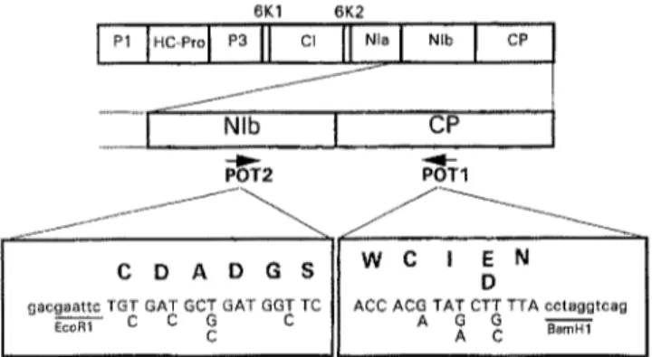
_ I N,b I cP POT1 ° II , , = . ,Fig. 1. Amino acid and nucleotidesequence ofthedegenerate primers POT1 a n d P O T 2 a n d their localization in the potyvirus genome. PI First protein; HC-Pro helper component- protease; P3 third protein; 6KI first 6K peptide; CI cytoplasmic inclusion protein; 6K2 second 6K peptide; NIa nuclear inclusion a protein; NIb nuclear inclusion b protein (RNA
polymerase); CP coat protein
RNAs were purified by two phenol/chloroform extractions. After ethanol precipitation, the RNA was resuspended in 50 ~tl of DEPC treated water.
Single stranded cDNAs were synthesized from 5 pg of total or viral RNA using an oligo-dT primer according to the Superscript Preamplification System protocol (Gibco- BRL). The resulting cDNAs were diluted 5-fold with sterile water. Five microliter from these preparations were then used for the PCR amplifications.
PCR experiments
Two degenerate primers (POT1 and POT2) were used for the amplification of cDNA corresponding to the 3'-terminal part of the NIb cistron and to the Y-terminal part of the CP cistron [3]. The sequence of the two degenerate primers and their location in the potyvirus genome are presented in Fig. 1. Amplification of the cDNA was carried out in a 50 gl volume using the Dynazyme DNA Polymerase kit. Five microliter of 10-fold Dynazyme PCR buffer, 100 ng of each POT1 and POT2 primers, 100 gM of each dATP, dCTP, dGTP, dTTP and 1 unit of Dynazyme DNA polymerase were added to 5 pl of the cDNA preparation. Sterile water was added to a final volume of 50 gl.
The following thermal cycle scheme was used for 37 reaction cycles (Biometra cycler): template denaturation at 94 °C for 30 sec, primer annealing at 40 °C (cycles 1 5) or 50 °C (cycles 6-37) for 1 min and DNA synthesis at 72 °C for 2 min. After a final 10 min elongation step at 72 °C at the end of these 37 cycles, amplification products were analysed by electrophoresis in a 1% agarose gel, in Tris-acetate-EDTA buffer [ 15]. Bands were visualised by ethidium bromide staining.
Cloning and sequencin 9 of PC R-amplified fragments
After electrophoresis, bands of expected lengths were excised and eluted with the QIAEX Gel Extraction Kit from QIAGEN. For the cloning of the amplified fragments, we used the EcoRI and BamHI restriction sites located respectively in the POT1 and POT2 primers. The YMV-BU2 fragment was cloned in a EcoRI-BamHI digested pBluescript vector (Strat- agene). The other amplified fragments were cloned in two separate fragments (due to the presence of an internal EcoRI restriction site for YMV-COT, YMV-BU1, YMV-CAR and
YMV-TOG, and due to the presence of a BamHI restriction site for YMV-12). Those digested fragments were inserted in their corresponding sites in the pBluescript vector.
For the sequencing reactions, the T7 primer, the primer Reverse or an internal synthetic otigonucteotide derived from the sequences were used. The double-stranded DNA se- quencing by the dideoxy chain termination method was carried out using T7DNA poly-
merase (Pharmacia) according to manufacturer's instructions.
Comparison of the sequences
The nucleotides and the derived amino acid sequences were compared to the E M A L L and SWALL library from the EMBL using the Fasta program [11]. Multiple alignment and dendogram were performed with the Clustal V program [-5, 6].
Results
R T-PCR amplifications
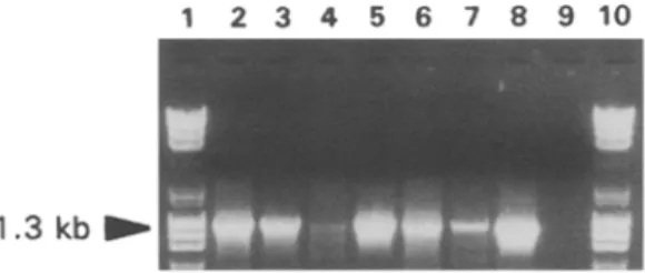
RT-PCR were carried out using the two degenerate primers, POT1 and POT2, designed to amplify cDNA fi'agments spanning the Y-terminal part of the NIb cistron and the Y-terminal part of the potyvirus CP cistron [3]. RT-PCR assays were carried out on RNA extracted from plants infected with the YMV-TOG, YMV-COT, YMV-CAR, YMV-BU 1, YMV-BU2 isolates and on a purified virus preparation of YMV-12. Total RNA isolated from N. tabacum cv Samsun infected with PVY was used as a positive control. Amplification with primers POT1 and POT2 yielded a fragment of about 1.3 kb for the six YMV isolates and a fragment of about 1.2 kb for PVY (Fig. 2, lane 2-8) whose length was in agreement with the known sequence of that virus [13]. No fragments were amplified from RNA extracted from healthy N. benthamiana (Fig. 2, lane 9).
Clonin9 and sequence analysis
The various PCR fragments corresponding to the 5'-terminal part of the CP cistron were cloned in the pBluescript vector and their nucleotide sequence were determined from three independent clones obtained from a single amplifi- cation.
Fig. 2. Electrophoretic gel of the RT-PCR assays performed with primers POT1 and POT2
on RNA extracted from a healthy N. benthamiana (9) and from plants infected with:
YMV-TOG (2); YMV-COT (3); YMV-12 (4); YMV-CAR (5); YMV-BU1 (6); YMV-BU2 (7)
Taxonomic position and characterization of YMV isolates 1071
N I b ~ I C P
Y M V - T 0 G : Y M V - C O T : Y M V - 1 2 : Y M V - C A R : Y M V - B U I : Y M V - B U 2 : P V Y - D : Q / A D T Q P D D D G Q T I D A G E S T E T K N K G K D E V G T Q S E R N Y N E Q K F P V S K D G N E T S K A Q S S ~ * * * * * * * * * * * * * * * * * * * * * * * * * * * * * * * * * * * * * * * * * * * * * * * * * * * * * * * * * * * * * * * * * * * * * * * * * * * * * * * * * * * * * * * * * * - - * * * * * * * * * * * * * * * * * * * * * * * * * * * * * * * * * * * * * * * * * * * * * * * * * * * * * * * * * * * * - - * * * * * * * * * * * * * * * * * * * * * * * * * * * * * * * * * * * * * * * * * * * * * * * * * * * * * * - - * * * * . . . * R D * M * * * * * * * D R A * * * * * * * * * * * * * * * * * * * * * * * * * * * * * * * * * * . . . * R D G * * * * * * * * D R A * / * N . . . D * * * * * * * S . . . K K D A R P * Q G . . . * I * V N P Y M V - T O G : Y M V - C O T : Y M V - 1 2 : Y M V - C A R : Y M V - B U I : Y M V - B U 2 : P V Y - D : 6 1 V A T R D R D I D A G T V G S - V L P R L A R K S L S N K M V L P T I R G K P I L N L D H L V K Y E P A Q V H L S N T R 6 1 * * * * * * * * * * * * * * * * * * * * * * * * * * * * * * * * * * * * * * * * * * * * * * * * * * * * * * * * * * * * 6 1 * * * * * * * * * * * * * * * * * * * * * * * * * * * * * * * * * * * * * * * * * * * * * * * * * * * * * * * * * * 6 1 * * * * * * * * * * * * * * * * * * * * * * * * * * * * * * * * * * * * * * * * * * * * * * * * * * * * * * * * * * * 4 9 * * * * * * * * * * * * * * * * * * * * * * * * * * * * * * * * * * * * * * * * * * * * * * * * * * * * * * * * * * * * 4 9 * * * * * * * * * * * * * * * * * * * * * * * * * * * * * * * * * * * * * * * * * * * * * * * * * * * * * * * * * * * * 2 7 M K G K * K * V N * * * S * T H T V * * I - - * A I T A * * R M * R S K * A T V * H * E * * L E * A * Q * I D I * * * * Y M V - T O G : Y M V - C O T : Y M V - 1 2 : Y M V - C A R : Y M V - B U I :YMV-
B U 2 : P V Y - D : 1 2 0 A T Q A Q F E V W Y M A I K S F Y E F E D E Q ~ I G M I M N G I I 4 V 1 2 0 * * * * * * * * * * * * * * * * * * * * * * * * * * * * * * * * * p r i m e r P O T 1 1 2 0 * * * * * * * * * * * * * * * * * * * * * * * * * * * * * * * * * 1 2 0 * * * * * * * * * * * * * * * * * * * * * * * * * * * * * * * * 1 0 8 * * * * * * * * * * * * * * * * * * * * * * * * * * * * * * * * * 1 0 8 * * * * * * * * * * * * * * * * * * * * * * * * * * * * * * * * * 8 5 * * * * * * * * * * * - - * * * * - - * - - * * * * * *Fig. 3. Amino acid sequence alignment of the N-terminal part of the coat protein of the six YMV isolates and of PVY-D./: presumed cleavage site between the NIb and the CP cistron. The trypsin-resistant core region of the CP begins at the amino acid residue equivalent to
D33 in PVY [28]
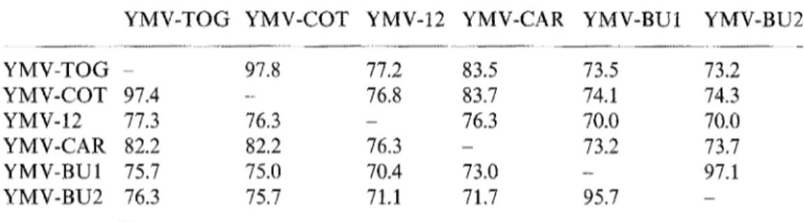
Table 1. Sequence identity (in percent) between the N-terminal part of the coat protein of the six YMV isolates
YMV-TOG YMV-COT YMV-12 YMV-CAR YMV-BU1 YMV-BU2
YMV-TOG - 97.8 77.2 83.5 73.5 73.2 YMV-COT 97.4 - 76.8 83.7 74.1 74.3 YMV-12 77.3 76.3 - 76.3 70.0 70.0 YMV-CAR 82.2 82.2 76.3 - 73.2 73.7 YMV-BU1 75.7 75.0 70.4 73.0 - 97.1 YMV-BU2 76.3 75.7 71,1 71.7 95.7 -
Above the diagonal: nucleotide sequence identity. Below the diagonal: amino acid sequence identity
T h e deduced amino acid sequences obtained for the six Y M V isolates were aligned with that of the N-terminal region of the C P of P V Y - D [16] (Fig. 3). This alignment revealed in all the Y M V isolates the presence of consensus motifs characteristic of the potyvirus genus. A m o n g them is the " D A G box" (position 13 to 15, Fig. 3) probably involved in the aphid transmissibility of the virus [1] and the putative cleavage site (Q/A, Fig. 3) between the N I b and the C P cistron. O t h e r consensus motifs were also present in the C-terminal part of the N I b cistron (data not shown).
YMV-TOG
~ YMV-
COT
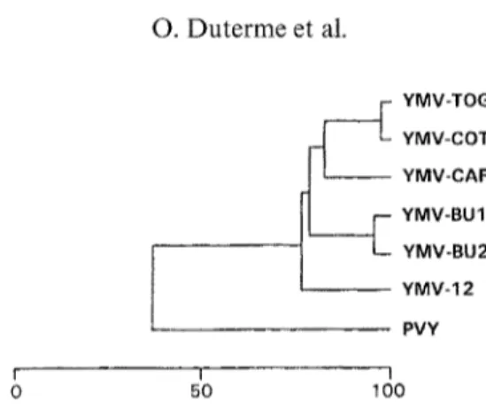
YMV-CAR YMV-BU1 YMV-BU2 YMV-12 PVY I I I 0 50 100Fig. 4. Dendrogram built from the amino acid sequence alignment presented in Fig. 3. Vertical distances are arbitrary, horizontal distances are proportional to percent amino acid
sequence divergence, shown in the scale below the figure
The sequence comparisons of the N-terminal parts of the CP of the six YMV isolates revealed identities ranging from 70.0% to 97.8% and from 70.4% to 97.4% at the nucleotide and at the amino acid level respectively (Table 1). At the amino acid level, high sequence identity were observed between YMV-BU1 and YMV-BU2 (95.7%) and between YMV-TOG and YMV-COT (97.4%). The other sequence identities observed between the six YMV isolates ranged from 70.4% to 82.2% (Table 1).
The comparison of the six YMV sequences revealed that the N-terminal part of the CP encompass a 152 aa fragment for the YMV-TOG, YMV-COT, YMV-12 and YMV-CAR isolates. However, this fragment is only 140 aa long for the YMV-BU1 and YMV-BU2 isolates. Indeed, the YMV-BU1 and YMV-BU2 isolates lack a stretch of 12 aa from position 34 to 45 in comparison to the four other isolates (Fig. 3).
In order to show the relationships between the six YMV isolates and PVY, a dendrogram was built from the multiple amino acid sequence alignment (Fig. 4). This dendrogram showed that YMV-TOG and YMV-COT and that YMV- BU1 and YMV-BU2 were respectively closely related. YMV-CAR shared an average sequence identity of 82.2% in comparison to YMV-TOG and YMV- COT while YMV-BU1 and YMV-BU2 presented 78.8 % sequence identity with YMV-CAR, YMV-COT and YMV-TOG. The average sequence identity be- tween YMV-12 and the five other YMV isolates was 75.9%. PVY, the type member of the
Potyvirus
genus, shared an average sequence identity of 35 % with the six YMV isolates.Amino acid sequence comparison between the six YMV isolates and 28 potyviruses previously sequenced in the considered region (listed in [20]) exhibit a broader divergence (identity level below 50%) (data not shown).
Discussion
The aim of our study on the N-terminal part of the CP cistron was to analyse the taxonomical relationship between YMV and members of the
Potyvirus
genusTaxonomic position and characterization of YMV isolates 1073 and to evaluate the variability between YMV isolates of different origins. The comparison of the entire CP cistron has been widely used to establish taxonomic relationships among potyvirus members [14, 17-19, 26]. The sequence data show a bimodal distribution of CP sequence identities [1%19]. Known distinct potyviruses vary in CP amino acid sequence identity between 38 % to 71%, while known strains of viruses range from 90% to 99%.
The PCR procedure utilizing degenerate primers designed to amplify the region coding for the C-terminal half of the Nib polymerase and the N-terminal half of the CP is a rapid and sensitive technique for detecting and characterizing members of the
Potyvirus
genus [3]. The cloning and sequencing of the six YMV amplified fragments obtained with the degenerate primers POT1 and POT2 revealed potyvirus specific consensus motifs, thus confirming the classification of YMV as a potyvirus member on the base of its biological, serological and microscopical properties [20]. Among these motifs, the "DAG box" [1] is in agreement with the aphid transmissibility of the YMV [24].YMV-BUt and YMV-BU2 lacked a stretch of 12 aa from position 34 to 45 within the CP cistron in comparison to the four other isolates. Similar deletions have been observed within some strains of P P V [7-] and for SMV-N and WMV-2 [28]. Examination of the nucleotide and amino acid sequences of the six YMV isolates revealed no evidence for partial CP duplication t-4, 9, 10, 27].
Since the sequence identities between the six YMV isolates and all the other potyviruses were below 50%, we can conclude that YMV is a distinct potyvirus. The high sequence identity observed between YMV-BU1 and YMV-BU2 and between YMV-TOG and YMV-COT (95.5% and 97.4% respectively) suggests that those YMV isolates are closely related strains of YMV. The other sequence identities between the six YMV isolates ranged from 70.4% to 82.2%, within the range of variation seen for this region in other potyvirus strains [20]. These identities were calculated for the N-terminal half of the CP cistron which includes the most variable region [17, 18, 20, 26]. As shown in Fig. 3, most of the sequence differences are located in the first 67 aa domain of the protein which is known to differ markedly between individual viruses. The core region of the CP (residues 67-152), shows much fewer amino acid substitutions in the YMV isolates compared to those seen in comparison with PVY (Fig. 3). These observations lead us to suggest that the six YMV isolates should be considered as different strains of a single virus.
References
1. Atreya P, Atreya C, Pirone T (1991) Amino acid substitutions in the CP result in the loss of insect transmissibility of a plant virus. Proc Natl Acad Sci USA 88:7887-7891 2. Chirgwin J, Przybyla A, MacDonald R, Rutter W (1979) Isolation of biologically
active ribonucleic acid from sources enriched in ribonuclease. Biochemistry 18: 5294- 5299
3. Colinet D, Kummert J (1993) Identification of a sweetpotato feathery mottle virus isolate from China (SPFMV-CH) by the polymerase chain reaction with degenerate primers. J Virol Methods 45:149-159
4. Frenkel M, Jilka J, McKern N, Strike P, Clark Jr J, Shukla D, Ward C (1991) Unexpected sequence diversity in the amino-terminal ends of the coat protein of strains of sugarcane mosaic virus. J Gen Virol 72:237-242
5. Higgins D, Sharp P (1989) Fast and sensitive multiple sequence alignments on a micro- computer. CABIOS 5:151-153
6. Higgins D, Bleasly A, Fuchs R (1991) Clustal V: improved software for multiple sequence alignment. CABIOS 8:189-191
7. Maiss E, Timpe U, Jelkmann W, Casper R (1989) Comparison of two different plum pox virus isolates on nucleic acid level. Acta Horticult 235:313-317
8. Marchoux G (1980) Pathologie des ignames en Guadeloupe; maladies virales. In: Les colloques de I'I.N.R.A., l'Igname. Seminaire International de Pointe-fi-Pitre, 28 July-02 August 1980, 93-105
9. Pappu S, Pappu H, Rybicki E, Niblett C (1994) Unusual amino-terminal sequence repeat characterizes the capsid protein of dasheen mosaic potyvirus. J Gen Virol 72:239-242 10. Pappu S, Pappu H, Lastra R, Niblett C (1994) Variability in the length of the amino
terminal sequence contributes to the capsid protein diversity among dashen mosaic potyvirus isolates. Arch Virol 136:407-413
1 t. Pearson W, Lipman D (t988) Improved tools for biological sequence comparison. Proc Natl Acad Sci USA 85:2444-2448
12. Reckhaus P, Nienhaus F (1981) Etiology of a virus disease of white yam D. rotundata in Togo. J Plant Dis Prot 88:492-509
13. Robaglia C, Durand-Tardif M, Tronchet M, Boudazin G, Astier-Manifacier S, Casse- Delbart F (1989) Nucleotide sequence of potato virus Y (N strain) genomic RNA. J Gen Virol 70:935-947
14. Rybicki E, Shukla D (1992) Coat protein phylogeny and systematics of potyviruses. In: Barnett OW (ed) Potyvirus taxonomy. Springer, Wien New York, pp 283-297 (Arch Virol [Suppl] 5)
15. Sambrook J, Fritch E, Maniatis T (1989) Molecular cloning: a laboratory manual. Cold Spring Harbor Laboratory Press, New York
16. Shukla D, Thomas J, McKern N, Tracy S, Ward C (1988) Coat protein of potyviruses. 4. Comparison of biological properties, serological relationships, and coat protein amino acid sequences of four strains of potato virus Y. Arch Virot 102:207-219
17. Shukla D, Ward C (1988) Amino acid sequence homology of coat proteins as a basis for identification and classification of the potyvirus group. J Gen Virol 69: 2703-2710
18. Shukla D, Ward C (1989) Structure of potyvirus coat proteins and its application in the taxonomy of the potyvirus group. Adv Virus Res 36:273-314
19. Shukla D, Ward C (1989) Identification and classification of potyviruses on the basis of coat protein sequence data and serology. Arch Virol 106:171-200
20. Shukla D, Ward C, Brunt A (1994) The potyviridae. CAB International, Wallingford 21. Terry E (1976) Incidence, symptomatology and transmission of a yam virus in Nigeria.
Proceedings of the Fourth Symposium of the International Society of Tropical Roots Crops. Call, Columbia, pp 170-173
22. Thouvenel J-C, Fauquet C (1977) Une mosaique de l'igname (Dioscorea cayenensis) caus6e par un virus filamenteux en C6te d'Ivoire. Comptes Rendus hebdomadaires des Seances de l'Acad~mie des Sciences 284:1947-1949
23. Thouvenel J-C, Fauquet C (1979) Yam mosaic, a new potyvirus infecting Dioscorea cayenensis in the Ivory Coast. Ann Appl Biol 93:279-283
24. Thouvenel J-C, Fauquet C (1986) Yam mosaic virus. AAB Description of Plant Viruses, No. 314
Taxonomic position and characterization of YMV isolates 1075 25. Ward C, Shukla D (1991) Taxonomy of potyviruses: current problems and some
solutions. Intervirology 32:269-296
26. Ward C, McKern N, Frenkel M, Shukla D (1992) Sequence data as the major criterion for potyvirus classification. Arch Virol [Suppl] 5:283 297
27. Xiao X, Frenkel M, Teakle D, Ward C, Shukla D (1993) Sequence diversity in the surface-exposed amino-terminal region of the coat protein of seven strains of sugarcane mosaic virus correlates with their host range. Arch Virol 132:399-408
28. Yu M, Frenkel M, McKern N, Shukla D, Strike P, Ward C (1989) Coat protein of potyviruses. 6. Amino acid sequences suggest watermelon mosaic virus 2 and soybean mosai virus-N are strains of the same potyvirus. Arch Virol 105:55-64
Authors' address: Dr. O. Duterme, Facult6 Universitaire des Sciences Agronomiques, Laboratoire de Pathologie V6g6tale, Passage des, D6port6s 2, B-5030 Gembloux, Belgium.