ISMRE2018/XXXX-2018 ALGERIA
Elaboration of Hydroxyapatite Bioceramics by
Sintering
Attabi Selma
1*, Majda Mokhtari
2, Hicham Elmsellem
3, Abed Elaziz Himour
11
Laboratory of Surface Engineering LIS, department of chemistry , University of Badji Mokhtar, BP 233 RP, Annaba, Algeria
2
University of Echahid Hamma Lakhdar, PO Box 789,El-Oued, Algeria. 3
Laboratory of Analitical Chemistry, Materials, and Environment (LC2AME), Faculty of Sciences, University of Mohammed Premier, B.P. 717, 60000 Oujda, Morocco
Address Email * [email protected]
Abstract— Thanks to its good properties of biocompatibility and bioactivity, hydroxyapatite (Ca10(PO4)6(OH) 2) is largly used to make functional
materials in biomedical applications. This inorganic material can be used either as a thin film on metallic alloys or to make bulk ceramic bodies[1]. Hydroxyapatite (HAP) has also a good thermal stability and better physicochemical properties thanks to its molar ratio Ca/P which is always fixed at 1,67 [2]. Actually, the powder based on hydroxyapatite can be obtained by various methods. We chose in this work to elaboratie the HAP powder by the chemical method of double decomposition ,which is very much used by industries thanks to its facility and its moderate cost. Thereafter, the powder was characterized by diffraction of x-rays, a thermal analysis TGA-DSC, and Fourier transform infrared spectroscopy to examine its characteristics. For this part, results confirm the obtention of well crystilled one single phase. In the next step of work, we aim to develop in details the elaboration of sintered bodies based on hydroxyapatite. For that, the powder sintered at 1150 ͦ C for two hours. Scaning electron microscopy images of final samples show the presence of porous structure genertated thanks to the additifs added to the powder before sintering.
Keywords— Hydroxyapatite, powder, sintering, biomaterial, porosity.
I. INTRODUCTION
Hydroxyapatite, with chemical formula Ca10(PO4)6(OH) 2,
is a calcium phosphate compound regrouped with bioactive ceramics. Thanks to its best properties of biocompatibility, ostéoconduction, ostéoinduction, and of thermal stability, this mineral material was used intensively in the fabrication of functional biomaterials. Hydroxyapatite (HAP) has better physicochemical properties thanks to its molar ratio Ca/P which is always fixed at 1,67. The pulverulent substance and the bioceramics based on hydroxyapatite can be obtained by various methods. Indeed, the final properties of the bioceramics depend on the method of synthesizing of the powder and on the process of elaboration of cermic bodies. Howerver, the porosity, especially nanoporosity of theses bodies is an important factor which influence on its properties. Generating nanoporosity is not as always possible, this demands a careful control of elaboration process.
II. EXPERIMENTAL PROCEDURE
A. Powder Synthesizing
A simple method of co-precipitation was proposed for the preparation of HAP powder. Firstly, the Ca and P precursor
aqueous solutions were separately prepared by dissolving
23,54 g of calcium nitrate tetrahydrate [Ca(NO3)2.4H20] and
6,87 g of diammonium hydrogen phosphate NH4H2PO4 in
bidistilled water.
On a magnetic stirrer (400tour/min), the solution of
diammonium hydrogen phosphate was added to the calcium
nitrate solution drop wise, and the PH of the solution was adjusted to 10 by adding ammonia solution (NH4OH).After
3 hours of maturation, the solution is maintained at the ambient temperature for precipitation. Thereafter, the precipitate was filtered and washed with bidistilled water. This operation was repeated several times in order to eliminate the ions NH4+ and NO3-.The final precipitate was dried to 70 ° C during 24h.The powder obtained was ground in a mortar and then calcined to 700 °C during 2 hours.
The stability of the phases of the calcined powder was studied by the method of the diffraction of x-ray (XRD) in a diffractometer of x-ray (Rigaku, Ultima IV, Japan) with CuKα = 1.5418 Å radiation produced with a tension of 40 kilovolts and a current of 40 my.
The thermal analysis of the powder was made by analysis (TG) thermogravimetric by using a thermal analyzer (Netzsch, 449C, Germany).To identify the functional groups, the analysis of FTIR was made in a spectrometer of FTIR (PerkinElmer, spectrum two 95277).
B. Sintering
Before sintering, the hydroxyapatite powder was mixed with 10% of starch and well grinded in a mortar. The mixture was pressed into pellets using a hydraulic press. The pellets were then sintered in a muffle furnace at 1150 C for 2 hours.
III. RESULTS
A. Phase analysis
Phase analysis of HAP prepared by co precipitation route is plotted in FIGURE 1. The figure show the presence of one single phase “HAP” thermally stable and well crystallized. The pattern confirms also the absence of any other phase.
Fig. 1. XRD patterns for HAP synthesized powder B. Thermal analysis
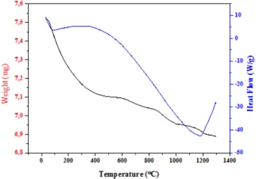
Thermogravimetric analysis, shown in FIGURE 2, revealed weight losses associated to 100 °C and 300 °C which corresponds to the evaporation of water absorbed (4,49% of water by weight). At low temperature, we observed an endothermic peak, which corresponds to the evaporation of water, and at high temperature we observed another endothermic peak, which corresponds to the transformation of precursors to the crystalline phase “hydroxyapatite”. The results obtained was similar to many recent works [8,9].
Fig. 2. Thermo graphics TGA-DSC of synthesized HAP powder
C. Fourier transform infrared spectroscopy:
The infra-red spectrum (IR) of the prepared and dried product is shown in FIGURE 3.The spectrum shows the presence of the bands relating to hydroxyapatite. Thus the hydroxyapatite is identifiable by the vibration bands of PO43- that appear to 550 and 600 cm-1, 960 cm -1, 1030-1120
cm-1. The bands with 560 and 630 cm -1 correspond to the stretching and vibration modes of ions OH- [8].
Fig. 3. IR spectrum of calcined HAP powder D. Structural Analysis of sintered samples:
Both Fig 4 shows the scanning electron micrographs of sintered sample. We can notice the presence of highly porous structure generated thanks to the addition of starch before sintering. The sintered sample preserve its highly porous structure having diameter ranging from 542 nm to 1.75 µm. Also, it can be noticed that the shape of pores is spherical throughout the entire sample. This remark was observed also in Ahmed's work [10], and it was reported that the pores preserve their spherical shape when adding just small quantities of starch .
Fig. 4. Scanning electron micrographs of sintered ceramics on cross section .
IV. CONCLUSION
In this work, we presented the method of elaboration of both powder and bulk bodies of hydroxyapatite. Sintered samples showed homogenous shape and diameter of pores.
REFERENCES
[1] L. Saïd. Étude comparative des caractéristiques physicochimiques et mécaniques des biomatériaux à base de phosphate de calcium, d’alumine et de zircone: Caractérisation et modélisation », 2013 [2] L. El Hammari, « Synthèse et études physico-chimiques des
phosphates de calcium poreux greffes par des molécules organiques: Structure et processus d’adsorption », 2007.
[3] S. Mollazadeh, J. Javadpour, A. Khavandi, «In situ synthesis andcharacterization of nano-size hydroxyapatite in poly (vinyl alcohol) matrix»,Ceram. Int. 33 (2007) 1579–1583.
[4] P.X. Ma, «Biomimetic materials for tissue engineering», Adv. Drug Del. Rev. 60(2008) 184–198.[5] J.R. Jones, L.M. Eh.
[5] J. C. Elliot, «Structure and chemistry of apatites and other calcium orthophosphate», Amsterdam, Ed. Elsevier, 1994.
[6] R. A. Young, D. W. Holcomb, « Variability of hydroxyapatite preparations », Calcified Tissue International, Vol. 34, Pages 17-32, 1982.
[7] J. Arends, J. Christoffersen, M.R. Christoffersen, H. Eckert, B.O. Fowler, J.C. Heughebaert, G.H. Nancollas, J.P. Yesinowski, S.J. Zawacki, « Calcium Hydroxyapatite precipitated from aqueous solution: An international multimethod analysis », J. of Crystal Growth, Vol. 84, Issue 3, Pages 515-532, 1987.
[8] A. Massit, A. Yacoubi, B. Chafik El Idrissi, et K. Yamni, « Synthèse de nanoparticules de phosphate tricalcique β par voie aqueuse », 2015.
[9] M. Mbarki, P. Sharrock, M. Fiallo, et H. ElFeki, « Hydroxyapatite bioceramic with large porosity », Materials Science and Engineering: C, vol. 76, p. 985-990, juill. 2017.
[10] Ahmed Y.M.Z, Ewais E.M.M, El-Sheikh S.M. Porous hydroxyapatite ceramics fabricated via starch consolidation technique, Journal of Ceramic Processing Research. Vol. 16, No. 00, pp. 1~10 (2015).

![Risiko- & [und] Schutzfaktoren der psychischen Gesundheit humanitärer Einsatzhelfer : eine systematische Literaturübersicht](data:image/gif;base64,R0lGODlhAQABAIAAAP///wAAACH5BAEAAAAALAAAAAABAAEAAAICRAEAOw==)