Université de Montréal
NUCLEAR-CYTOPLASMIC INTERACTIONS IN
RAT
OOCYTES AND RECONSTRUCTED EGGS DERIVED
BY SOMATIC CELL NUCLEAR TRANSfER
par JAE GYU YOO
Département de 3 iornédecine Vétérinaire faculté de Médecine Vétérinaire
Thèse présentée à la Faculté des Études Supérieures
en vue de l’obtention du grade de Philosophiae Doctor (Ph.D.) en Sciences Vétérinaires
option reproduction
Décembre, 2006
o
7
Université
(III
de Montréal
Direction des bibliothèques
AVIS
L’auteur a autorisé l’Université de Montréal à reproduire et diffuser, en totalité ou en partie, par quelque moyen que ce soit et sur quelque support que ce soit, et exclusivement à des fins non lucratives d’enseignement et de recherche, des copies de ce mémoire ou de cette thèse.
L’auteur et les coauteurs le cas échéant conservent la propriété du droit d’auteur et des droits moraux qui protègent ce document. Ni la thèse ou le mémoire, ni des extraits substantiels de ce document, ne doivent être imprimés ou autrement reproduits sans l’autorisation de l’auteur.
Afin de se conformer à la Loi canadienne sur la protection des renseignements personnels, quelques formulaires secondaires, coordonnées ou signatures intégrées au texte ont pu être enlevés de ce document. Bien que cela ait pu affecter la pagination, il n’y a aucun contenu manquant.
NOTICE
The author of this thesis or dissertation has granted a nonexciusive license allowing Université de Montréal ta reproduce and publish the document, in part or in whole, and in any format, solely for noncommercial educational and research purposes.
The authoc and co-authors if applicable retain copyright ownership and moral rights in this document. Neither the whole thesis or dissertation, nor substantial extracts from it, may be printed or otherwise reproduced without the author’s permission.
In compliance with the Canadian Ptivacy Act some supporting forms, contact
information or signatures may have been removed from the document. While this may affect the document page count, it does flot represent any Ioss of
Faculté des études supérieures
Cette thèse intitulée
NUCLEAR-CYTOPLASMIC INTERACTIONS IN RAT OOCYTES AND
RECONSTRUCTED EGGS DERIVED BY SOMATIC CELL NUCLEAR
TRANSFER
présentée par:
JAE GYU YOO
a été évaluée par tin jury composé des personnes suivantes
Bruce D. Murphy, président-rapporteur Lawrence C. Smith, directeur de recherche
Alan K. Goff, membre du jury Jay M. Baltz, examinateur externe Mario Jacques, représentant du doyen de la FES
111
RÉSUMÉ
Le clonage par transfert de noyau de cellule somatique (TNCS) permet la génération
de modèles animaux transgéniques à partir de cellttles génétiquement modifiées et constitue ainsi une alternative potentielle à la technologie des cellules souches embryonnaires, qui n’est toujours pas établie chez le rat. Cependant, il existe plusieurs problèmes à étudier pour l’établissement de protocoles stables de TNCS chez le rat. Les objectifs de cette étude sont 1) d’étudier le mécanisme d’activation spontanée constaté dans l’ovule de la ratte 2) d’optimiser le développement des ovules parthénogénétiques et issues du procédé de TNCS chez le rat en étudiant les changements nucléaires, les changements dans l’activité de Maturation-Prornoting factor (MPF) et de Mitogen activated-Protein (MAP) kinase, et le développement in vitro des embryons après divers traitements d’activation, et 3) d’étudier la
coordination optimale du cycle cellulaire entre les cellules donneuses et les ovules receveuses pour le TNCS chez le rat et d’analyser l’expression des gènes et des protéines impliqués dans le cytosquelette des ovules reconstruits cultivés in vitm.
Peu après avoir été exposés à l’environnement in vitro, les ovules de la ratte
deviennent activés spontanément. Cette activation spontanée des ovules est caractérisée par la reprise de la division méiotique suivie de la dispersion cytoplasmique des chromosomes. Ni la présence prolongée in vivo dans les oviductes après l’ovulation, ni le
traitement à l’hyaluronidase n’ont affecté l’activation spontanée des ovules. L’inhibiteur de
dependent kinase II (CaMKII) ont empêché l’activation spontanée des ovules dans le milieu contenant du calcium. L’activité de la CaMKII a augmenté à 20 minutes et est demeurée haute pendant 30 minutes, suivi d’une activité diminuée à 60 minutes après la récolte des ovules. La forme constitutivement active de la CaMKII était localisée près du fuseau rnéiotique après la récolte des ovules. Nos résultats indiquent que les ovules de la ratte sont très sensibles au calcium extracellulaire sous des conditions in vitro et la CaMKII est l’un des signaux précoces qui activent les ovules de la ratte spontanément après la récolte.
L’activation des ovules est une étape essentielle lors du clonage par TNCS. Dans notre étude, des ovules ont été activés par la stimulation électrique (EST) seule ou en combinaison avec la culture de courte durée en présence de 6-dimethylaminopurine (DMAP), de cyclobeximide (CHX)/cytochalashin B (CB), et de roscovitine (toute la combinaison de ROS)/CB. Tous les groupes ont efficacement induit l’inactivation de l’activité de MPf. L’activation de la MAP kinase varie selon les différents groupes de traitement. DMAP induit une inactivation plus rapide de la MAP kinase que les groupes de traitement à la CHX/CB et ROS/CB. Les ovules du groupe CHXICB ont procédé à la dissolution de la membrane nucléaire et au clivage de manière synchronisée après le traitement d’activation, tandis que les ovules des groupes DMAP et ROS/CB n’étaient pas synchronisés. Bien que le développement in vitro jusqu’au stade blastocyste ait été
efficace après la parthénogenèse. le développement des embryons issus du TNCS a bloqué au stade 2-cellules dans tous les régimes examinés.
La micromanipulation, la coordination du cycle cellulaire entre les noyaux des celltiles donneuses et les ovules recevetises, et l’activation artificielle des ovules sont des
V
étapes très importantes de la procédure du clonage animal. Des ovules au stade métaphase II (MII) et des ovules pré activés au stade télophase II (Tu) ont été employés comme cytoplasme receveur avec des cellules donneuses aux phases GO/Gl, M, et S/G2. D’ailleurs, des pronucléi et des blastomères provenant d’embryons issus du TNCS au stade 2-cellules ont été employés comme cellules donneuses en combinaison avec des ovoplastes zygotiques et parthénogénétiques énucléés pour le clonage dit en série. Aucune différence significative dans le taux de clivage n’a été observée parmi les groupes d’activation après le TNCS. Les cellules donneuses en phase M ont eu un taux sensiblement plus élevé de clivage que les cellules dormeuses en phase GO/Gl avec des ovules en Mfl et les cellules donneuses en phase G2 avec des oocytes de Tu. Cependant, aucun embryon reconstruit par TNCS n’a tL se développer au-delà dci stade 2-cellules pendant la culture in vitro.
D’ailleurs, les embryons reconstruits cultivés in vivo, c’est-à-dire après le transfert embryonnaire dans l’oviducte de femelles porteuses, n’ont également pas pu se développer plus loin. Pour mieux comprendre les causes de l’arrêt du développement, des embryons reconstruits par TNCS au stade 2-cellules ont été analysés pour examiner la distribution des protéines dti cytosquelette et la transcription des ARN messagers. La distribution anormale de microtubules et l’expression diminuée de plusieurs transcrits du cytosquelette ont été montrées dans les embryons au stade 2-cellules reconstruits par TNCS. Ces résultats indiquent que l’arrêt du développement des embryons issus dci TNCS chez le rat est associé à la transcription inexacte de gènes du cytosquelette, vraisemblablement ayant pour résultat la distribution anormale de microtubules.
Mots clés: cacium/ca1rnoduÏin-dependent kinase II (CaMKII), activation spontanée des ovules, activation parthénogénétique, transfert de noyau de cellule somatique (TNCS), rat
vii
ABSTRACT
Somatic ceil nuclear transfer (SCNT) enables the generation of transgenic animal models from geneticafly modifled celis and it is a potential alternative to the ES celi technology, which is stifl not established yet in rats. However, there are rnany problems to resolve to establish stable protocols for SCNT in rats. The objectives of this study are 1) to investigate the possible reasons and rnechanism of rat spontaneous oocyte activation, 2) to optimize development of parthenogenic and SCNT-derived oocytes in rats by investigating the patterns of nuclear changes, the changes of MPF and MAP kinase activities, and in vitro embryo development after various activation treatments, and 3) to
investigate optimal cdl cycle coordination between donor celis and recipient oocytes for rat SCNT and to analyse the expression of genes and proteins involved in the cytoskeleton of
in vitro cultured reconstructed eggs.
Soon after exposure to an in vitro environment, ovtilated rat oocytes are activated spontaneously; this spontaneous oocyte activation is characterized by resumption ofmeiotic division followed by the cytoplasmic scattering of chromosomes. Neither in vivo aging in oviducts after ovctlation nor hyaluronidase treatment affected spontaneous oocyte activation. L-type calcium channel blocker, IP3R inhibitor and inhibitor of calcium/calmodulin dependent kinase II (CaMKII) prevented spontaneous oocyte activation in calcium containing medium. The activity of CaMKII increased at 20 min and remained high for 30 min followed by decreased activity by 60 min after oocyte recovery. Constitutively
findings indicate that rat oocytes are very sensitive to extraceilular calcium in vitro
conditions and CaMKII is one of the upstrearn signais that activate rat oocytes spontaneously after recovery.
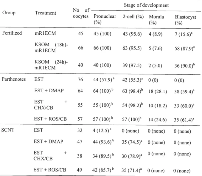
Oocyte activation is an essential step in successful cloning by SCNT. In our study, oocytes were activated by electrical stimulation (EST) alone or in combination with 6-dimethylaminopurine (DMA?), cyciohexirnide (CHX)/cvtochaiashin B (CB), and roscovitine (ROS)/CB. Ail combination groups effectively induced inactivation of MPF activity. The patterns of MAP kinase varied in different treatment groups. DMAP induced faster inactivation of MAP kinase than CHX/CB and ROS/CB treatment groups. CHX/CB-treated oocytes showed synchronous nuclear breakdown and cleavage after activation treatrnent, whereas DMAP and ROS/CB treated groups showed asynchronous patterns. Although in vitro deveiopment to the blastocyst stage vas efficient after
parthenogenesis, development of SCNT-derived embryos was arrested at 2-ceil stage in ail regimens examined.
The procedure of micromanipulation, coordination of celi cycle between donor nuciei and recipient oocytes, and artificial oocyte activation are very important steps in the procedure for cloning animais. Metaphase II (MII) stage and pre-activated telophase II (Tu) stage oocytes were used as a recipient cytoplasm with GO/G1, M, and S/G2-phases donor ceHs. Moreover, pronuclear and 2-ecu stage blastorneres derived from SCNT were
used as donor celis with enucleated zygotic and parthenogenetic ooplasts for serial cloning.
ix SCNT. M-phase donor celis had a significantly higher cleavage rate than GO/G1-phase donor ceils with MII oocytes and G2-phase donor celis with Tu oocytes. However, no reconstructed ernbryo vas able to develop beyond the 2-celi stage during in vitro culture. Moreover, reconstructed embryos cultured in vivo, i.e. after transfer to the oviduct of
surrogate females, were also unable to develop further. To better understand the causes of deveÏopmental atTest, reconstructed 2-celi stage embryos were analyzed to examine the distribution of cytoskeletal proteins and transcription of mRNAs. Abnorrnal microtubule distribution and downregulated expression ofseveral cytoskeletal transcripts were shown in 2-ceil stage reconstructed embryos. These resttlts indicate that the developmental arrest of rat SCNT embryos is associated with improper transcription of cytoskeleton genes, presumably resulting in abnormal microtubule distribution.
Key Words: Ca2/caImodutin-dependent protein kinase II, Spontaneous oocyte activation, Parthenogenic activation, SCNT, Rat
ACKNOWLEDGMENTS
I would like to express my gratitude to my supervisor, Dr. Lawrence C. Smith, whose
expertise, understanding, and patience, added considerably to my graduate experience. I
appreciate his vast knowledge and skill in many areas.
I would like to thank members ofjury for my dissertation, Dr. Brnce Murphy, and Dr.
Àlan Goff I also would like to thank Dr. Jay M. Baltz from the University of Ottawa for taking tirne out from his busy schedule to serve as myextemal examiner.
I must also acknowledge Dr. Sang-Yong Choe and Dr. Gyu-Jin Rho from
Gyeongsang National University in Korea for their support and advice. I sincerely thank ail of the cunent and former members of CRRA for their help and friendship over the last four and a haif years, especially Cannen Leveillee, Micheline Sicotte, Jacinthe Iherrien, france Filion, Mira Dobias, Patrick Vincent and Micheline St-Gerrnain. Without their help I wouÏd flot be writing this thesis today. I spent a great deal of time at CRRA and ail people are like rny family.
I would also like to thank my father and mother for the support they provided me
TABLE 0F CONTENTS
INDENTIFICATION 0F JURY ii RESUME iii ABSTRACT vii ACKNOWLEDGMENTS x DEDICATION xiTABLE 0f CONTENTS xii
LIST 0f TABLES xvi
LIST 0F FIGURES xvii
LIST 0F ABBREVÏATIONS xix
INTRODUCTION 1
CHAPTER 1- LITERATURE REVIEW 4
I. Importance of Rat in Biomedical Research 4
2. Mechanisms Involved in Metaphase II Arrest 6
2.1. Cytostatic Factor(CSF) 7
2.2.Maturation Promoting factor (MPf) 8
2.3. Mitogen-ActivatedProtein Kinase (MAP kinase) 10
3. The Resumption ofrneiosis from MII 14
4. Ca21Ca1modu1in—depeiident Protein Kinase 11 15
xiii
5.1. Calcium ionophores 18
5.2. Strontium 19
5.3. Ethanol 19
5.4. Protein Synthesis Inhibitors 19
5.5. Protein Kinase Inhibitors 20
5.6. Cdc2 Kinase Inhibitors 21
5.7. MEK inhibitor 21
6. General Introduction to Nuclear Transfer 22
6.1. Artificial Oocyte Activation ofNuclear Transfer Ernbryos. 26
6.2. In vitro culture system 27
6.3. Ccli Cycle Coordination 28
6.3.1. Ceil Cycle Synchronization 31
7. Problems encountered in Nuclear Transfer 33
7.1. Low Efficiency 33
7.2. Epigenetic Modification 34
7.3. Developmental Anomalies 35
8. Probiem, Hypothesis, and Objectives 36
CHAPTER II Extracellular calcium induces activation ofCa2*/Calmodulin_
dependent protein kinase II mediates spontaneous oocyte activation in the rat. 40
Abstract 41
Materials and Methods 45
Resuits 49
Discussion 54
References 72
CHAPTER III- Changes to nuclear status, maturation-promoting factor, and
mitogen-activated protein kinase activity foflowing different parthenogenetic
activation regimens in rat oocytes. 78
Abstract 79
Introduction 80
Materials and Methods 83
Resuits 88
Discctssion 92
References 104
CHAPTER W - Developmental aiiest and cytoskeletal anomalies of rat ernbryos
reconstructed by somatic ceil nuclear transfer. 108
Abstract 109
Introduction 110
Materials and Methods 112
Resuits 121
xv Acknowledgrnents 131 References 142 GENERAL DISCUSSION 146 GENERAL CONCLUSIONS 155 REFERENCES 157
LIST 0F TABLES
CHAPTER I
Table 1. Celi cycle coordination between donor nuclei and recipient oocytes — 29
CHAPTER III
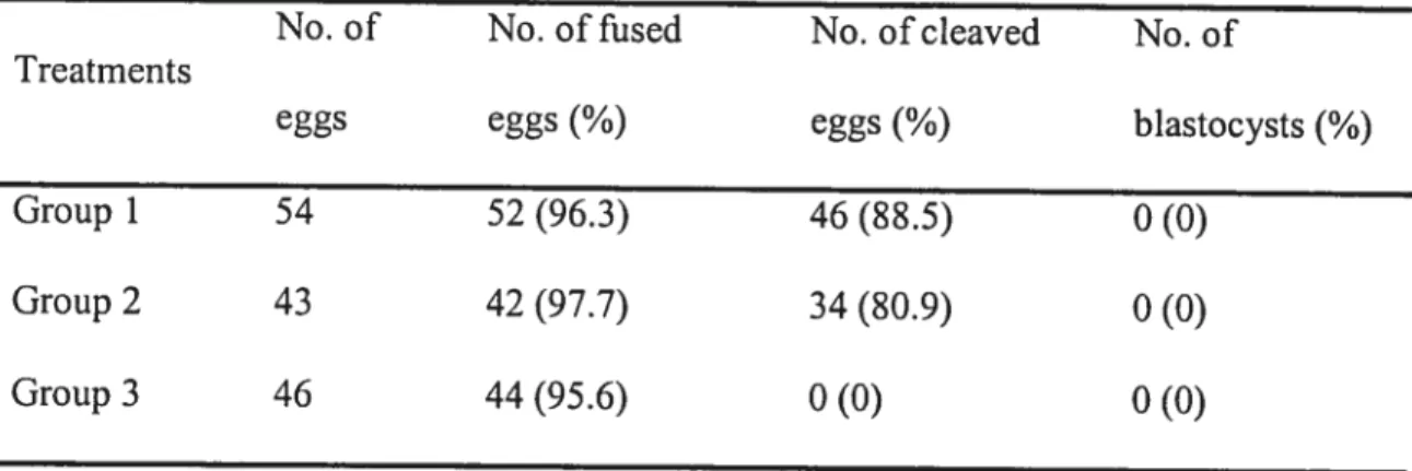
Table 1. Developrnentin vitro of rat oocytes obtained afterin vivo
fertilization, parthenogenic activation and sornatic ceil nuclear
transfer (SCNT). 97
CHAPTER IV
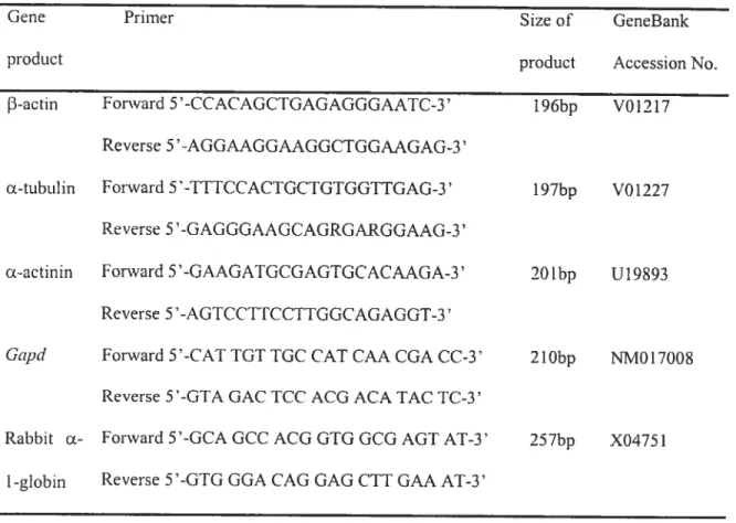
Table 1. Primers for real-timePCR. 132
Table 2. Sornatic Cell Nuclear Transfer (SCNT) with different celi cycles of
donor ceils and recipient oocytes. 133
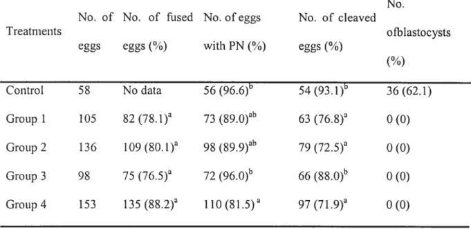
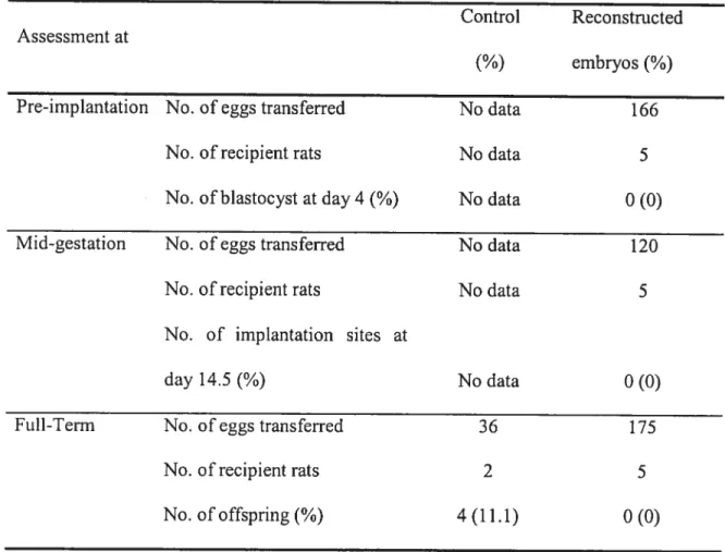
Table 3. Serial cloning with zygotic and parthenogenetic cytoplasm. 134 Table 4. fii vivo development ofreconstructed and fertilized ernbryos
xvii
LIST 0F FIGURES
CHAPTER IFigure 1. Cytostatic factor (CSF), Maturation-promoting factor (MPF), and Mitogen-Activated Protein (MAP) kinase activity during meiosis,
fertilization and early development. 13
Figure 2. Schematic drawing ofthe nuclear transfer procedure by
microinjection in mice. 25
CHAPTER II
Figure 1. The distribution ofthe insensitive and sensitive fernale rats. 59 Figure 2. Pattems of rat spontaneous oocyte activation. 61 Figure 3. Effect of hyaluronidase (A) and calcium in medium (B) depends
onovulated rat oocyte aging in oviducts. 63 Figure 4. Effect ofnifedipine (A) and xestospongin (B) on spontaneous
activation of rat oocytes. 65
Figure 5. CaMKII activity ofspontaneous and chernical indctced activation withlwithout different conditions (calciurn-free, nifedipine, and
xestospongin) in rat oocytes. 67
Figure 6. Distribution of active CaMKII in rat oocytes. 69 Figure 7. Effect ofmyr-AIP (Inhibitor ofCaMKII) on spontaneous
CHAPTER III
Figure 1. Changes inhistone Hi and MAP kinase activities in rat oocytes
following EST alone (A), EST plus DMAP (B), EST plus
CHX/CB (C), and EST plus ROS/CB (D). 99
Figure 2. Kinetics of cellular events in rat oocytes activated with EST alone ta), EST plus DMA? (b), EST plus CHX/CB (e), and EST plus
ROS/CB (d). 101
figure 3. Morphology ofeggs at one and two-cell stages followingin vivo
fertilization (A and A’), EST plus DMAP (3 and 3’), EST plus
CHX/CB or ROS/C3 (C and C’) and SCNT (D and D’). 103
CHAPTER IV
Figure 1. Distribution ofcelÏ-cycle stages of fetal flbroblasts in different
culture conditions measured by flow cytornetry. 137 Figure 2. Relative amount of-actin, a-tubulin, Œ-actinin, and GapcÏ mRNA
in rat two-ceIl stage ernbryos derived from SCNT and in vivo
fertilized and in vitro cuÏtured. 139
Figure 3. Microtubule organization in in vivo fertiÏized and SCNT-derived
xix
LI$T 0F ABBREVIATIONS
6-DMAP 6-dimethyaminopurine
CaMKII Calcium/caÏmodulin-dependent protein Kinase II
CG Cortical Granule
CHX Cyclohexirnide
cDNA cornplementary Deoxyribonucleic Acid
CSF Cytostatic Factor
ES ceils Embryonic Stem celis
Gapd Glyceraldehyde 3-phosphate dehydrogenase
h hour
hCG human Chorionic Gonadotropin
IVF In Vitro Fertilization
KSOM potassium simplex optimized medium
LOS Large Offspring Syndrome
MAP kinase Mitogen-Activated Protein Kinase
mRlBCM modified Rat l-ecu Embryo CuÏture Medium rnRNA messenger Ribonucleic Acid
MPf Maturation-Promoting Factor
ROS Roscovitine
RT-PCR Reverse Transcription-Ployrnerase Chain Reaction
PJVISG Pregnant Mare’s Serum Gonadotropin QTL Quantitative Trait Locus
SCNT Somatic Celi Nuclear Transfer ZGA Zygotic Genorne Activation
1
INTRODUCTION
Transgenesis has been used to generate appropriate models in biomedical research in
many species, including rats. Many transgenic rat strains have been ‘humanized” by using a hurnan gene. These humanized transgenic rats provide a bridge between genetic
Iinkage stuclies in humans, and have been used to dissect complex diseases such as heart hypertrophy (Tian et al., 2004), end-organ damage (Hocher et aI., 1996), and hypertension (Bohiender et al., 2000; Liefeldt et aI., 1999). Human disease modelling in rats is valuable, especially with hurnanized rats; it is possible to investigate specific disease progression through in vivo studies. However, since real ES ccli unes remain unavailable in rats, it is difficult to develop knockout and knock-in technology in this species.
Cloning through somatic ccli nuclear transfer (SCNT) with gene-targeted somatic celis can be used to develop model systems to investigate the function ofgenes involved in complex traits. Several factors are known to be important for successful development of embryos reconstructed by SCNT. inciuding preparation of matured oocytes by in vitro
oocyte maturation or superovulation, in vitro culture system, oocyte activation, and ceil
cycle coordination between donor and recipient celis (Fuika et al., 1998). Especially in rat, although the flrst cloned offspring have been obtained by SCNT (Zhou et al., 2003), numerous steps remain to be optirnized to improve the accessibility ofcloning technologies.
Instead of rernaining arrested at the metaphase II (MII) stage, most rat oocytes undergo a rapid spontaneons activation soon after oviductai recovery and handiing under
an in vitro condition. Uniike oocyte activation by sperm, this spontaneous activation is not complete. Most oocytes undergo another metaphase-like anest after extrusion of the second polar body (Keefer and Schuetz, 1982; Zeilmaker et al., 1974). Sorne candidate factors affecting the spontaneous activation include the length of the time that oviducts containing ovulated oocytes rernain in the animais after cervical dislocation, oxygen deprivation and ion concentration (Keefer and Schuetz, 1982). After sperm attachrnent to the oocyte, Ca2/calmoduiin-dependent protein kinase II (CaMKII) function as the downstream effector of Ca2 action and is closely invoived in the release ftom MII stage
anest (Lorca et ai., 1994; Winston and Maro, 1995). It has been reported that CaMKII is
deepiy involved in rat oocyte spontaneous activation (Ito et al., 2006). It is very important to understand the reasons of spontaneous activation before oocytes can be used as a recipient for rat SCNT.
For the conversion ofthe oocyte into a pronuclear zygote at fertilization, the action of
Ca2 is essential to trigger a variety of signalling pathways. At fertilization, an increase in
intracellular calcium induces cortical granule (CG) exocylosis and ccli cycle progression rnediated by decreases in the activities of maturation promoting factor (MPf) and mitogen activated protein (MAP) kinase, and recruitment of maternal rnRNAs, leading to the formation of pronuclci (Runft et ai., 2002; Schultz and Kopf, 1995). After SCNT, reconstructed eggs need to be activated artiflciallv to undergo further developrnent. It is very important to optimize artificial activation methods, which vary considerably among different species.
3
Celi cycle coordination between donor nucÏei and the recipient oocyte is an important factor to maintain normal ploidy and induce successful reprogramming aCter SCNT (Campbell et al., I 996a). The level of MPF activity in the recipient cytopÏasm is a key factor to maintain normal ploidy of donor nuclei after SCNT. A direct comparison between SCNT with different combinations of recipient and donor celis has not yet been conducted in rats. Moreover, celi cycle coordination is particularly important for rat SCNT due to spontaneous oocyte activation. By understanding the interactions between nucleus and cytoplasrn, we can improve assisted reproductive techniques such as somatic ceil nuclear transfer in rats.
CHAPTER I
LITERATURE REVIEW
1. Importance of the Rat in Biomedical Research
Among Ïaboratory mammals, the ratvas the first dornesticated species to be used in
scientific research (Lindsey, 1979) and the species of rats that has been for rnost experimental research is the Norway rat (Ratttts norvegicits). In 1903, Williarn Bateson used the rat to show the concepts ofMendel’s laws. In 1909, King established the first inbred rat strain, PA, and the flrst inbred mouse, DBA1, was also set up at the sarne year (see revietv, Jacob and Kwitek, 2001). Currently, according to the Rat Genome Database, 538 established inbred strains for complex traits are available (Lazar et al., 2005).
In humans, to recapitulate the clinical outcorne of diseases, rats serve as an important animal moUd, although species-specific differences exist. Further, they can provide access to clinically appropriate pathways, especially when little is known about the basis of a disease (Jacob and Kwitek, 2002). During the Iast 14 years there has been a constant increase in the use of the rat for genornic and genetic studies and nearly every drug has been tested in the rat before human application (Lazar et aI., 2005). Thus, most rat research is ultimately aimed at improving human health through the understanding of key genetic and physiologicaÏ factors in common disease pathways.
5
The size ofthe rat provides better access for microsurgery (intravenous cannulation, vascularized organ transplantation), enables tissue and organ sampling (pituitary, area of the central nervous system), multiple sampling and in vivo function analyses (Tesson et al., 2005). So far, most rat models have phenotypic characteristics that are relevant to a particular human condition (Jacob and Kwitek, 2002). These were initially induced surgically or pharmacologically, but eventually they were developed by phenotypic selection for certain traits, such as. hypertension (Rapp, 2000), and generating inbred strains; isolation of spontaneous mutants for hurnan disease model, such as type I diabetes mellitus (Colle et al., 1983; Mordes et al., 1987), and transgenesis (Mullins et al., 1990). In general, these models give a chance to advance biomedical research, but they do not always recapitulate the clinical outcomes of human disease due to species-specific di fferences.
Quantitative trait loctis (QTL) mapping is the statistical study of alleles to identify chromosomal regions that contain genes affecting complex phenotypes. QTL mapping is a proven useful resource to assign the biology of the rat onto the genornic sequence by
identifying chromosomal regions that contain genes affecting complex phenotypes. Most
rat models reflect a clinical phenotype, and several comparative mapping studies have determined that common phenotypes often rnap to conserved genomic regions between rat and human. The ultimate goal of QTL mapping is to identify the genes that underlie complex phenotypes and diseases and to gain a better understanding of their physiology and pathophysioogy (Lazar et al.. 2005). The QTL in rats match the evolutionarily conserved regions where the QTL map in human, irnplying that the genes found in the rat
have increased iikelihood to contribute to the disease process in humans. Genomic sequencing of the rat was 90% identical to that of the hciman genorne (Jacob and Kwitek, 2002; Lazar et ai., 2005).
Generation of animais with a gain or loss of gene function wouid be a direct way to understand the function of a gene and to associate it with a particular pathophysiologicai process. In the early 1980s, pronuclear injection vas rapidlv adopted and established as the method ofchoice for generating transgenic mice (Hammer et al., 990; Mullins et al., 1990). Although more than 200 transgenic rats have been generated, knockout and
knock-in technology are unavailabie due to the absence of viable embryonic stem (ES) celi tines. A potential alternative to the ES ceil technoiogy would be cloning through somatic celi nuclear transfer (SCNT), since it ailows the use of any strain of rat, type of ceils and even genetically modifi ed fibroblasts.
2. Mechanisms Involved in Metaphase II Arrest
Before finishing the second rneiosis, the oocyte must stably retain a high MPF activity to remain arrested at metaphase II (MII) untii fertilization. Maintenance of cyciin
Bi and securin activity is the ultimate control point in the maintenance of MII arrest (Joncs,
2005). There are 3 possible rnechanisms of cytostatic factor (CSF) induced MII anest. First, increased synthesis ofcyclin B1/securin maintains MII an-est, which means a control at the most upstream point with control at the level of cyclin 31 and secunn synthesis. The second control point is at the level of the Anaphase-Promoting Complex/Cyclosome
7
(APC/C), a multi-subunit F3 ligase complex, either directly by negative regulation of the APC/C or indirectly by affecting the ability ofcdc20 to switch on the APC/C. finally, the level of the 26S proteasorne affects MPF activity by degradation of polyubiquitinated cyclin 31/securin (Joues, 2005). It has been reported that APC is regulated in Xenopus oocytes extracts via the binding of Cdc2O by the early mitotic ihibitor (Ernil) (Reirnann et al., 2001). However, reduction ofsubfunctional levels ofErnil in Xenopîts prornetaphase contradicts a contribution to metaphase anest (Ohsumi et al., 2004), and the relationship between Ernil and CSF-rnediated MII arrest remains unclear (Tung et al., 2005). Recently, it has been reported that a conserved mammalian orthologue ofXenopus XErpl/ endogenous meiotic inhibitor 2 (Erni2) is an essential CSF component and Emi2 is required to tiaintain mammatian MII atTest (Shoji et al., 2006).
2.1. Cytostatic factor (CSF)
The elevated levels of MPF activity that enable the oocyte to remain arrested at MII stage are rnaintained by a factor known as CSF (Masui and Markert, 1971), which prevents cyclin B degradation (Murray and Kirschner, 1989). The proto-oncogene c-ntos gene product vas the first molecule irnplicated in CSf activity (Sagata et al., 1989). In frog and mouse, mos injection into embryos induces a metaphase arrest and removal of c-rnos induces metaphase release (Colledge et al., 1994; O’Keefe et al., 1989). Mos is an upstream kinase of downstream mediators including a rnitogen-activated protein (MAP) kinase module containing the MEK and Erkl/2 kinases, the 90 kDa ribosomal subunit S6
kinase (p901SK) and components of the spindle-assembly checkpoint (SAC), particularly the vertebrate orthologues of the yeast mitotic alTest deficient (Mad) and budding uninhibited by benzirnidazole (Bub) proteins (Bhatt and Ferrell, 1999; Tunquist et al., 2003; Tunquist et al., 2002). Ail CSF pathways are thouglit to ultimately inhibit a ubiquitin ligase called the APC/C (Schmidt et al., 2006). The APC/C is a large assembiy of proteins that associates with one of at Ieast two activators, Cdc2O or Cdlii, to direct regulation for subsequent degradation by the proteasome (Schmidt et al., 2006). The Mos/MAP kinase/P90 pathway inhibits APC/C by activating a subset of components of SAC, which norrnally prevents the onset of anaphase (Musacchio and Hardwick, 2002).
2.2. Maturation-Promoting Factor (MPf)
Meiosis is arrested at the second metaphase stage (MII) in mature mammalian oocytes. Maturation-prornoting factor (MPF), a heterodimeric proteiri kinase, maintains suspension of the oocyte ccli cycle. MPF is highly conserved and consists of a reguÏatory subunit comprised of cyclin B (Gautier et ai., 1990) and a catalytic subunit comprised of a cyclin-dependent protein kinase that is fric 34 kDa product of the cdc2 gene (p342) (Dunphy et al., 1988; Gautier et al., 1988). Activation ofMPF is induced by cyclin B and requires phosphorylation of threonine residue 161 and dephosphorylation of tyrosine residue 15 ofthe p342 subunit (King et al., 1994; Murray and Hunt, 1993).
Active MPF induces nuclear envelope breakdown (NEBD), chromosome condensation and assernbly of the metaphase spindie in eukaryotic ceils (Munay and
9
Kirschner, 1989). Histone Hi, the chromosomal packaging protein, is a major substrate of active MPF (Murray and Kirschner, 1989). Phosphorylation of histone HI by cellular extracts forns the hasis of an assay used to deteniiine the level of MPf activity during meiosis and mitosis (Ivlurray and Kirschner, 1989). As the oocyte transit between rneiosis I and rneiosis II, cyclin degradation is initiated, but new synthesis of cyclin serves to
hamper the drop in MPF activity (Winston, 1997). Continued degradation and synthesis at meiotic Ivili serves to explains why, when protein synthesis inhibitors are applied to eggs, the eggs subsequently-activate (Moses et al., 1995). This is because, in the absence of new cyclin synthesis, cyclin continues to be degraded and subsequently the level of cyclin drops so low that MPf activity becomes insufficient to maintain the anest at MII. The degradatioH of MPF within the fertilization-competent egg at MII is dependent on the integrity of an architectural element ofthe celi, the rneiotic spindie. If the meiotic spindle is disrupted with a microtubule-disassembling agent, cyclin degradation is inhibited and the egg remains arrested in MII (Kubiak et al., 1993; Moses et al., 1995; Verihac et al., 1993). In contrast, the degradation of MPF between meiosis I and meiosis II does flot appear to be linked to the integrity of the spindie microtubules, but in this case the degradation of MPF is much siower (Winston, 1997). This suggests that in the egg at MII the presence of an architectural element, the meiotic spindie, is associated with components that provide a mechanism to increase the efficiency of cyclin degradation.
The level ofMPf activity is relativety tow at the germinal vesicle stage and steadily increases as meiosis progresses to the first metaphase (lvii) stage. The level of MPf
activity then decreases markedly at the anaphase I (AI) and telophase I (TI), but increases
againby the MII stage to a level sirnilar to that in MI oocytes.
2.3. Mitogen Activated Protein Kinase (MAP kinase)
Activation of MAP kinase occctrs when oocytes are triggered to resume meiosis from the arrest at prophase I. The initial activation of the kinase is dependent on protein synthesis, because ifpuromycin, an inhibitor of protein synthesis, is applied, MAP kinase
does not become active in mouse or rat oocytes (Verifiac et al., 1993; Zemicka-Goetz et al.,
1997). However, once MAP kinase is activated, protein synthesis is no longer necessary to maintain MAP kinase activation (Verihac et al., 1993; Zemicka-Goetz et al., 1997).
Activation of MAP kinase in the oocyte appears to be responsible for the major changes in organizational state of the microtubutes (i.e. the switch from the interphase to
the M-phase configuration of microtubules) as well as the disassembly of the germinal vesicle (Chesnel and Eppig, 1995; moue et al., 1998; Verihac et al., 1993; Verihac et al.,
1994) that is required for the formation of MII egg.
In the fertilization-competent egg anested at MII, MAP kinase is enriched on the entire meiotic spindie, and the MAP kinase located on this architectural element has been shown to be the active form of tue kinase (Hatch and Capco, 2001). The presence ofthe active form of the kinase vas demonstrated at the immunocytochemical level using antibodies that bind sites on MAP kinase that are phosphorylated only when the kinase is active.
1l
This xvas compared with the distritubtion ofboth the active and inactive forrns ofthe kinase using an antibody that recognized both forrns of the kinase (i.e. total MAP kinase)
figure Ï. Cytostatic factor (CSF), Maturation-prornoting factor (MPF), Mitogen-Activated protein MAP) kinase activity during meiosis, fertilization and early ernbryo development.
CSf is an activity that maintains high levels ofMPF in arrested oocytes. The molecules
that make up CSf have been a rnystery, although a role lias been proposed for the Mos protein. The activity of MPF rises abniptly at germinal vesicle breakdown (GVBD). Its activity declines brieflv at the time of flrst polar body extnision (PBI) and during the short interkinesis period MPF activity is re-established at a higli level (the first division of meiosis). MPf activity remains high during MII arrest until a spenri-derived Ca2 signal (sp.) induces Ca2 oscillation and degradation ofcyclin 31 and so loss in MPF activity by the time of second polar body extrusion (PB2). MAPK activity rises with a Iag as compared to MPF, but its activity remains stable and does flot fali until just before pronucleus formation (PN) (modification of Duesbery and Vande Woude, 2002; Joncs, 2005).
liii
— o an
—
cc N — o 4’ e: t — —3. The resumption of meiosis from MII
APC/C has an important role in achieving exit from MII arrest. It has the abiiity to tag its substrates with ubiquitin (Morgan, 1999; Peters, 2002). Cyciin Bi and securin are two key APC/C substrates. Polyubiquitination of cyclin Bi by APC/C rapidiy decreases
MPf activity. MII anest of oocytes is released by a sperm-derived Ca2 signal and is
dependent on a Ca2 rise in the cytoplasm of oocytes. In mammalian eggs, the 3-5 minute first Ca2 rise is generated by sperrn, and the rise is foiiowed by a series of Ca2* spikes until pronuclear formation. It is interesting that in mammals, the Ca2 signal is oscillatory and lasts severai hours. The Iong-lasting Ca2 ociilations are required for eggs to activate fuily (Jones, 199$; Jones, 2005; Jones et aï., 199$). A single Ca2 peak can be an effective stimulus, but only in aged oocytes, which have ïess capacity to synthesize cyclin B. However, in fresh oocytes, a single Ca2 peak induces oniy partial egg activation, ieading to the extrusion of the second polar body with chrornatin re-arrested on a monopolar third spindie (Kubiak, 1989).
Cyclin BI and securin are two important downstream targets of Ca2 action at fertilization. It ïs possible that MPF levels are decreased and the ccli cycle is restarted by polyubiquitination and proteolysis of cyclin B (Joncs, 2005). Using a cRNA construct coupled with GFP, it has been shown that a large degradation of cyciin Bi and securin occurs soon after spenn attachrnent and just before second polar body extrusion, and that an oscillatory signal is needed to obtain prolonged cyclin Bi destruction (Nixon et al., 2002).
15
4. Ca2t’Calmodulin-dependent protein kinase II
Calcium/calmodulin-dependent protein kinase II (CaMKII) is a large kinase with a molecular weight of 500 kDa. CaMKII has several isotypes: a,
f3,
y, and 3; and the functional enzyme is itself a heterornultimer composed of several catalytic subunits (Kanaseki et al., 1991). Calcium binds to one or more ofthe calmodulin binding sites, in association with the kinase, activates it and permits phosphorylation on serine/threonine at consensus phosphorylation sites. After activation of CaMKII, it can undergo autophosphorylation on 1286/287, which pemiits the kinase to remain active in the absenceof calcium and calmodulin (Hanson and Schulman, 1992). CaMKII is thought to be
involved with the exit from M phase in both somatic celis (Obta et al., 1990) and in eggs (Lorca et al., 1993; Lorca et aI., 1991; Winston and Maro, 1995). In amphibian eggs, CaMKII activates the ubiquitin-dependent cyclin degradation pathway as a response to the fertilization-indticed elevation in Ca2(Lindsay et al., 1995; Lorca et al., 1994) and also acts on c-mos degradation (Lorca et al., 1993; Lorca et aï., 1991; Winston and Maro, 1995). In mammalian eggs, an increase in activity of CaMKII can be rneasured at egg activation
t Winston and Maro, 1995).
CaMKII is present in the fertilization-competent mouse egg (Hatch and Capco, 2001; Johnson et al., 1998; Winston and Maro, 1995) and it is also enriched on the meiotic spindie (Johnson et al., 199$). Biochemical assays have demonstrated that the level of CaMKII activity will greatly increase after fertilization and artificial oocyte activation with calcium ionophore (Johnson et al., 1998; Winston and Maro, 1995). Peaks in CaMKII
activity are associated with the peaks in Ca2 (Markoulaki et al., 2004). n living eggs application of membrane penneant CaMKII inhibitors biocked the transit into anaphase II when the eggs were subsequently activated with calcium ionophore.
During the latter part of M phase, interpolar microtubules fonn an overlapping assembly in the midzone region of the spindle (Rattner, 1992). Midzone microtubles contain rnany spindle-related proteins (Martineau et al., 1995; Wheatley and Wang. 1996), and it has been proposed that these proteins have a role in the formation of the contractile ring during cleavage (Cao and Wang, 1996; Wheatley and Wang, 1996). The colocalization of CaMKII and MAP kinase provides the opportunity for interaction between the two kinases. This may be particularly important for CaMKII since it becomes active on the spindle 5 min after egg activation at the same time that MAP kinase
also is active and associated with the meiotic spindie (Hatch and Capco, 2001). There is a basal levet ofCaMKII activity in the mouse egg prior to fertilization (Johnson et al., 1998; Winston and Maro, 1995) due presumably to the endogenous level of Ca2, which may promote the stability ofMAP kinase in the MII egg.
CaMKII activity is increased during fertilization and CaMKII inhibitors are able to block activation in mouse eggs (Markoulaki et al., 2004; Tatone et al., 2002). Increased CaMKII activity is Iikely to activate the APC/C at fertilization by directly activating the APCi’C or possibly by phosphorylating one or more APC/C subunits, thereby stimulating degradation of cyclin Bi, securin and possibly other stibstrates important in MII arrest
17
During the first meiotic division, sister centromeres from homologous chromosomes are held together by chissmata (Petronczki et al., 2003). This process is essential for the traction of maternai and patemal kinetochores toward opposite poTes ofthe mitosis I spindie. Sister chromatid cohesion is mediated by a multisubunit complex called cohesion (Nasmyth and Haering, 2005). A site-specific protease called separase mediates resolution of chiasmata in yeast (Kitajima et aI., 2003; Waizenegger et al., 2000). The activity of separase is kept in check by the binding of an inhibitory chaperone cailed securin (Ciosk et al., 1998). The sudden destruction of securin by the APC/C and Cdc2O activates separase at the onset of anaphase. Recently, Liu and Maller (2005) characterized XErpl (Emi2), an inhibitor of the APC/C and key component of CSF activity in Xenopus egg extract. Activated CaMKII triggers exit from MII arrest by sensitizing Xerpl by phosphorylation at 1195, which then leads to enhanced binding of Pixi to Xerpl/Erni2 (Liu and MaÏler, 2005; Rauh et aI.. 2005).
5. Artifïcial Oocyte Activation
Parthenogenetic activation is the activation and embryonic development of eggs without participation of sperm. At first, parthenogenetic activation was studied to understand moiecutar mechanisms of fertilization and early embryonic development (Steinhardt et al.. 1974) and more recently it bas been applied to induce further development after nuclear transfer (NT). Varions different activation treatments have been used in NT procedures. The first somatic celi cloned animal was produced by
activating the reconstructed ernbryos with a series of electric pulses (Wilmut et al.. 1997). The concentration of intraceÏlular-free calcium can be elevated in mammalian oocytes by many different treatments without sperm attachment to oocytes. Several reagents have been used to induce an intracellular calcium increase such as strontium (Kiine and Kiine, 1992), ethanol (Shiina et al., 1993), and calcium ionophores (Kiine and Kline, 1992; Mehlrnann and Kline, 1994). In the case of freshly ovulated oocytes, only a partial activation (MIII arrest) occurs with a short Ca2 signal, while aged oocytes can be readily activated by a short Ca2 signal, i.e. single pulse.
5.1. Calcium ionophores: Calcium ionophores, like A23187 and ionornycin, are able to
form a complex with a calcium ion and transport it through a biological membrane by a camer-type mechanism. Incubation of mature mammalian oocytes with A23 187 or ionomycin generates a single transient calcium increase (Kiine and Kline, 1992; Wang et aI., 1999). Treatment of bovine oocytes with A23187 alone induces a decrease in the level of MPF activity within 30 min (Liu et al., 1998) and therefore meiotic resumption, pronuclear formation, and further preimplantation development (Wang et al., 199$; Wang et al., 1999; Ware et al., 1989). The concentration and duration of A231$7 exposure affects the efficiency of oocyte activation (Wang et al.. 199$; Wang et al., 1999; Ware et al., 1989). In rat, ionomycin treatment of oocytes induces the cortical reaction (Raz et al., 199$).
19
5.2. Strontium: Strontium in solution exists as a divalent cation, which in muscle ceils, can be taken up by the sarcoplasmic reticulum (SR) via the ATP-dependent transport system (Grupen et al., 2002). In the presence of strontium, calcium is released from isolated SR (Kiine and Kïine, 1992). In calcium-free medium, strontium generates repetitive and regular calcium oscillations, but at a low dose (1 mM) of strontium induces a single intracellular calcium increase (Cuthbertson et al., 1981; Kiine and Kline, 1992). Strontium is less efficient in calcium-containing medium, therefore it is thought that strontium activates oocytes by dispiacing bound calcium (Fraser, 1987; Whittingham and Siracusa, 1978). Strontium induces Ca2 oscillations in immature and mature mouse oocytes. The action of strontium involves phospholipase C activation and requires a synergistic activation oflnsP3 to generate Ca2 oscillations (Zhang et al., 2005).
5.3.Ethanol: Ethanol interacts with celi membranes directly, polarizing the membrane and displacing calcium from membrane phospholipids (Whittingham, 1980). It causes a greater and longer increase of intracellular calcium than the first increase of fertilization (Nakada and Mizuno, 199$; Shiina et al., 1993). Ethanol treated bovine oocytes do flot develop to blastocysts, therefore oocytes activated with ethanol require an additional treatment with inhibitors ofprotein synthesis or protein kinase (Liu et al., 1998).
5.4. Protein synthesis inhibitors: Oocytes have been successfully activated by treatment with protein synthesis inhibitors such as cycloheximide (CHX) (Siracusa et al., 1978) and
puromycin (Balakier and Casper. 1993). CHX bas been used to induce pronuclear development in matured mouse and bovine oocytes (Clarke and Masui, 1983; Sirard et al., 1989). CHX, a glutaramid antibiotic, restrict the synthesis or re-accumulation ofcycÏin B, thereby preventing the re-synthesis of MPF activity (Lévesque and Sirard, 1996; Presicce and Yang, 1994) and CHX treatment oocytes quickly resume their maturation (Saeki et al., 1998).
5.5. Protein kinase inhibitors: Protein kinase inhibitors suppress the level of MPF activity in oocytes directly by blocking phosphorylating activity of p342, or indirectly by inhibiting MAPK, which regulates p342 activity (Grupen et al., 2002). 6-dimethylaminopurine (6-DMA?) bas been shown to enhance the activation stimulus and to accelerate pronuclear formation and parthenogenetic development in mouse and bovine oocytes (Moses et aI., 1995; Susko-Parrish et al., 1994; Szotlosi et al., 1993). Compared to protein synthesis inhibitors, protein kinases inhibitors used with a calcium stimulus such as ionornycin induce a more effective activation rate of oocytes (Liu et al., 1998; Rho et al., 199$) and reconstructed oocytes after nuclear transfer (Galli et aÏ., 2002; Loi et al., 1998). However, 6-DMA? causes the second meiotic spindie to disintegrate and the oocyte to pass directiy into interphase (Navara et aI., 1994). 6-DMAP enhances the speed ofpronuclear formation compared to CHX treatment in sheep and bovine parthenogenesis (Alexander et al.. 2006; De La Fuente and King, 1998). 6-DMAP treated oocytes have a shorter period of Gi-phase of the ccli cycle, resulting in earlier S-phase entry and premature DNA
21
svnthesis (De La fuente and King, 1998; Loi et al., 1998: Winger et aI., 1997). In 6-DMA? treated parthenote embryos, tetrapÏoidy was the most common abnorrnaÏity in bovine and sheep (Alexander et al., 2006; De La Fuente and King, 1998; Winger et al., 1997). It has been proposed that the ernbryonic ceils do not possess the ecU cycle checkpoint controls or they are restricted during early developrnent (Delhanty and Handyside. 1995).
5.6. Cdc2 kinase inhibitors: Roscovitine is a selective cdc2 kinase inhibitor, which lias been reported to anest ceils in late GI and at G2/M celI cycle transition. It acts as a conipetitive inhibitor for ATP and when cornplexed with cdk2, it binds to tlie ATP-binding pocket ofcdk2 (Albanacin et aI., 2005). Roscovitine parthenogenetically activates mouse eggs (Phillips et al. 2002) and it bas been successfully used to prevent the resumption of rneiosis or GV3D in cow (Donnay et aI., 2004; Merrnillod et al., 2000), pig (Krischek and Meinecke, 2001; Schoevers et al., 2005) and liorse oocytes (Franz et al., 2003). Bohemine, the related cdk inhibitor, parthenogenetically activates bovine eggs (Alberio et al., 2000), and another reÏated cdk inhibitor, olomoucine, accelerates pronucÏear formation in mouse eggs (Abraham et al., 1995).
5.7. MEK inhibitor: Inhibition of MEK using 10 bIM U0126 induces only partial parthenogenetic activation in pig (Tatemoto and Muto, 2001). However, in mice, 50 iM U0126 effectively induced the inactivation of both MAP kinase and p342 kinase,
resulting in an induction of the pronuclear formation but no development to the blastocyst stage (Phillips et al. 2002). Differences in the resuits obtained inpig and mouse oocytes may 5e due to the different concentrations ofU0126.
6. General Introduction to Nuclear Transfer
Sornatic ccli nuclear transfer (SCNT or somatic celi cloning) is a tecimique in which the nucleus of a sornatic celi is transferred into an enucieated matured oocyte for the generation ofa new individital, genetically identical to the somatic ccli donor. SCNT may be used to generate multiple copies of genetically dite farm animais. to produce transgenic animais for pharmaceutical protein production or xeno-transplantation, or to preserve endangered species. In addition to its practicai applications, cloning has become an essential tool for studying gene function, genomic imprinting, genomic reprogramming, regulation ofdevelopment, genetic diseases, and gene therapy, as weIl as rnany other topics.
Although the experiment was proposed by Spernarm (193$), due to technical reasons, the flrst nuclear transfer experiments were performed in arnphibian 14 years later (Briggs and King, 1952). Even though they couid not produce a successful aduit, they showed the developmental potential of embryonic nuclei from early embryos to develop to a tadpole. In mammals, similar studies were repeated inrabbit (Bromhall, 1975) and mice (lllmensee and Hoppe, i9$1 Modlinski, 1978). Finally, Willadsen (1986) cioned a sheep with cleavage stage embryonic nuciei and this report was quickly followed by production
23
of cÏoned cattie (Prather and First, 1987), pig (Prather et al., 1989), and rabbit (Stice and RobI, 1988).
Recent work on sornatic ceil cloning lias indicated that the procedure can achieve success in far more mammalian species than in lower vertebrates where the expeHments first started and with far better resuits wlien early ernbryo-derived celis are used, with the notable exception in the frog of the “fertile nuclei” taken from the intestinal epithelium of feeding larvae (Gurdon and Uehuinger, 1966). Ruminants (Baguisi et al., 1999; Cibelli et al., 1998; Wilmut et al., 1997) and mice (Wakayama et aI., 1998) are the mammalian species where most of the initial work on somatic ceil nuclear transfer was initiated with reasonably good resuits judging from the number of offspring obtained. To this list other domestic mammals were added: pig (Polejaeva et al., 2000), rabbit (Chesne et al., 2002), cat (Shin et aI., 2002), mule (Woods et al., 2003), horse (Galli et al., 2003), rat (Zhou et al., 2003), and dog (Lee et al., 2005).
Figure 2. Schematic drawing of the mice nttclear transfer procedure by microinjection in
mice. Briefly, the metaphase plate is removed from a MII oocyte (enucleation). Then the nucleus of a donor ccli is ether injected directly into the cytoplasrn or injected into
perivitefline space (space between the cytoplasm and zona-pellucida of the enucleated oocyte). If it is necessary, fusion is conducted. foiÏowing chemicai activation, the reconstructed embryo is cuitured in vitro until the 2-ecu stage or blastocyst for embryo
transfer to generate offspring. Altematively, the inner ccli mass (1CM) of the blastocyst
can give rise to embryonic stem (ES) ceils. The outer ceils of the biastocyst, the
trophectodemi (TE), wiil give rise to the extraembryonic tissues (placenta) and 1CM celis wiIl gencrate the embryo (taken from Meissner and Jaenisch, 2006).
z
- ‘F.s:’
Ip
Ic
o
1f
n
U) = U) G)Z5
D U) ‘f C > oE
G) o (O 1n
G) G) Q o C o Q G) C—1--6.1. Arti ficial Oocyte Activation of Nuclear Transfer Ernbryos.
The activation of the reconstrticted embryo is an essential step to overcome the meiotic alTest and allow subsequent development. As rnentioned before, the activation protocols are usually assessed on the ability to induce parthenogenetic activation of the metaphase oocyte and subsequent ernbryo development. Exposing the oocyte to a direct
current (DC) pulse ofelectricityin the presence of calcium also increases the concentration
of intracellular-free calcium (Bodo et aI.. 1998). DC pulses enable the recipient oocyte
and donor ceil to fuse and, at the sarne time, it is an effective activation treatment. Therefore, simultaneous fusion and activation in the presence of calcium is a feature of
many NT procedures (Baguisi et al., 1999; Kato et aI., 1998; Polejaeva et al., 2000; Wells et aI., 1997).
Strontium activates mouse oocytes with a high success rate and is commonly used for cloning mice (Wakayama et al.. 1998; Whittingham and Siracusa, 1978). In ruminants, the use of a calcium ionophore (ionomycin) followed by a treatment with kinase inhibitors like 6-DMAP (Susko-Parrish et al., 1994) or protein synthesis inhibitors (Presicce and Yang, 1994; Siracusa et al.. 1978) for 4—6 h are the most effective treatments available
today. In the pig, repeated electrical stimulation alone or in combination with the above mentioned inhibitors is used (Mayes et al., 1995). However. insufficient or non physiological activation could cause failure of developrnent even after implantation. To
mimic the calcium oscillations in fertilization, spemi extract has been used for activation
27
place during nuclear transfer and activation is relevant to a successful outcome. Control of ploidy shouid also be a priority when activation follows nuclear transfer. for this reason, chernicals for activation should be carefully chosen according to the ccli cycle. for instance, with GO/G1-phase donor celi, 6-DMAP or other protocol with cytoskeletai inhibitors such as a CB should be used to prevent any extnision of chromosomes. With donor celis in G2/M, the extrusion ofpseduo polar body is necessary to re-establish normal ploidy and, therefore, neither 6-DMAP nor cytochalasin B (CB) should be used.
6.2. In vitro culture system
The need for an in vitro culture system for early stage embryos is desirable in any species. Fspecially, in large animaIs, because of the difficulties of ernbryo transfer (ET) into the oviducts and the excessive costs ofrecipients, it is necessary to set up an optimal in
vitro culture system for animal cloning. Initially, embryo ctilture was performed in vivo
in the sheep oviduct after agar embedding (Wiltadsen, 1986). More recently, with the refinement of in vitro protocols, embryo culture is carried out alrnost exclusively in vitro.
Although the developmental rate to blastocyst is usually used as an indicator of culture efficiency, it would be appropriate to have a culture system permissive for embryos that have higher competence to develop to term. This would reduce the number of embryos available for ET, the number ofrecipients, and the costs.
6.3. Ccli Cycle Coordination
The celi cycle of donor celis is important to maintain normal ploidy (2N) and induce successful reprogramming. To complete reprogramming, donor nuclei arrested at GO by serum starvation are needed (Baguisi et al., 1999; Kato et al., 1998; Wakayama et al., 1998; Wilmut et aÏ., 1997). Live offspring have been produced also with cyciing celis in presumptive Gi (Cibelli et al., 1998). The ceil cycle stage of the recipient is aiso ofmajor importance, as in vitro development is significantly improved with MII cytoplasts compared to preactivated interphasic cytoplasts (Heyman et al., 2002).
The activity ofMPF in the recipient cytoplasrn is a key factor to maintain normal ploidy of donor nuclei after nuclear transfer. MII alTested oocytes maintain a high MPF activity. When MPF is active, it induces nuclear envelope breakdown (NEBD), chromosome condensation and re-organization of the cytoskeleton. G2/M and GO/G1 phases may promote epigenetic reprogramming by releasing chromatin-associated factors during chromosome condensation with MII airested oocytes (Oback and Wells, 2002). As mentioned above, MPF activity can be decreased by artificial oocyte activation with an increase of intracellular calcium. Therefore. two types of oocyte cytoplasms can be used as recipients for NT, and different phases of donor ccli cycle should be aUj usted for these cytoplasms. Table 1 summarizes ccli cycle coordination between donor nuclei and recipient oocytes.
o
o
Table 1. Ccli cycle coordination hetween donor nuclei and recipient oocytes GO/G]-pliase•
After ccli injection or fusion to MII cytoplasm: DNA bas not yet replicated in GO/G1 (2N) stage clonur nuclei nuclei, and chromosomes are not ready to seegate. tutu In fl-ie mouse, 3 h aftei-emry of nucleus into the cytoplast, disanayed single chromatids become Nonucttvated attached to a single pole ofa newiy formed spindie apparattLs (Wakayama et al.. 1998; Wakayama et Çïtojlusts al., 1999). • After activation: The chromosomes segregate randomly and unequally in a pseudo-mitotic event. The inhibitor of actin-filament polyinerlization, cytochalasin B (CB), prevents cytokinesis and expulsion of a pseudo-polar body containing chromatin. thereby maintaining normal diploid status. G2/M-phuse When G2-pbase ceils are used as a donor with MII recipient oocytes, no segregation occurs, therefore ctonor nucici DNA replication will induce abnoniaI ploidy in the reconstructed embryo. itito•
After ceil injection or fcïsion to MII cytoplasrn: The chromosome of G2/M-phase donor nuclei is Aonctctnuted ready to segregate after DNA replication. The chromatin forms double-stranded condensed CïtopÏctsts chromatids (Collas et aI., 1992). • After activation: In the absence oC CB, segregated sister chromatids were extruded, resulting in a single diploid pseudo-polar body and pronucleus (Cheong et al.. 1993; Wakayama et al., 1)99). M-phase nuclei were used in the first step For serial NT with blastomeres (Kwon and Kono, 1996), -ceHaiet al., andta1fibrohlast (OnoetaL,)O1). ____ _--]o
©
clom)r The S-phase nuclei have between 2-4N amounts of DNA and the chromosomes are not yet ready to 1?uclci mb segregate. Nonaciivcued•
Afier ccli injection or fusion to Mli c1oplasm: The chromatin of S-phase nuclei is tvpically C’toplusi.v framented and shows a hih incidence of chromosomai abnormaiities (Collas et al.. 1992). I Afler activation: DNA replication occurs again. resulting in incorrect p1oid and deve[opmental failure. NT mb Pre-I G l—phase donor nuclei: it may initiate DNA replication aiid or S—phase donor nuclei continue DNA ciebivaled repi ication atter transfer. C’ ‘tOJ)/USlS • G2-phase donor nuclei: No other round ot DNA synthesis is ohserved. When using somatic ce!! nuclei in either 00/01—phase or are randomly selected. pre-activated cytoplasm resuits in very poor emhryo development (Wakayama and Yanagimachi. 2001). Low level of MPf activitv in pre-activated oocytes maintains an intact nuclear envelope. so it does not allow chromatin remodelling to promote embryo development. Pre-activated telophase Il oocytes have successful resuits with G0/G1-phase donor nuclei in goats (Baguisi et aI.. 1999). and with 02—phase donor nuclei in bovine (Bordignon and Srnith. 2006). J6.3.]. Ce!! ciclesvnchroiii:atioii
As mentioned before, ceil cycle coordination is important to improve cloning efficiency, but there is culTently no optimal system that provides 100% synchronization of somatic celis in a defined stage of celi cycle. Synchronization is achieved by inducing metabolic blocks that satisfy following criteria: (1) arrest at a specific point in the cell cycle (checkpoint), (2) normal cdl progress through the ceil cycle until they reach the arresting point, and (3) reversible block with minimal side effects on proliferation and differentiated phenotype (Krek and DeCaprio, 1995; Stem and Dulic, 1998). The degree of synchronization must be monitored with flow cytometry and appropriate molecular markers. GO-phase. There are two ways to obtain quiescent GO-phase donor cells: starvation in low senim for several days (Campbell et al., 1996b) and culture to confluency (Campbetl et al., 1996a). The serum starvation method may have different responses depending on cdl type and ceil unes, so it takes more time to set up the protocol (Oback
and Wells, 2002). This rnethod should be reversible, i.e., cells must be serum-stirnulated and resume normal ccli cycle progression. To classify the GO stage. many negative markers have been used; 5 -bromo-2-deoxyuridine (Brd U), proliferating celi nuclear antigen (PCNA) (Larsen et al., 2001), and the downregulation ofKi-67 antigen (Pellicciari et al., 1995). Since actual positive markers have not yet been found and negative rnarkers are flot vcry informative, it is currently impossible to identify the GO-phase clearly. The accumulation of free p27Kipl and a stable E2F-p 130 complex are one ofthe best indicators for entry into GO stage (Smith et al., 1996). Growth-arrest-specific genes are aiso good indicators (Peilicciari et al., 1995).
G1-phctse. There are different development rates after nuclear transfer with Gi stage donor celis. Roscovitine-treated G0/G1 ceils (Gibbons et al., 2002) or early Gi-phase ceils derived from mitotic celis improved fetal and calf survival (Kasinathan et aI., 2001; Urakawa et al., 2004). Sufficient numbers of early Gi ceils can be obtained for NT by selection of mitotic ceils followed by allowing them to divide. Low dose of kinase inhibitors, such as staurosporine and butyrolactone I, block ceils in early or late GI, respectively (Kues et al., 2000). The accumulation of D-type cyclins (Dl, D2, and D3) is used as molecular markers for early G 1-phase and cyclin E for late GI ceils.
S-phase. A double thyrnidine block (an inhibitor of DNA synthesis) method possibly arrests cefls at the GUS-phase and reversible inhibitors can also be used, such as thymidine, aphidicotin, mimosine or hydroxyurea. Aphidicolin, a powerfuÏ inhibitor of DNA polymerase alpha and nuclear DNA replication, is the least cytotoxic drug and produces the highest synchrony (Brachet et al., 1981). However, the efficiency of this protocol in synchronizing celis has recently been questioned (Shedden and Cooper, 2002).
G2-phase. CeIl populations in G2 are most difficuit to obtain. An efficient method involves the double thymidine bÏock, followed by incubation with the topoisornerase II inhibitor Hoechst 33342 (Tobey et al., 1990). High concentrations ofButyrolactone I also
alTest at the G2/M-boundary (Kues et al.. 2000), which can be detected by cyclin B
accumulation.
AI-phase. Synchronization at M-phase is the easiest. Several methods enable to
33
trypsin. Microtubule depolymerising agents (nocodazole, demecolcine or colcemid) and neutral cystein protease inhibitor N-acetyl-leucyl-leucyl-norleucinal (ALLN) reversibly anest the ceils in metaphase (Sherwood et al., 1993; Urbani et ai., 1995; Zhoti et al., 2001). It is easy to monitor mitotic arrested ceils with a light microscope and verified in control celis using Hoechst 33342 staining with UV light.
7. Problems eucountered in Nuclear Transfer
7.1. Low Efflciency
Although live offspring have been produced by SCNT, the overail efficiency of generating viable cloned animais rernains extrernely low with high incidence of developmentai abnormalities. Pre- and perinatal death rates are significantly higher in clones compared to controls regardless of species (Wells et al., 2004). Species-specific differences in techniques like micromanipulation (enucleation or injection), isolation. and type of the donor celi as well as the subsequent activation and culture conditions, probably impact the development of the reconstructed embryo (Meissner and Jaenisch. 2006). Especially in mice and rats, zygotic gene activation (ZGA) may be related to the low efficiency. These species undergo ZGÀ at an carTier time compared with bovine and porcine (2-ecu in mice and rat vs. 8-16 cdl in bovine and porcine), which rnight allow less
time for the somatic genome to be reprograrnrned (Rideout et al., 2001). The abnormai expression of genes crctcial for early developrnent in NT blastocyst (Boiani et al., 2002;
Bortvin et al., 2003; Kishigami et aÏ., 2006) due to aitered epigenetic reprogramming (DNA methylation and chrornatin modifications) of the donor genome is ciosely reiated to the developmental abnormalities in clones (Meissner and Jaenisch, 2006).
7.2. Epienetic modification
Epigenetic modifications sitch as histone acetylation and DNA methylation areheritabie modification of the chromatin that is not encoded in the nucleotide sequence. Epigenetic modification is responsibie for a range of ceilular functions such as tissue specific gene expression, ccli differentiation, genomic imprinting, X-chromosome
inactivation and so on (Bird, 2002). DNA methyltransferases such as Dnmtl, Dnmt3a, and Dnmt3b have an important role for establishment and maintenance of DNA
methylation (Bestor, 2000; Bird, 2002). Dnmtl has two isoforms: an oocyte-specific isoform (Dnmtlo) and a somatic isoform. Dnmtlo is believed to be responsible for maintaining but not for establishing imprints. Somatic Dnrntl seems to be responsibie for copying methylation pattems after DNA replication, so it is often referred to as the “maintenance” methyltransferase. The Dnrnt3 fami Ïy (Dnmt3 a, 3b, 31, and several isoforms) is required for the de novo methylation that occurs after implantation. No viable offspring and eariy embryonic death (Dnmtl and Dnmt3b) or death shortly after birth (Dnmt3a) vas shown with mutantmice lacking each ofthe enzymes by gene targeting
J
Abnorrnalities in DNA methylation in NT embryos have been reported by several groups (Bourchis et al., 2001; Kang et al.. 2002; Dean et al., 2001; Kang et al., 2001). The cloned bovine ernbryos did not undergo normal global demethylation in early ernbryogenesis and even showed precocious de novo methylation (Dean et al., 2001), with euchromatin being abnormally hypomethyl ated and centromeric heterochromatin being abnormally hyperniethyÏated (Bourc’his et al., 2001). Therefore, different chromosomal regions might respond differently to dernethylation in the egg cytoplasrn. In mice, several imprinted genes in cloned blastocysts showed that most of’ the examined genes displayed aberrant methylation and expression pattems (Maim et al., 2003). It is difficuit to explain the reason for abnorrnal DNA methylation pattems in cloned ernbryos clearly. Because of the epigenetic difference between the sornatic donor celi and the gametes, it is likely that the somatic nucleus responds differently to the egg cytopÏasrn, affecting subsequent events during embryogenesis (Meissner and Jaenisch, 2006).
7.3. Developmental anomalies
Originally, “large offspring syndrome” (LOS) was described after in vitro embryo
cu]ture in ruminants (Walker et al., 199$). LOS was also caused by in vitro maturation, in
vitro fertilization, the vitrification of oocytes and some components such as serum in the medium (Jacobsen et al., 2000; Sinclair et aI., 1997). LOS is now used to describe a number of malformations and diseases. Increased birth weight is just one of the manifestations LOS caused by NT in sheep. cows and mice. Others include placenta!